Scholarship@Western
Scholarship@Western
Electronic Thesis and Dissertation Repository
8-5-2016 12:00 AM
Gene Discovery in Mendelian and Complex Diseases
Gene Discovery in Mendelian and Complex Diseases
Sali Farhan
The University of Western Ontario Supervisor
Dr. Robert A. Hegele
The University of Western Ontario Graduate Program in Biochemistry
A thesis submitted in partial fulfillment of the requirements for the degree in Doctor of Philosophy
© Sali Farhan 2016
Follow this and additional works at: https://ir.lib.uwo.ca/etd
Part of the Computational Biology Commons, Genetics Commons, Genomics Commons, and the
Molecular Genetics Commons
Recommended Citation Recommended Citation
Farhan, Sali, "Gene Discovery in Mendelian and Complex Diseases" (2016). Electronic Thesis and Dissertation Repository. 4077.
https://ir.lib.uwo.ca/etd/4077
This Dissertation/Thesis is brought to you for free and open access by Scholarship@Western. It has been accepted for inclusion in Electronic Thesis and Dissertation Repository by an authorized administrator of
Through the Finding of Rare Disease Genes in Canada (FORGE Canada) initiative,
individuals affected with rare Mendelian diseases were clinically ascertained with a goal of
identifying the genetic origin of their disease. Herein, I describe the methods for identifying the genetic basis of four Mendelian diseases. The application of next generation sequencing led to the discovery of non-synonymous variation in the DNA of individuals affected by rare diseases. The effects of the candidate variants were assessed using a series of functional experiments to complement the human genetics data. The variants observed in patients’ cells are extremely rare, were consistently predicted to be pathogenic by multiple in silico
predictive programs, segregated with disease status in the family, and affected the biological properties of their respective gene products, as measured by functional assays.
Having successfully identified genetic variants underlying the Mendelian diseases, we sought to use the same approach to extract the genetic variation that may predispose
individuals to complex diseases, primarily neurodegenerative disorders. We designed a neurodegeneration specific gene panel that utilizes next generation sequencing chemistry. We sequenced patients diagnosed with one of five neurodegenerative diseases: 1)
Alzheimer’s disease; 2) amyotrophic lateral sclerosis (ALS); 3) frontotemporal dementia (FTD); 4) Parkinson’s disease; or 5) vascular cognitive impairment, as part of the Ontario
Neurodegenerative Disease Research Initiative (ONDRI). We were successful in detecting rare variants in a large fraction of cases that may be related to the neurodegenerative phenotypes.
ii
Keywords
iii
Co-Authorship Statement
In all manuscripts listed here, I am the first author and I performed the experiments, unless specified in the Materials and methods section of each Chapter. I also analyzed the data and wrote each manuscript with guidance from Dr. Robert A. Hegele and Dr. Michael J. Strong.
Dr. Robert A. Hegele (primary supervisor) provided funding, supervision, contributed to study design, manuscript preparation, and critical revision for all Chapters. Dr. Michael J. Strong (co-supervisor) provided funding, supervision, contributed to study design,
manuscript preparation, and critical revision for Chapters 6 and 7.
Chapter 2 contains material from the manuscript entitled, ‘Exome sequencing identifies NFS1 deficiency in a novel Fe-S cluster disease, infantile mitochondrial complex II/III deficiency’ published in Molecular Genetics & Genomic Medicine on January 10, 2014 and co-authored by Dr. Jian Wang, John F. Robinson, Dr. Piya Lahiry, Dr. Victoria M. Siu, Dr. Chitra Prasad, Dr. Jonathan B. Kronick, Dr. David A. Ramsay, Dr. C. Anthony Rupar, and Dr. Robert A. Hegele (PMID: 24498631).
Chapter 3 contains material from the manuscript entitled, ‘Old gene, new phenotype: mutations in heparan sulfate synthesis enzyme, EXT2 leads to seizure and developmental
disorder, no exostoses’ published in Journal of Medical Genetics on August 5, 2015 and co-authored by Dr. Jian Wang, John F. Robinson, Dr. Asuri N. Prasad, Dr. C. Anthony Rupar, Dr. Victoria M. Siu, and Dr. Robert A. Hegele (PMID: 26246518).
Chapter 4 contains material from the manuscript entitled, ‘Linkage analysis and exome sequencing identify a novel mutation in KCTD7 in patients with progressive myoclonus epilepsy with ataxia’ published in Epilepsia on July 24, 2014 and co-authored by Lisa M.
iv
Chapter 5 contains material from the manuscript entitled, ‘TMTC3 is a synaptic protein involved in seizure susceptibility and intellectual disability’ which has been submitted for publication, and co-authored by Kevin C.J. Nixon, Michelle Everest, Tara Edwards, Shirley Long, Dmitri Segal, Maria J. Knip, Dr. Heleen H. Arts, Dr. Rana Chakrabarti, Dr. Jian Wang, John F. Robinson, Dr. C. Anthony Rupar, Dr. Victoria M. Siu, Dr. Michael O. Poulter, Dr. Robert A. Hegele, and Dr. Jamie M. Kramer.
Chapter 6 contains material from the manuscript entitled, ‘The ONDRISeq panel: custom designed next generation sequencing of genes related to neurodegeneration’ accepted in npj Genomic Medicine on August 5, 2016 and co-authored by Allison A. Dilliott, Dr. Mahdi Ghani, Christine Sato, Eric Liang, Dr. Ming Zhang, Adam D. McIntyre, Dr. Henian Cao, Dr. Lemuel Racacho, John F. Robinson, Dr. Michael J. Strong, Dr. Mario Masellis, Dr. Peter St. George-Hyslop, Dr. Dennis E. Bulman, Dr. Ekaterina Rogaeva, and Dr. Robert A. Hegele.
v
vi
Acknowledgements
During graduate school, I have had the privilege to work with and be mentored by excellent scientists and clinicians, extraordinary teachers, inspiring trainees, and I have also developed incredible friendships that I hope to maintain throughout my life.
First, I would like to thank my supervisor, Dr. Rob Hegele, for providing me with an excellent training environment. Thank you for your guidance and support throughout
graduate school. I sincerely appreciate the autonomy you have given me during experimental planning and project management. On a personal note, your resiliency and composure during the most difficult situations are the greatest lessons you have unknowingly taught me.
I would also like to acknowledge my co-supervisor, Dr. Michael Strong, for welcoming me into his lab and teaching me about amyotrophic lateral sclerosis and frontotemporal dementia. Thank you for always including me in lab meetings and social gatherings. I sincerely appreciate your willingness to mentor me throughout graduate school and in my career.
Thank you to Dr. Dave Litchfield and Dr. Greg Gloor, for providing me with excellent mentorship and support during committee meetings and elsewhere. I genuinely value the feedback and support you have provided me while I prepared for my qualifying exam and completing my Doctoral dissertation. Sincere thanks to Dr. Murray Huff for also providing me with spontaneous mentorship in the Robarts hallways, and for allowing me to
borrow various lab tools that I fully intend on returning one day.
vii
To the members of the Strong lab, thank you for welcoming me into your lab. Thank you Dr. Kathryn Volkening, Cheryl Leystra-Lantz, and Wendy Strong, for guiding me during experimental planning and data analysis. Thank you Dr. Danae Campos-Melo and Dr.
Cristian Droppelmann for being inspirational mentors during lab meetings, journal clubs, and elsewhere.
I am grateful to the Department of Biochemistry for providing me with research and
administrative support. Thank you to Barb Green and Melita Hayes for their guidance throughout graduate school.
I would also like to thank my Robarts friends: Amy, Alex, Kevin, Michael, Arielle, Jacqueline, Zack, and Josh for first being great colleagues and then great friends. Thank you for the laughs, the memories, and the great conversations whether it was at 2 pm or 2 am. To all of my wonderful friends outside of graduate school who are always eager to listen to me talk science, I am forever grateful to you.
To my family, thank you for always supporting me to pursue higher education and for guiding me to a life of happiness. Thank you to my loving parents who have sacrificed their careers, home, and dreams for their children. Your comfort and happiness are always my priorities. Thank you to my sweet and caring big brother, Doureid, for being a wonderful part of my life. To my big sisters, Sara and Jasmine, thank you for your love, support, and
friendship.
viii
Funding Acknowledgements
The work completed in this PhD thesis was supported by grants from the Canadian Institutes of Health Research (CIHR), Finding of Rare Disease Genes in Canada (FORGE Canada), and Genome Canada, and the Ontario Neurodegenerative Disease Research Initiative.
ix
Table of Contents
Title Page i
Abstract ii
Keywords iii
Co-Authorship Statement iv
Dedication vi
Acknowledgements vii
Table of Contents x
List of Tables xix
List of Figures xx
List of Appendices xxii
List of Abbreviations xxiii
Chapter 1 - Introduction
1 Overview 1
1.1 Variation in the human genome 1
1.1.1 Inherited and de novo variation 2
1.1.2 Germline and somatic variation 2
1.1.3 Structural variation
1.1.3.1 Chromosomal abnormalities: numerical and structural chromosomal
aberrations 3
1.1.3.2 Copy number variation 3
1.1.3.3 Variable number of tandem repeats 4
1.1.3.4 Insertions and deletions 5
1.1.4 Single nucleotide variation 5
1.2 Genetic diseases in humans
1.2.1 Monogenic (Mendelian) diseases 6
1.2.2 Digenic diseases 7
1.2.3 Polygenic (complex) diseases 8
1.3 Phenotypic variation 9
1.3.1 Heritability 10
1.4 Epigenetics and human diseases 10
1.5 Approaches to studying genetic diseases 11
1.5.1 Family studies
1.5.1.1 Linkage studies 12
1.5.1.2 Candidate gene approaches 13
1.5.2 Population studies
1.5.2.1 Association studies 13
1.6 Tools to study genetic variation in disease 14
1.6.1 Sequencing versus genotyping: benefits and pitfalls 14
x
1.6.3 Molecular studies and model organisms 15
1.7 Variant causality criteria 16
1.8 Thesis outline 19
1.8.1 Finding of Rare Disease Genes (FORGE) Canada
1.8.1.1 Project rationale 20
1.8.1.2 Project overview 20
1.8.1.3 Project aims 20
1.8.1.4 Project significance 21
1.8.1.5 My role in the project 21
1.8.2 The Ontario Neurodegenerative Disease Research Initiative (ONDRI)
1.8.2.1 Project rationale 22
1.8.2.2 Project overview 22
1.8.2.3 Project aims 23
1.8.2.4 Project significance 24
1.8.2.5 My role in the project 25
1.8.3 Familial ALS and FTD study: defining the role of oligogenic inheritance in neurodegenerative disease
1.8.3.1 Project rationale 25
1.8.3.2 Project overview 26
1.8.3.3 Project aims 26
1.8.3.4 Project significance 27
1.8.3.5 My role in the project 27
1.9 Thesis hypotheses 27
1.10 Thesis aims 28
1.11 Conclusion 28
1.12 References 29
Chapter 2 - Novel phenotype, novel gene: NFS1 deficiency in a novel Fe-S cluster disease, infantile mitochondrial complex II/III deficiency
2 Study rationale 36
2.1 Overview 36
2.2 Introduction 37
2.3 Materials and methods
2.3.1 Ethics 38
2.3.2 Patients and biological materials 38
2.3.3 DNA isolation 39
2.3.4 Enzymology assays 39
2.3.5 Histology 39
2.3.6 SNP genotyping 40
2.3.7 Autozygosity mapping 40
2.3.8 Exome sequencing 40
xi
2.3.10 Variant calling and annotation 41
2.3.11 Variant discovery
2.3.11.1 Prioritization of homozygous, non-synonymous, and
rare variants 42
2.3.11.2 in silico analyses 42
2.3.12 Variant validation 43
2.3.12.1 PCR 43
2.3.12.2 Imaging, purifying, and sequencing of PCR products 43
2.3.13 Population screening 44
2.3.13.1 Genotyping of local population 44
2.3.13.2 Genotyping of an ethnically diverse cohort 45
2.3.13.2.1 TaqMan genotyping assay 45
2.3.13.2.2 Pooling samples 45
2.3.13.2.2.1 Restriction enzyme digestion 46
2.3.13.2.2.2 Visualizing and inferring genotypes 46
2.3.14 Cell culture 46
2.3.15 Immunoblotting 46
2.3.16 Quantitative PCR 47
2.3.17 Co-immunoprecipitation 47
2.3.18 Antibodies 48
2.3.19 Statistics 48
2.4 Results
2.4.1 Clinical characterization of IMC23D 48
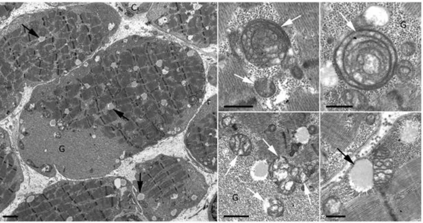
2.4.2 Histopathological evaluation of affected individuals 55
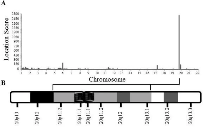
2.4.3 Autozygosity mapping generated 20p11.2-q13.1 as a
candidate region 57
2.4.4 Exome sequencing and in silico analyses generate candidate
variants 59
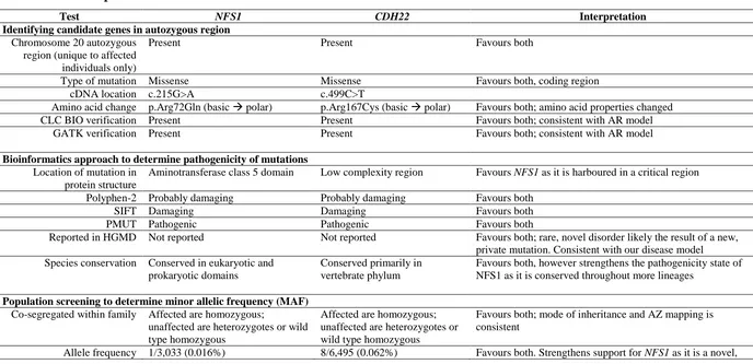
2.4.5 Population screens demonstrate the rarity of NFS1 p.Arg72Gln 62
2.4.6 Deficiency in NFS1 expression in patients with IMC23D 62
2.4.7 Characterization of IMC23D disease summary: comparing NFS1
and CDH22 65
2.5 Discussion 68
2.6 Conclusion 71
2.7 References 71
Chapter 3 - Novel phenotype, known gene: mutations in heparan sulfate synthesis enzyme, EXT2 leads to seizure and developmental disorder, no exostoses
3 Study rationale 76
3.1 Overview 76
3.2 Introduction 77
xii
3.3.1 Ethics 78
3.3.2 Patients and biological materials 78
3.3.3 DNA isolation 79
3.3.4 Genotyping 79
3.3.5 Autozygosity mapping 79
3.3.6 Exome sequencing 79
3.3.7 Sequence alignment 79
3.3.8 Variant calling and annotation 80
3.3.9 Variant discovery
3.3.9.1 Prioritization of homozygous, non-synonymous,
and rare variants 80
3.3.9.2 in silico analyses 80
3.3.10 Variant validation
3.3.10.1 PCR 81
3.3.10.2 Imaging, purifying, and sequencing of PCR products 81
3.3.11 Population screening
3.3.11.1 Genotyping of local population 81
3.3.11.2 Identifying overall MAF of EXT2 variants 81
3.3.12 Cell culture
3.3.12.1 Patient cells 82
3.3.12.2 Mutagenesis 82
3.3.12.3 Transfection 82
3.3.13 Protein isolation 83
3.3.14 Immunoblotting 83
3.3.15 Reverse transcriptase (RT)-PCR 83
3.3.16 Antibodies 83
3.3.17 Statistics 84
3.4 Results
3.4.1 Clinical description of patients with SSM syndrome 84
3.4.2 Genome-wide autozygosity mapping reveals highly significant
homozygous regions 91
3.4.3 Compound homozygous EXT2 variants in patients with
SSM syndrome 93
3.4.4 EXT2 variants segregate in SSM syndrome family 95
3.4.5 Population screening reveals EXT2 variants are ultra-rare 97
3.4.6 Decreased EXT2 expression and activity in patients with
SSM syndrome 97
3.4.7 Both EXT2 variants are necessary for the development of
SSM syndrome 99
3.5 Discussion 102
3.6 Conclusion 108
xiii
Chapter 4 - Known phenotype, known gene: linkage analysis and exome sequencing identify a novel mutation in KCTD7 in patients with progressive myoclonus epilepsy with ataxia
4 Study rationale 112
4.1 Overview 113
4.2 Introduction 113
4.3 Materials and methods
4.3.1 Ethics 114
4.3.2 Patients and biological materials 115
4.3.3 DNA isolation 115
4.3.4 Genotyping 115
4.3.5 Autozygosity mapping 115
4.3.6 Exome sequencing 115
4.3.7 Sequence alignment 116
4.3.8 Variant calling and annotation 116
4.3.9 Variant discovery
4.3.9.1 Prioritization of homozygous, non-synonymous,
and rare variants 116
4.3.9.2 in silico analyses 116
4.3.10 Variant validation 116
4.3.10.1 PCR 117
4.3.10.2 Imaging, purifying, and sequencing of PCR products 117
4.3.11 Population screening
4.3.11.1 Identifying overall MAF of KCTD7 variant 117
4.4 Results
4.4.1 Clinical description of patients with progressive myoclonus
epilepsy with ataxia 117
4.4.2 Electroencephalogram features 123
4.4.3 Genome-wide autozygosity mapping generated a high
priority region on chromosome 7p12.1-7q11.22 127
4.4.4 KCTD7 p.Tyr276Cys in patients with progressive myoclonus
epilepsy with ataxia 129
4.4.5 KCTD7 p.Tyr276Cys segregates with disease phenotype in
the family 131
4.4.6 KCTD7 is a known disease gene 133
4.5 Discussion 135
4.6 Conclusion 138
4.7 References 139
xiv
5 Study rationale 143
5.1 Overview 143
5.2 Introduction 144
5.3 Materials and methods
5.3.1 Ethics 146
5.3.2 Patients and biological materials 146
5.3.3 DNA isolation 146
5.3.4 Exome sequencing 146
5.3.5 Sequence alignment 146
5.3.6 Variant calling and annotation 146
5.3.7 Variant discovery
5.3.7.1 Prioritization of autosomal recessive, non-synonymous,
and rare variants 147
5.3.7.2 in silico analyses 147
5.3.8 Variant validation
5.3.8.1 PCR 147
5.3.8.2 Imaging, purifying, and sequencing of PCR products 147
5.3.9 Cell culture 148
5.3.10 Protein isolation 148
5.3.11 Immunoblotting 148
5.3.12 Reverse transcriptase (RT)-PCR 148
5.3.13 Model organisms 148
5.3.13.1 Gateway cloning technology 148
5.3.13.2 Drosophila stocks 149
5.3.13.3 RT-qPCR 149
5.3.13.4 Bang sensitivity 150
5.3.13.5 Immunohistochemistry, image acquisition, and analysis 150
5.3.14 Antibodies 152
5.3.15 Statistics 152
5.4 Results
5.4.1 Clinical presentation of patients with nocturnal seizures with
developmental delay 153
5.4.2 Compound heterozygous TMTC3 variants in patients with
nocturnal seizures with developmental delay 156
5.4.3 Loss of TMTC3 in patients with nocturnal seizures and
developmental delay 158
5.4.4 Neuronal knockdown of Drosophilatmtc3 causes increased
susceptibility to mechanically induced seizures 161
5.4.5 TMTC3 is localized at presynaptic terminals in rat brains 167
5.5 Discussion 170
5.6 Conclusion 172
xv
Chapter 6 - The ONDRISeq panel: custom designed next generation sequencing of genes related to neurodegeneration as part of the Ontario Neurodegenerative Disease Research Initiative
6 Study rationale 176
6.1 Overview 176
6.2 Introduction 177
6.3 Materials and methods
6.3.1 Design of ONDRISeq 179
6.3.2 Sample collection and DNA isolation 187
6.3.3 Library preparation 187
6.3.4 Next generation sequencing 188
6.3.5 Sequence alignment 188
6.3.6 Variant calling 189
6.3.7 Variant annotation 189
6.3.8 Variant classification and prioritization 189
6.3.9 APOE genotyping 190
6.3.10 Variant validation 191
6.3.10.1 Variant validation 1: NeuroX 191
6.3.10.2 Variant validation 2: TaqMan allelic discrimination 192
6.3.10.3 Variant validation 3: Sanger sequencing 192
6.3.10.3.1 PCR 193
6.3.10.3.2 Imaging, purifying, and sequencing of
PCR products 193
6.3.10.4 Variant validation 4: SOD1 testing 193
6.3.11 C9orf72 genotyping 193
6.3.12 Statistical analysis 194
6.4 Results
6.4.1 Study subjects 194
6.4.2 Quality assessment of ONDRISeq data 196
6.4.3 ONDRISeq is concordant with NeuroX, TaqMan allelic
discrimination assay, and Sanger sequencing 198
6.4.4 Genetic variation in patients with neurodegenerative disease 198 6.4.4.1 C9orf72 hexanucleotide expansion in patients with
neurodegenerative disease 199
6.4.4.2 Genetic variation identified in patients with
neurodegenerative disease using ONDRISeq 199
6.4.4.3 APOE genotypes in patients with neurodegenerative
disease 200
6.4.4.4 Case study: strong evidence of pathogenicity for
APP p.Ala713Thr in AD patient 203
6.5 Discussion 206
xvi
6.7 References 208
Chapter 7 - Oligogenic inheritance in families with amyotrophic lateral sclerosis and frontotemporal dementia
7 Study rationale 212
7.1 Overview 213
7.2 Introduction 213
7.3 Materials and methods
7.3.1 Ethics 215
7.3.2 Sample collection clinical assessment 215
7.3.3 DNA isolation 215
7.3.4 C9orf72 testing 216
7.3.4.1 Amplicon length analysis 216
7.3.4.2 Repeat-primed PCR 216
7.3.4.3 Immunohistochemistry 217
7.3.4.4 Southern immunoblotting 217
7.3.5 ATXN2 expansion testing 218
7.3.6 Next generation sequencing 218
7.3.7 Sequence alignment 219
7.3.8 Variant calling 219
7.3.9 Variant annotation 219
7.3.10 Variant classification and prioritization 219
7.3.11 Variant validation 219
7.3.11.1 PCR 220
7.3.11.2 Imaging, purifying, and sequencing of PCR products 220
7.4 Results
7.4.1 Clinical description 220
7.4.2 Variants identified in patients with ALS 227
7.4.3 C9orf72 dipeptide immunostaining 236
7.4.4 Oligogenic inheritance in patients 238
7.5 Discussion 241
7.6 Conclusion 245
7.7 References 245
Chapter 8 - Discussion
8.0 Summary 248
8.1 Context of study findings
8.1.1 Chapter 2: NFS1, a novel disease gene underlying a novel
metabolic disease 248
8.1.2 Chapter 3: Expanding the biological function of EXT2,
xvii
8.1.3 Chapter 4: Confirming the association of KCTD7 in
progressive myoclonus epilepsy 250
8.1.4 Chapter 5: The application of a model organism to understand
the function of a novel disease gene TMTC3, in neurodevelopment 251 8.1.5 Chapter 6: Developing a custom sequencing based method to
study neurodegeneration 252
8.1.6 Chapter 7: The effect of multiple genetic variants on
neurodegenerative disease risk 253
8.2 Methodological considerations 254
8.2.1 Study strengths and implications 255
8.2.2 Additional study caveats 256
8.3 Future directions 257
8.4 Final conclusions 260
8.5 References 260
Appendices 266
xviii
List of Tables
Table 1.7.1 Population databases of healthy controls 17
Table 1.7.2 Disease databases 17
Table 1.7.3 Examples of in silico predictive tools 18
Table 2.4.1 Clinical and biochemical findings of patients with IMC23D 53
Table 2.4.7 Comparison of NFS1 and CDH22 as the cause of IMC23D 65
Table 3.4.1 Clinical description of patients with SSM syndrome 87
Table 3.5.1 Hereditary diseases with abnormal heparan sulfate levels 104
Table 4.4.1 Clinical description of patients with epilepsy with ataxia 121
Table 4.4.6 Comparison of all reported patients with KCTD7 variants 134
Table 5.4.1 Clinical features of patients with nocturnal seizures with ID 155
Table 6.3.1 Genes associated with amyotrophic lateral sclerosis, frontotemporal dementia, Alzheimer’s disease, Parkinson’s disease, or vascular cognitive impairment as represented on
the ONDRISeq targeted resequencing panel 181
Table 6.4.1 Patient demographics 195
Table 6.4.2 Quality control metrics for sequencing runs on ONDRISeq 197
Table 6.4.4.1 Other risk variants identified in a cohort of 216 disease cases 201
Table 6.4.4.2.1 Diagnostic yield of ONDRISeq in a cohort of 216 disease cases 201
Table 6.4.4.2.2 Variants identified in a cohort of 216 disease cases as
detected by ONDRISeq 202
Table 7.4.4 Summary of clinical and genetic information 240
xix
List of Figures
Figure 2.4.1 Pedigree of IMC23D 52
Figure 2.4.2 Pathological findings in patients with IMC23D 56
Figure 2.4.3 Autozygosity mapping of IMC23D family 58
Figure 2.4.4 Mapping and exome sequencing of IMC23D family identifies a highly
conserved and destabilizing missense mutation, p.(Arg72Gln) in NFS1 61
Figure 2.4.6 Depletion in NFS1 Protein and Transcript Levels in
Patients with IMC23D 64
Figure 3.4.1 Pedigree with four children affected with SSM syndrome 86
Figure 3.4.2 Autozygosity mapping generates highly significant
homozygous regions 92
Figure 3.4.3 Schematic of the genetic and bioinformatic studies in the
SSM family 94
Figure 3.4.4 EXT2 variants segregate with affected individuals in the family 96
Figure 3.4.6 Decreased EXT2 expression and activity in patients with
SSM syndrome 98
Figure 3.4.7 Both EXT2 variants are necessary for the development of
SSM syndrome 101
Figure 4.4.1 Pedigree with three daughters affected with epilepsy with ataxia 120
Figure 4.4.2 EEG recording of the affected individuals 126
Figure 4.4.3 Homozygous region on chromosome 7 unique to
affected individuals 128
Figure 4.4.4 Schematic of mutation discovery 130
Figure 4.4.5 KCTD7 g.661041A>G segregates with affected individuals
xx
Figure 5.4.1 Pedigree with four children affected with nocturnal seizures with
developmental delay 154
Figure 5.4.2 Compound heterozygous TMTC3 variants in patients with
nocturnal seizures with developmental delay 157
Figure 5.4.3 Depleted TMTC3 protein and reduced transcript expression in cells of
patients affected by nocturnal seizures with developmental delay 160
Figure 5.4.4 Tmtc3 deficiency confers susceptibility to seizures in
Drosophila melanogaster 165
Figure 6.4.4.4 APP variant in AD case 205
Figure 7.4.1 Pedigrees of three families affected with ALS or ALS-FTD 226
Figure 7.4.2.1 Genetic analyses of C9orf72 genotypes of representative individuals from each
family 229
Figure 7.4.2.2 Genetic analyses of C9orf72 expansion profiles of representative individuals
from each family 231
Figure 7.4.2.3 Genetic analysis of ATXN2 genotyping profiles of family 2 233
Figure 7.4.2.4 Validation of variants in OPTN and ARHGEF28 235
xxi
List of Appendices
Appendix A - Ethics approval 266
Appendix B - Chapter 6: Primer list 269
Appendix C - Chapter 7: Expanded genetic and clinical information on 270
all individuals within the study
xxii
List of Abbreviations
ACD, autosomal co-dominant
ACMG, American College of Medical Genetics and Genomics AD, Alzheimer’s disease
ADm, autosomal dominant
ALS, amyotrophic lateral sclerosis ALSoD, ALS online database
aMCI, amnestic single or multidomain mild cognitive impairment AMRF, action myoclonus-renal failure
AR, autosomal recessive
AST, aspartate aminotransferase bvFTD, behavioural variant FTD BWA, Burrows-Wheeler Aligner
CADASIL, cerebral autosomal dominant arteriopathy with subcortical infarcts and leukoencephalopathy
CARASIL syndrome, cerebral autosomal recessive arteriopathy with subcortical infarcts and leukoencephalopathy
CH, compound heterozygous CK, creatine kinase
CLIA, clinical laboratory improvement amendments CMT disease, Charcot-Marie-Tooth disease
CNV, copy number variation COX, cytochrome C oxidase
dbGaP, the database of Genotypes and Phenotypes DIF, digoxigenin
xxiii
ENFL, nocturnal frontal lobe epilepsy ExAC, Exome Aggregation Consortium FORGE, Finding of Rare Disease Genes FTD, frontotemporal dementia
GATK, Genome Analysis Toolkit gDNA, genomic DNA
GMT, Gomori modified trichrome
GTEx, Genotype-Tissue Expression project GWAS, genome-wide association study
HDLS, leukoencephalopathy, diffuse hereditary, with spheroids HET4, hereditary essential tremor, 4
HGMD, Human Gene Mutation Database HME, hereditary multiple exostoses
HMN7B, neuropathy, distal hereditary motor, type VIIB HPS, hematoxylin, phloxin, and saffron
HSN1E, hereditary sensory neuropathy type 1E HZ, homozygous
IMC23D, infantile mitochondrial complex II/III deficiency Indels, insertions and deletions
IR, inverted repeats
LBD, Lewy body dementia
LINEs. long interspersed transposon derived elements LOD, logarithm of odds
MAF, minor allele frequency
MELAS, mitochondrial encephalomyopathy, lactic acidosis, and stroke-like episodes MERF, myoclonic epilepsy with ragged red fibers
MND, motor neuron disease
MoCA, Montreal Cognitive Assessment
xxiv
NARP, neurogenic muscle weakness, ataxia, and retinitis pigmentosa NBIA2A, neurodegeneration with brain iron accumulation 2A
NBIA2B, neurodegeneration with brain iron accumulation 2B NCL, neuronal ceroid lipofuscinoses
NHLBI ESP, National Heart, Lung, and Blood Institute Exome Sequencing Project OMIM, Online Mendelian Inheritance in Man
ONDRI, Ontario Neurodegenerative Disease Research Initiative PAS, periodic acid-Schiff (PAS)
PCR, polymerase chain reaction PD, Parkinson’s disease
PKU, phenylketonuria
PMID, PubMed identification
PolyPhen-2, Polymorphism Phenotyping version 2 PSS Potocki-Shaffer syndrome
qRT-PCR, quantitative reverse transcriptase PCR RNA, ribonucleic acid
RT-PCR, reverse transcriptase PCR SDH, succinic dehydrogenase SDS, sodium dodecyl sulfate
SIFT, Sorting Intolerant From Tolerant
SINEs, short interspersed transposon derived elements SNP, single nucleotide polymorphism
SNV, single nucleotide variant SSC, standard sodium citrate
SSM, seizures-scoliosis-macrocephaly STR, short tandem repeats
TCAG, The Centre for Applied Genomics VCF, variant calling format
xxv
Chapter 1 - Introduction
1
Overview
Materials from the following texts with appropriate modifications, were incorporated in Chapter 1:
(1) Farhan, SMK., and Hegele, RA. “Genetics 101 for cardiologists: Rare genetic variants and monogenic cardiovascular disease”. Canadian Journal of Cardiology. 2013; 29(1): 18-22 (PMID: 23200093).
(2) Farhan, SMK., and Hegele, RA. “Exome sequencing: new insights into lipoprotein disorders”. Current Cardiology Reports. 2014; 16(7): 507-517 (PMID: 24893940).
(3) Farhan, SMK., and Prasad, AN. “Exploring the Epilepsiome I: Genetics of Age dependent Epileptic Encephalopathies”. Pediatric Epilepsy, 4th edition, Chapter 7.
(4) Prasad, AN., and Farhan, SMK. “Exploring the Epilepsiome II: Approaching the Complex Epilepsies”. Pediatric Epilepsy, 4th edition, Chapter 8.
(5) Farhan, SMK., ONDRI Investigators, Strong, MJ. “The Ontario Neurodegenerative Disease Research Initiative (ONDRI)”. Canadian Journal of Neurological Sciences. 2016; Accepted.
1.1
Variation in the human genome
Variation in the human genome, which is composed of approximately three billion base pairs, can lead to benign or pathogenic biological effects. These DNA variations often classified as neutral, adaptive, or deleterious, can be under positive selection (Darwinian
selection) or negative selection (purifying selection) based on their effect on an
individual’s fitness (Fay et al., 2001). DNA variations that increase an individual’s fitness
individual’s fitness, are under negative selection. Importantly, some pathogenic variants
can escape natural selection by exerting their effects post sexual reproduction. In this section, I will introduce the various classes of genetic variation observed in the human genome and provide examples of diseases caused by each class of variation.
1.1.1
Inherited and
de novo
variation
Inherited variations are DNA changes transmitted through the germline from parent to offspring. Conversely, de novo or sporadic variants spontaneously arise in the offspring’s germ cells and are absent from the parents’ germ cells. Importantly, if de novo variations
are not deleterious, they can be transmitted to the progeny and are reclassified as inherited variation. De novo variations are the most rare class of genetic variation and have recently been of great interest in neurodevelopmental diseases, primarily autism spectrum disorders (Ku et al., 2012; Neale et al., 2012; Yuen et al., 2015).
1.1.2
Germline and somatic variation
Errors during DNA replication that escape polymerase proofreading can occur within germ or somatic cells. Unlike somatic variation, germline variation can be transmitted to the progeny. Familial diseases, which reappear in multiple generations in pedigrees, are often linked to germline variation. An example of a familial disease is Marfan syndrome (OMIM 154700), an autosomal dominant disorder characterized by defective connective tissue leading to increased height, disproportionate limbs, and multiple cardiovascular
1.1.3
Structural variation
1.1.3.1 Chromosomal abnormalities: numerical and structural chromosomal
aberrations
Chromosomal abnormalities can be either numerical or structural aberrations, which can
be easily visualized using cytogenetic techniques. Numerical chromosomal aberrations also termed chromosomal aneuploidy, are any cases when the karyotype is not 46, XY or
46, XX for males and females, respectively. Therefore, they are caused by the absence (monosomy) or addition (trisomy, triploidy) of chromosomes in cells. An example of a numerical chromosomal aberration is Down syndrome (Trisomy 21, OMIM 190685), a neurodevelopmental disorder characterized by distinct facial features and intellectual disability in addition to multiple systemic anomalies, due to three copies of chromosome 21 (or chromosome 21q) (Wiseman et al., 2009). Chromosomal abnormalities are usually the result of nondisjunction at the first or second meiotic division (Ioannou and Tempest, 2015).
Individuals with structural chromosomal aberrations may still have a normal karyotype however, sizable portions of chromosomes have been deleted, duplicated, or inverted. Additionally, portions of DNA may have merged and translocated with other chromosomes (Weckselblatt and Rudd, 2015). An example of a disease caused by structural chromosomal abnormalities is DiGeorge syndrome (22q11.2 deletion
syndrome, OMIM 188400), a characteristic dysmorphic facial syndrome with congenital heart defects (Michaelovsky et al., 2012).
1.1.3.2 Copy number variation
Copy number variation (CNV) were originally classified as structural DNA segments that are >10 Kb in the genome and are variations that are more frequently observed than
if encompassing the entire gene or localized within regulatory elements (Stranger et al., 2007). CNV can also influence select regions within genes such as exons, and can sometimes encode an abnormal protein. Among a multitude of examples, CNVs within chromosome 16p13.1 have been associated with schizophrenia (Ingason et al., 2011), autism spectrum disorders, and intellectual disability (Ullmann et al., 2007).
1.1.3.3 Variable number of tandem repeats
One third of the human genome is comprised of repetitive DNA sequences, which are in
the form of large segmental duplications (low CNVs) or long and short interspersed transposon derived elements (LINEs and SINEs) (Kozlowski et al., 2010). In addition, variable number of tandem repeats (VNTR) are a class of genetic variation that are due to multiple nucleotides repeating consecutively within genes. There are two main types: minisatellites and microsatellites, which typically occur outside of coding regions, in non-coding sequences that often flank genes. Minisatellites are sometimes referred to as VNTR and range in size from 10-60 nucleotides and have been observed in thousands of locations in the human genome (Kozlowski et al., 2010). Similarly, microsatellites, often referred to as short tandem repeats (STR), are typically composed of 2-10 nucleotides and are hypermutable sites across the genome (Kozlowski et al., 2010). Interestingly, these common repeat regions served as markers for genome-wide linkage studies prior to the emergence of the International HapMap Project (International HapMap, 2005).
Importantly, when these repeats are located in regulatory or coding regions and exceed the normal, stable range, they can result in aberrant gene products, eventually causing disease (Lopez Castel et al., 2010). Examples of diseases due to multinucleotide expansions include Fragile X syndrome (OMIM 300624), an intellectual disability caused by CGG repeats within the 5’-untranslated region of the FMR1 gene (Bagni et al., 2012). In addition, Huntington’s disease (OMIM 143100) is a neurodegenerative disease caused
1.1.3.4 Insertions and deletions
Insertions or deletions (indels) of nucleotides in the DNA sequence may have an impact on gene expression or protein function. Indels can range from 1-50 base pairs with the majority consisting of <10 base pair changes. Depending on their location, indels can cause inframe or frameshift variants, or splicing variants. For example, the most common variant in cystic fibrosis (OMIM 219700), an autosomal recessive respiratory disease characterized by defective mucociliary clearance, is a three-nucleotide deletion in the CFTR gene, causing the loss of phenylalanine at amino acid position 508, within the
ABC transporter domain (Flume and Van Devanter, 2012). While >90% of intron-containing genes undergo alternative splicing to regulate gene expression and protein synthesis, indels within or near the exon-intron junctions alternating the 5’, 3’ splice sites or the branch point, can cause disease (Singh and Cooper, 2012). These DNA changes can potentially prevent the spliceosomes, the splicing machinery, from recognizing these elements ultimately inducing inadvertent exon skipping or intron retention (Singh and Cooper, 2012)
1.1.4
Single nucleotide variation
Single nucleotide variation (SNV), which is also referred to as point mutation or
substitution, is the most common class of variation. They can be either transitions where the SNV is an interchange between purines (adenine, C <-> guanine, G) or pyrimidines (cytosine, C <-> thymine, T); or transversions, which interchanges purine and
pyrimidines (A<->C, A<->T, G<->C, or G<->T). If an SNV is within the codon frame, it can result in synonymous or non-synonymous changes. The degeneracy of the genetic
code allows for multiple unique codons to generate the same amino acid. Therefore, SNVs within codons, especially in the third nucleotide position may not alter the amino acid as proposed in ‘the wobble hypothesis’ (Crick, 1966). These synonymous changes
termed missense or nonsense variants, respectively. SNVs can also cause splicing defects if located within or near the exon/intron boundaries and can alter the splice donor or acceptor recognition sequence, rendering them unrecognizable by splicing machinery (Cartegni et al., 2002). An example among many SNV-mediated human diseases is Duchenne muscular dystrophy (OMIM 310200), an X-linked muscle disorder, most often caused by nonsense variation within the DMD gene leading to protein truncation
(Hoffman et al., 1987).
1.2
Genetic diseases in humans
1.2.1
Monogenic (Mendelian) diseases
Mendelian traits, first characterized by Austrian monk Gregor Mendel in the mid-1800s, explain simple and apparent parent to child transmission patterns (Henig, 2000; van der Waerden, 1968). Mendel first studied these traits in plants by observing height and colour differences in subsequent generations (Henig, 2000; van der Waerden, 1968). In doing so, the laws of Mendel emerged, which attempt to explain these noticeable differences. The
first law, known as ‘the law of independent assortment’, states that during gamete formation, different pairs of alleles segregate independently of each other (Castle, 1903; Monaghan and Corcos, 1984). The second law, ‘the law of segregation’, suggests that the
two alleles for each trait separate (segregate) during gamete formation, and then unite at random (Castle, 1903; Monaghan and Corcos, 1984). The third law, ‘the law of
dominance’,states that recessive alleles are always masked by dominant alleles that determine the phenotype (Castle, 1903; Monaghan and Corcos, 1984).
Although additional complexities and exceptions have been recognized since they were originally proposed, these principles are still in use and are applied today in
disorders, one variant is sufficient to cause disease; this is in contrast to recessive disorders, where two defective alleles are necessary.
Mendelian diseases are typically not affected by exogenous factors such as the patient’s environment, gene by environment interactions, or epigenetic modifications
however; these factors can exacerbate or alleviate the phenotype. For example, individuals diagnosed with phenylketonuria (PKU, OMIM 261600), an autosomal recessive, metabolic disease commonly caused by variations in the PAH gene, can exacerbate or alleviate the intellectual disability phenotype caused by an accumulation of
tyrosine, if an affected individual maintains a high or low protein diet, respectively (Demirkol et al., 2011). Mendelian diseases are mostly rare in the general population as they are the result of a highly penetrant variant in a highly conserved and physiologically essential protein (Farhan and Hegele, 2013). Individuals affected by severe Mendelian diseases such as Tay-Sachs disease (OMIM 272800), a severe early-onset neurological disorder caused by variants in the HEXA gene, do not reach the age of sexual maturity; and if they do, are unable to reproduce due to many biological and social factors (Fernandes Filho and Shapiro, 2004).
1.2.2
Digenic diseases
Digenic inheritance, considered the simplest form of oligogenic inheritance, is characterized by the presence of two independent variants that jointly modify the phenotype. Digenic inheritance is a relatively unrecognized type of disease inheritance, as its definition has not been clearly defined and often mistaken with epistasis, the interaction of genes. In 2013, Alejandro Schäffer suggested a universal definition of
digenic inheritance in medical genetics: ‘inheritance is digenic when the variant genotypes at two loci explain the phenotypes of some patients and their unaffected (or
more mildly affected) relatives more clearly than the genotypes at one locus alone’ (Schaffer, 2013). In recognition of the importance of digenic inheritance in human diseases, the ‘digenic disease database’ was created with the intent of aggregating
An example of a disease with digenic inheritance is long QT syndrome (OMIM 192500), a congenital heart disease characterized by a prolonged QT interval as detected by electrocardiography (Millat et al., 2006). While variants in either KCNQ1 or KCNH2 can cause long QT syndrome, the condition can also result from co-occurrence of variants in both genes (Berthet et al., 1999). Furthermore, the digenic inheritance observed in long QT syndrome, has been replicated in other studies (Millat et al., 2006; Tester et al., 2005). Importantly, digenic traits can also be considered complex diseases as they can be modulated by non-genetic factors, as described in the subsequent section.
1.2.3
Polygenic (complex) diseases
Complex diseases are affected by various factors including a burden of genetic variation, the environment, gene-environment interactions, or epigenetic modifications (Cordell, 2009; Iyengar and Elston, 2007; Petrovski and Kwan, 2013). Complex diseases are more prevalent in the population therefore, they often follow a ‘common variant, common disease’ genetic principle. Traditionally, common variants are primarily studied in
complex diseases however, they only marginally contribute to disease susceptibility. Potentially, the presence of multiple genetic variants with modest but cumulatively, significant effects can confer risk to disease (Do et al., 2012; Petrovski and Kwan, 2013). Despite these biological consequences, low effect variants are able to persist in the population, as they are individually not large enough to be targeted by natural selection.
Based on these observations, conventional genome-wide association studies (GWAS) used single nucleotide polymorphism (SNP) arrays, which did not capture any rare variation and were based on a priori knowledge of common susceptibility SNPs
(Koboldt et al., 2013). However, there has been growing interest in assessing the effects of rare variants in complex diseases to identify the missing heritability (discussed in
As initially observed by Francis Galton and Karl Pearson in the 1900s,
quantitative traits typically follow a Gaussian (normal) distribution (Galton and Galton, 1997). Individuals residing at the tail ends of the distribution are often studied to identify any genetic variation contributing to the trait of interest. This approach, referred to as extreme phenotype sampling, is robust in detecting rare variation enriched in cases (extreme phenotypes), than controls (individuals within the normal range) (Auer and Lettre, 2015). Moreover, in the absence of large effect variants, which can underlie Mendelian diseases as previously discussed, individuals with extreme phenotypes may
carry a burden of multiple small effect variants that collectively increase risk for disease. The burden of genetic risk is often quantified by summation of risk alleles to produce a ‘polygenic risk score’ (Dudbridge, 2013). Finally, in addition to genetic predisposition,
age, sex, puberty, pregnancy, or diet can influence disease manifestation.
1.3
Phenotypic variation
As discussed in the preceding section, complex disorders are influenced by multiple factors and therefore, can have multiple etiologies. The phenotypic variation in an individual with a complex trait can be attributed to genetic variation, environmental conditions, or an interplay of both potential influences (Zuk et al., 2012). Simply, the phenotypic value (P) is equal to the sum of the genotypic value (G) and the
environmental deviation (E). Therefore, changes in G and/or E influence the variability in P:
P = G + E
Importantly, G is comprised of three components: 1) the additive genetic value (A); 2) the dominance component (D); and 3) the epistasis (I, interaction of genes).
G = A + D + I
These genetic influences make up the heritability of a quantitative trait, which I will discuss next.
1.3.1
Heritability
To determine the effect of genetic variation on phenotypic variation (VP), we often rely
on the heritability of the phenotype of interest. Heritability is the variation in an
individual’s phenotype that is attributed to the genetic variation transmitted through the parents’ germ cells (Zuk et al., 2012). Specifically, heritability is the ratio of genetic
variance (VG) to phenotypic variance (VP). There are two main types of heritability:
broad sense and narrow sense. Broad sense heritability (H2) is all the genetic variance: H2 = VG/VP = (VA + VD + VI)/VP.
Alternatively, narrow sense heritability (h2) is only the additive genetic variance: h2 = VA/VP
1.4
Epigenetics and human diseases
In addition to genetic variations that contribute to benign human diversity or disease, the expression of genes can be modulated by epigenetic modification machinery. In general,
epigenetics is the study of mechanisms that control gene expression without altering the DNA sequence. These potentially heritable processes are stable and are particularly important during development. There are multiple types of epigenetic modifications
grouped into three main categories: 1) DNA methylation; 2) histone modification; 3) and nucleosome positioning (Handy et al., 2011; Portela and Esteller, 2010).
An example of a disease with epigenetic modification is ATR-X syndrome (OMIM 301040), an X-linked dominant disease characterized by intellectual disability and alpha-thalassemia (Badens et al., 2006). ATR-X syndrome is caused by heterozygous (hemizygous) variants in the ATRX gene and males are predominantly affected however, carrier females may have skewed X-inactivation (Badens et al., 2006). ATRX binds DAXX to form a chromatin-remodeling complex, and pathogenic variants in either protein can lead to telomere dysfunction, genomic instability, or altered gene expression (Leung et al., 2013; Watson et al., 2013).
1.5
Approaches to studying genetic diseases
The relationship between disease penetrance and allele frequencies can help in prioritizing which approach to pursue when studying genetic diseases. As previously discussed, highly penetrant phenotypes are more likely to be caused by very rare genetic variation. While low-to-intermediate penetrant phenotypes can also be attributed to very rare genetic variation, these phenotypes are typically not severe enough to be clinically ascertained (McCarthy et al., 2008). In contrast, common variants typically result in low penetrant phenotypes, and are often the targets of GWAS (McCarthy et al., 2008).
In this section, I will introduce conventional approaches to study genetic diseases in related individuals (family studies applying forms of linkage analysis) and in unrelated individuals (population studies applying forms of association analysis). While linkage studies, candidate gene approaches, and GWAS have been successful in identifying loci
that are causal or confer risk to disease, massively parallel sequencing is able to rapidly and economically identify causal variants, which I will discuss in subsequent sections.
1.5.1
Family studies
1.5.1.1 Linkage studies
from parent to offspring, more often than expected (Dawn Teare and Barrett, 2005; Edwards, 2012). Across a population, when alleles at separate loci are associated with each other at a significantly higher frequency than would be expected by chance, they are in linkage disequilibrium and therefore, can be evidence of common ancestry (Dawn Teare and Barrett, 2005). Using linkage maps, which were initially developed by Thomas Hunt Morgan and his student, Alfred Sturtevant, helped us further refine linkage studies. Morgan and Sturtevant hypothesized that recombination of genes was dependent on the physical distance between them, measured in centimorgans (cM; 1 cM is approximately 1
million base pairs) (Sturtevant et al., 1919). This proposition allowed for linkage studies to be applied in humans to identify regions of the genome containing disease-associated genes. Furthermore, a specific subtype of linkage analysis known as the parametric of inheritance model-based analysis, determines co-segregation of genetic markers in families (Dawn Teare and Barrett, 2005). As the proximity of loci on a chromosome reduces the probability that they will separate by recombination, it is likely that a closely related disease cohort would co-inherit an entire section within the chromosome. As aforementioned, this phenomenon known as ‘linkage disequilibrium’ defies Mendel’s law of segregation. Using this approach, a genetic region unique to affected individuals and absent from healthy controls, can be identified. This approach also assigns a logarithm of the odds (LOD) score for each linkage event with significantly large LOD scores
indicative of linkage; and low or negative values are evidence of genetic recombination. LOD scores of ≥3 are equivalent to P-values of ≤0.001, and are traditionally considered
statistically significant.
1.5.1.2 Candidate gene approaches
Prior knowledge of genes implicated in a disease has a central advantage over traditional linkage studies and GWAS. Candidate gene approaches are hypothesis driven and are
based on a well-established relationship of a gene with a disease, prior knowledge from genetic analysis (genetic variant present in affected individuals); or a high likelihood
can significantly expedite genetic discoveries. However, candidate gene approaches are generally not suited for novel diseases or diseases exhibiting locus heterogeneity.
1.5.2
Population studies
1.5.2.1 Association studies
Association approaches are more useful for elucidating susceptibility alleles underlying
complex diseases in multiple unrelated individuals. This unbiased approach facilitated the era of GWAS, which used genotyping arrays to screen a large number of cases and controls, for thousands of common variants across the genome (Auer and Lettre, 2015). To identify genuine disease-associated variants, the variant frequency in cases must be significantly higher than in controls, which at first glance, likely favours linkage disequilibrium (Auer and Lettre, 2015). The International HapMap Project assessed common variation in a global, ethnically diverse cohort, and determined that when
considering linkage disequilibrium, there are approximately one million unique loci in the human genome (International HapMap, 2005). Following a Bonferonni correction for the million unique loci, which are treated as independent tests, a genome-wide significant P -value = <5×10⁻⁸, became the standard threshold. In addition to observing genome-wide significance of an association between an allele and a phenotype, it is important to replicate the association in an unbiased population sample to eliminate possible confounding variables such as population stratification, which may occur when subpopulations within the study cohort carry the allele due to differences in ancestral origin, rather than disease state (McCarthy et al., 2008). Furthermore, association of the variant and disease should be followed by ensuring the signal is replicable and harboured
in or near a gene biologically relevant to the disease mechanism.
1.6
Tools to study genetic variation in disease
1.6.1
Sequencing versus genotyping: benefits and pitfalls
To identify the genetic basis of human diseases, we often use sequencing or genotyping-based assays. In sequencing approaches, we indiscriminately identify the full nucleotide
sequence of genes. In genotyping approaches, we use a known set of genetic markers to determine their presence or absence in individuals. Each approach is selected based on
the study hypothesis and objectives. Traditionally, GWAS used arrays to rapidly genotype cases and controls, as previously discussed. Knowledge of the entire genetic sequence is preferred however, genotyping approaches are often selected to reduce costs. In my PhD thesis, I used sequencing approaches to discover novel disease loci and genotyping approaches to screen for the newly discovered loci in cases and controls.
1.6.2
Massively parallel sequencing
Today, we are in the era of massively parallel DNA sequencing often referred to as next generation sequencing. Next generation sequencing technologies is a broad umbrella term for technologies that facilitate rapid, efficient sequencing of multiple genomes by
performing millions of reactions simultaneously leading to high throughput of data (Farhan and Hegele, 2014). This is in contrast to Sanger sequencing, now considered first generation sequencing, which has been widely and successfully used in genetic studies most notably in elucidating the code of the first human genome (Lander et al., 2001). However, Sanger sequencing is labour intensive and can be prohibitively expensive for
studies with a large sample size. The advent of next generation sequencing technologies has accelerated gene discoveries in virtually all diseases. There are three main types of next generation sequencing applications for sequencing DNA variants: 1) whole genome sequencing; 2) exome sequencing; and 3) targeted gene panels.
Whole genome sequencing is an indiscriminate approach that decodes the genetic information in an individual’s genome. In contrast, exome sequencing targets only the
to the exons. Targeting only the protein-coding regions of the genome originates from the observation that a large fraction of human genetic diseases are caused by
non-synonymous variants in evolutionarily considered protein-coding genes (Chong et al., 2015; Ng et al., 2009). Moreover, the difference in cost between the two methods; and the computational power necessary to reassemble the human exome (1-2% of the human genome), is significantly less resource intensive (Farhan and Hegele, 2014).
Notably, while next generation sequencing has led to the discovery of genes not previously implicated in human diseases, in general, the majority of findings are novel
variants in known disease-causing genes. These trends have consequently led to the development of custom designed next generation sequencing gene panels where disease-specific genes are preselected and are screened without sequencing other regions of the genome. This allows for a prioritized, economical, and rapid genetic diagnostic approach without the burden of incidental and secondary findings in known disease-causing genes not relevant to the disease of interest. I describe the approach of developing a targeted neurodegeneration gene panel in Chapter 6.
1.6.3
Molecular studies and model organisms
The use of molecular approaches and model systems allows us to recapitulate genetic variation found in human diseases and provides a clearer understanding of the biological consequence exerted by the variants (Nabbout and Dulac, 2011). There are multiple molecular biology approaches to investigate the effect of variants beginning primarily with the patients’ tissue (ex vivo experiments). Gene expression and protein interaction
assays are examples of experimental approaches to investigate the pathogenicity of
candidate genetic variants within patient derived cells. However, ex vivo experiments are not always possible especially when studying neurological diseases as many neurological
system using established cell lines. Using this approach, we can determine whether the variant exerts a loss or a gain of protein function and the resulting effect on the
connecting pathways.
Finally, in vivo models, where the candidate gene is disrupted or the variant is genetically engineered and implanted into embryos that develop into model organisms, have led to the advancement of human genetics research. Specifically, various non-mammalian organisms including Amoeba proteus (amoeba), Caenorhabditis elegans (worms), Drosophila melanogaster (fruit flies), and Danio rerio (zebrafish) have been
routinely used as model organisms in research given their relatively simple development and short lifespan. The absence of these characteristics in transgenic non-human animal models makes them significantly more challenging to study (Cunliffe et al., 2014). However, despite the long generation times of non-human animal model organisms such as Mus musculus (mice), Sus scrofa (pigs), or Pan troglodytes (chimpanzee), they are more related genetically and physiologically, to humans and therefore, these animals are more likely to generate a phenotype consistent with the human disease (Kullmann et al., 2014). The application of both types of model organisms has greatly advanced our knowledge of human diseases and has ushered the successful development of drugs used to treat the patients.
1.7
Variant causality criteria
Naturally, a single human genome contains numerous rare and common variations.
Therefore, prioritization criteria are needed to distinguish a potentially causative variant from merely common benign genetic variation. Being able to ascribe causality to a particular DNA variant with a high degree of likelihood is essential for genetically diagnosing patients with inherited diseases. In this PhD thesis, I have applied the
(1) The genetic variant is rare in the population
Allele frequency is an important metric of variant deleteriousness. Intuitively, rare diseases are more likely to be caused by rare variants, as they are relatively depleted in the population, as previously discussed. Upon obtaining next generation sequencing data, I prioritized rare variants, which often have a minor allele frequency (MAF) <<1% or may not have a MAF, in ascertained samples in population databases. To determine the allele frequency, I surveyed multiple population databases containing thousands of healthy controls (Table 1.6.1.1) as well as disease specific databases (Table 1.6.1.2). It is
important to mention that some of the databases listed here were not initially available during the duration of my PhD studies. Furthermore, in addition to incorporating thousands of individuals, it is important to ensure the reference databases used are ethnically diverse and accurately catalogue human diversity. Population ascertainment bias can occur and can lead to misinterpretation of the allele frequency. For example, European populations are enriched in currently available reference sources. Therefore, the absence of a variant in European populations should be interpreted with caution, as the same variant can be more frequent in African or Middle Eastern populations, which are currently not adequately represented.
Table 1.7.1 Population databases of healthy controls.
Databases Sample # Type of sequence Reference
ExAC 60,706 Exome Lek et al., 2015
NHLBI ESP 6,500 Exome Fu et al., 2013
1000 Genomes 2,500 (originally 1092) Genome Abecasis et al., 2012
dbSNP Multiple Sherry et al., 2001
ExAC, Exome Aggregation Database; NHLBI ESP, National Heart, Lung, and Blood Institute Exome Sequencing Project; dbSNP, database of single nucleotide polymorphisms. Empty cells depict unknown
information.
Table 1.7.2 Disease databases.
Databases Type of disease Type of sequences Reference ClinVar Mendelian and complex Multiple Landrum et al., 2014
OMIM Mendelian Multiple Hamosh et al., 2002
OMIM, Online Mendelian Inheritance in Man; HGMD, Human Gene Mutation Database; DECIPHER,
Database of genomic variation and phenotype in humans using Ensembl resources.
(2) The genetic variant is predicted to affect the protein function using in silico
predictive programs
Conservation is another important metric of variant deleteriousness. The conservation of the wild type allele throughout evolution illustrates its importance in gene function. Accordingly, I used multiple in silico predictive software (Table 1.6.1.3) to assess the conservation of the wild type residues and also the potential biological effect of the amino acid change. Intuitively, a drastic change in amino acid properties is more likely to affect protein function potentially by disrupting folding, ultimately altering or abolishing ligand-binding sites. Ultimately, the results of these programs are merely prediction and were always interpreted with caution, as their algorithm basis is the same across all genes, independent of gene function or expression.
Table 1.7.3 Examples of in silico predictive tools.
Tool Category Algorithm basis Reference
PolyPhen-2 Missense Protein structure and conservation Adzhubei et al., 2013
SIFT Missense Conservation Ng et al., 2003
MutationTaster Missense Protein structure and conservation Schwarz et al., 2010 CADD Missense Simulated variants and fixed alleles Kircher et al., 2014 Condel Missense Implements PolyPhen-2, SIFT, and
MutationAssessor
González-Pérez et al., 2011
MutationAssessor Missense Conservation Reva et al., 2011
PANTHER Missense Conservation Mi et al., 2016
Asseda Splicing Combined gene information content Mucaki et al., 2013 Human Splicing Finder Splicing Proximity to exon/intro Desmet et al., 2009
MaxEntScan Splicing Entropy Yeo et al., 2004
GERP Conservation Genomic conservation rates Cooper et al., 2005 phyloP Conservation Alignment and phylogenetic trees Pollard et al., 2010 ClustalW* Conservation Alignment and phylogenetic trees Larkin et al., 2007
*Not predictive software, through alignment, researchers can observe the conservation of wild type
residues.
Association of the genetic variant with disease status is important in establishing causality. Herein, I prioritized variants that were significantly overrepresented in cases than controls. I screened all available family members to determine whether the variants observed segregate with the disease status in the pedigrees, in the predetermined mode of inheritance.
(4) Functional validation of the genetic variant
Appropriate assays should be used to experimentally validate the pathogenicity of the
candidate variants. These experiments can demonstrate whether the variant exerts a gain or loss of protein function and how this is related to the biology of disease. Experiments using patient-derived tissue (ex vivo), genetically engineered cell lines expressing the mutants (in vitro), or model organisms (in vivo) that clearly recapitulate the human disorder, are useful in assessing variant pathogenicity. This is often the rate-limiting step in implicating DNA variation in human disease. In Chapters 2, 3, and 5, I used multiple experimental approaches to objectively evaluate the effect of the candidate variants.
1.8
Thesis outline
In this section, I will introduce the projects and opportunities to study monogenic diseases and complex diseases (familial and sporadic). More details are provided in Chapters 2-7.
1.8.1
Finding of Rare Disease Genes (FORGE) Canada
1.8.1.1 Project rationale
Rare genetic diseases affect the lives of approximately 500, 000 children in Canada (Mackenzie and Boycott, 2012). While individually rare, collectively they account for a significant proportion of hospitalizations (Farhan and Hegele, 2013). Their
disproportionate prevalence in certain communities is often the result of years of
disease-causing variant is harbored within a highly homozygous region in affected individuals and is transmitted through asymptomatic carriers (Broman and Weber, 1999; Lander and Botstein, 1987; Puffenberger et al., 2012). Previously, the use of standard genetic techniques such as homozygosity genetic mapping and candidate gene
sequencing have been successful although costly and laborious, in identifying the genetic basis of monogenic disorders (Lahiry et al., 2009; Puffenberger et al., 2004). The
application of next generation sequencing has identified hundreds of disease-causing genes and has greatly advanced the study of rare inherited disorders.
1.8.1.2 Project overview
The FORGE Canada Consortium was a nationwide initiative to ascertain and study the genetic basis of multiple inherited pediatric onset rare genetic disorders (Beaulieu et al., 2014). The Children’s Hospital of Eastern Ontario Research Institute at the University of Ottawa was established as the lead institution. Gene discovery was an overall objective of the consortium. Accordingly, there were five categories in which to group the multiple disorders submitted by Canadian clinicians. These include: 1) multiple unrelated individuals affected by the same rare disorder; 2a) consanguineous families; 2b)
autosomal dominant families; 3) non-consanguineous families with two or more affected individuals; and 4) single affected individuals with no family history. Overall, 264 projects were funded to study their genetic basis. The 264 disorders were distinct clinical phenotypes affecting multiple systems and observed in either single or multiple
pedigrees.
1.8.1.3 Project aims
The overall aims of FORGE, which are recapitulated here, are to:
1) Assist clinicians and researchers in recruiting patients with rare genetic diseases.
3) Organize and facilitate a national data coordination centre to accelerate and improve the interpretation of large-scale sequence data.
4) Establish ethical guidelines for analyzing and interpreting sequence data for the purposes of genetic counseling.
1.8.1.4 Project significance
Identifying disease genes is important for several reasons. First, it can help us understand the dynamic function and mechanism of the gene product in the normal state. Second, we
can implement presymptomatic testing and carrier screening for high-risk populations. Third, depending on the biochemical pathway, the gene may be druggable. While these disorders are each clinically distinct, the process used to identify the causative gene is the same in each case. Publishing causative genes and variations for which the confidence in their causality is high will allow research groups locally and globally to move the
discovery process forward using different lines of experimentation in which they are expert. These efforts can eventually lead to targeted therapeutic interventions.
1.8.1.5 My role in the project
Members of the Hegele lab worked with clinicians and scientists who have ascertained multiple, distinct rare disorders that became approved by the FORGE Canada