S H O R T R E P O R T
Open Access
Comparative genomic analyses of a virulent
pseudorabies virus and a series of its
in vitro passaged strains
Chao Ye
1†, Jiqiang Wu
1†, Wu Tong
1,2, Tongling Shan
1,2, Xuefei Cheng
1, Jingjing Xu
1, Chao Liang
1, Hao Zheng
1,2,
Guoxin Li
1,2*and Guangzhi Tong
1,2*Abstract
Background:Pseudorabies virus (PRV) of the familyHerpesviridaeis the causative agent of Aujeszky’s disease. Attenuation of PRV by serial passaging in vitro is a well-established method; however, the dynamic variations occurring on viral genome during this process have not been characterized.
Methods:Genome sequencing and comparative genomic analyses of a virulent pseudorabies virus and a series of its plaque-purified strains via serial passaging in vitro were performed, and the properties in vitro and in vivo of which were further characterized.
Results:Compared to the parental virus, replication in vitro was enhanced in the highly passaged F50, F91, and F120. In contrast, lethality in mice decreased gradually with passage number. Genome sequencing of F50, F91, and F120 showed deletion of a large fragment containing gE, which is likely related to their attenuation. In addition, single nucleotide variations were identified in many genes of F50, F91, and F120. In-frame and frameshift indels were also detected in specific genes of passaged strains. Particularly frameshift mutations were observed in highly passaged strains, resulting in a truncated but overexpressed pUL46.
Conclusion:During attenuation of PRV by serial passaging in Vero cells, dynamic variation patterns including a large deletion, single nucleotide variations, small in-frame indels, and also frameshifts mutations successively emerged, contributing to evolution of the viral population and enabling the gradual attenuation of the virus. These data provide clues to better understand PRV attenuation during passaging.
Keywords:Pseudorabies virus, Genomic analyses, Passaging, Attenuation, Dynamic variation
Introduction
Pseudorabies virus (PRV) of the family Herpesviridae, subfamily Alphaherpesvirinae[1], is the causative agent of Aujeszky’s disease, a major viral disease in pigs, the virus natural reservoir. It causes severe neurological dis-ease and high mortality in newborn piglets, and repro-ductive failure in sows [2], resulting in significant economic losses to the pig industry worldwide. Besides pigs, PRV can infect numerous mammals causing neuro-logical disease and acute death [3].
Effective vaccines have long been available for PRV [4]. Among the various vaccines used, the attenuated Bartha vaccine strain, a derivative of a virulent strain generated by extensive passage, has been the most com-monly used [5]. Thus far, large-scale vaccination com-bined with the implementation of effective diagnostic tests to differentiate infected from vaccinated animals has allowed eradicating circulating PRVs from domestic pigs in many countries [6]. However, since 2011, a re-emergence of pseudorabies has occurred in vaccinated pigs in China. Genomic analysis of PRV variants isolated from these outbreaks has shown that they are evolution-arily divergent from European-American strains [7], and lack of complete protection by the Bartha-K61 vaccine has been experimentally confirmed [8, 9]. This has
* Correspondence:407314184@qq.com;guoxinli@shvri.ac.cn;
gztong@shvri.ac.cn
†Chao Ye and Jiqiang Wu contributed equally to this work.
1Shanghai Veterinary Research Institute, Chinese Academy of Agricultural
Sciences, No. 518, Ziyue Road, Minhang District, Shanghai 200241, China Full list of author information is available at the end of the article
prompted the need to fully comprehend virus attenu-ation for the development of new vaccine candidates.
Since the 1980s, a number of studies have been under-taken to identify the genome-wide mutations in Bartha and other vaccine strains, thereby elucidating the genetic basis of their attenuation. And a large deletion containing two nonessential glycoproteins (gI and gE) within the unique short (US) portion of the viral genome was identi-fied and proven to contribute to virus attenuation [10– 12]. Subsequent studies further showed that defects in other genes of live vaccine strain Bartha also contributed to its attenuated phenotype [13, 14]. More recently, Illu-mina high-throughput sequencing (HTS) was applied to determine the genomic diversity in this vaccine strain, resulting in the discovery of many previously unknown coding differences between Bartha and PRV virulent strains [15]. These studies have provided very important clues to understand attenuation and variation in PRV. However, if inclusion of viruses from the intermediate passages of the attenuation process in this type of studies might allow connecting more genetic variation informa-tion with specific phenotypic differences, thereby gaining clear insights into attenuation at the genetic level.
In our previous work we developed an attenuated PRV by serial passage of the variant JS-2012 in Vero cells at 40 °C for 120 generations [16]. Pathogenicity in piglets of the resulting strains F50, F91, and F120 (named ac-cording to passage number) showed that pathogenicity gradually declined as the number of passages increased, with JS-2012-F120 being avirulent in 2-week-old piglets [16]. But the relationship between virus attenuation and genetic variation during the process of serial passaging is still not clear.
To better understand the relationship between genetic variation and virus attenuation, in the present work we have further characterized these JS-2012 passaged PRV strains in vitro and in vivo. Compared to the parental virus, strains from passages 50 to 120 produced higher titers but relatively smaller plaques. Lethality of the vi-ruses in a mouse model gradually decreased as the passage number increased. Genome-wide sequencing showed the presence of a large deletion, including genes US8, US9, and US2, in all the passaged strains analyzed. In addition, a variable number of single nucleotide varia-tions were detected in many genes, mostly in the UL re-gion of the genome. In addition, small in-frame and frameshift indels were identified in some genes. In par-ticular, frameshift mutations were observed in genes UL16 and UL46, the latter producing a truncated but overexpressed pUL46, which might contribute to the en-hanced virus replication in vitro and attenuation in ani-mals. These data provide important clues to better understand attenuation and variation in PRV, and offer further insights into the evolution of the virus.
Materials and methods Viruses and cells
PRV JS-2012 is a PRV variant isolated from a diseased new-born piglet. Strains JS-2012-F50, -F91, and -F120, named ac-cording to their respective passage, were purified from the corresponding virus stocks by three rounds of single-plaque cloning at 37 °C. Vero and PK-15 cells were grown in Dul-becco’s modified Eagle’s medium (DMEM) (Gibco, USA) supplemented with 10% fetal bovine serum (Gibco, USA).
Infection of mice
A total of 50 SPF BALB/c mice from six- to eight-week-old were divided into 5 groups (10 mice per group). Groups 1 to 4 were inoculated intramuscularly with a 100-μL inocu-lum containing 104 50% tissue culture infective dose (TCID50) of JS-2012, F50, F91, and F120, respectively. Mice in group 5 were inoculated with 100μL of DMEM and constituted the control group. Neurological symptoms and survival status of mice were observed every 12 h for the first 2 days after challenge, because during this period the mice generally did not show significant symptoms. During the remaining time of the monitoring period, mice gradually developed neurological symptoms and therefore were ob-served every 6 h for the rest 8 days in order to capture the changes of survival status of each mice. Meanwhile the neurological symptoms score level was determined for each mice in experimental groups. Specifically, mice with the ab-sence of neurological symptoms were scored as 0, mild neurological symptoms such as unrest, excitation and occa-sional itching were scored as 1, and severe neurological symptoms (severe pruritus and self-mutilation) were scored as 2. Mice with a score of 2 were considered“dead”, and euthanized for animal welfare reasons [17]. All animal ex-periments were performed in accordance with the Guide-lines for the Care and Use of Laboratory Animals of the institute and under the protocols approved by the Institu-tional Animal Care and Use Committee.
Illumina library preparation and sequencing
The complete genome sequence of the JS-2012 strain (GenBank Accession no. KP257591) has been previously described [7]. The genomic DNA of the other strains was prepared as previously described [15]. Genomic li-brary was prepared using Nextera XT DNA Sample Preparation Kit (Illumina, USA) and sequencing was performed on an Illumina Miseq platform, at the Shang-hai Majorbio Bio-pharm Technology Co., Ltd. (ShangShang-hai, China). The number of sequence reads generated for each strain is listed in Additional file1: Table S1.
PCR amplification and sanger sequencing
2μL of template DNA (50 ng), 25μL of 2 × GC buffer II (Takara), 0.5μL of Ex Taq polymerase (Takara), 0.5 mM primers, deoxynucleoside triphosphates and distilled water. PCR products of the expected sizes were cloned into the pMD-18 T vector (Takara), and three randomly selected clones per PCR product were sequenced by Sanger sequencing.
Annotation of genes and analysis of protein-coding sequences
The raw Illumina reads of F50, F91, and F120 were firstly deadaptered and merged into single longer reads by Seq-Prep program, then the quality-controlled sequencing fragments were aligned with the reference genome (Gen-Bank Accession no. KP257591) by BWA software and then assembled by geneious 8, respectively. PCR amplifi-cation and Sanger sequencing were used to determine the gaps. The genomes of F50, F91, and F120 were annotated by BLAST analysis of each viral ORF, with manual adjust-ment to the JS-2012 reference sequence. The annotated genomic sequences were deposited into the GenBank se-quence database with accession numbers MG551316 (F50), MG551317 (F91), and MG589642 (F120). For single nucleotide variations analysis, the nucleotide sequence of each ORF of the three passaged strains and JS-2012 were aligned to identify the number and frequency of single nu-cleotide variations in each passaged strain using the algo-rithm Muscle (Codons) implemented in MEGA v.5.0 [18]. Meanwhile, all the variations identified in F50, F91, and F120 compared to JS-2012 at both the amino acid and nu-cleotide levels are summarized in Table 1 and Add-itional file2: Table S2, respectively, and the in-frame and frameshift indels are described in Table2.
3′rapid amplification of cDNA ends (RACE)
The total RNA of PK-15 cells infected with the indicated virus was extracted using the RNeasy Mini Kit (Qiagen) and treated with RNase-free DNase I (Ambion) followed by re-verse transcription with the primer QT 5’-CCAGTGAGC AGAGTGACGAGGACTCGA(T16)-3′. The PCR reactions for obtaining the mRNA 3′ends of UL16 and UL46 were performed with the resulting cDNA product and the follow-ing primers: UL16 F597/Q0 (5’-CGAGTGCCGCGTGG ACCAC-3′ and 5’-CCAGTGAGCAGAGTGACG-3′) and UL46 F453/Q0 (5’-GCACCCGTTCAAGCACAAG-3′ and 5’-CCAGTGAGCAGAGTGACG-3′). UL16 F597 and UL46 F453 are the oligos annealing with the UL16 and UL46 genes, while Q0is the anchor primer of QT. The PCR frag-ments were sub-cloned into the TA cloning vector pMD-18 T (Dalian, China) and subjected to DNA sequencing.
Western blot analysis
At 24 h post infection, cells were collected into ice-cold PBS, centrifuged, and lysed with RIPA buffer for 30 min
followed by centrifugation at 4 °C for 3 min. The super-natants were collected, boiled for 10 min, separated by SDS-PAGE on a 10% polyacrylamide gel, and transferred to a nitrocellulose membrane using a Bio-Rad semi-dry transfer cell. The membranes were blocked using 5% non-fat milk in TBS-T buffer, incubated with mice poly-clonal antibodies for VP5 (UL19) (1:1000), UL16 (1:500), and UL46 (1:500), and mice monoclonal antibodies for gE (1:1000) and β-actin (1,6000), diluted in 2% non-fat milk in TBS-T buffer, followed by incubation with goat horseradish peroxidase-conjugated secondary antibodies diluted in TBS-T buffer. Protein band intensities were measured using the ImageJ (NIH) Gel Analyzer module.
ORF analysis of UL46 and UL16 containing frameshift mutations
The ORFs of UL46 and UL16 containing frameshift muta-tions were analyzed using ORF Finder at the National Center for Biotechnology Information (https://www.ncbi.nlm.nih. gov/orffinder/) with the default setup, and EditSeq (DNAS-TAR) was used to manually calculate the molecular weights of the corresponding proteins of each virus strain.
Results
Serial passaging at high temperature generated attenuated strains with high titer
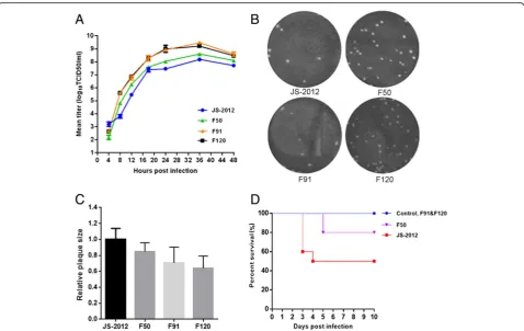
To further characterize the replication properties of the passaged strains, strains from various points of the serial passage were selected and purified for evaluation. One-step growth kinetics in PK-15 cells showed that the PRV strains from higher passages (F50 to F120) exhibited a higher repli-cation rate within the first 12 h post infection and higher viral titers at the later time points analyzed (Fig.1a). In con-trast, the plaques of F50, F91 and F120 grew a little smaller than that of JS-2012 gradually (Fig.1b-c). Furthermore, in a mouse model, lethality of the passaged strains gradually de-creased as passaging inde-creased, with F50 still retaining a certain degree of lethality, while F91 and F120 showed no lethality in mice (Fig.1d).
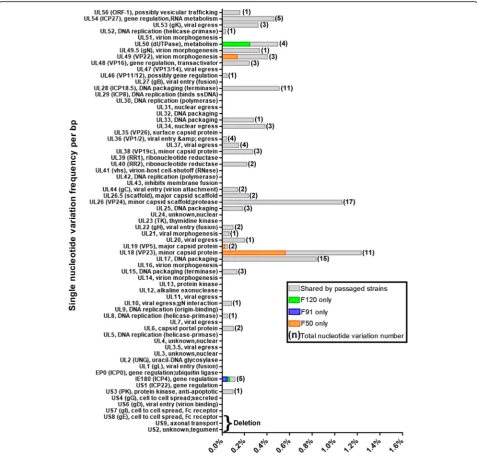
Single nucleotide variations occurring during PRV passage
Table 1Amino acid variations identified in the F50, F91 and F120 passages of PRV JS-2012
Gene Amino acid variations found in F50, F91, and F120 compared to JS-2012a
F50 F91 F120
UL6 P542S, P553S P542S, P553S P542S, P553S
UL8 S542A S542A S542A
UL10 (gM) V256A V256A V256A UL15 G169E, P669D G169E, P669D G169E, P669D UL16 ND 317(+LPRH), S322P, 325–327
(IPE > NKR), 329–330(IN>LK), 331–332(DY>△)
317(+LPRH), S322P, 325–327(IPE > NKR), 329–330(IN>LK), 331–332(DY>△) UL17 G237D, A241S, T249A,
P253△, A255P, D258A, 259(+GGG), N263D, P273L, P374L
G237D, A241S, T249A, P253△, A255P, D258A, 259(+GGG), N263D, P273L, P374L
G237D, A241S, T249A, P253△, A255P, D258A, 259(+GGG), N263D, P273L, P374L UL18 (VP23) H47Y, P60A, H69Q, S79G,
A142V, T270 M
H47D, P60A, S79G, A142G, T270 M
H47D, P60A, S79G, A142G, T270 M UL19 (VP5) I178M, I1315T I178M I178M UL22 (gH) P433L, A618V P433L, A618V P433L, A618V
UL25 L23P L23P L23P
UL26 (VP24) M124 T, I125L, R131L, S132C, Q136R, S137R, R139G, L143 V, T146A, V153A, Q161R
M124 T, I125L, R131L, S132C, Q136R, S137 V, R139G, L143 V, T146A, V153A, Q161R, A455△
M124 T, I125L, R131L, S132C, Q136R, S137R, R139G, L143 V, T146A, V153A, Q161R
UL26.5 ND A216△ ND
UL28 (ICP18.5) A413P, D414E, D425G, 425(+GA), V430G, D432G, A522V
A413P, D414E, D425G, V430G, D432G, A522V
A413P, D414E, D425G, 425(+GA), V430G, D432G, A522V
UL33 P39A P39A P39A
UL34 A177V, T178S A177V, T178S A177V, T178S UL36 (VP1/2) T2832A T2832A T2832A
UL37 E240D, F629 L, G762R E240D, F629 L, G762R E240D, F629 L, G762R UL38 (VP19c) A218V A218V A218V
UL40 (RR2) A176T A176T A176T UL44 (gC) R107H G90D, R107H G90D, R107H
UL46 (VP11/12) ND ND 599–626(PLTRHGSMRTSFRRG VRAAQRFVRRRLS>△), 629–631 (SAE > TTT), A633P, 635–674 (RASGDSASAAAPAAASARGET DHVYQHPRPRTRADDGLYQ>△), Q675G, 678–695(PVIDLTGHRA SRRKSWRV>△)
UL48 (VP16) R39Q,P89A R39Q,P89A R39Q,P89A UL49 (VP22) R168H, N198D R168H R168H UL49.5 (gN) T87A T87A T87A UL50 (dUTPase) S209A S209A G191R,S209A UL53 (gK) P164L, P171L P164L, P171L P164L, P171L
UL54 (ICP27) W20R, C48R, S156F,Q182R W20R, C48R, S156F,Q182R W20R, C48R,S156F,Q182R US8 (gE), US9, US2 Deletion Deletion Deletion
IE180 (ICP4) P468S, G1385R S187 L, P468S, G1385R L76P, P468S, G1385R
NDreferred to no difference
a
more than 10 single nucleotide variation sites (Fig. 2). Consequently, the single nucleotide variations frequency (number of single nucleotide variations/ORF length) of UL28, UL26, UL18, and UL17 were very high at 0.5, 1.0, 1.2, and 0.8%, respectively (Fig.2). No more than 5 vari-ation sites were found in any other gene and, therefore, their incidence frequency were below 0.5% (Fig.2).
Large deletions and small indels are involved in the evolution of PRV during serial passages
One of the most notable variations identified was a large deletion covering the region of US8 (gE), US9, and US2 (Table1). The US8 (gE) deletion is also present in the gen-ome of Bartha and other vaccine strains attenuated by ex-tensive in vitro passage. In addition, we found small indels in the genomes of passaged strains. In-frame indels were present in the ORFs UL17, UL26, UL26.5, and UL28, and some, but not all, were related to number variation of short sequence repeats. For instance, a 3-nt deletion in UL26 of strain F91 was apparently caused by deletion of one repeat of the short sequence repeat“GCC”(Table2).
Frameshift mutations were also identified. Sequencing at both the DNA and mRNA levels confirmed the pres-ence of frameshift mutations in the UL16 and UL46 genes of some passaged strains. Specifically, the muta-tion (an insermuta-tion of a cytosine) in UL16 of F91 and F120 occurred within a homopolymer (CCCCCCC) nu-cleotide stretch, whereas the frameshift mutation in UL46 of F120 was caused by the insertion of a 29-nt
short sequence repeat (GGACGACGACGACGGCGCC CCCTGGGCCC) (Fig. 3a). Specific 3’ RACE of these genes further revealed that the two frameshift mutations had no effect on transcription termination of the two genes; that is, the UL16 and UL46 ORFs of all the strains studied were transcribed and terminated at the same position (Fig. 3b). However, bioinformatic analysis of these ORFs indicated that the mutations resulted in a premature termination codon in the UL46 of F120 (Fig.
3c). Indeed, the predicted 65.9-kDa truncated UL46 product was detected by Western blot analysis of the cell extracts of F120-infected cells (Fig. 3d). On the other hand, as predicted by ORF Finder the frameshift muta-tion in UL16 gene resulted in a delayed terminamuta-tion codon in that of F91 and F120 (Fig. 3c). However, this mutation resulted in only a 0.3-kDa change in protein size, which cannot be detected by western blot analysis (Fig. 3d). In addition, the frameshift mutation in UL46 were associated with relatively higher expression levels of the corresponding product in F120 compared to the other strains, while the mutation in UL16 had no signifi-cant effect on the expression of its protein product in F91 and F120 (Fig.3d).
Discussion
[image:5.595.69.518.101.348.2]Viruses can acquire mutations as they replicate in cell cul-ture, while only mutations favorable to viral replication in vitro are rapidly selected and enriched in the population. This is particularly significant during extensive passage in Table 2Indels identified in passaged strains compared to the parental virus (JS-2012)
a
culture, resulting in viruses with novel acquired genetic al-leles and evolved phenotypes that usually less pathogenic in the natural host but better adapted for replication in cells [19,20].
In our previous study, the PRV JS-2012 strain was serially passaged at high temperature for 120 generations. As the passage number increased, viruses showed a progressive at-tenuation in the piglet infection model, up to the point of completely losing its ability to cause death in piglets by pas-sage 120 [16]. To characterize the evolution and variation of the viral population during passaging, in this study, sev-eral representative strains at various steps of the serial pas-sage were characterized in vitro and in vivo, and the genome of F50, F91, and F120 was sequenced and further analyzed. The growth curves and plaque assays showed that, during passaging the viruses were better adapted for replication and release in the cell line, but seem that the ability to spread were a little weakened (Fig.1). In addition, the lethality of the passaged strains in mice decreased
progressively as the passage number increased, which is consistent with our previous results in piglets (Fig. 1). Therefore, during in vitro passaging the virus gained better replication capacity in cells while becoming largely attenu-ated in animals, probably as a result of genetic variation and rapid evolution of the virus.
To gain insights into the evolution of JS-2012 during continuous in vitro passage, three representative strains (F50, F91, and F120) were sequenced and analyzed. Not-ably, a deletion of large DNA fragment including US8 (gE), US9, and US2 was identified in the genomes of all three strains. Absence of gE expression in the three pas-saged strains was further confirmed by Western blot ana-lysis (Fig.3d). Several studies have shown that deletion of gE from the PRV genome causes attenuation of the virus [21, 22]. Therefore, attenuation of JS-2012 during passa-ging is likely to be related to deletion of the fragment con-taining gE. The presence of large deletions or deletion of some genes is very common in other herpesviruses and in
Fig. 1In vitro and in vivo characteristics of the JS-2012 and its passaged strains.aOne-step growth curves. PK-15 cells were infected at an MOI of 1 with each virus. Cell culture supernatants were harvested at 4, 8, 12, 18, 24, 36, and 48 h post infection. Virus titers at each time point were determined by the TCID50assay in Vero cells. The data represented means ± SD for 2 independent experiments per data point.bPlaques of JS-2012 and the serial
passaged strains generated in infected PK-15 cells cultured at 37 °C for 4 days.cRelative plaque diameters of each virus were calculated and compared to those of PRV JS-2012. Meanwhile the average plaque diameter of PRV JS-2012 were set as 1.dSurvival percentages of mice inoculated with 104 TCID50of each virus per mouse. In JS-2012-infected group, a total of 2 mice were heavily infected and dead at 3 days post inoculation, and another 3
[image:6.595.59.538.86.388.2]large DNA viruses extensively passaged in culture [23], suggesting that this might be a unique feature of large DNA viruses evolving in cell culture. Besides the large de-letion, single nucleotide variation was a dominant vari-ation pattern in many genes, particularly in the UL region of the virus genome. In addition, small in-frame indels identified in genes UL17, UL26, UL26.5, and UL28 con-tributed to evolution of the virus. Some of these indels seemed to have occurred by contraction of specific short sequence repeats, most likely through recombination or polymerase slippage mechanisms [15]. But the remaining
indels were not related to short sequence repeats, suggest-ing other mechanisms might be involved in the generation of indels during the evolution of PRV.
Previously frameshift mutations have been observed in the genome of PRV by serial passaging in rabbit kidney cells [24], and also in the TK gene of Acyclovir-resistant HSV-1 strains and the RL5A, RL13, UL131A and UL130 genes of human cytomegalovirus [25, 26]. And it was confirmed that the frameshift mutations might be rele-vant for the adaptation of PRV in cell culture [24]. Here frameshift mutations also occurred during serial
[image:7.595.59.537.87.544.2]passaging of PRV JS-2012 in Vero cells. The presence of the frameshift mutations was confirmed at both the DNA and mRNA levels for the UL16 and UL46 genes in the corresponding passaged strains. An insertion of a cytosine within one homopolymer nucleotide stretch (CCCCCCC) generated a frameshift in the UL16 gene of F91 and F120, and the frameshift in the UL46 gene of F120 was generated by an insertion of a 29-nt short se-quence repeat (GGACGACGACGACGGCGCCCCCTG GGCCC). Therefore, both mutations were related to al-terations in short sequence repeats. Specific 3’ RACE further revealed that the two frameshift mutations had no effect on transcription termination of any of the two genes, suggesting that the transcriptional stop signal of the viral gene had been conserved. Further analysis sug-gested that the mutations resulted in a premature ter-mination codon in the UL46 of F120, and a delayed termination codon in the UL16 of F91 and F120 (Fig.
3c). Accordingly, the predicted 65.9-kDa truncated UL46 product was detected by Western blot analysis in F120-infected cell extracts (Fig.3d). Moreover, the trun-cated UL46 product expressed by F120 was expressed at higher level than the full-length protein produced by the lower passage viruses. The effect of these changes on virus growth and attenuation deserves further investigation.
In conclusion, extensive passaging of PRV in vitro can result in a great deal of variation, dramatically changing the biological characteristics of the virus. In this study, we showed that during attenuation of PRV by serial pas-saging in Vero cells, dynamic variation patterns includ-ing a large deletion, sinclud-ingle nucleotide variations, small in-frame indels, and frameshifts mutations successively emerged contributing to evolution of the viral popula-tion and enabling the gradual attenuapopula-tion of the virus. In particular, the frameshift mutation in UL46 affected the
[image:8.595.58.537.86.401.2]size and expression level of the corresponding proteins and could have a potentially important effect on the virus characteristics. All these data provide important clues to better understand attenuation and variation in PRV, and further offer insights into the evolution of the virus.
Conclusions
During attenuation of PRV by serial passaging in Vero cells, dynamic variation patterns including a large dele-tion, single nucleotide variations, small in-frame indels, and frameshifts mutations successively emerged contrib-uting to evolution of the viral population and enabling the gradual attenuation of the virus. In particular, the frameshift mutation in UL46 affected the size and ex-pression level of the corresponding proteins and could have a potentially important effect on the virus charac-teristics. All these data provide important clues to better understand attenuation and variation in PRV, and fur-ther offer insights into the evolution of the virus.
Additional files
Additional file 1:Table S1.The raw data of illumina sequencing.
(DOCX 13 kb)
Additional file 2:Table S2.Nucleotide variation in ORFs of F50, F91,
F120 compared to JS-2012. (DOCX 16 kb)
Abbreviations
DMEM:Dulbecco’s modified Eagle’s medium; HTS: High-throughput sequencing; ORF: Open reading frame; PRV: Pseudorabies virus; RACE: Rapid amplification of cDNA ends; TCID50: 50% tissue culture infective dose
Acknowledgements
We thank Editage (https://www.editage.cn/) for editing the text of this manuscript.
Funding
This study was supported by the National Key Research and Development Program of China (2016YFD0500100), China Agriculture Research System (NYCYTX-009), and Shanghai Municipal Agriculture Science and Technology Key Project (2015, 1–7 and 2016, 4–2).
Availability of data and materials
The datasets contained in this study are available from the corresponding author upon request.
Authors’contributions
GT and GL designed this study and CY wrote the manuscript. CY, JW, WT, CL, XC, JX, and HZ performed the relevant experiments and analyses. And TS provided some helpful suggestions for improving the manuscript. All authors read and approved the final manuscript.
Ethics approval
This study was performed according to the Guide for the Care and Use of Laboratory Animals of the Shanghai Veterinary Research Institute, Chinese Academy of Agricultural Sciences, China.
Consent for publication Not applicable.
Competing interests
The authors declare that they have no competing interests.
Publisher’s Note
Springer Nature remains neutral with regard to jurisdictional claims in published maps and institutional affiliations.
Author details
1Shanghai Veterinary Research Institute, Chinese Academy of Agricultural
Sciences, No. 518, Ziyue Road, Minhang District, Shanghai 200241, China.
2
Jiangsu Co-innovation Center for Prevention and Control of Important Animal Infectious Diseases and Zoonoses, Yangzhou 225009, Jiangsu, China.
Received: 10 May 2018 Accepted: 29 November 2018
References
1. Pellett PE, Davison AJ, Eberle R, Ehlers B, Hayward GS, Lacoste V, et al. Herpesvirales. In: King AMQ, Adams MJ, Carstens EB, Lefkowitz EJ, editors. Virus taxonomy: ninth report of the international committee on taxonomy of viruses. London: Elsevier academic press; 2011. p. 99–107.
2. Pejsak ZK, Truszczyni MJ. Truszczynsk: Aujeszky’s disease (pseudorabies). In: Straw BE, Zimmerman JJ, D’Allaire S, Taylor DJ, editors. Diseases of swine. 9th ed. Ames: Blackwell publishing ltd; 2006. p. 419–33.
3. Freuling CM, Müller TF, Mettenleiter TC. Vaccines against pseudorabies virus (PrV). Vet Microbiol. 2017;206:3–9.
4. Mettenleiter TC. Immunobiology of pseudorabies (Aujeszky’s disease). Vet Immunol Immunopathol. 1996;54:221–9.
5. Robbins AK, Ryan JP, Whealy ME, Enquist LW. The gene encoding the gIII envelope protein of pseudorabies virus vaccine strain Bartha contains a mutation affecting protein localization. J Virol. 1989;63:250–8.
6. Müller T, Hahn EC, Tottewitz F, Kramer M, Klupp BG, Mettenleiter TC, et al. Pseudorabies virus in wild swine: a global perspective. Arch Virol. 2011;156:1691.
7. Ye C, Zhang QZ, Tian ZJ, Zheng H, Zhao K, Liu F, et al. Genomic characterization of emergent pseudorabies virus in China reveals marked sequence divergence: evidence for the existence of two major genotypes. Virology. 2015;483:32–43.
8. An TQ, Peng JM, Tian ZJ, Zhao HY, Li N, Liu YM, et al. Pseudorabies virus variant in Bartha-K61-vaccinated pigs, China, 2012. Emerg Infect Dis. 2013;19:1749–55. 9. Luo Y, Li N, Cong X, Wang CH, Du M, Li L, et al. Pathogenicity and genomic
characterization of a pseudorabies virus variant isolated from Bartha-K61-vaccinated swine population in China. Vet Microbiol. 2014;174:107–15. 10. Lomniczi B, Blankenship ML, Ben-Porat T. Deletions in the genomes of pseudorabies virus vaccine strains and existence of four isomers of the genomes. J Virol. 1984;49:970–9.
11. Mettenleiter TC, Lukacs N, Rziha HJ. Pseudorabies virus avirulent strains fail to express a major glycoprotein. J Virol. 1985;56:307–11.
12. Petrovskis EA, Timmins JG, Gierman TM, Post LE. Deletions in vaccine strains of pseudorabies virus and their effect on synthesis of glycoprotein gp63. J Virol. 1986;60:1166–9.
13. Lomniczi B, Watanabe S, Ben-Porat T, Kaplan AS. Genome location and identification of functions defective in the Bartha vaccine strain of pseudorabies virus. J Virol. 1987;61:796–801.
14. Klupp BG, Lomniczi B, Visser N, Fuchs W, Mettenleiter TC. Mutations affecting the UL21 gene contribute to avirulence of pseudorabies virus vaccine strain Bartha. Virology. 1995;212:466–73.
15. Szpara ML, Tafuri YR, Parsons L, Shamim SR, Verstrepen KJ, Legendre M, et al. A wide extent of inter-strain diversity in virulent and vaccine strains of alphaherpesviruses. PLoS Pathog. 2011;7:e1002282.
16. Liang C, Tong W, Zheng H, Liu F, Wu J, Li G, et al. A high-temperature passaging attenuated pseudorabies vaccine protects piglets completely against emerging PRV variant. Res Vet Sci. 2017;112:109–15.
17. Klopfleisch R, Teifke JP, Fuchs W, Kopp M, Klupp BG, Mettenleiter TC. Influence of tegument proteins of pseudorabies virus on neuroinvasion and transneuronal spread in the nervous system of adult mice after intranasal inoculation. J Virol. 2004;78:2956–66.
18. Tamura K, Peterson D, Peterson N, Stecher G, Nei M, Kumar S. MEGA5: molecular evolutionary genetics analysis using maximum likelihood, evolutionary distance, and maximum parsimony methods. Mol Biol Evol. 2011;28:2731–9.
20. Wilkinson GWG, Davison AJ, Tomasec P, Fielding CA, Aicheler R, Murrell I, et al. Human cytomegalovirus: taking the strain. Med Microbiol Immunol. 2015;204:273–84.
21. Tong W, Li GX, Liang C, Liu F, Tian Q, Cao YY, et al. A live, attenuated pseudorabies virus strain JS-2012 deleted for gE/gI protects against both classical and emerging strains. Antivir Res. 2016;130:110–7.
22. Wang CH, Yuan J, Qin HY, Luo Y, Cong X, Li Y, et al. A novel gE-deleted pseudorabies virus (PRV) provides rapid and complete protection from lethal challenge with the PRV variant emerging in Bartha-K61-vaccinated swine population in China. Vaccine. 2014;32:3379–85.
23. Renner DW, Szpara ML. The impacts of genome-wide analyses on our understanding of human herpesvirus diversity and evolution. J Virol. 2017; 92:e00908–17.
24. Grimm KS, Klupp BG, Granzow H, Müller FM, Fuchs W, Mettenleiter TC. Analysis of viral and cellular factors influencing herpesvirus-induced nuclear envelope breakdown. J Virol. 2012;86:6512–21.
25. Sasadeusz JJ, Tufaro F, Safrin S, Schubert K, Hubinette MM, Cheung PK, et al. Homopolymer mutational hot spots mediate herpes simplex virus resistance to acyclovir. J Virol. 1997;71:3872–8.