CSEIT11831235 | Received : 02 Feb 2018 | Accepted : 13 Feb 2018 | January-February-2018 [(3) 1 : 1101-1105 ]
International Journal of Scientific Research in Computer Science, Engineering and Information Technology © 2018 IJSRCSEIT | Volume 3 | Issue 1 | ISSN : 2456-3307
1101
Brain Tumor Detection Using Genetic Algorithm
M. Poornima
*1, C. Kuyin
2, M. Revathy
3*1Assistant Professor, Department of Commerce with Computer Applications, Jayaraj Annapackiam College for
Women (Autonomous), Periyakulam, Tamilnadu, India
2Assistant Professor, Department of Commerce with Computer Applications, Jayaraj Annapackiam College for
Women (Autonomous), Periyakulam, Tamilnadu, India
3Student, III B.Com (CA), Department of Commerce with Computer Applications, Jayaraj Annapackiam
College for Women (Autonomous), Periyakulam, Tamilnadu, India
ABSTRACT
Detection of brain tumor is very common fatality in current scenario of health care society. Image segmentation is used to extract the abnormal tumor portion in brain. Brain tumor is an abnormal mass of tissue in which cells grow and multiply uncontrollably, apparently unregulated by mechanisms that control cells. Several techniques have been developed for detection of tumor in brain. Our main concentration is on the techniques which use image segmentation to detect brain tumor. Tumor classification and segmentation from brain computed tomography image data is an important but time consuming task performed by medical experts.
Keywords : Brain Tumor, GA, Image Segmentation
I.
INTRODUCTION
Segmentation of medical images is challenging due to poor image contrast and artifacts that result in missing or diffuse tissue boundaries. We present a discrete wavelet based genetic algorithm is proposed to detect the MR brain Images. First, MR images are enhanced using discrete wavelet descriptor, and then genetic algorithm is applied to detect the tumor pixels. A genetic algorithm is then used in order to determine the best combination of information extracted by the selected criterion. The present approach uses k-Means unsupervised clustering methods into Genetic Algorithms for guiding this last Evolutionary Algorithm in his search for finding the optimal or sub-optimal data partition (Harris and Buxton 1996, Kim et al 2000, Li Zhijun et al 2006) task that as we know, and requires a non-trivial search because of its intrinsic NP-complete nature. To solve this task, the appropriate genetic coding is also
discussed since this is a key aspect in the implementation. Our purpose is to demonstrate the efficiency of Genetic Algorithms to automatic and unsupervised image segmentation. Some examples in human MRI brain tumor segmentation are presented and overall results discussed.
Medical imaging is performed in various modalities, such as magnetic resonance imaging (MRI), computed tomography (CT), ultrasound etc. Segmentation is typically performed manually by expert physicians as a part of treatment planning and diagnosis. Due to the increasing amount of available data and the complexity of features of interest, it is becoming essential to develop automated segmentation methods to assist and speed-up image- understanding tasks.
A. Discrete Wavelet Transform
enhancement which is done by noise removing and edge enhancement. Wavelet basis function enables DWT based filtering procedures to adapt to spatial variations. Wavelets are functions generated from one single function Ψ by dilations and translations. The basic idea of the wavelet transform is to represent any arbitrary function as a superposition of wavelets. Any such superposition decomposes the given function into different scale levels where each level is further decomposed with a resolution adapted to that level.
The DWT is identical to a hierarchical sub band system where the sub bands are logarithmically spaced in frequency and represent octave-band decomposition. By applying DWT, the image is actually divided i.e., decomposed into four sub bands. These four sub bands arise from separable applications of vertical and horizontal filters. Figure 1.1.(a) shows the sub bands labeled LH1, HL1 and HH1 represent the finest scale wavelet coefficients, i.e., detail images while the sub band LL1 corresponds to coarse level coefficients, i.e., approximation image. Figure 1.1. (b) shows the next coarse level of wavelet coefficients, the sub band LL1 alone is further decomposed and critically sampled.
Wavelet Based Denoising method relies on the fact that noise commonly manifests itself as fine-grained structure in the image and DWT provides a scale based decomposition. Thus, most of the noise tends to be represented by wavelet co-efficient at the finer scales. Discarding these coefficients would result in a natural filtering of the noise on the basis of scale. Because the coefficients at such scales also tend to be the primary carriers of edge information, this method threshold the DWT coefficients to zero if their values are below a threshold. These coefficients are mostly those corresponding to noise. The edge relating
threshold. The Inverse DWT of the thresholded coefficients is the denoised image.
Wavelet thresholding is a signal estimation technique that exploits the capabilities of wavelet transform for signal denoising. It removes noise by killing coefficients that are insignificant relative to some threshold, and turns out to be simple and effective, depends heavily on the choice of a thresholding parameter and the choice of this threshold determines, to a great extent the efficacy of denoising. Threshold Selection plays main role in denoising. A small threshold may yield a result close to the input, but the result may still be noisy. A large threshold on the other hand, produces a signal with a large number of zero coefficients. This leads to a smooth signal. Paying too much attention to smoothness, however, destroys details and in image processing may cause blur and artifacts. Some of thresholding methods are: (i) Hard thresholding, (ii) Soft thresholding, (iii) Semi-soft Thresholding and (iv) Quantile thresholding. In our implementation, soft thresholding method is used to analyze the performance of denoising system for DWT decomposition, since soft thresholding results in better denoising performance than other denoising methods.
Soft thresholding leads to less severe distortion of the object of the interest than other thresholding methods. Several approaches have been suggested for setting the threshold for each band of the wavelet decomposition. A common approach is to compute
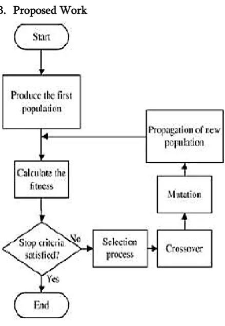
B. Proposed Work
Figure 1.2. Flow for Genetic Algorithm
II.
GENETIC ALGORITHM FOR IMAGE
SEGMENTATION
Genetic algorithms determine the optimal value of a criterion by simulating the evolution of a population until survival of best fitted individuals. The survivors are individuals obtained by crossing-over, mutation and selection of individuals from the previous generation. We think that GA is a good candidate to find out the optimal combination of segmentation results for two main reasons. First one is due to the fact that an evaluation criterion is not very easy to differentiate. GA is an optimization method that does not necessitate to differentiate the fitness function but only to evaluate it. Secondly, if the population is important enough considering the size of the search space we have good guarantees that we will reach the optimal value of fitness.
GA is a special form of local search that models our own understanding of evolution. In essence a number of simultaneous agents (the population) each having an encoded state (the chromosome) perform a random walk (mutations) around the search space, while forming new solutions from combinations of existing solutions (crossover) and, thus adjusting and refocusing the efforts of the search on exceptionally good areas once located. A few important choices are made during any application of genetic algorithms, involving how to encode the population (binary, integer, decimal, etc), how to mutate the population (mutate all genes, some genes, etc), how to select the parents for crossovers (roulette wheel, tournament selection), how to perform those crossovers (uniform, single-point), and finally what fitness function to use for evaluation. Though these choices seem complex, in situations where the energy functional has hundreds or even thousands of dependent variables and parameters these few choices can yield nearly optimal values for all variables and parameters concerned.
Execution of the genetic algorithm is carried out in four steps:
A. Definition of the genotype
Initial population (segmentation results) and computation of the fitness function (evaluation criterion) of each individual.
1) Genotype: the K-means segmentation result of an image S1 is considered as an individual described by the class of each pixel.
2) Initial population: a set of individuals characterized by their genotypes. It is composed of the segmentation results to combine.
B. Selection of individuals
The selection process selects chromosomes from the mating pool directed by the survival of the fittest concept of natural genetic systems. In the proportional selection strategy adopted in this article, a chromosome is assigned a number of copies, which is proportional to its fitness in the population, which then goes into the mating pool for further genetic operations. Roulette wheel selection is one common technique that implements the proportional selection strategy.
C. Mutation and Cross-over of individuals
Individual mutation: individual’s genes are modified for better adaptation in the environment. We use the non-uniform mutation process which randomly selects one chromosome xi, and sets it as equal to a non-uniform random number:
xi+( bi - xi)f(G) if r1 <0.5
xi -( xi + ai)f(G) if r1>=0.5 (1.6) f(G)=(r2(1-G/Gmax))b (1.7)
r1, r2 : numbers in the interval [0, 1]
ai, bi : lower and upper bound of chromosome xi G : the current generation
Gmax : the maximum number of generations b : a shape parameter
Crossover is a probabilistic process that exchanges information between two parent chromosomes for generating.
Two child chromosomes we use the arithmetic crossover which produces two complementary linear combinations of the parents
X′ = aX + (1 − a)Y
Y ′ = (1 − a)X + aY (1.8) where
X, Y : genotype of parents a : a number in the interval [0, 1]
D. Evaluation of individual /Termination criterion
This criterion allows stopping the evolution of the population. We can consider the stability of the fitness function f=1/M of the evaluation criterion of the population or set a maximal number of iterations (Gmax =1000: the maximum number of generations).
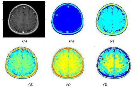
The segmentation results of the wavelet based genetic algorithm are shown in Figure 1.4. Figure 1.4.a-d shows segmented images using the K-means algorithm with mean and variance as attributes with different numbers of clusters (NC) 2, 3, 6, 9, which constitutes the initial population for the GA.
Figure 1.4 (a) Enhanced T2 weighted MRI brain tumor image, (b-e) is segmented image using wavelet based GA algorithm with NC=2,3,6,9 (Number of cluster) and (f) is a resultant segmented image with four cluster (white matter, gray matter, CSF and tumor(red)).
III.
CONCLUSION
achieved SNR value from 20 to 44 and segmentation accuracy from 82 percent to 97 percent of detected tumor pixels based on ground truth.
IV.
REFERENCES
[1]. Minglun Gong and Yee-Hong Yang (2001), 'Genetic-based multiresolution color image segmentation', Vision Interface, pp. 141-148. [2]. Nordin P. and Banzhaf W. (1996), 'Programmatic
compression of images and sound', In Genetic Programming 1996,Proceedings of the First Annual Conference, Koza J. R. Editors, MIT Press, pp. 345-350.
[3]. Ou Y., Cheng W. and Han Ferng-ching (2004), 'Based on genetic algorithm fuzzy c- means clustering algorithm', Journal of Chongqing University, Vol. 27 No. 6, pp. 89 - 92.
[4]. Philippe Andrey (1999), 'Genetic algorithms