The neural induction process; its morphogenetic aspects
PIETER D. NIEUWKOOP
Hubrecht Laboratory, Netherlands Institute for Developmental Biology, Utrecht, The Netherlands
Edited by:
RICHARD GORDON
1* and NATALIE K. BJÖRKLUND
21Department of Radiology and 2Department of Human Genetics, University of Manitoba, Winnipeg, Canada
ABSTRACT This posthumous review of early embryonic inductions concludes: 1) the amphibian egg has only two distinct components, animal and vegetal. Interactions at their mutual boundary forms meso-endoderm. This is “meso-endoderm induction”, not just “mesoderm induction”. 2) The dorso-ventral polarity of the yolk mass implies a dorsally situated inducing centre. 3) Accumulation of cells into one, two, three or many cell masses [problastopores] along the circumference of the meso-endoderm results in as many axes, implying a self-organizing capacity of meso-endoderm. 4) Induction of the meso-endoderm is slow, spreading cell to cell through the animal moiety from the boundary of the vegetal yolk mass towards the animal pole. 5) Interaction between mesoderm and ectoderm is a separate step leading to cranio-caudal differentiation of the archenteron roof. 6) The initial invaginating endoderm and mesoderm, representing the future pharynx endoderm and prechordal plate mesoderm, first contacts the most posterior presumptive neurectoderm after having passed the still uninvaginated trunk mesoderm. At that moment an antero-posterior level neural induction actually starts. 7) The ectoderm contraction wave coincides spatially and temporally with the induced neural plate. 8) Two successive homoiogenetic waves of inductive activity pass through the presumptive neurectoderm in the anterior direction, the first one, “activation”, giving rise to neural differentiation and ultimately forebrain, the second one, “transformation”, to more caudal CNS structures. These are separate, successive steps in CNS regional induction. 9) The midbrain represents a secondary formation in the neural plate. 10) The observed changes in morphogenesis may depend upon separate, successive binary decisions via [cell and] nuclear state splitters [involving differentiation waves].
KEY WORDS:
differentiation, ectoderm, mesoderm, endoderm, polarity
0214-6282/99/$15.00
© UBC Press Printed in Spain
www.ehu.es/ijdb
*Address for reprints: Department of Radiology, University of Manitoba, Health Sciences Centre, 820 Sherbrook Street, Winnipeg, MB R3A 1R9 Canada. FAX: (204) 787-2080. e-mail: [email protected]
Editors’ note: This manuscript was found in January, 1999, in Winnipeg, on a diskette dated 1992, probably left by the late Pieter D. Nieuwkoop during his first visit to us that year. (Another, identical diskette was subsequently found in Utrecht.) We cannot recall that he ever mentioned the manuscript either then, nor on his second visit in 1995. However, much of the manuscript outlines specific ideas he discussed during our preparation of Björklund and Gordon (1994), particularly those involved in early mesoderm induction, forcing N.K.B. back to the lab to make more observations, and thereby vastly improving that paper. All our additions to the text are placed in square brackets: []. None of the original text was deleted. As none of the figures or figure captions for this manuscript could be found, we have copied seemingly corresponding figures from existing publications. A long list of references was on the same diskette, but it only partially overlapped the text citations. Thus we had to make educated guesses for some of the citations. There was no abstract, and so the abstract is by us, extracted from the text and condensed.
Abbreviations used in this paper: CAMP, cyclic adenine monophosphate; CNS, central nervous system; FGF, fetal growth factor; IMP, intramembrane particle; PKC, protein kinase C; TGFbeta, transforming growth factor beta.
The morphogenetic interactions preceding the neural
induction process
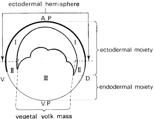
From isolation experiments of successive animal-vegetal zones of the urodele blastula it could be concluded that the upper, animal half of the blastula forms only atypical ectoderm and the vegetal yolk mass only atypical endoderm, whereas the intermediate, subequatorial zone forms ecto-, meso- and endodermal struc-tures. Recombination of the animal cap and the vegetal yolk mass leads again to the development of a complete embryo with all
This interaction of the two moieties is restricted to the peripheral region of the blastula, the blastocoelic cavity preventing contact between the moieties in the central region of the blastula (see Fig. 1).
The urodele as well as anuran fertilised egg –and likewise the full grown oocyte– shows a visible subdivision in a pigmented animal half and an unpigmented or lightly pigmented vegetal half. The animal half is relatively rich in cytoplasmic organelles and relatively poor in yolk granules, whereas the vegetal half is rich in yolk granules and relatively poor in cytoplasm. Studying the membrane properties of the amphibian egg with freeze-fracture techniques, Bluemink and Tertoolen (1978) found a statistically significant difference in IMP [intramembrane particle] sizes be-tween animal and vegetal halves of the fertilised egg with more small IMP’s in the animal half. Dictus et al. (1984), studying the membrane fluidity of the fertilised egg, found that the animal half has a rigid membrane, whereas the vegetal half has a much more fluid membrane with a higher particle mobility, the two different moieties showing a sharp mutual boundary at the equator of the egg. The amphibian egg therefore starts development from a minimum of the spatial heterogeneity, consisting essentially of only two different components, an animal and a vegetal one. These differ both in cytoplasmic composition and in outer membrane properties, thus confirming the conclusion taken from the above-cited isolation and recombination experiments.
As already mentioned, recombination of animal caps and veg-etal yolk masses leads to normal embryo formation due to the induction of the meso-endoderm in the animal moiety under an inductive influence exerted by the vegetal yolk mass (Nieuwkoop, 1969a). This conclusion is further corroborated by quantitative measurements of the recombinate blastula components, using the anuran Xenopus laevis (Sudarwati and Nieuwkoop, 1971) as well as by a qualitative analysis of the resulting larvae, using different urodele species with different embryonic pigmentation and yolk granule size (Nieuwkoop and Ubbels, 1972). The latter
experi-ments were supplemented with recombinates of 3H-thymidine
marked and unmarked ectodermal caps and vegetal yolk masses. These recombination experiments show that not only the entire mesoderm, but also the dorsal pharyngeal endoderm and the most dorsal intestinal endoderm are being induced in the totipotent animal “ectodermal” moiety by an inducing influence exerted by the vegetal yolk mass (Nieuwkoop and Ubbels, 1972). In the urodele gastrula these presumptive endodermal components are localized outside the blastoporal groove formed at the periphery of the vegetal yolk mass (see Fig. 2). It is therefore more correct to speak of endoderm induction” than the widely used term “meso-derm induction”.
Recombination experiments with animal caps and vegetal yolk masses, recombined in different mutual orientation (d/d [dorsal/ dorsal], d/l [dorsal/lateral] and d/v [dorsal/ventral]), demonstrated that the d/v polarity of the resulting larva depends upon an invisible d/v polarity of the vegetal yolk mass and not, or at least no longer, upon the apparent visible polarity of the animal moiety, which often shows a lighter pigmentation at its dorsal side due to the location of the grey crescent (Nieuwkoop, 1969b). Without going into detail about the origin of the d/v polarisation in the amphibian egg, it is evident that the grey crescent, which forms opposite the sperm-entrance point in the monosperm anuran egg, represents the visible manifestation of the d/v polarisation of the animal half of the fertilised amphibian egg (see among others Ubbels et al. [1979, 1983]). The transfer of the d/v polarity from the animal to the vegetal half of the egg is still unknown.
Fig. 1. Diagram of the subdivision of the amphibian blastula into two functionally different moieties and three successive regions, viz. animal, ectodermal hemisphere (I), subequatorial zone (II) and veg-etal yolk mass (III). A.P., animal pole; D., dorsal; V., ventral; V.P., vegetal pole.” (From Fig. 2.1, p. 11 in Nieuwkoop and Sutasurya, 1979, with permission of Cambridge University Press.)
Using separate dorsal, lateral and ventral portions of the vegetal yolk mass as meso-endoderm inducer acting upon animal ectoder-mal caps, it could be demonstrated that the d/v polarity of the yolk mass is chiefly due to the presence of a dorsally situated inducing centre. Whereas lateral and ventral yolk mass portions induce only ventral meso-endodermal structures, the dorsal yolk mass induces chiefly axial meso-endodermal structures in competent late blastula/ early gastrula ectoderm (Boterenbrood and Nieuwkoop, 1973).
I am very skeptical about the possible identification of natural inducers, since fully competent “ectoderm” needs only a trigger to switch from its primary, epidermal, pathway into that for meso-endodermal or for neural development. The specificity of the reaction depends for the far greater part on the specific properties of the reacting ectoderm, its competence, than on the specificity of the inducing factor. We know from 50 years of research that many, chemically quite different and even foreign substances can trigger the same induction process. There is, however, reason to assume that the natural inducer for the meso-endoderm pathway differs from that of the neural pathway.
At present, a large number of institutes are working on the mesodermal inducer. It may either be a single graded factor, or a number of different, possibly related, factors (see Melton 1991, [for a] review). They are at present sought among the FGF and the TGFβ growth factor families. It is evident that signal transduction pathways play an important role in the passage of the signal from the cell exterior to the cell interior, thus traversing the cell mem-brane for [before] ultimately reaching the nucleus.
There is a general feature of the induced meso-endoderm which is of great importance for embryonic axial system formation, viz. the tendency of mesodermal cells to accumulate –in normal development at the dorsal side by means of dorsal convergence during gastrulation– and to organise themselves into a complete axial system with notochord and somites as well as prechordal mesoderm by means of self-organisation. This could be demon-strated in des- [disaggregation] and reaggregation experiments performed by Nieuwkoop in the seventies, but only recently pub-lished in extenso (Nieuwkoop, 1992). He used separate cell suspensions of animal caps and vegetal yolk mass obtained by treatment with Ca-free culture medium. The two cell suspensions, after being separately thoroughly stirred, were subsequently trans-ferred to a semi-spherical depression in the agar bottom of an operation dish containing a Ca-enriched culture medium. This transfer was done in such a way, so that the endodermal cells were at the bottom and the ectodermal cells on top, thus creating a “normal” animal-vegetal polarity. The two cell suspensions re-aggregated and formed a normal-looking blastula, but without d/v polarity. Nevertheless, the blastulae subsequently started to gas-trulate, ultimately forming a properly organised embryo. These embryos either showed a single dorsal axis system, like in the normal embryo, or formed double, triple or even multiple axis systems. This presumably occurs by accumulation of the majority of the initially evenly induced meso-endodermal cells into either a single cell mass, into two cell masses usually opposing each other or into triple or multiple cell masses forming along the circumfer-ence of the induced meso-endoderm (see Fig. 3). This behaviour can only be explained by assuming an intrinsic self-organising capacity of the induced meso-endoderm. By increasing the inten-sity of the meso-endoderm inducing capacity of the yolk mass – using dorsal instead of whole yolk mass and two instead of a single
dorsal yolk mass– a more complex nature of the resulting embryos is obtained, viz. in the form of more complete axial structures and of more complex axial systems (double, triple or multiple ones) (Nieuwkoop, 1992).
Induction of the meso-endoderm is a slow process, spreading from cell to cell through the animal moiety from the boundary of the vegetal yolk mass in the direction of the animal pole. Induction starts around the 64 cell stage (Nakamura and Takasaki, 1970) and extends till about the mid-gastrula stage when the mesodermal competence of the reacting “ectoderm” falls off rapidly. Kanéda and Hama (1979), studying the origin of the trunk organizer in the posterior half of the marginal zone, found that at the early gastrula stage the meso-endodermalisation of the animal, “ectodermal” moiety has only progressed over the future anterior half of the marginal zone. (The marginal zone represents the ring-shaped zone of the blastula/early gastrula which gives rise to all the future meso-endodermal structures of the embryo: see Vogt, 1929.) This means that at the early gastrula stage only the anterior half of the future archenteron roof (see Nieuwkoop, 1973, 1974 [the latter citation could not be identified]) shows meso-endodermal differen-tiation tendencies upon isolation, whereas the future posterior half of the archenteron roof, the later trunk organizer, does not yet differentiate into meso-endoderm upon isolation.
The cranio-caudal differentiation of the invaginating dorsal, axial meso-endoderm evidently depends upon a further inductive interaction. Whereas the fully invaginated archenteron roof differ-entiates into the anterior prechordal plate meso-endoderm and the more posterior chorda-mesoderm of trunk and tail, the correspond-ing zones of the archenteron roof do not show the same differen-tiation tendencies before and after invagination. At the early gastrula stage the presumptive prechordal plate meso-endoderm, Fig. 3. Formation of the mesoderm in the wake of the mesoendoderm expansion wave and appearance of the problastopores. Stage 9.5+ (24 h at 20oC). As the animal cap expansion wave enters the mesoendoderm
located directly above the dorsal blastoporal groove, does not differentiate into prechordal plate meso-endoderm upon isolation, but forms notochord and somites, presenting the character of a trunk organizer. Directly after passing the dorsal blastoporal lip the same material begins to form prechordal plate meso-endoderm besides chorda-mesoderm. This transformation process is com-pleted at about stage 10 3/4 when topographical contact is estab-lished between the anterior archenteron roof and the overlying ectoderm, the future trunk organizer, which has not yet developed axial differentiation tendencies at that stage. This interaction causes both a change in differentiation tendencies of the most anterior portion of the archenteron roof, viz. from trunk into head organizer, as well as the appearance of chordamesodermal differ-entiation tendencies in the future trunk organizer (Hoessels, 1957; Kanéda and Hama, 1979).
These changes are due to a perpendicular interaction between the anterior archenteron roof and the overlying posterior uninduced “ectoderm” (Kanéda, 1980) as well as to a further tangential spreading of the meso-endoderm inducing action around the dorsal blastoporal lip (Kanéda, 1981) (see Fig. 3). This interaction between the meso- and ectoderm forms a separate step in the formation of the meso-endoderm leading to the characteristic cranio-caudal differentiation of the mesodermal archenteron roof as necessary preparation for the subsequent regional neural induction process.
The induced meso-endoderm comprises the endodermal pha-ryngeal and dorsal intestinal anlagen and the entire dorsal, lateral and ventral mesodermal anlagen. The dorsal pharyngeal endo-derm as well as the dorsalmost intestinal endoendo-derm directly bor-ders the vegetal yolk mass endoderm, while the dorsal, lateral and ventral mesoderm is formed at some distance from the yolk mass endoderm. Hama et al. (1985) actually consider the pharyngeal endoderm as the initiator of the dorsal axial mesoderm, Spemann’s well known amphibian “organizer” or “organization centre”. (see Fig. 2).[For further comments by Nieuwkoop on Hama etal. (1985) in personal correspondence, see p. 90-91 in Gordon (1999).]
The gastrulation process starts with blastoporal groove forma-tion, due to flask cell formation at the periphery of the yolk mass endoderm. This flask cell formation leads to an inward extension of the peripheral yolk mass cells and a reduction of its outer surface of the yolk mass, enabling the epiboly of the animal cap to proceed further towards the blastopore. In the double-layered anuran gastrula, with Xenopus laevis as the most extreme example, the rolling-in of the fully internally situated mesoderm –internal mar-ginal zone– actually starts independent of the invagination of the archenteron, for which flask cell formation in the external layer is an essential prerequisite. In the single-layered urodele gastrula the induced endo- and mesoderm invaginates together around the blastoporal lip, segregating only later into the endodermal and mesodermal components during endodermal tube formation in the trunk and during the ultimate segregation of the prechordal meso-and endoderm (Vogt, 1929). The rolling-in of the mesoderm, responsible for the formation of the mesodermal archenteron roof, which is going to underlie the future neural plate, is an active process in which the anterior edge of the mesodermal sheet plays a leading role, pulling the mesoderm towards the animal pole along the inner surface of the ectoderm (Nakatsuji 1984; Nakatsuji and Johnson, 1984).
When the entire yolk mass endoderm is isolated at successive stages of development its behaviour is different. Whereas the yolk mass endoderm of stage 7 2/3 does not show any flask cell formation at its periphery, flask cells begin to form at the dorsal side of the isolated yolk mass endoderm taken from stage 8. The older the isolated yolk mass the more extensive flask cell formation at the dorsal side and the further the process spreads along the lateral and ventral periphery of the yolk mass endoderm. The full autono-mous programming of the yolk mass around its entire periphery is only reached at a late blastula stage (Doucet-de Bruïne, 1973, see also Nieuwkoop, 1973). This behaviour of the yolk mass endoderm strongly suggests a flask cell inducing action by the adjacent marginal zone meso-endoderm. Although [the] dorsal marginal zone favours flask cell formation in the yolk mass endoderm, a direct correlation between marginal zone formation and flask cell formation could not be demonstrated (Doucet-de Bruïne, 1973), so that a more autonomous process of flask cell formation was suggested. This outcome may, however, be partially due to opera-tional difficulties in these recombination experiments.
Fig. 4. Diagrammatic sketch of cross-section through normal neural plate of host and longitudinal section through elongated implant, showing equivalent extension of activating principle in a horizontal direction through neural plate of host and in a vertical direction through implant. Arrows indicate directions of morphogenetic move-ments which lead to the closure of the neural tube and to the later shape of the activated part of the implant. (From Fig. 13, p. 21 in Nieuwkoop et al., 1952a, with permission of John Wiley and Sons.)
The neural induction process and the regional
forma-tion of the CNS [central nervous system]
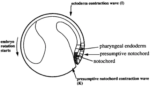
After formation of the dorsal blastoporal lip and the beginning invagination of the dorsal meso-endoderm a constriction [surface contraction] wave apparently appears in the overlying ectoderm, indicating the initiation and spatial extension of the neural induction process (Brodland et al., 1991 [an early draft of Brodland et al., 1994]). The [ectoderm contraction] wave front initially [actually, 4 h after launching: Fig. 17 in Gordon, Björklund and Nieuwkoop, 1994] covers a semi-circular area at some distance in front of the dorsal blastopore. The wave moves outwards and forwards in the direction of the animal pole and becomes ellipsoid in shape. The wave front finally disappears near the anterior boundary of the future neural plate. The first visible feature of the future neural anlage appears around stage 12 by the formation of a dorso-median groove, extending from the neighbourhood of the blasto-pore to the centre of the future neural plate anlage. The plate itself is subsequently outlined by a flattening of the antero-dorsal surface in the nearly spherical embryo. The median groove corresponds with the position of the underlying notochordal anlage and repre-sents the area of firm attachment of archenteron roof and overlying neurectoderm (stage 13). Slightly later, the outer boundary of the neural plate becomes visible by the elevation of its outer boundary, formed by the neural folds (stage 14). The neural anlage gradually changes its form from an initial horse-shoe-shape into a key-hole-shape by means of the considerable lengthening of its posterior half (Burnside and Jacobson, 1968). The anterior portion of the neural plate shows its maximal width at stage 15, at which stage the neural folds begin to protrude markedly. Subsequently, the CNS anlage steadily shrinks in medio-lateral direction when the lateral neural folds begin to approach each other while the anterior neural fold curves caudalwards. Simultaneously, the caudal half of the neural anlage forms a deep groove flanked with protruding neural folds (stage 16/17). The lateral folds first make contact with each other in the future hindbrain region. The closure of the neural tube
subsequently spreads both in anterior and in posterior direction. The anterior neural pore closes finally over the caudal boundary of the forebrain. The complete closure of the neural tube is only achieved at an early tail bud stage around stage 20/21 when the epidermis of L [left] and R [right] sides of the neural anlage have fused.
For a proper understanding of the neural induction process in the amphibian embryo it is essential to realise that an interaction occurs between the invaginating and upwards moving archenteron roof and the simultaneously downwards moving epibolic spreading of the ectodermal moiety during the gastrulation process. (M. Jacobson, who suggested a clonal development of the CNS from predetermined cells (Jacobson, 1982, 1984; Jacobson and Hirose, 1981), questioning the existence of a neural induction process, had, however, to recant his statements (Jacobson, M., ...).[Nieuwkoop did not specify the citation here. Our reading of Jacobson, (1985) and Sheard and Jacobson, (1987, 1990) does not suggest “recanting”.]) The first invaginating endo- and meso-derm, representing the future pharynx endoderm and the pre-chordal plate mesoderm, comes first into contact with the most posterior presumptive neurectoderm after having passed the still uninvaginated trunk mesoderm. At that moment and at that antero-posterior level the neural induction process actually starts. The archenteron roof subsequently shifts anteriorwards and comes into contact with ever more anterior presumptive neurectoderm. In the urodeles the two oppositely directed movements finally come to a standstill when the prechordal mesoderm reaches the original animal pole region, so that the future antero-posterior axis spatially coincides with the original animal-vegetal axis. (In the anuran embryo the invagination process stops at about 30o from the animal
pole, so that in the anurans the antero-posterior axis of the embryo does not coincide with the original animal-vegetal axis of the egg.) As soon as this definitive position is reached, the two layers firmly attach to each other in the dorsal midline forming the functional Fig. 6. Cross-section through hind-brain region of Ambystoma
mexicanum 20, tail bud stage, showing unilateral folding up of proximal portion of implant. Enlargement ±67X.” (From Fig. 11, p. 18 in Nieuwkoop et al., 1952a, with permission of John Wiley and Sons.)
mesodermal prechordal plate/notochordal and neural notoplate complex.
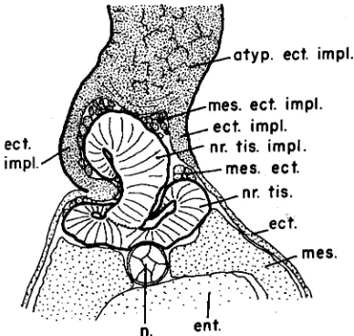
Eyal-Giladi (1954) isolated consecutive cranio-caudal regions of the presumptive neurectoderm at successive stages of develop-ment and reared them in a neutral environdevelop-ment (without mesoder-mal substrate) of host embryos. She could show that two succes-sive waves of inductive activity pass through the presumptive neurectoderm in anterior direction, the first one, called “activation” giving rise to neural differentiation and ultimately leading to fore-brain formation, and the second one, called “transformation”, to the partial transformation of these forebrain differentiation tendencies into those for more caudal structures of the CNS (Nieuwkoop et al., 1952 [three articles: Nieuwkoop, 1952; Nieuwkoop et al., 1952a,b]). These experiments formed an excellent confirmation of the conclusions made from earlier fold-implantation experiments by Nieuwkoop et al. (1952) [Nieuwkoop, 1952; Nieuwkoop et al., 1952a,b], in which longitudinal strips of double-layered competent gastrula ectoderm were attached at one end to the midline of a host neural plate or to the corresponding area of the presumptive neurectoderm at an earlier developmental stage. This procedure leads to neuralisation of the attached ectodermal fold over a length (see Figs. 4,5) equivalent with half the width of the host neural plate at the level of implantation.
These experiments demonstrate that neural induction is a homoiogenetic induction process, spreading from cell to cell in proximo-distal direction in the attached fold and, very likely in a similar manner in medio-lateral and anterior directions in the normal neural plate. Implantation of folds in the forebrain region of
the host neural plate only gives rise to forebrain structures in the fold. Implantation in the hind brain region, however, leads to complex brain formations with proximally situated hind-brain and more distally fore-brain structures (see Figs. 6,7). Implantation in the anterior spinal cord region of the host neural plate leads to smaller neural structures with proximally spinal cord and more distally hind-brain character, while implantation in the posterior spinal cord region causes only the formation of small spinal cord structures in the fold.
These results can only be explained by assuming the presence of two successive inductive actions, viz. an initial one, called activation, evoking neural differentiation tendencies, and ultimately leading to fore-brain formation, and a secondary inductive action, called transformation, and being superimposed upon the first one, transforming the initially evoked fore-brain differentiation tenden-cies into those for more posterior structures of the CNS. The two inductive actions show a different cranio-caudal distribution in the archenteron roof and subsequently in the overlying neural plate: viz. a strong activating activity is found in the prechordal mesoderm and in the anteriormost chordamesoderm, reducing in caudal direction, whereas a high transforming activity is present in the most posterior portion of the archenteron roof, tapering off in anterior direction to a nearly complete absence in the prechordal plate mesoderm. (Sala, 1955) (see Fig. 8).
The two inductive actions correspond with different periods of competence of the reacting and neurectoderm. The ecto-derm is competent for the activating influence from an early blastula (stage 8) (Chuang, 1955) up to the mid-gastrula stage (stage 11/12) when the archenteron roof has reached its definitive position near the original animal pole (see Fig. 9). In the anuran amphibia where the invaginating archenteron roof stops at about 30o short of the animal pole, the ventral ectoderm has moreover a
lower neural competence than the dorsal ectoderm, due a.o. [account of] to the presence of different isozymes active in signal transduction (Otte et al., 1990). Competence for the transforming Fig. 8. Diagrammatic sketch of primary activation and subsequent
counteraction by transforming mesodermal influences in folds of competent ectoderm attached to prospective prosencephalic, rhombencephalic and spino-caudal regions of a host embryo. Subdi-vision of folds into a proximal neural (continuous line), an intermediate mesectodermal (interrupted line and slightly hatched) and a distal ectoder-mal region (pointed line) by the penetration of a primary activating principle which liberates prosencephalic differentiation tendencies in the activated regions. Subsequent counteraction between prosencephalic differentia-tion tendencies (tapering continuous lines) and the transforming influences from the mesodermal substrate which penetrates into the proximal portion of the attached folds (indicated by small crosses). Dorsal mesodermal substrate: darkly hatched, prechordal substrate: white. (From Fig. 1, p. 88 in Nieuwkoop, 1952, with permission of John Wiley and Sons.)
Fig. 9. The notochord starts forming in the wake of the presumptive notochord contraction wave and the center of gravity starts shifting. Stage 11 (38 h at 20oC)....Sagittal section. The presumptive notochord is
action arises in the activated neurectoderm at about stage 11/12 and lasts till the late neural plate stage (stage 15/16) (Nieuwkoop and Albers, 1990). The two inductive actions therefore form separate, successive steps in the regional induction process of the CNS.
The nature of the natural neural inducer is even more question-able than that of the mesodermal inducer, since very atypical stimuli, like e.g. high or low pH or Ca-free culture medium, are able to release neural differentiation in the highly competent gastrula ectoderm. This makes it much more difficult to analyse the nature of the natural inducing signal.
It is evident from Otte et al’s (1988, 1989, 1990) work, that both the PKC and the cAMP signal transduction pathways play an important role in the transfer of the signal over the cell membrane. Otte and Moon (1992b) demonstrated that the different degrees of competence of dorsal and ventral ectoderm in the anuran, Xeno-pus laevis, gastrula is based upon different PKC isozymes. Whereas the PKCβ is uniformly distributed, the PKCα is predominantly localised in the more competent dorsal ectoderm. The level of PKCα seems to be crucial for the level of neural competence, while the PKCβ is involved in mediating neural induction as such.
Very recent work, of Otte and Moon (1992a), showed that injection of Xwnt-8 RNA in ventral vegetal blastomeres, of the 32-cell embryo, did not only lead to the formation of a second meso-endoderm inducting centre in the vegetal blastomeres, giving rise to a second axial mesoderm formation, but also to the elevation of the neural competence of the ventral ectoderm to the level of the dorsal ectoderm. They concluded that the presence of dorsal mesoderm is a prerequisite for establishing the differences in neural competence between dorsal and ventral ectoderm. This means that another fast-spreading signal must have first spread in front of the signal for dorsal meso-endoderm induction through the animal ectoderm of the blastula. The question now arises why no differences in competence between dorsal and ventral gastrula ectoderm have ever been found in urodele ectoderm, in which meso-endoderm and neural induction have been so extensively studied.
Activated neurectoderm, formed under the natural inductive influence of the prechordal mesoderm or obtained by artificial exposure of its inner surface to the saline solution (Holtfreter, 1944, in Ambystoma and Nieuwkoop, 1963, in Rana pipiens and Am-bystoma) develops autonomously into a well-organised forebrain with telencephalon and olfactory placodes and diencephalon with eye structures. After des- [disaggregation] and reaggregation of presumptive forebrain and adjacent ectoderm the resulting re-aggregate gives again rise to forebrain formations, the complexity of which depends upon the state of sorting-out of the ecto- and neurodermal cells (Townes and Holtfreter, 1955) at the beginning of forebrain segregation by means of self-organisation.
The transformed part of the neural plate compromises the mid-brain, hindbrain and spinal cord as well as the tail somite region. Hindbrain and spinal cord, both underlain by notochord and somites, form nearly identical segmental structures, whereas the midbrain differs markedly from the forebrain as well as from the hindbrain in spatial organisation. It turns out that transformation actually directly leads to hindbrain/spinal cord formation, while the midbrain repre-sents a secondary formation due to the spatial overlap of the forebrain and hindbrain/spinal cord domains in the neural plate (Nieuwkoop, 1991).
The somites of the tail develop out of the most posterior portion of the neural plate (Bijtel, 1931, 1936, 1958; Nakamura, 1942,
1952) under a special inductive influence exerted by the underly-ing section of the archenteron roof (Spofford, 1945, 1948, 1959 [probably 1953]). Niazi (1969), when working at the Hubrecht Laboratory, could show that the presumptive tail somite material still differentiates into spinal cord when isolated at stage 13/14, but into tail somites when isolated at stage 16/17 with a partial transformation around stage 15. This means that the presumptive tail somite material goes first through the neural activation pro-cess and subsequently through a gradual, possibly stepwise transformation process via rhombencephalon, spinal cord and finally into tail somites. It must be assumed that a separate, secondary competence for somitic mesoderm formation arises in the transforming neurectoderm, since the primary mesodermal competence is already abolished at an early to mid-gastrula stage (see Fig. 10).
induction process. This conclusion is further supported by the demonstration of the markedly different inductive capacity of median and more lateral regions of the archenteron roof, the inductive capacity falling off rapidly from the dorsal midline in [the] lateral direction (Leussink, 1970). Albers (1987) could recently demonstrate that the formation of the outer boundary of the neural plate does not depend upon a presumed threshold value of a medio-lateral diffusion gradient as suggested by many authors (a.o. Wolpert, 1985), but upon the loss of competence of the reacting ectoderm during the slow lateral homoiogenetic propaga-tion of the inductive acpropaga-tion. Loss of competence for transformapropaga-tion plays likewise a dominating role in the regional segregation of the hindbrain/spinal cord region of the CNS (Nieuwkoop and Albers, 1990) and probably also in the segregation of the neural crest along the periphery of the mid/hindbrain spinal cord region of the CNS (Nieuwkoop, 1992).
The development of special structures in the successive brain segments depends upon local inductive interactions between the different regions of the neural plate and the corresponding regions of the underlying archenteron roof. E.g., the segregation of the telencephalon into separate left and right hemispheres as well as the bilateral development of the eyes in the diencephalon are due to a suppressive action of the median prechordal plate mesoderm (Adelman, 1932, 1936; Boterenbrood, 1962). The formation in the infundibulum in the ventral diencephalon is likewise due to an interaction with the underlying substrate. The formation of the complex eye depends upon a series of inductive interactions between the evaginating primary eye vesicle and the overlying epidermis (lens induction and secondary eye cap formation) and surrounding head mesenchyme (tapetum versus retina differentia-tion) (Hoperskaya and Golubeva, 1982). The formation of the regional segments of the hindbrain and spinal cord are likewise based upon regional interactions between the neurectoderm and the underlying archenteron roof, in particular by the segmented somites and migrating neural crest.
The cephalic placodes –the olfactory placodes bordering the telencephalon, the lens placode of the eye anlage, the ear placode bordering the hindbrain as well as the placodes of the cephalic ganglia and lateral line placodes– develop out of the so-called placodal ectoderm which directly surrounds the neural plate an-lage. Placodal ectoderm formation actually represents a separate step in the development of the competence of the ectoderm for respectively neural, placodal and epidermal differentiation (Nieuwkoop, 1963). The various placodes develop locally under the simultaneous or sequential influences of adjacent meso-, endo- and neurodermal anlagen (Jacobson, 1963a,b,c).
Although a whole scala of influences seem to be at work at particular stages of development in the organisation of the CNS, the observed changes in morphogenesis may depend upon sepa-rate, successive binary decisions, as assumed by Gordon in his [cell and] nuclear state splitter hypothesis [Gordon and Brodland, 1987; Björklund and Gordon, 1993].
Acknowledgments
[No acknowledgments were included with the draft manuscript. The editors would like to thank Daniel W. Rickey for translating the computer documents from PC WordPerfect to Macintosh Word, Jenny Narraway for retrieving figures, Randall T. Moon for checking a block of the text for accuracy, and Oeke Kruythof, Makoto Asashima, Antone G. Jacobson and Lewis Wolpert for assistance in confirming references.]
References
ADELMANN, H.B. (1932). The development of the prechordal plate and mesoderm of Amblystoma punctatum. J. Morphol. 54: 1-68.
ADELMANN, H.B. (1936). The problem of cyclopia. Q. Rev. Biol. 11: 161-182, 284-304.
ALBERS, B. (1987). Competence as the main factor determining the size of the neural plate. Dev. Growth Differ. 29: 535-545.
BIJTEL, J.H.I. (1931). Über die Entwicklung des Schwanzes bei Amphibien. W. Roux’ Arch. Entwicklungsmech. Org. 125: 448-486.
BIJTEL, J.H.I. (1936). Die Mesodermbildungspotenzen der hinteren Medullarplattenbezirke bei Ambystoma mexicanum in bezug auf der Schwanzbildung. W. Roux’ Arch. Entwicklungsmech. Org. 134: 262-283. BIJTEL, J.H.I. (1958). The mode of growth of the tail in Urodele larvae. J. Embryol. Exp.
Morphol. 6: 466-478.
BJÖRKLUND, N.K. and GORDON, R. (1993). Nuclear state splitting: a working model for the mechanochemical coupling of differentiation waves to master genes (with an Addendum). Russian J. Dev. Biol. 24: 79-95 .
BJÖRKLUND, N.K. and GORDON, R. (1994). Surface contraction and expansion waves correlated with differentiation in axolotl embryos. I. Prolegomenon and differentiation during the plunge through the blastopore, as shown by the fate map. Comput. Chem. 18: 333-345.
BLUEMINK, J.G. and TERTOOLEN, L.G.J. (1978). The plasma-membrane IMP pattern as related to animal/vegetal polarity in the amphibian egg. Dev. Biol. 62: 334-343.
BOTERENBROOD, E.C. (1962). On Pattern Formation in the Prosencephalon. An Investigation on Disaggregated and Reaggregated Presumptive Prosencephalic Material of Neurulae of Triturus alpestris. Ph.D. Thesis, University of Utrecht, Utrecht.
BOTERENBROOD, E.C. and NIEUWKOOP, P.D. (1973). The formation of the meso-derm in urodelean amphibians. V. Its regional induction by the endomeso-derm. W. Roux’ Arch. Entwicklungsmech. Org. 173: 319-332.
BRODLAND, G.W., GORDON, R., SCOTT, M.J., BJÖRKLUND, N.K., LUCHKA, K.B., MARTIN, C.C., MATUGA, C., GLOBUS, M., VETHAMANY-GLOBUS, S. and SHU, D. (1994). Furrowing surface contraction wave coincident with primary neural induction in amphibian embryos. J. Morphol. 219: 131-142.
BURNSIDE, M.B. and JACOBSON, A.G. (1968). Analysis of morphogenetic move-ments in the neural plate of the newt Taricha torosa. Dev. Biol. 18: 537-552.
CHUANG, H.H. (1955). Untersuchungen über die Reaktionsfähigkeit des Ektoderms mittels sublethaler Cytolese/Investigations on the reactivity of the ectoderm by means of sublethal cytolysis. Chin. J. Exp. Biol. (J. Acad. Sinica) 4: 151-186.
DICTUS, W.J.A., VAN ZOELEN, E.J.J., TETTEROO, P.A.T., TERTOOLEN, L.G.J., DE LAAT, S.W. and BLUEMINK, J.G. (1984). Lateral mobility of plasma membrane lipids in Xenopus eggs: regional differences related to animal/vegetal polarity become extreme upon fertilization. Dev. Biol. 101: 201-211.
DOUCET-DE BRUÏNE, M.H.M. (1973). Blastopore formation in Ambystoma mexicanum. W. Roux’ Arch. Entwicklungsmech. Org. 173: 136-163.
EYAL-GILADI, H. (1954). Dynamic aspects of neural induction in Amphibia. Arch. Biol. (Liège) 65: 179-259.
GORDON, R. (1999). The Hierarchical Genome and Differentiation Waves: Novel Unification of Development, Genetics and Evolution. World Scientific, Singapore.
GORDON, R. and BRODLAND, G.W. (1987). The cytoskeletal mechanics of brain morphogenesis: cell state splitters cause primary neural induction. Cell Biophys. 11: 177-238.
GORDON, R., BJÖRKLUND, N.K. and NIEUWKOOP, P.D. (1994). Dialogue on embryonic induction and differentiation waves. Int. Rev. Cytol. 150: 373-420.
HAMA, T., TSUJIMURA, H., KANÉDA, T., TAKATA, K. and OHARA, A. (1985). Inductive capacities for the dorsal mesoderm of the dorsal marginal zone and pharyngeal endoderm in the very early gastrula of the newt, and presumptive pharyngeal endoderm as an initiator of the organization center. Dev. Growth Differ. 27: 419-433.
HOESSELS, E.L.M. (1957). Evolution de la plaque préchordale d’ Ambystoma mexicanum; sa différenciation propre et sa puissance inductrice pendant la gastrulation/Evolution of the prechordal plate of Ambystoma mexicanum; its own differentiation and its inductive power during gastrulation. Ph.D. Thesis, University of Utrecht, Netherlands.
HOPERSKAYA, O.A. and GOLUBEVA, O.N. (1982). Mechanisms of melanophore induction in amphibian development. Dev. Growth Differ. 24: 245-257.
JACOBSON, A.G. (1963a). The determination and positioning of the nose, lens and ear. I. Interactions within the ectoderm, and between the ectoderm and underlying tissues. J. Exp. Zool. 154: 273-284.
JACOBSON, A.G. (1963b). The determination and positioning of the nose, lens and ear. II. The role of the endoderm. J. Exp. Zool. 154: 285-291.
JACOBSON, A.G. (1963c). The determination and positioning of the nose, lens and ear. III. Effects of reversing the antero-posterior axis of epidermis, neural plate and neural fold. J. Exp. Zool. 154: 293-304.
JACOBSON, M. (1982). Origins of the nervous system in amphibians. In Neuronal Development, (ed. N.C. Spetzer), Plenum Press, New York, pp. 45-99.
JACOBSON, M. (1984). Cell lineage analysis of neural induction: origins of the cells forming the induced nervous system. Dev. Biol. 102: 122-129.
JACOBSON, M. (1985). Clonal analysis and cell lineages of the vertebrate central nervous system. Annu. Rev. Neurosci. 8: 71-102.
JACOBSON, M. and HIROSE, G. (1981). Clonal organization of the central nervous system of the frog. II. Clones stemming from individual blastomeres of the 32- and 64-cell stages. J. Neurosci. 1: 271-284.
KANÉDA, T. (1980). Studies on the formation and state of determination of the trunk organizer in the newt Cynops pyrrhogaster. II. Inductive effect from the underlying cranial archenteron roof. Dev. Growth Differ. 22: 841-849.
KANÉDA, T. (1981). Studies on the formation and state of determination of the trunk organizer in the newt Cynops pyrrhogaster. III. Tangential induction in the dorsal marginal zone. Dev. Growth Differ. 23: 553-564.
KANÉDA, T. and HAMA, T. (1979). Studies on the formation and state of determina-tion of the trunk organizer in the newt Cynops pyrrhogaster. Roux’ Arch. Dev. Biol. 187: 25-34.
KOEBKE, J. (1977). Über das Differenzierungsverhalten des mesodermalen Keimbezirks verschieden alter Prägastrulationsstadien von Ambystoma mexicanum. Z. Mikrosk. Anat. Forsch. 91: 215-228.
LEUSSINK, J.A. (1970). The spatial distribution of inductive capacities in the neural plate and archenteron roof of urodeles. Neth. J. Zool. 20: 1-79.
MELTON, D.A. (1991). Pattern formation during animal development. Science 252: 234-241.
NAKAMURA, O. (1942). Die Entwicklung der hinteren Körperhälfte bei Urodelen. Annot. Zool. Jap. 21: 169-236.
NAKAMURA, O. (1952). Extirpation experiments of the presumptive rudiments in the caudal region of the newt. Annot. Zool. Jap. 25: 105-112.
NAKAMURA, O. and TAKASAKI, H. (1970). Further studies on the differentiation capacity of the dorsal marginal zone in the morula of Triturus pyrrhogaster. Proc. Japan Acad. 46: 546-551.
NAKATSUJI, N. (1984). Cell locomotion and contact guidance in amphibian gastru-lation. Am. Zool. 24: 615-627.
NAKATSUJI, N. and JOHNSON, K.E. (1984). Experimental manipulation of a contact guidance system in amphibian gastrulation by mechanical tension. Nature 307: 453-455.
NIAZI, I.A. (1969). Differentiation capacities of the prospective tail somite region of the neural plate in the embryos of Ambystoma mexicanum. J. Embryol. Exp. Morphol. 22: 1-14.
NIEUWKOOP, P.D. (1952). Activation and organization of the central nervous system in amphibians. Part III. Synthesis of a new working hypothesis. J. Exp. Zool. 120: 83-108.
NIEUWKOOP, P.D. (1963). Pattern formation in artificially activated ectoderm (Rana pipiens and Ambystoma punctatum). Dev. Biol. 7: 255-279.
NIEUWKOOP, P.D. (1969a). The formation of mesoderm in urodelean amphibians. I. Induction by the mesoderm. W. Roux’ Arch. Entwicklungsmech. Org. 162: 341-373.
NIEUWKOOP, P.D. (1969b). The formation of mesoderm in urodelean amphibians. II. The origin of the dorso-ventral polarity of the mesoderm. W. Roux’ Arch. Entwicklungsmech. Org. 163: 298-315.
NIEUWKOOP, P.D. (1973). The ‘organisation center’ of the amphibian embryo: its origin, spatial organisation and morphogenetic action. Adv. Morphogen. 10: 1-39.
NIEUWKOOP, P.D. (1991). The successive steps in the pattern formation of the amphibian central nervous system. Dev. Growth Differ. 33: 149-154.
NIEUWKOOP, P.D. (1992). The formation of the mesoderm in urodelean amphibians
VI. The self-organizing capacity of the induced meso-endoderm. Roux’s Arch. Dev. Biol. 201: 18-29.
NIEUWKOOP, P.D. and ALBERS, B. (1990). The role of competence in the cranio-caudal segregation of the central nervous system. Dev. Growth Differ. 32: 23-31.
NIEUWKOOP, P.D. and SUTASURYA, L.A. (1979). Primordial Germ Cells in the Chordates: Embryogenesis and Phylogenesis. Cambridge University Press, Cambridge.
NIEUWKOOP, P.D. and UBBELS, G.A. (1972). The formation of mesoderm in urodelean amphibians. IV. Quantitative evidence for the purely “ectodermal” origin of the entire mesoderm and pharyngeal endoderm. W. Roux’ Arch. Entwicklungsmech. Org. 169: 185-199.
NIEUWKOOP, P.D., BOTERENBROOD, E.C., KREMER, A., BLOEMSMA, F.F.S., HOESSELS, E.L.M., MEYER, G. and VERHEYEN, F.J. (1952a). Activation and organization of the central nervous system in amphibians. I. Induction and activation. J. Exp. Zool. 120: 1-31.
NIEUWKOOP, P.D., BOTERENBROOD, E.C., KREMER, A., BLOEMSMA, F.F.S., HOESSELS, E.L.M., MEYER, G. and VERHEYEN, F.J. (1952b). Activation and organization of the central nervous system in amphibians. II. Differentiation and organization. J. Exp. Zool. 120: 33-81.
NIEUWKOOP, P.D., JOHNEN, A.G. and ALBERS, B. (1985). The Epigenetic Nature of Early Chordate Development: Inductive Interaction and Competence. Cam-bridge University Press, CamCam-bridge.
OTTE, A.P. and MOON, R.T. (1992a). Ectopic induction of dorsal mesoderm by overexpression of Xwnt-8 elevates the neural competence of Xenopus ectoderm. Dev. Biol. 152: 184-187.
OTTE, A.P. and MOON, R.T. (1992b). Protein kinase C isozymes have distinct roles in neural induction and competence in Xenopus. Cell 68: 1021-1029.
OTTE, A.P., KOSTER, C.H., SNOEK, G.T. and DURSTON, A.J. (1988). Protein kinase C mediates neural induction in Xenopus laevis. Nature 334: 618-620.
OTTE, A.P., KRAMER, I.M., MANNESSE, M., LAMBRECHTS, C. and DURSTON, A.J. (1990). Characterization of protein kinase C in early Xenopus embryogenesis. Development 110: 461-470.
OTTE, A.P., VAN RUN, P., HEIDEVELD, M., VAN DRIEL, R. and DURSTON, A.J. (1989). Neural induction is mediated by cross-talk between the protein kinase C and cyclic AMP pathways. Cell 58: 641-648.
SALA, M. (1955). Distribution of activating and transforming influences in the arch-enteron roof during the induction of the nervous system in amphibians. I. Distribution in cranio-caudal direction. Proc. Kon. Ned. Akad. Wet. C58: 635-647.
SHEARD, P. and JACOBSON, M. (1987). Clonal restriction boundaries in Xenopus embryos shown with two intracellular lineage tracers. Science 236: 851-854.
SHEARD, P. and JACOBSON, M. (1990). Analysis of frequency of intermingling between labeled clones in Xenopus embryos. Ann. N.Y. Acad. Sci. 599: 141-157.
SPOFFORD, W.R. (1945). Observations on the posterior part of the neural plate in Amblystoma. I. J. Exp. Zool. 99: 35-52.
SPOFFORD, W.R. (1948). Observations on the posterior part of the neural plate in Amblystoma. II. The inductive effect of the intact posterior part of the chorda-mesodermal axis on competent prospective ectoderm. J. Exp. Zool. 107: 123-164.
SPOFFORD, W.R. (1953). Observations on the posterior part of the neural plate in Amblystoma. III. The differentiation of neural plate grafts after translocation of mesodermal and neural primordia. Arch. Biol. 64: 439-493.
SUDARWATI, S. and NIEUWKOOP, P.D. (1971). Mesoderm formation in the anuran Xenopus laevis (Daudin). W. Roux’ Arch. Entwicklungsmech. Org. 166: 189-204.
TOWNES, P.L. and HOLTFRETER, J. (1955). Directed movements and selective adhesion of embryonic amphibian cells. J. Exp. Zool. 128: 53-120.
UBBELS, G.A., GERHART, J.C., KIRSCHNER, M.W. and HARA, K. (1979). Determi-nation of dorso-ventral polarity in the anuran egg: reversal experiments in Xenopus laevis. Arch. Anat. Microsc. Morphol. Exp. 68: 211.
UBBELS, G.A., HARA, K., KOSTER, C.H. and KIRSCHNER, M.W. (1983). Evidence for a functional role of the cytoskeleton in determination of the dorsoventral axis in Xenopus laevis eggs. J. Embryol. Exp. Morphol. 77: 15-37.
VOGT, W. (1929). Gestaltungsanalyse am Amphibienkeim mit örtlicher Vitalfärbung. II Teil: Gastrulation und Mesodermbildung bei Urodelen und Anuren/Analysis of the organization of the amphibian embryo with local vital dyes. Part II: Gastrulation and mesoderm development in urodeles and anurans. W. Roux’ Arch. Entwicklungsmech. Org. 120: 384-706.