Comparative Study on Ovarian Follicle
Detection Using Segmentation Techniques
Aarti M. Parekh#1, Prof. Nidhi B. Shah#2
M.E Student, Dept. of Computer Engineering, Sardar Vallabhbhai Patel Institute of Technology, Vasad, Gujarat, India
Assistant Professor, Dept. of Computer Engineering, Sardar Vallabhbhai Patel Institute of Technology, Vasad,
Gujarat India.
ABSTRACT: Image segmentation is one of the most critical tasks in ultrasound image analysis. It simply indicates partitioning the image into different regions which contain similar attributes which may be color, intensity or texture.
Ultrasound imaging is the hardest medical imaging modality upon which to perform segmentation.Ultrasound images
are difficult to segment due to presence of speckle noise and the boundaries of abnormal regions are also difficult to recognize due to similarity. This paper surveys various image segmentation techniques for the detection of the follicles presents in ovary to recognize and diagnose the various forms of ovulatory failure that can contribute to infertility. Performances of some of the previous works are identified and compared.
KEYWORDS: Segmentation, Ultrasound, Follicle detection, ovary, Infertility
I. INTRODUCTION
Ultrasound or sonography has helped in diagnosis and treatment of infertile patients. Ultrasound machine is used to image internal parts of the body non-invasively. This machine is widely used in medical imaging because it is inexpensive, not harmful or easy to use. Therefore, Ovary is imaged by this machine to detect pathological changes
such as tumour, cancer and polycystic ovary. Transvaginal ultrasound imaging of the follicles in the ovary gives very
important information about the ovary such as number of follicles, size, position and response to hormonal imbalance. Manual analysis of follicles is error-prone.
Image analysis simply refers to processing of images with the aim of finding what objects are presented in the image. So Image segmentation is one of the most critical tasks in automatic image analysis [1]. Image segmentation process is done by separating or grouping an image into different regions. There are currently many different ways of performing
image segmentation.
An ovarian cyst is simply a collection of fluid within the normal ovary. Identification of ovarian status and follicle monitoring becomes the most important step in the evaluation of an infertile woman. The ovary is imaged for its morphology (normal, cystic, polycystic or multicystic), for its abnormalities (cysts, dermoids, endometriomas, Hemorrhagic etc), for its follicular growth in ovulation monitoring [2]. Ovulation scans allow the doctor to determine accurately when the egg matures and when it ovulates. A normal ovary consists of 8-10 follicles from 2mm to 28mm in size. The group of follicles with less than 18mm in size are called antral follicles, and the size in the range of
18-28mm are known as dominant follicles. Ovary containing 12 or more follicles measuring 2–9mm is considered as poly
cystic ovary. This is the basic procedure for most infertility treatment, since the treatment revolves around the ovulation [3]. Daily scans are done to visualize the growing follicle, which looks like a black bubbles on the screen of the ultrasound imaging machine.
II. RELATED WORK
In [1] authors used K-Means Clustering algorithm for segmentation of follicles presents in ovary. Discrete wavelet
transform is used for despeckling the ultrasound images. Structural Similarity (SSIM), False Acceptance Rate (FAR)
compared with the existing algorithm. In [3] authors proposes an image clustering approach for follicles segmentation using Particle Swarm Optimization (PSO) method with a new modified nonparametric fitness function which uses Mean Structural Similarity Index (MSSIM) and Normalized Mean Square Error (NMSE) to produce more convergent
cluster.PSO image clustering with contrast enhancement produce larger intracluster distance, intra-cluster distance and
quantization error than PSO image clustering which not preceded by contrast enhancement. In [4] authors used the multi-scale morphological process for noise reduction and for the contrast enhancement, and horizontal & vertical scanline thresolding is used for segmentation. These parameters considered for the segmentation are major axis, minor axis, area and perimeter. In [5] authors used multiscale morphological approach for contrast enhancement & preprocessing. Then a scanline thresholding is used to extract the contours of the follicles. In [6] authors proposed enhanced labeled watershed algorithm with object growing for detection of follicles and the speckle noise is removed using median filter. In [7] authors used Gaussian low pass filter for image denoising. And region based active contour without edges is used for the segmentation, then morphological operations are applied for removing unwanted regions which gives better result within small amount of time. In [8] authors proposed A soft thresholding function is proposed for the process of removing noise. Then segmentation of follicles from the ultrasonic image is done by fuzzy c-means
clustering.. The efficiency of the algorithm depends on the value of Mean Square Error (MSE) and Peak Signal to
Noise Ratio (PSNR). In [9] authors proposed algorithm uses edge based method for segmentation. The preprocessing is done by gaussian low pass filter or contourlet transform for despeckling the ultrasound images of ovaries. In [10] authors used a thresholding function is for denoising the image in the wavelet domain. Morphological approach is used for implementing contrast enhancement. Fuzzy c-means clustering algorithm is applied to the image for follicle segmentation which takes less time than existing method.
III. IMAGE SEGMENTATION
Image Segmentation concern about dividing whole image into the sub parts that may be similar or dissimilar with
respect to features.Image segmentation may possibly derive into such types like local and global segmentation. Local
segmentation has relates to the small area of an image and global segmentation has focused on segmenting whole image. Global segmentation deals with segments which contains large amount of pixels. So the aim of image segmentation is the partition of the image into a set of regions, which are visually distinct and uniform with respect to some property, such as grey level, texture, shape or colour [11]. This partitioning is domain independent. It gives more information in the region of interest in an image and clearly differentiate the object and the background from the image. The follicles are basically the regions of interest (ROI) in an ovarian ultrasound image, which need to be detected by using image segmentation techniques. This is basically an object recognition problem. Thus, the basic image processing steps, namely, preprocessing, segmentation, feature extraction and classification. Segmentation of follicles from the
ultrasound image of ovary gives the knowledge about ovarian cysts[12]. There are many techniques for ovarian cysts
detection by image segmentation in the literature, such as thresholding, edge-based, region growing and clustering etc. The following figures shows the normal ovary with antral and dominant follicle , cystic ovary and poly cystic ovary with 10 or more follicles:
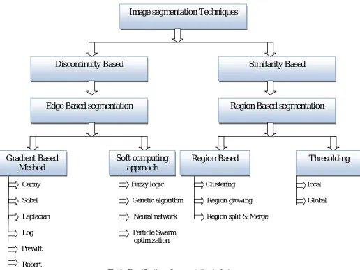
IV. IMAGE SEGMENTATION TECHNIQUES
Image segmentation techniques or methods are classified into two main categories as follows:
Canny Fuzzy logic Clustering local
Sobel Genetic algorithm Region growing Global
Laplacian Neural network Region split & Merge
Log Particle Swarm optimization
Prewitt
Robert
Fig 1: Classification of segmentation techniques
A. SEGMENTATION BASED ON SIMILARITIES
Segmentation based on similarities seems to the division of images in areas which are similar according to pre-defined criteria. It is similar according to the pre-pre-defined condition. That includes Region based segmentation in which region in any image is a subset of the image with respect to some criteria. The pixels with similar behaviour are grouped together in one region [13] . The similarity may be based on for color, texture, gray level or shape. Region based segmentation algorithms mainly includes the following methods:
1. Segmentation based on clustering
Clustering refers to a process of grouping pixels based on the similarity. It is basically grouping the data. The pixels having same attributes are grouped together to form clusters. Same attributes may have same colour, size, texture, shape etc. So the effectiveness of this algorithm depends on which similarity criteria used. A good clustering
Image segmentation Techniques
Edge Based segmentation
Region Based segmentation
Discontinuity Based
Similarity Based
Gradient Based
Method
Soft computing
approach
method will produce high quality clusters with high intraclass similarity. The clustering algorithm includes K-means
,fuzzy clustering and fuzzy c-means clustering etc.In the K-means algorithm initially we define the number of clusters
k. Then k-cluster center are chosen randomly. The extension of K means algorithm is called fuzzy K means or fuzzy C means algorithm [12]. Fuzzy C means generally preferred, like the knowledge element easily fit in with many Cluster and relate to each element.
2. Region Growing method
Region growing is one of popular methods of region based segmentation which Starts with a pixel and works by adding the pixels based on similarity, to the region, repeat until all pixels belong to same region. The region growing is a widely used segmentation method due to its computational simplicity and stability sensitive to noise. The method is
further classified as seeded region growing method (SRG) and unseeded region growing method (USRG). The main
advantage of this method is connected regions are guaranteed, multiple criterions at the same time and give very good results with less noisy[12]. And the disadvantages are over segmentation when the image is noisy or has intensity variations, cannot distinguish the shading of the real images and power and it is very time consuming.
3. Region merging & splitting
The whole image is treated as single region and then divides it into four quadrants based on predefined criteria. Then further divide the quadrant into other four quadrants for the same criteria. So it simply partition the image consecutively into minor and minor quadrant regions.The process will continues till further division is possible. manually interaction will not be required in this technique but it requires the input to be organized into a pyramidal grid structure which may be difficult.
4. Thresolding
Thresolding is simply separating foreground or object from the background . It provides groups having pixels with similar intensities pixels and useful for extracting boundaries from images on backgrounds. The algorithm converts a multilevel image into a gray level image by choosing a proper threshold value T. Then we can divide the pixels into multiple different regions so as to separate more useful objects from background. Thresolding techniques classify into 2 techniques:
i. Local thresolding
When the threshold value is not fixed that approach is called local thresolding. In local thresholding, different threshold value allocate for every single area of image. The main drawback of this approach is that it is not applicable to multichannel images and moreover the spatial characteristics of an image are not taken into the account. So sensitive to noise is high.
ii. Global thresolding
When the threshold value is fixed that approach is called global thresolding. The value of thresolding which is depend on the properties of pixels. In global thresholding, only single global threshold value is positioned to the
complete image [13]. This approach is used when the difference between background and foreground objects is
very distinct.
B. SEGMENTATION BASED ON DISCONTINUITIES
Segmentation based on discontinuities needs edge detection so subdivision can be performed based on the intensity of an image changed. Edge detection is to locate the pixels on the region edge. That includes segmentation algorithm using operator like canny, sobel, prewitt etc. Edge detection techniques transform images to edge images using the changes of grey lavels in the images. Edges occur on the boundary between two regions. There are many edge detection methods like canny, sobel , prewitt etc.
1. Gradient Based segmentation
operators with the image. Commonly used operators in gradient based edge detection method are sobel’s operator, canny operator, Laplace operator, Laplacian of Gaussian (LOG) operator etc. These operators work well for images with sharp edges and less amounts of noise.
2. Segmentation based on Soft computing approach
i. Fuzzy logic
Fuzzy image processing is the collection of all approaches that understand, represent and process the images, their segments and features as fuzzy sets. The representation and processing will be depend on the selected fuzzy
technique and on the problem to be solved.The advantages in fuzzy is membership function which may be utilized
to symbolize some amount of properties, and fuzzy IF-THAN rules can be used for to achieve conclusion. There are many fuzzy properties, inference rules, fuzzy operators presenting many difficulties due to uncertainty[8]. Disadvantages are the determination of fuzzy membership is not an limited work and the totalling related with fuzzy approaches might possibly to be rigorous. Pixels are divided into fuzzy sets i.e each pixel may belong to multiple sets and regions of image.
ii. Genetic algorithm
Genetic Algorithms (GAs) form a type of popular metaheuristic algorithms which can be evolutionary technique which follows different steps. GAs also used to segment images through equal bi-level and multi-level thresholding. The GA-based clustering technique is adopted for solving image segmentation problems, primary the input patterns are generated corresponding to every pixel of the image. The generated input patterns integrated neighbouring
pixels information to take contextual information of the image. Genetic algorithm Derives from the evolution
theory, consists of three major operations like selection, crossover, and mutation which is used in pattern's recognition applications.
iii. Neural network
The Neural Network is artificial demonstration of human brain. Artificial Neural Network also known as a neural network. It has been widely used in medical image analysis such as an example segmentation, data compression, image enhancement and noise suppression. Multi-layer perceptron (MLP), self-organizing maps (SOM), Hopfield and pulse coupled neural networks[14]. SOM network is the most suitable networks useful for segmentation. This is an unsupervised network on the basis of the competitive learning and discovering topological structure hidden in the input data for visual display in a single two dimensional display several dimensional the input data for visual display in a couple of dimensional spaces.
iv. Particle swarm optimization
Swarm Intelligence which is originated from the study of swarms of social organisms. Studies of the social behavior of organisms in swarms with the design of very efficient optimization and clustering algorithms. Particle Swarm Optimization (PSO) is an evolutionary computation technique which is developed by Kennedy and Eberhart in 1995. PSO is a optimization approach which build on the social behavior of birds flocks . It is a population based search procedure where the individuals, referred to as particles, are grouped into a swarm. Each particle in the swarm represents a candidate solution to the optimization problem. The particle swarm optimization is definitely evolutionary technique to solve the constant and discrete problems and it can be referred to population-based stochastic approach for solving computational problems[15]. The particles which move around the search space to resolve the optimization problems. it is straightforward and fast technique.
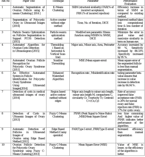
V. REVIEW TABLE
Summary of some follicle segmentation techniques and their performance as follows in table 1:
Table 1
Ref.No Paper Title Technique Parameter Performance
Evaluation 1 Automatic Segmentation of
Ovarian Follicle using K-means Clustering [ 2014 ]
K-Means Clustering
SSIM (structural similarity) FAR(% of incorrect acceptance), FRR (% of incorrect rejection)
Efficiency increase in terms of SSIM as compared to existing method.
2 Segmentation of Polycystic Ovary in Ultrasound Images [2014]
Active counter without edge
method
Time, No. of Iteration, DICE
Improved method takes less computational time & less number of iteration
3 Particle Swarm Optimization on Follicles Segmentation to Support PCOS Detection [2015]
Particle swarm optimization
method
Modified non parametric fitness function using MSSIM & NMSE,
DICE
Minimize the error in pixel value and maximize similarity in term on human eye. 4 Automated Algorithm for
Ovarian Cysts Detection in Ultrasonogram [2015]
Thresolding ( Based on horizontal & Vertical
Scan-line)
Major axis, Minor axis, Area, Perimeter Accuracy increased by 90 %, Sensitivity increased by 88.33% ,Specificity increased by 95 %.
5 Automated Ovarian Follicle Recognition for
Polycystic Ovary Syndrome [2011]
Scanline Thresolding
MSE (Mean square error) Mean square error of the segmented follicle is less than manual segmentation. 6 An Effective Automated
System in Follicle
Identification for Polycystic Ovary Syndrome
Using Ultrasound Images[2014]
Enhanced Watershed Algorithm
Recognition rate , Misidentification rate tuning parameter beta value ensures that increase in value increase in detection rate by 84.04 % .
7 Detection of cysts in medical ultrasound images of ovary [2014]
Region based active contour method without
edges
Major axis length to minor axis length, minor axis length M, compactness C, circularity Cr, Tourtocity Tr, Centroid
C= (Cx,Cy)
Rate of accuracy improved false acceptance rate (FAR) is 20% for normal ovary and false rejection rate (FRR) is 17% for cystic ovary. 8 Detection of Cysts in
Ultrasonic Images of Ovary [2013]
Fuzzy C-Means Clustering
PSNR (Peak Signal to Noise Ratio) ,MSE(Mean Square Error)
less time consuming And higher value of PSNR indicates better performance of the algorithm.
9 Automatic Detection of Follicles in Ultrasound Images of
Ovaries using Edge Based Method [2011]
Edge Based Method (Canny
operator)
FAR(Type I error) ,FRR(Type II error) Increased efficiency than manual segmentation
10 Ovarian Follicle Detection for Polycystic Ovary Syndrome using Fuzzy C-Means Clustering [2013]
Fuzzy C-Means Clustering
VI. CONCLUSION AND FUTURE WORK
In this review paper, we have discussed all the major image segmentation techniques and algorithms. An efficient noise reduction technique needs to be developed and texture features need to be investigated for the follicle detection .The detection of pattern and recognition using edges and points is all possible by these methods. Since number of parameters like colour, intensity, noise etc. affect these algorithms, there is a challenging task to select an appropriate algorithm for a ultrasound image. Classification will be used to classify the ovary based on the extracted follicles.
ACKNOWLEDGEMENT
The authors would like to thank HOD of department ,Prof. Bijal J. Talati for her continuous support, encouragement and cooperation throughout the progress of the work and our special thanks to Dr. Bhavesh Patel, Department of
radiology, Sir Sayajirao General Hospital (SSG Hospital), Baroda, for providing his guidance, eternal help and
providing dataset for our future work.
REFERENCES
1. Kiruthika.V, M.M.Ramya,” Automatic Segmentation of Ovarian Follicle using K-means Clustering”, Fifth International Conference on Signals and Image Processing, pp.137-141,2014
2. H Prasanna Kumar, S Srinivasan,”Segmentation of Polycystic Ovary in Ultrasound Images”, 2nd International Conference on Current Trends in Engineering and Technology, ICCTET, vol.8, pp.237-240,2014
3. E.Setiawati, Adiwijaya, Tjokorda A.B.W, “Particle Swarm Optimization on Follicles Segmentation to Support PCOS Detection”, 3rd International Conference on Information and Communication Technology (ICoICT), pp.369-374, 2015.
4. Sandy RIHANA, Hares Moussallem, Chiraz Skaf, “Automated Algorithm for Ovarian Cysts Detection in Ultrasonogram”,2nd International Conference on Advances in Biomedical Engineering, pp. 219-222, 2013.
5. Palak Mehrotra , Chandan Chakraborty, Biswanath Ghoshdastidar, Sudarshan Ghoshdastidar,”Automated Ovarian Follicle Recognition for Polycystic Ovary Syndrome”, International Conference on Image Information Processing (ICIIP ),2011.
6. Ranjitha Sitheswaran, Dr.S.Malarkhodi,”An Effective Automated System in Follicle Identification for Polycystic Ovary Syndrome Using Ultrasound Images”, International Conference on Electronics and Communication System (lCECS ),2014.
7. Prema t. akkasaligar, girijamma v. malagavi,”detection of cysts in medical ultrasound images of ovary”,Proceedings of 5th SARC-IRF International Conference, Bangalore,Vol.4, ISBN: 978-93-84209-13-1, pp.57-62,2014.
8. Ashika Raj,”Detection of Cysts in Ultrasonic Images of Ovary”, International Journal of Science and Research (IJSR),vol 2 ,issue 8 ISSN: 2319-7064, pp. 185-189, 2013.
9. P.S.Hiremath, Jyothi R. Tegnoor,”Automatic Detection of Follicles in Ultrasound Images of Ovaries using Edge Based Method”, IJCA Special Issue on “Recent Trends in Image Processing and Pattern Recognition” RTIPPR ,pp.120-125, 2010
10. Ashika Raj,” Ovarian Follicle Detection for Polycystic Ovary Syndrome using Fuzzy C-Means Clustering”, International Journal of Computer Trends and Technology (IJCTT) ,volume 4 Issue 7,pp.2146-2149,2013
11. P. S. Hiremath and Jyothi R. Tegnoor,” Follicle Detection and Ovarian Classification in Digital Ultrasound Images of Ovaries”,pp168-199,2013.
12. M.S. Sonawane, C.A. Dhawale, Ph.D., “A Brief Survey on Image Segmentation Methods”, International Journal of Computer Applications (0975 – 8887) National conference on Digital Image and Signal Processing, pp. 1-5, 2015
13. Kanchan Kumari*, Dr. Kuljit Kaur, “Image Segmentation : Review On Existing Techniques”, International Journal of Advance Foundation And Research In Science & Engineering (IJAFRSE) ,Volume 1, Special Issue , ICCICT,pp.1-6, 2015.
14. Vijai Singh, Shivangi Gupta, Shrutika Saini,” A Methodological Survey of Image Segmentation Using Soft Computing Techniques”, International Conference on Advances in Computer Engineering and Applications (ICACEA), pp.419-422,2015
15. I. O. Rabiu, A. D. Usman, A. M. S. Tekanyi,’ A Review on Computer Assisted Follicle Detection Techniques and Polycystic Ovarian Syndrome (PCOS) Diagnostic Systems, International Journal of Computer Trends and Technology (IJCTT) ,volume 28 Number 1 – October ,pp.41-45, 2015
BIOGRAPHY