ABSTRACT
HO, TRICIA CHRISTINE. Defining the Mannitol Dehydrogenase (MTD) Interactome during Unconventional Protein Secretion. (Under the direction of Dr. Michael B. Goshe and Dr. John D. Williamson).
Although Golgi-dependent secretion was long believed to be the sole protein secretory pathway in cells, unconventional, non-Golgi secretion has been more recently confirmed to occur in both animals and plants. Specific mechanisms for such unconventional secretion, however, were transformed to express celery mannitol dehydrogenase (MTD), a protein have yet to be completely elucidated. To characterize these mechanisms Arabidopsis thaliana cells known to be secreted by unconventional/leaderless means, to identify components of its secretory
Defining the Mannitol Dehydrogenase (MTD) Interactome during Unconventional Protein Secretion
by
Tricia Christine Ho
A thesis submitted to the Graduate Faculty of North Carolina State University
in partial fulfillment of the requirements for the Degree of
Master of Science
Biochemistry
Raleigh, North Carolina
2016
APPROVED BY:
__________________________ __________________________ Dr. Michael B. Goshe Dr. John D. Williamson
Co-Chair of Advisory Committee Co-Chair of Advisory Committee
ii
BIOGRAPHY
Tricia Christine Ho was born in Columbia, Maryland. She attended the University of Virginia and obtained her Bachelor of Science in Biology in 2011. After working in an environmental laboratory for 2 years, she began her graduate studies in the
iii
TABLE OF CONTENTS
LIST OF TABLES………vii
LIST OF FIGURES……….viii
CHAPTER 1: Mapping a Protein’s Journey: The Highways and Byways of Protein Trafficking……….………1
1.1 Conventional, Golgi-Dependent Secretion………...2
1.1.1 Protein Insertion into the ER………...2
1.1.2 Trafficking from the ER via the Golgi/TGN………..…….3
1.1.2.1 ER to the Golgi……….6
1.1.2.2 Maturation through the Golgi……….…….7
1.1.2.3 Exit from the TGN……….…………8
1.1.3 Protein Modifications in the ER and Golgi………9
1.1.4 Export and Retention Signals……….……..10
1.2 Intracellular Vesicular Trafficking……….14
1.2.1 Endosomal Trafficking Pathways……….14
1.2.2 Vesicle Formation………...17
1.2.2.1 COPI, COPII………..17
1.2.2.2 MVB and the ESCRT Pathway……….18
1.2.3 Vesicle Fusion……….……19
1.2.4 Autophagy………20
1.2.4.1 Inductionof Autophagosome Formation………...……..21
1.2.4.2 Cargo Recognition/Selection……….…..21
1.2.4.3 Autophagosome Formation………...…..22
1.2.4.4 Fusion and Degradation………...……22
1.3 Intracellular Protein Targeting/Transport………23
1.3.1 Targeting/Transport to Proteasomes……….23
1.3.2 Targeting/Transport to Mitochondria and Chloroplasts…………...23
1.3.2.1 Protein Import into Mitochondria……….……24
1.3.2.2 Protein Import into Chloroplasts………...……..25
1.3.3 Targeting/Transport to Vacuoles………..………26
iv
1.3.3.2 Golgi-Independent Vacuolar Trafficking………....…28
1.4 Unconventional, Golgi-Independent Secretion………..……31
1.4.1 Leaderless Secretion in Mammalian, Yeast, and Parasitic Cells….31 1.4.1.1 Type I – Direct Translocation Across the PM…………..……….32
1.4.1.2 Type II – ABC Transporter-Based………..…….…..33
1.4.1.3 Type III – Autophagy-Based………..………….…35
1.4.1.4 Type IV – Golgi Bypass………...…..………..36
1.4.2 Unconventional Protein Secretion in Plants………40
1.4.2.1 Storage Proteins………..…….40
1.4.2.2 Lectins………...……….41
1.4.2.3 Superoxide Dismutases (SODs)………...…42
1.4.2.4 Mannitol Dehydrogenase (MTD)………....……..42
1.4.2.5 Hygromycin Phosphotransferase (HYGR)………...…..43
1.4.3 Mechanisms of Unconventional Secretion/Trafficking in Plants...44
1.4.3.1 Golgi-Independent Transport from ER to Vacuoles……...….44
1.4.3.2 Fusion of Vacuole and PM………...…46
1.4.3.3 Vesicles/MVBs (Multivesicular Bodies)………...………46
1.4.3.4 EXPO (Exocyst Positive Organelle)………...….…….48
1.5 Methods of Protein-Protein Interaction Analysis………51
1.5.1 Cell Fractionation and Immunoblot Analysis…………...……..51
1.5.2 Electron Microscopy………...……..52
1.5.3 Confocal Microscopy………...………54
1.5.4 Mass Spectrometry in Proteomics………...……..55
1.5.4.1 Proteomic Approaches………...…….55
1.5.4.2 Data Acquisition Methods………...…….56
1.5.4.3 Separation, Ionization, and Mass Analyzers………57
1.5.4.4 Fragmentation Methods………..57
1.5.5 Summary...……….58
v
CHAPTER 2: Elucidating the Mannitol Deyhdrogenase (MTD) Interactome
Associated with Unconventional Secretion………...78
2.1 Abstract………...…….78
2.2 Introduction………...…..78
2.3 Experimental………...…….82
2.3.1 Materials....………82
2.3.2 Construction and Transformation of 35S-MTD-FLAG……...82
2.3.3 Cell Maintenance and Treatment………...….83
2.3.4 Protein Extraction………...….83
2.3.5 Co-Immunopurification (Co-IP) of MTD-Complex…...83
2.3.6 Immunoblotting………...……84
2.3.7 In-Gel Tryptic Digestion and LC/MS/MS………...…….84
2.3.8 LC/MS/MS Data Acquisition...86
2.3.9 Data Normalization and Comparisons……...…86
2.3.10 Data Filtration and Ratio Calculations…………...………88
2.4 Results and Discussion………...…91
2.4.1 SA Triggers Formation of an MTD-Interacting Complex; Crosslinking Preserves the MTD-Interactome...91
2.4.2 Assessing the MTD Interactome following SA Treatment...93
2.4.3 The MTD Interactome 20 minutes after SA Treatment...95
2.4.4 Changes in the SA-Specific MTD Interactome between 10 and 20 Minutes...98
2.4.5 SA-Induced Signaling Proteins...104
2.4.6 Initial Secretory Complex...105
2.4.7 MTD-Interacting Proteins with Maximum Presence after 10 Minutes...108
2.5 Conclusions and Future Perspectives……….……..………111
vi
APPENDICES………...……119
Appendix A...120
Appendix B...121
Appendix C...123
vii
LIST OF TABLES
Table 2.1 – The MTD Interactome 10 minutes after SA Treatment
(SA10)...95
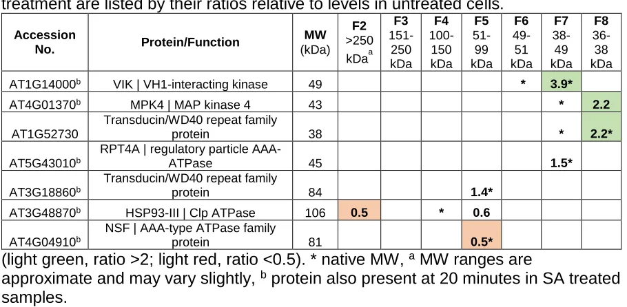
Table 2.2 – MTD Interacting proteins 20 minutes after SA Treatment (SA20)...97
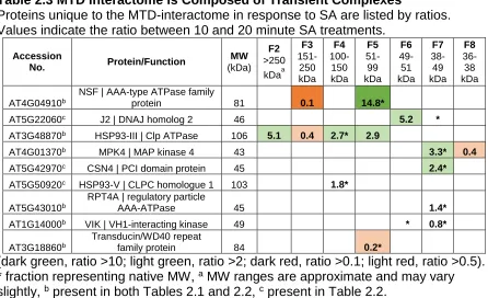
Table 2.3 – MTD Interactome is Composed of Transient Complexes...99
Table 2.4 – Interactors in the Transient Complexes Classified by Previously Reported Function...102
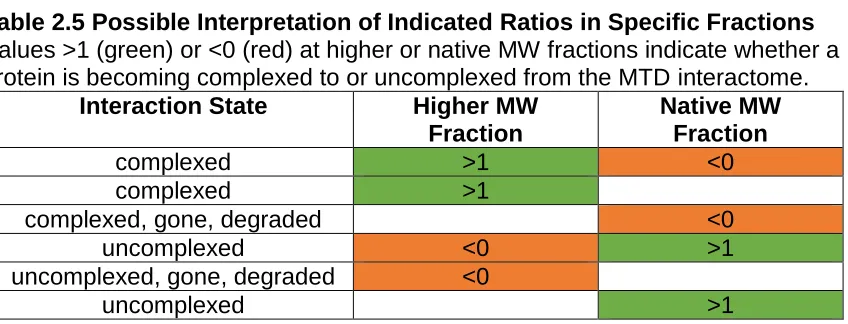
Table 2.5 – Possible Interpretation of Indicated Ratios in Specific Fractions...104
Table D.1 – Proteins Unique to SA10...125
Table D.2 – Ratios of Unique SA10 Proteins...127
Table D.3 – Proteins Unique to SA20...128
viii
LIST OF FIGURES
Figure 1.1 – Final Targeting of Proteins...4
Figure 1.2 – Trafficking From the ER to the TGN…………...12
Figure 1.3 – Endosomal Trafficking Pathways………...15
Figure 1.4 – Intracellular Targeting………...29
Figure 1.5 – Unconventional Secretory Pathways in Animal Cells………...38
Figure 1.6 – Unconventional Secretory Pathways in Plant Cells………….…...49
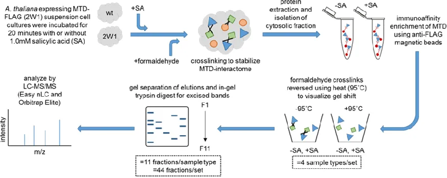
Figure 2.1 – Sample Preparation Workflow...85
Figure 2.2 – Data Analysis Workflow...89
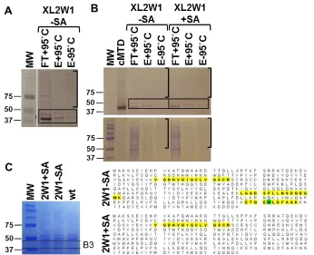
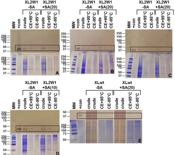
Figure 2.3 – MTD is Present in Immunopurified, Crosslinked Complexes...92
Figure A.1 – MTD-Complex Co-Immunopurification...126
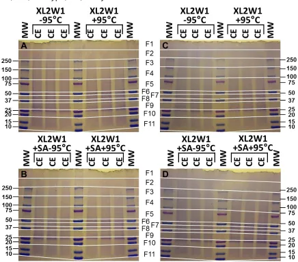
Figure B.1 – In-Gel Digestion Fractions...127
1
CHAPTER 1
Mapping a Protein’s Journey: The Highways and Byways of Protein Trafficking
Protein trafficking is required for almost all cellular processes, making it a crucial part of cell function and survival. Cell maintenance, metabolism, secretion, defense, and response to stimuli are only a few of an extensive list of processes that involve protein trafficking. In animals for instance, neurotransmitters secreted into the synaptic cleft are recognized by receptors on the postsynaptic target that could be involved in the control of many pathways and systems (Hyman, 2005). If
neurotransmitters are packaged and secreted inefficiently, or not at all, a number of processes would be affected. In both plants and animals, environmental signals can trigger events that require protein trafficking. For example, a plant senses it is under pathogen attack via receptors on the cell surface, targeting transport of specific transcription factors into the nucleus and triggering expression of required defense proteins. Once defense proteins are made, they must be directed toward the point of pathogen attack. Not only is accurate and timely protein trafficking required for cell survival, but often for survival of the organism as a whole.
Trafficking of proteins that are initially co-translated into the lumen of the endoplasmic reticulum (ER) can be divided into two major classes: Golgi-dependent trafficking (classical) and Golgi-independent trafficking (nonclassical), often referred to as conventional and unconventional protein secretion/trafficking, respectively. Other proteins, such as mitochondrial and chloroplast proteins, are initially translated in the cytosol and then transported directly into their respective organelles
independent of the ER/Golgi apparatus. Classical, Golgi-mediated secretion largely restricts secretory responses to regulation by activation of gene expression. This is, or can be, relatively slow, because not only does it require gene activation and production of a new protein, it also requires trafficking through the entire Golgi pathway prior to secretion. To enter the Golgi stacks, proteins must first be
2
and utilize an unconventional/nonclassical secretion pathway. There are also
proteins that undergo constitutive or stress/stimulus-regulated “leaderless” secretion (Nickel, 2011).
1.1 Conventional, Golgi-Dependent Secretion
1.1.1 Protein Insertion into the ER
Transport and localization of proteins begins as early as translation, where signal sequences often are responsible for targeting proteins to specific cellular compartments or the plasma membrane (PM). In prokaryotes, secretory and membrane proteins are targeted directly to the PM by a 20 to 30 amino acid, N-terminal signal sequence. Secreted proteins can be delivered to the PM by a post-translational or co-post-translational mechanism (Cross et al., 2009). In eukaryotes, an analogous signal sequence first targets nascent proteins to the ER for later sorting via the Golgi (Luirink and Sinning, 2004). Signal sequences in prokaryotes and eukaryotes have the same general structure consisting of three domains: an N (N-terminal) domain, an H (hydrophobic) domain, and a C (C-(N-terminal) domain (Hegde and Bernstein, 2006). While the basic and slightly polar natures of the N and C domains, respectively, are present in all signal peptides, it is the hydrophobic H domain that appears to be essential for binding the signal recognition machinery (Gierasch, 1989; von Heijne, 1990). Of the 3 domains, the N domain is the most variable in length and composition, ranging from 1-5 residues. It does, however, typically have a net positive charge. The H domain, ranging from 7-15 residues, can contain Pro, Gly, Ser, or Thr residues, but is generally rich in Leu, Ala, Met, Val, Ile, Phe, and Trp residues. The C domain, ranging from 3-7 residues, has a higher polarity than those in the N domain and determines the cleavage site.
As soon as the signal sequence emerges from the exit tunnel of the ribosomal complex during translation (Halic and Beckmann, 2005; Hegde and Bernstein,
3
bind their own GTP molecule before the SRP/ribosome-nascent chain (RNC)
complex can bind to the SR on the target ER membrane (Luirink and Sinning, 2004; Halic and Beckmann, 2005). At this point the RNC is transferred to the translocon, an integral membrane protein-conducting channel, and the signal peptide enters the channel (Halic and Beckmann, 2005). GTP hydrolysis then drives SRP/SR
dissociation and allows translation to proceed into the lumen of the ER (Luirink and Sinning, 2004; Halic and Beckmann, 2005).
1.1.2 Trafficking from the ER to the Final Destination via the Golgi/TGN
In eukaryotic cells, once newly synthesized proteins enter the lumen of the ER, resident lumenal proteins catalyze or mediate modification and folding. Upon leaving the ER, proteins then pass through the vesicular-tubular clusters (VTCs) or intermediate compartment (IC) (more recently termed the ER-Golgi intermediate compartment [ERGIC]) (Duden, 2003), followed by transport through a series of stacked, membrane-bounded Golgi compartments (cis-, medial-, and trans-), or cisternae, where further modification occurs. Each Golgi compartment also has its own set of resident protein processing enzymes. The trans-Golgi face gives rise to the trans-Golgi network (TGN), where mature proteins are packaged into vesicles for secretion or transport to other cellular compartments (Figure 1.1). This is the longest route from the ER, with proteins passing through all of these compartments before reaching their final destination, the extracellular space or a specific organelle. There are a variety of coated and non-coated vesicles specific to each segment of
4
Figure 1.1 Trafficking of Proteins from the TGN
There are a number of pathways a protein can take from the TGN as well as a variety of final destinations. A majority of the classical transport pathways transit the TGN. From the TGN, proteins can be targeted directly to the PM (1) or secreted via secretory granule formation (2). Alternately, DVs formed at the TGN first deliver cargo to the MVB then to the PSV (3), or CCVs formed at the TGN can also deliver cargo to MVBs/LEs/PVCs. Both pathways eventually lead to the lysosome. CCVs can also deliver cargo directly to the LV (Golgi-dependent) (4). PACs can originate from either the TGN (Golgi-dependent) or directly from the ER (Golgi-independent) to transport cargo to the PSV (5, 6). Proteins can be directly taken to the LV from the ER bodies (not shown) that originate from the ER (Golgi-independent) (7). Some of these pathways (3 and 4) are connected to the endocytic pathway (8), where
proteins endocytosed at the PM form EEs before fusing with/into MVBs, also called LEs. Mitochondrial (9) and chloroplastic (10) proteins are translated in the cytosol and delivered directly to their respective organelles without passing through the ER-Golgi.
(Details for each pathway can be found in the text).
a found only in plant cells
6
1.1.2.1 ER to the Golgi
As translation continues, the signal sequence is fully extruded into the ER, where it is then removed by a membrane-bound signal sequence peptidase complex (SPC) (Braakman and Hebert, 2013). Once translation is complete, proteins are processed, folded, and then either retained in the ER (as discussed below) or packaged and directed to the Golgi. Proteins exit the ER at ER export sites (ERES), or the transitional ER (tER), membrane domains where vesicles containing proteins targeted for secretion begin to form (Watson and Stephens, 2005). COPII proteins (coat proteins), including Sar1p, Sec23/24p, and Sec13/31p, mediate vesicle formation for anterograde transport of proteins from the ER to the Golgi (Duden, 2003). Before reaching the Golgi, the COPII coat dissociates, resulting in naked vesicles that then fuse to form the VTCs/ERGIC (Duden, 2003; De Matteis and Luini, 2008). Proteins in the ERGIC can then either continue on to the Golgi or undergo retrograde transport (i.e. be transported back to the ER); both options are thought to involve formation of COPI-coated vesicles (Szul and Sztul, 2011). COPI proteins mediate the formation of vesicles in a manner similar to COPII vesicle formation, both of which are described below.
There are currently two models that describe cargo vesicle transport from the ER to the Golgi: the transport complex model and the stable compartment model (Brandizzi and Barlowe, 2013). In the transport complex model, ERGIC
compartments track on microtubules from ERES to the Golgi. The stable
compartment model proposes that ERES-Golgi transport is a two-step process: the first being a microtubule-independent, short-range transport from ER to ERGIC, followed by microtubule-dependent, long-range transport from ERGIC to the Golgi. In both models, dynein and kinesin motors are thought to be responsible for
anterograde and retrograde transport, respectively, along microtubules. In mammalian cells, COPII-coated vesicles are known to be associated with
7
TRAPPI (transport protein particle I) tethering complex, located on the ERGIC, and resulting in a partially microtubule-independent transport. In plant cells, however, the ER and Golgi are in much closer proximity, and are thought to be stabilized in the correct orientation for vesicular exchange by a matrix of tubular connections or tethering factors. Thus, ER to Golgi transport does not rely on microtubule transport, and additionally, plants lack an identifiable ERGIC (Hwang and Robinson, 2009). In addition, the role of COPII-coated vesicles in this process in plants is itself under debate (Brandizzi and Barlowe, 2013). The precise mechanism whereby cargo vesicles reach the cis-Golgi from ERGIC is still unclear. From ERGIC, COPI-coated vesicles are responsible for anterograde transport to the cis-Golgi (Brandizzi and Barlowe, 2013). ER and Golgi resident proteins, such as those comprising the trafficking machinery itself, are recycled from the trans- to the cis-Golgi
compartments via retrograde COPI-coated vesicles in both plant and mammalian cells (Nakano and Luini, 2010).
1.1.2.2 Maturation through the Golgi
Proteins destined for secretion or further transport move through the Golgi stack from the cis- to the medial- and then to the trans-Golgi face, before moving on into the TGN (De Matteis and Luini, 2008) (Figure 1.2). Proteins entering the TGN are segregated from resident proteins and undergo modifications and processing as they mature through the various Golgi compartments. Four models have been proposed for Golgi trafficking in animal cells, differing primarily with regard to the mechanism by which cisternae transition from the cis- to the trans-Golgi (Moremen et al., 2012). The two classic models are the cisternal maturation model and the vesicular transport model. The cisternal maturation model proposes that cisternae undergo translocation as they mature, with processing enzymes being delivered to each compartment and with mature vesicles eventually budding off at the TGN. Transport vesicles enriched with processing enzymes have been identified,
8
vesicular transport model, in contrast, describes cisternae as stable or fixed, and that processing enzymes constantly undergo anterograde/retrograde transport. These models, however, appear to be inconsistent with more recent data suggesting that new proteins can quickly access all the different stages of the Golgi cisternae. This observation gave rise to the rapid partitioning model, which proposes that specific lipid composition regulates the polarity of the Golgi and the distribution of resident proteins, either involved in transport or localization of resident proteins, respectively. This model is supported by the observation that the membrane composition of the cis-Golgi more closely resembles the ER membrane, being enriched in glycerophospholipids (GPLs) (Patterson et al., 2008). The membrane of the trans-Golgi, however, is enriched in sphingolipids (SLs) and is thus more similar to the PM (Patterson et al., 2008). Cargo proteins and resident proteins are believed to have affinity for either SL or GPL domains, respectively, which then retain specific proteins using thermodynamic partitioning (Patterson et al., 2008).
1.1.2.3 Exit from the TGN
9
aggregation of secretory cargo molecules, separating them from the other soluble proteins (Robinson and Pimpl, 2014). These clusters interact with specific lipid domains in the TGN, causing budding and formation of immature secretory granules (ISGs) that develop into mature secretory granules (MSGs) (Robinson and Pimpl, 2014). Proteins destined for the apical/basolateral membranes also leave the TGN through lipid domain-mediated budding, although the sorting mechanisms for these proteins are currently unclear (Kienzle and von Blume, 2014). Lastly, vacuolar-targeted proteins also exit the TGN via CCVs, forming multivesicular bodies (MVBs) (Robinson and Pimpl, 2014), more details of which are discussed below.
1.1.3 Protein Modifications in the ER and Golgi
Protein processing and post-translational modifications (PTMs) that are essential for folding and protein maturation can occur as early as co-translation into the ER and continue through arrival at the TGN (Figure 1.2). Glycosylation, for instance, can occur as soon a suitable glycosylation site (sequon), is far enough into lumen of the ER (Braakman and Bulleid, 2011) to be accessible to a membrane integral oligosaccharyltransferase (OST) that is proximal to the translocon. OSTs covalently transfer preassembled glycans to the asparagine (Asn) residue within the sequon via an amide linkage (Moremen et al., 2012). The consensus sequence for a standard N-glycosylation sequon is Asn-X-Ser/Thr, where X can be any amino acid except for proline, and is the most common glycosylation consensus in nascent polypeptides (Moremen et al., 2012). Simultaneously, protein disulfide isomerases (PDIs) and prolylpeptidyl cis-trans isomerases (PPIases) can catalyze the formation of disulfide bonds and proline-isomerization, respectively, in the elongating
10
in Moremen et al. (2012), several different glycosyltransferases catalyze various O-glycosylation reactions, each targeting a specific consensus sequence containing Ser or Thr. The branched glycan modifications themselves can then be modified many times before the protein arrives at the TGN. Glycosyltransferases catalyze the transfer of sugars, glycosidases (glycoside hydrolases) remove sugars, and
epimerases and sulfotransferases modify the sugars on the target protein (Stanley, 2011). These enzymes are found in cis-, medial-, trans-Golgi compartments, or in the TGN, and although they may be transported to neighboring compartments, they are eventually recycled back to their original Golgi compartment as resident proteins (Stanley, 2011).
1.1.4 Export and Retention Signals
Resident proteins in both the ER and Golgi that assist in processing newly synthesized proteins must be recycled back to their original compartments. This is mediated by retention signals, amino acids in the proteins that allow them to be identified and returned to the ER/Golgi (Figure 1.2). Most ER-synthesized, transmembrane proteins contain ER export signals. The most common export sequences contain di-acidic (D/EXD/E), di-hydrophobic (FF, YY, LL, FY), and aromatic motifs, that allow direct binding of Sec24 through the membrane of the budding vesicle (Gillon et al., 2012). Soluble ER-synthesized proteins, however, lack the ability to bind Sec24, and require transmembrane cargo receptors that bind both Sec24 and the soluble protein itself (Gillon et al., 2012). Cargo receptors also
11
12
Figure 1.2 Trafficking from the ER to the TGN
Nascent polypeptides enter the ER lumen cotranslationally, and undergo initial modification, such as N-glycosylation by oligosaccharyltransferases (OSTs) and processing by PDIs and PPIases (1). Anterograde transport from the endoplasmic reticulum (ER) to the ER-Golgi intermediate compartment (ERGIC) occurs via coat protein complex II (COPII)-coated vesicles (2a). Recruitment of the COPII complex involves Sec12, Sar1, Sec13/31p, and Sec23/24p. Proteins bound for the ERGIC are have export signals: D/EXD/E, FF, YY, LL, FY, or aromatic motifs. Retrograde transport from ERGIC to the ER occurs via COPI-coated vesicles (2b). Recruitment and disassembly of the COPI complex involves Arf1, a guanine nucleotide exchange factor (GEF), and a GTPase activating protein (GAP). Resident ER proteins are recognized by K/HDEL, diLys, hydrophobic, or N-terminal Arg motifs. Cargo proteins undergo anterograde transport in COPI-coated vesicles to the Golgi; retrograde transport also uses COPI-coated vesicles to return to the ERGIC and ER (3).
Proteins are modified and processed as they mature and transit the Golgi stack (4). O-glycosylation is the most common modification; branched glycans can also be modified by glycotransferases (GTs), glycosidases (GHs), epimerases, and sulfotransferases (SULTs). Resident Golgi proteins, with KXD/E motifs, undergo retrograde transport to the cis-Golgi via COPI-coated vesicles. Proteins arriving at the TGN have several ways of being transported to their final destination (5). Resident Golgi proteins are returned to the cis-Golgi via retrograde transport.
* Two models describe transport from ERGIC to Golgi: the transport complex model and the stable compartment model.
** Four models describe maturation through Golgi: the cisternal maturation model, the vehicular transport model, the modified cisternal maturation model, and the rapid partition model.
Details concerning each step can be found in text.
14
1.2 Intracellular Vesicular Trafficking
1.2.1 Endosomal Trafficking Pathways
Endosomes in eukaryotic cells are involved in a number of cellular trafficking pathways, including protein transport to the lysosome/vacuole for degradation or in returning integral membrane proteins to the PM (Figure 1.3). Vesicles forming at the PM and blebbing into the cytoplasm, classified as either clathrin-independent or clathrin-dependent endocytosis (Conner and Schmid, 2003; Doherty and McMahon, 2009), deliver cargo to the early endosome (EE) for sorting (Grant and Donaldson, 2009). From the EE, the cargo can take several routes: to lysosomes/vacuoles via late endosomes, directly to the TGN, or back to the PM via recycling endosomes (Grant and Donaldson, 2009). In the recycling pathway, EEs/recycling endosomes are responsible both for returning recycled membrane proteins to the PM and for returning cargo receptors to the TGN (Reyes et al., 2011). From the EE, proteins that must be returned to the PM are first routed to the juxtanuclear endocytic recycling compartment (ERC), from which recycling endosomes are then formed (Grant and Donaldson, 2009). Clathrin-dependent cargo can also be returned to the PM using a rapid recycling pathway from the EEs (Grant and Donaldson, 2009). Membrane proteins bound for lysosomal or vacuolar degradation are sorted in the late endosomes (LEs), also called MVBs or prevacuolar compartments, prior to transport into endosomal intraluminal vesicles (ILVs) (Reyes et al., 2011). LE’s also transport newly synthesized lysosomal and vacuolar proteins to their respective organelles (Reyes et al., 2011). Certain proteins are known to associate with specific types of vesicles depending on its cargo and destination; for example, Rab5 is found on early endosomes and Rab11 is found on recycling endosomes (Grant and
15
Figure 1.3 Endosomal Trafficking Pathways
Vesicles formed at the PM, both clathrin-dependent and -independent, deliver cargo to the EEs for sorting. From the EE, they can be transported to the lysosome,
vacuole, or TGN (1) via MVBs/LEs/PVCs or directly to the TGN (2). Proteins
returned to the PM can undergo rapid or slow recycling. Rapid recycling (3) involves direct transport of cargo proteins to the PM, while in slow recycling (4), proteins are first targeted to the ERC, where they are returned to the PM in recycling endosomes. ILVs in the MVB can also be secreted into the extracellular space (5) after MVB fusion with the PM. To initiate the MVB pathway (grey box) (6), ESCRT-0 (green) binds ubiquitinated proteins and PtdIns3P on endosomal membranes, resulting in recruitment of ESCRT-1 (purple). ESCRT-1, which interacts with both ESCRT-0 and proteins modified with ubiquitin (brown circle), recruits ESCRT-II (yellow). ESCRT-II interacts with ESCRT-I, ubiquitinated proteins, and PtdIns3P (blue circle) on the endosomal membrane. Vps25, a subunit of ESCRT-II, is the nucleation point where Vps20, a subunit of ESCRT-III (orange), initiates assembly of the filamentous ESCRT-III complex by triggering oligomerization of Snf7 (orange triangle), another ESCRT-III subunit. Oligomerization terminates when Vps24 (ESCRT-III) (orange square) binds to the last Snf7, completing vesicle formation. ESCRT-III recruits deubiquitinases that deubiquitinate the cargo proteins. Vps2 (ESCRT-III) and accessory proteins recruit the Vps4 complex (cream), which disassembles ESCRT-III via ATP hydrolysis, terminating cargo sorting and vesicle formation.
17
1.2.2 Vesicle Formation
Proteins are transported within the cell in various types of vesicles. COPI and COPII proteins are key players in vesicular protein transport, interacting with and sorting specific cargo proteins. Vesicle formation can also occur via the MVB
pathway, mediated by ESCRT (endosomal sorting complexes required for transport) complexes, or via autophagosome formation during autophagy.
1.2.2.1 COPI, COPII
As previously mentioned, the COPII proteins, Sar1p, Sec23/24p, and Sec13/31p, are responsible for membrane deformation and vesicle budding at the ER. Sec12, an integral ER membrane protein, acts as a guanine nucleotide exchange factor (GEF) for Sar1, a small GTPase (Lee et al., 2004). Sar1-GTP binds to the ER membrane and recruits Sec23/24p, a heterodimeric protein complex that interacts with cargo protein sorting signals (Lee et al., 2004). The Sar1-Sec23/24p complex then recruits Sec13/31p, another heterodimer, which leads to polymerization of COPII complexes. The subsequent deformation and budding of the membrane that is then triggered leads to formation of the COPII vesicle (Lee et al., 2004).
18
1.2.2.2 MVB and the ESCRT Pathway
Although the ESCRT complexes are involved in a number of cellular
processes (e.g. cytokinesis and HIV [human immunodeficiency virus] budding), this section will focus mainly on their role in the MVB pathway as described by Schmidt and Teis (2012) (Figure 1.3). ESCRT protein complexes assemble at the cytosolic membrane, rather than at the lumenal membrane, of the MVB and are crucial to the formation and budding of vesicles from the MVB membrane into the MVB lumen. There are five ESCRT complexes, listed in the order in which they assemble: ESCRT-0 ESCRT-I, ESCRT-II, ESCRT-III, and the Vps4 complex (vacuolar protein sorting). Protists and plants lack ESCRT-0, which is found only in fungi and animals. ESCRT-III and the Vps4 complex are highly conserved and found in all eukaryotic organisms as well as a few prokaryotes. While both cytokinesis and HIV budding require only three ESCRT complexes, the MVB pathway utilizes all five. This pathway is the best described in yeast, but is also present in higher organisms.
The ESCRT-0 complex, a Vps27/Hse1 (vacuolar protein sorting HSE1)
heterodimer (also called HRS/STAM1/2 [hepatocyte growth factor-regulated tyrosine kinase substrate/signal transducing adaptor molecule1/2] in humans) (Henne et al., 2011), binds phosphatidylinositol 3 phosphate (PtdIns3P) and ubiquitinated proteins on endosomal membranes to initiate the MVB pathway. Once bound to the
endosome and cargo, ESCRT-0 recruits ESCRT-I. Although the MVB pathway exists in protists and plants, they are assumed to use a different complex to recruit ESCRT-I (Schmidt and Teis, 2012). ESCRT-I, a heterotetramer of
Vps23/Vps28/Vps37 (Tsg101/hVps28/Vps37[A-C] in humans) and Mvb12 (hMvb12[A,B] in humans) (Henne et al., 2011) or UBAP1 (ubiquitin-associated protein), interacts with both ESCRT-0 and ubiquitinated proteins (Schmidt and Teis, 2012). ESCRT-I recruits ESCRT-II, another heterotetramer composed of
(CHMP6/CHMP4[A-19
C]/CHMP3/CHMP2[A,B] in humans) (Henne et al., 2011), and accessory
components. Vps25 (ESCRT-II) is the nucleation point where Vps20 (ESCRT-III) initiates assembly of the filamentous ESCRT-III complex by triggering
oligomerization of Snf7. The binding of Vps24 to the last Snf7 protomer terminates oligomerization, and results in recruitment and binding of Vps2. ESCRT-III recruits deubiquitinases, resulting in deubiquitination of cargo and recycling of ubiquitin prior to MVB vesicle formation. Vps2, along with ESCRT-III accessory proteins, recruits the Vps4 complex, composed of Vps4 (an AAA [ATPase associated with various cellular activities]-ATPase) and Vta1 (Henne et al., 2011; Schmidt and Teis, 2012). The Vps4 forms a homododecamer, two hexameric rings stacked together with a central pore, and is activated by Vta1 binding (Schmidt and Teis, 2012). After Vps4 disassembles ESCRT-III via ATP hydrolysis, the Vps4 complex dissociates and becomes inactive once again.
There are two models that describe ESCRT-III-mediated vesicle maturation: the Snf7 oligomerization model and the dome model (Henne et al., 2011) (not shown). The Snf7 oligomerization model proposes that the tension of ESCRT-III rings cause the membrane to deform, resulting in an inverted, elongated tube. Membrane deformation allows the Snf7 rings to readjust to a preferred diameter, followed by capping of Vps24/Vps2 (Lenz et al., 2009). The dome model proposes that ESCRT-III rings define an area of the membrane, which is then capped by a Vps24/Vps2 protein hemisphere consisting of multiple subunits protruding into the neck of the budding vesicle (Fabrikant et al., 2009).
1.2.3 Vesicle Fusion
After coat proteins (COPI, COPII, and clathrins) are removed from transiting vesicles, the resulting naked vesicles approach their acceptor/destination
20
target/acceptor SNAREs (t-SNAREs) interact to initiate membrane fusion, followed by release of the vesicle cargo into the target compartment (Brandizzi and Barlowe, 2013).
The exocyst, a highly conserved complex of eight subunits: Sec3, Sec5, Sec6, Sec8, Sec10, Sec15, Exo70, and Exo84, mediates vesicle tethering to the PM, assisting in a number of cellular processes (TerBush et al., 1996; Hsu et al., 1996; Guo et al., 1999; Heider and Munson, 2012). The exocyst is a member of the CATCHR (Complex Associated with Tethering Containing Helical Rods) family; with all eight subunits having conserved helical bundles that form similar rod-like
structures (Heider and Munson, 2012).
1.2.4 Autophagy
21
1.2.4.1 Induction of Autophagosome Formation
The Ser/Thr kinase TOR (target of rapamycin), negatively regulates Atg1, another Ser/Thr kinase, under nutrient-rich conditions. During starvation or in the presence of rapamycin, however, Atg1 is derepressed and forms a scaffold with Atg13 and Atg17, causing recruitment of additional Atg proteins and initiation of autophagosome formation. In mammalian cells, Unc-51-like kinase 1 (ULK1) and Unc-51-like kinase 2 (ULK2), homologs of Atg1, and FIP200 (focal adhesion kinase family-interacting protein of 200 kDa, the homolog of yeast Atg17) form a complex during starvation that localizes to the phagophore (Hara et al., 2008; Jung et al., 2009).
1.2.4.2 Cargo Recognition/Selection
22
1.2.4.3 Autophagosome Formation
As indicated above, autophagosomes are assembled at the phagophore assembly site (PAS) by addition of new membranes, as opposed to vesicle budding from existing membranes. In yeast, assembly of the new phagophore membrane requires the class III phosphatidylinositol 3 kinase (PtdIns3K) complex, which consists of PtdIns3K Vps34 (vacuolar protein sorting 34), myristoylated Ser/Thr kinase Vps15, Atg14, and Atg6/Vps30 (Liang et al., 1999; Kihara et al., 2001; Itakura et al., 2008; Sun et al., 2008). In mammalian cells, Bcl-2 (B-cell lymphoma/leukemia-2) regulates Beclin 1, the homolog of yeast Atg6/Vps30, by binding and sequestering it during nutrient-rich conditions, but dissociating from it during starvation to trigger autophagosome formation (Kang et al., 2011). Atg proteins bind to PtdIns3P (phosphatidylinositol 3 phosphate), which is produced by the PtdIns3K complex (Nice et al., 2002; Stromhaug et al., 2004; Obara et al., 2008). Two ubiquitin-like (Ubl) conjugation systems (Atg12-Atg5-Atg16 and Atg8-phosphatidylethanolamine [PE]) are then recruited to the phagophore by the PtdIns3K complex and Atg proteins, including Atg9, an integral membrane protein needed for final autophagosome formation (Suzuki et al., 2001; Suzuki et al., 2007).
1.2.4.4 Fusion and Degradation
Once the autophagosome is formed, Atg proteins dissociate, Atg4 cleaves Atg8 from phosphatidylethanolamine (PE), releasing the autophagosome into the cytosol (Kirisako et al., 2000). The uncoating mechanism for the remaining Atg proteins is unknown. Release of the autophagosome into the cytosol allows it to fuse with the lysosome, and initiates cargo degradation. The machinery involved in
23
1.3 Intracellular Protein Targeting/Transport
1.3.1 Targeting/Transport to Proteasomes
Lemus and Goder (2014) describe the ER-associated protein degradation (ERAD) pathway, which is conserved in all eukaryotes. This pathway targets misfolded proteins within the ER lumen to the cytosol, a process known as relocation. In yeast, there are three ERAD pathways that recognize misfolded
proteins. The protein domain that is misfolded dictates which ERAD pathway is used to transport the protein to the cytosol, with the ERAD-L, ERAD-M, and ERAD-C pathways recognizing proteins with misfolding in their ER lumenal, transmembrane, or cytoplasmic domains, respectively. These three pathways have also been
identified in mammalian cells, although the preference for specific substrates does not appear to be as strong as in yeast. ERAD substrates (misfolded regions) are targeted to ER membrane-embedded E3 ligases that assist in ubiquitination of the substrate as it emerges into the cytosol. The precise mechanisms for substrate recognition and retrotranslocation have not yet been identified; however,
polyubiquitination in the cytosol is known to target the misfolded protein to the 26S proteasome for degradation in eukaryotic cells.
1.3.2 Targeting/Transport to Mitochondria and Chloroplasts
Although mitochondria and chloroplasts themselves contain small,
prokaryotic-like genomes called, mtDNA/mDNA or ctDNA/cpDNA, respectively, the majority of mitochondrial and chloroplastic proteins are encoded by the nuclear genome. Translation of nuclear-encoded mitochondrial and chloroplastic proteins occurs in the cytosol as described by Neupert and Herrmann (2007) and Soll and Schleiff (2004), respectively. Most of these proteins remain unfolded, supported by chaperones in a conformation that is later, post-translationally, transported across the organellar membrane. Cytosolic chaperones prevent degradation and
24
precursors (Becker et al., 2012). These precursor proteins contain cleavable presequences, commonly referred to as transit sequences, which direct them to specific locations within the organelle, much as secretory protein signal sequences target proteins to the ER. This presequence is usually located at the N-terminus, but internal sequences are observed as well, particularly in mitochondrial preproteins. Both mitochondria and chloroplasts have outer (OM) and inner membranes (IM), and therefore require two translocases for protein import into the inner matrix or stroma, respectively (Figure 1.4). The outer mitochondrial membrane translocase (TOM complex, consisting of Tom40, Tom70, Tom22, and Tom20) and the inner
mitochondrial membrane translocase (TIM23 or TIM22 complex) are responsible for importing the majority of proteins into the mitochondrial matrix (Becker et al., 2012). Tom40, Tim23, and Tim22 are the channel proteins that make up the TOM, TIM23 (consisting of Tim23, Tim17, Tim21, and Tim50), and TIM22 complexes (consisting of Tim22, Tim18, and Tim54), respectively (Becker et al., 2012). In chloroplasts, the main translocases are known as the TOC and TIC complexes, which also have associated channel proteins (Soll and Schleiff, 2004). The TOC complex consists of 2 receptors (Toc159 and Toc34), a channel (Toc75), and peripheral components (Toc64) (Lee et al., 2013; Flores-Pérez and Jarvis, 2013). The TIC complex is composed of multiple subunits, including, among others, Tic110, Tic62, Tic55, and Tic40; however, the subunits that form the actual channel have not been identified. The complexes mentioned above might contain additional subunits or accessory proteins, but only representative/main subunits are listed (Figure 1.4).
1.3.2.1 Protein Import into Mitochondria
Five pathways have been described for mitochondrial protein import, three only recently (see Becker et al. 2012) (Figure 1.4a). The two classical routes are the presequence pathway and the carrier pathway. The presequence pathway involves preproteins with positively charged, N-terminal presequences that target the
25
presequence pathway), preproteins with hydrophobic presequences are inserted into the IM, after which transit sequences are cleaved by an IM peptidase (IMP) in the inner membrane space (IMS), located between the OM and IM. The carrier pathway in contrast, targets hydrophobic preproteins that lack N-terminal presequences, but have internal presequences, that interact with cytosolic chaperones, Hsp70 and Hsp90 (heat shock proteins) (Neupert and Herrmann, 2007). The TOM complex recognizes the carrier precursor-chaperone complex and transports the precursor through the Tom40 channel into the IMS. In the IMS, cleavable precursor proteins cross the IM via the TIM23 complex, and hydrophobic carrier precursors are inserted into the IM via the TIM22 complex. Three newly described import pathways are applied to more specific types of proteins but also involve the TOM complex. The β-barrel pathway (SAM) imports β-β-barrel protein precursors that are inserted into the OM with help from the SAM (sorting and assembly machinery) complex (Sam50, Sam35, and Sam 37); the mitochondrial intermembrane space assembly pathway (MIA) imports cysteine-rich IMS proteins, which are folded by Mia40; and finally the α-helical insertion (Mim1) pathway is responsible for transporting OM proteins with multiple transmembrane α-helices via the Mim1 complex.
1.3.2.2 Protein Import into Chloroplasts
26
and targets the precursor protein to the peripheral channel protein Toc64 (Lee et al., 2013). Lastly, ankyrin repeat-containing protein 2 (AKR2), in conjunction with
Hsp17.8, escorts OM proteins to the Toc75 channel protein. The ankyrin repeat is a common structural motif in proteins, consisting of a helix-loop-helix-β-hairpin fold that mediates protein-protein interactions (Mosavi et al., 2004). Once precursors destined for the stroma cross the OM through their respective channels, they interact with Tic22, a subunit of the TIC complex in the intermembrane space (IMS) that guides proteins across the IM via the TIC channel. When the N-terminal transit peptide emerges into the stroma, it is cleaved by the stromal processing peptidase (SPP) and the emergent protein is then folded with the assistance of
chaperones/chaperonins, such as Hsp70, Cpn60/10, and cpSRP43.
Lumenal thylakoid proteins are transported using the cpSec and the cpTat pathway, while integral membrane proteins on the thylakoid are transported using the spontaneous pathway and the cpSRP pathway, as described by Schünemann (2007). Lumenal proteins contain a bipartite transit sequence that allows them to first traverse the envelope membrane into the stroma where the envelope transit
sequence is cleaved. The remaining transit sequence is required for transport across the thylakoid membrane by one of the two pathways mentioned above. The cpSRP (chloroplast signal recognition particle) pathway seems to be used exclusively for members of the light-harvesting chlorophyll binding proteins (LHCPs). A transit sequence targets the LHCPs into the stroma and is cleaved off; LHCPs then bind to the cpSRP to form a transit complex responsible for LHCP integration. Some
thylakoid membrane proteins insert into the membrane without energy or assistance from other proteins in the spontaneous insertion pathway. These proteins also contain a bipartite sequence that first targets them into the stroma and then to the thylakoid membrane.
1.3.3 Targeting/Transport to Vacuoles
27
was but a single type of vacuole that functioned in storage of proteins and pigments, and controlled cell osmotic potential by regulated transport of small ions. Although not universally accepted, the work of Paris et al. (1996) and others, suggested there are two types of vacuoles in plants, a protein storage vacuole (PSV) and lytic
vacuole (LV). Having a function complementary to that of the 26S proteasome, LVs, analogous to lysosomes in animal cells, are responsible for storage and degradation of certain classes of unwanted materials (Xiang et al., 2013). In addition, it was initially believed that transport to vacuoles occurred only through a Golgi-dependent mechanism. However, Golgi-independent vacuolar targeting was suggested when it was observed that transport of wheat seed storage proteins was not associated with Golgi complexes (Levanony et al., 1992). Subsequently, it was discovered that protein targeting to both of types vacuoles can occur via either Golgi-dependent or Golgi-independent mechanisms as reviewed by Xiang et al. (2013). Both
mechanisms require targeting signals called vacuolar sorting determinants (VSDs), as well as vacuolar membrane receptors that recognize and bind the VSDs. Three classes of VSDs have been identified: sequence-specific, C-terminal, and protein structure-dependent VSDs. Two families of membrane receptors have also been described: the vacuolar sorting receptor (VSR) and the receptor membrane ring-H2 (RMR).
1.3.3.1 Golgi-Dependent Vacuolar Trafficking
28
cytosolic tail of VSRs via a tyrosine motif (YMPL), allowing CCV formation and transport into LVs (Richter et al., 2009). The formation of vesicles at the TGN that are bound for the LV can also be mediated by the AP3 complex.
1.3.3.2 Golgi-Independent Vacuolar Trafficking
Golgi-independent transport can also occur via PACs that form when cargo proteins using this pathway aggregate (Xiang et al., 2013; Michaeli et al., 2014) (Figure 1.4c). These PACs, however, originate from the ER membrane, in a RMR-mediated process, and fuse directly with the storage vacuole (Xiang et al., 2013). RMRs as described above are transmembrane proteins that function as receptors for protein sorting by binding to VSDs on cargo proteins destined for the PSV. The Golgi-independent pathway utilized by LV resident proteins involves formation of ER bodies that apparently transport proteins directly to the LV, as no transport
29
Figure 1.4 Intracellular Targeting
Mitochondrial and chloroplastic proteins are translated in the cytosol, where they remain in an unfolded conformation supported by chaperones. This allows post-translational transport across the organellar membrane. These proteins have N-terminal or internal presequences (red line), known as transit sequences, that direct them to specific locations within the organelle. Mitchondria and chloroplasts both have outer and inner membranes, requiring two translocases for protein import to the inner matrix or stroma, respectively. Five pathways have been described for mitochondrial protein import (A): the presequence pathway (1), the carrier pathway (2), the β-barrel (SAM) pathway (3), the mitochondrial intermembrane space
assembly (MIA) pathway (4), and the α-helical insertion (Mim1) pathway (5). The details of each pathway are described in the text. In chloroplasts (B) there are several chaperones that bind and direct proteins to the TOC complex in the outer membrane for import. Each chaperone set (1, 2, and 3) targets precursor proteins to a different subunit or receptor of the TOC complex, as described in the text. Once in the IMS, proteins destined for the stroma bind Tic22 and are directed into the TIC channel. In the stroma, the N-terminal transit peptides are cleaved by SPP. In plants, proteins undergo both Golgi-dependent and Golgi-independent transport into the PSV and the LV (C). DVs and PACs are derived from the TGN and fuse with the PSV, although DVs do first transition through the MVB. Proteins with VSSs are recognized by VSRs and are packaged into vesicles, whose formation is facilitated by either AP1 or AP3 (not shown). CCVs are formed from AP1 mediated formation and fuse with the LV. In the Golgi-independent pathways, PACs and ER bodies form directly from the ER and fuse with the PSV and LV, respectively.
AKR2, Ankyrin repeat-containing protein 2; AP1, adaptor protein 1; CCV, clathrin coated vesicle; ER, endoplasmic reticulum; DV, dense vesicles; Hsp, heat shock protein; IM, inner membrane; IMP, inner membrane peptidase; IMS, inner
membrane space; LV, lytic vacuole; MIA, mitochondrial intermembrane space assembly pathway; MPP, mitochondrial processing peptidase; MVB, multivesicular body; OM, outer membrane; PAC; PM, plasma membrane; PSV, protein storage vacuole; SAM, sorting and assembly machinery; SPP, stromal processing peptidase; TGN, trans-Golgi network; TIC, translocon of the inner chloroplast membrane; TIM, translocon of the inner mitochondrial membrane; TOC, translocon of the outer
31
1.4 Unconventional, Golgi-Independent Secretion
1.4.1 Leaderless Secretion in Mammalian, Yeast, and Parasitic Cells
For decades the sole pathway for regulated protein secretion was believed to be via the ER/Golgi. However, in the mid-1980s, when Auron et al. (1984)
sequenced the human interleukin 1 (IL-1) cDNA they found that, although it is secreted from human monocytes in response to an Escherichia coli endotoxin, it does not have a recognized signal sequence. Several years later, IL-1β was found to be actively secreted from human monocytes in the presence of brefeldin A (BFA), a known inhibitor of COPII vesicle formation, which prevents ER to Golgi transport (Rubartelli et al., 1990). Secretion of IL-1β was also observed in the presence of monensin (Rubartelli et al., 1990), an ionophore that inhibits conventional protein secretion by neutralizing and thereby disrupting the function of normally acidic compartments such as the Golgi (Mollenhauer et al., 1990). Since these initial reports, a large number of eukaryotic proteins have been found to be secreted via unconventional or Golgi-independent pathways. Although not an endogenous protein in most organisms, when exogenously expressed in animal and plant cells, the jellyfish green fluorescent protein (GFP), although a normally cytosolic protein, was found to be secreted. As with proteins like IL-1β, GFP lacks a known signal sequence, and secretion can occur in the presence of BFA when expressed in CHO (Chinese hamster ovary) cells as well as in NIH 3T3 (a standard fibroblast cell line), HEK293 (a human embryonic kidney cell line), and some cancer cell lines (Tanudji et al., 2002).
Although the emerging diversity of mechanisms makes classification problematic, unconventional protein secretion in animal and yeast systems is
32
proteins either directly across the PM or via ABC transporters, respectively. Secretion of proteins via what is termed the autophagy pathway (i.e. secretory autophagy [Jiang et al., 2013]) is classified as Type III, while proteins secreted by Golgi bypass mechanisms are termed Type IV secretion; both Type III and Type IV are vesicular pathways, although Type III is used in secretion of cytoplasmic
proteins, while Type IV involves integral membrane proteins.
1.4.1.1 Type I – Direct Translocation Across the PM
The numerous studies on the secretion of fibroblast growth factor 2 (FGF2) have made it one of the best characterized examples of Type I unconventional secretion in the literature. An in vitro system of PM-derived, inside-out vesicles was used to assess FGF2 secretion. In this system, transport of the protein into the lumen of the vesicle is equivalent to export to the extracellular space (Schäfer et al., 2004). In these experiments, FGF2 was found to traverse the lipid bilayer
33
(Faham et al., 1996; Faham et al., 1998; Raman et al., 2003; Nickel, 2007). Two lines of evidence indicate that HSPG binding is necessary for FGF2 secretion. First, mutant FGF2 proteins with truncated C-terminal ends that were unable to bind HSPG could not be exported, while wild-type FGF2 could not be secreted from cells that lacked HSPG (Zehe et al., 2006). Extracellular FGF2 binds to HSPGs after which it interacts with high-affinity FGF receptors on the surface of target cells (Pellegrini et al., 2000; Schlessinger et al., 2000; Nickel, 2007). Finally, Tec kinase catalyzed phosphorylation of tyrosine 82 is also required for FGF2 secretion (Ebert et al., 2010), although the exact function of this phosphorylation is unknown (Nickel, 2011). Other proteins secreted by type I mechanisms include FGF1, HIV Tat, and annexins (Nickel, 2011). Membrane lipids, such as phosphoinositides or acidic phospholipids (e.g. phosphatidylserine), recruit these proteins to the cytoplasmic leaflet in preparation for translocation (Nickel, 2011). HIV Tat, FGF1, and annexin A2 have been shown to bind PtdIns(4,5)P2, while FGF1 and annexin A2 (Anxa2) bind phosphatidylserine to form the hetero-oligomeric complexes required for export (Rescher et al., 2004; Hayes et al., 2004; Hayes et al., 2009; Nickel, 2011).
1.4.1.2 Type II – ABC Transporter-Based
ABC (ATP-binding cassette) transporters are integral membrane proteins that use ATP (adenosine triphosphate) to move substrates across membranes
34
dissociation on the extracellular side of the membrane, and the subsequent return of the transporter to its former inward-facing conformation. ABC transporters have been implicated in mediating translocation of lipid-modified peptides and proteins across the PM during unconventional secretion (Rabouille et al., 2012).
ABC transporters are known to mediate unconventional secretion in several species. In Saccharomyces cerevisiae, Ste6p (sterile), is an ABC transporter that is normally responsible for export of the mating pheromone a-factor (McGrath and Varshavsky, 1989; Kuchler et al., 1989; Michaelis, 1993). Mature a-factor is a peptide pheromone that contains 12 amino acid residues with a farnesylated and methylated C-terminal cysteine (Anderegg et al., 1988; Schafer et al., 1989). In Ste6p mutant lines, a-factor is not secreted and the mutant is sterile. In
Schizosaccharomyces pombe, another peptide pheromone, M-factor, with the same C-terminal modifications as a-factor, is secreted by the ABC transporter Mam1 (mating type auxiliary gene, M cell specific) (Christensen and Nielsen, 1997). In Drosophila melanogaster, Mdr49 (multidrug resistance) mediates export of a
geranylated germ cell attractant (Ricardo and Lehmann, 2009). The Mdr (multidrug resistance) class of transporters mediates export of a broad range of molecules, such as antibiotics and cancer chemotherapeutics (Higgins, 2007). The ABC
transporters, Ste6, Mam1, and Mdr proteins, all contain 12 potential transmembrane domains and two ATP-binding sites (Christensen and Nielsen, 1997). When
conservative amino acid substitutions are permitted, Ste6 and Mdr proteins
have >60% sequence homology (Kuchler et al., 1989) and Mam1 has approximately 50% sequence homology when compared to Ste6 (a functional homolog of human Mdr3 [Ricardo and Lehmann, 2009]) and human Mdr1 (Christensen and Nielsen, 1997). Proteins exported by ABC transporters apparently require some sort of acylation for targeting and secretion. For example, HASPB (hydrophilic acylated surface protein B) (Denny et al., 2000; Stegmayer et al., 2005) and PfCDPK1 (Plasmodium falciparum Ca+2-dependent protein kinase 1) (Möskes et al., 2004),
35
parasitophorous vacuole, needs addition of three acyl groups at its N-terminus for maximum membrane anchoring capability: N-myristoylation, palmitoylation, and a basic motif (Möskes et al., 2004). This dual acylation is reminiscent of Src
(sarcoma)-family protein kinases anchoring to the membrane and associating with lipid rafts via myristoylation and palmitoylation; this is known as the two-signal model (Resh, 1999). It is possible that other lipid modifications are involved in export using ABC transporters, but only these few have been described to date.
1.4.1.3 Type III – Autophagy-Based
IL-1β (interleukin 1β), a cytoplasmic protein, was one of the first examples of unconventional secretion described (Rubartelli et al., 1990). IL-1β, IL-18, and IL-33 precursors are all cleaved by the protease caspase-1 prior to secretion (Nickel and Rabouille, 2009). Caspase-1 is known to be a key regulator of the inflammatory response and is activated by the inflammasome (Nickel and Rabouille, 2009), a multi-protein complex in the cell that becomes activated during infection or stress (Schroder and Tschopp, 2010). There are different types of inflammasomes,
classified based on their protein composition (Petrilli et al., 2005). The mature form of IL-1β, however, might not be required for secretion as the IL-1β precursor has also been identified in supernatants from cells with inactive inflammasomes (Keller et al., 2008; Rabouille et al., 2012). Although the mechanism of IL-1β secretion has been under investigation for more than 25 years, many questions remain, including the type of vesicle transporting IL-1β, although secretory lysosomes, MVBs, and microvesicles have all been suggested in this role (Andrei et al., 1999; MacKenzie et al., 2001; Andrei et al., 2004; Qu et al., 2007; Rabouille et al., 2012).
36
Rabouille et al., 2012). Grh1 and GrpA, orthologues of mammalian GRASP65 (Golgi reassembly stacking protein) and GRASP55, proteins involved in Golgi cisternae stacking (Barr et al., 1997), are also required for secretion of Acb1 and AcbA, respectively (Kinseth et al., 2007; Bruns et al., 2011). During nutrient starvation, Grh1 localizes near ERES and promotes formation of CUPS (compartments for unconventional protein secretion), enriched in PtdIns3P (phosphatidylinsotol 3 phosphate) (Bruns et al., 2011). Both PtdIns3P and an N-terminal acetylation is required for membrane association of Grh1, which in turn recruits Vps23 (ESCRT-I), Atg8, and Atg9 (Axe et al., 2008; Bruns et al., 2011). Snf7 (ESCRT-III), however, is not found within CUPS, indicating that not all proteins required for the MVB or autophagy pathway are required for CUPS formation (Bruns et al., 2011). Other examples include Grh1, Bug1, Vps25/36 (ESCRT-II), and Vps20/2 (ESCRT-III), proteins required for formation of CUPS, but not required for autophagy (Bruns et al., 2011). Ssol, a PM t-SNARE, is also required for Acb1 secretion (Duran et al., 2010). In mammalian cells, starvation also triggers formation of compartments similar to CUPS, termed omegasomes (Axe et al., 2008; Matsunaga et al., 2010). CUPS and omegasomes both form near or associated with the ER and have membranes enriched in PtdIns3P (Bruns et al., 2011).
1.4.1.4 Type IV – Golgi Bypass
Proteins that use the Golgi bypass mechanism are defined as those that undergo anterograde transport to the PM that is resistant to BFA inhibition. Several proteins (Nickel and Rabouille, 2009; Rabouille et al., 2012), such as Hsp150 (heat shock protein) (Fatal et al., 2004), Ist2 (increased sodium tolerance) in yeast
(Jüschke et al., 2005), CD45 (cluster of differentiation, a protein tyrosine
37
38
Figure 1.5 Unconventional Secretory Pathways in Animal Cells
Four unconventional secretory pathways have been described in animal cells: Type I (direct translocation across the PM), Type II (ABC transported-based, Type III
(autophagy-based), and Type IV (Golgi bypass mechanism). Proteins known to utilize each type of secretion (in colored boxes) and their corresponding transport components (in blue boxes) are listed; more details of each can be found in the text. FGF2, the best characterized protein to use Type I secretion (purple), requires integral and peripheral membrane proteins. FGF2 binds to PtdIns(4,5)P2 on the cytoplasmic leaflet of the PM and, using currently unknown mechanisms,
translocates across the PM in a folded conformation. In Type II secretion (brown) the cargo protein binds the cytoplasmic face of the ABC transporters (Ste6p, Mam1, Mrd49). Energy provided by an ATPase subunit drives a conformational change to translocate and release the cargo into the extracellular space. Acb1 and AcbA are othologs that undergo Type III secretion (red) during cell starvation. Proteins involved in Golgi cisternae stacking (Grh1/GrpA) and the ESCRT pathway
(Vps25/36 and Vps20/2) are required for formation of CUPS, enriched in PtdIns3P. Sso1, a PM t-SNARE, is also required for Acb1 secretion. Proteins that use Type IV secretion (green) undergo anterograde transport to the PM and are resistant to BFA inhibition. CFTR, for instance, is transported from the ER to the PM in COPII
vesicles, first passing through ERGIC and fusing with recycling endosomes.
ABC, ATP-binding cassette; Acb, Acyl-CoA-binding protein; ADP, adenosine diphosphate; Anxa2, annexin A2; Atg, autophagy; ATP, adenosine triphosphate; BFA, brefeldin A; CD, cluster of differentiation; CFTR, cystic fibrosis transmembrane conductance regulator; COPII, coat protein II; CUPS, compartments for
unconventional protein secretion; ER, endoplasmic reticulum; ERGIC, ER-Golgi intermediate compartment; ESCRT, endosomal sorting complexes required for transport; FGF2, fibroblast growth factor 2; HASPB, hydrophilic acylated surface protein B; Hsp, heat shock protein; IL-1β, interleukin 1-β; Ist, increased sodium tolerance; Mam1, mating type auxiliary gene, M cell specific; Mrd49, multidrug resistance 49; PfCDPK1, Plasmodium falciparum Ca+2-dependent protein kinase 1; PM, plasma membrane; PtdIns3P, phosphatidylinsotol 3 phosphate; PtdIns(4,5)P2, phosphatidylinositol 4,5-bisphosphate; Ste6p, sterile 6; TGN, trans-Golgi network; t-SNARE, soluble NSF (N-ethylmaleimide-sensitive factor) attachment protein
receptor; Vps, vacuolar protein sorting
40
1.4.2 Unconventional Protein Secretion in Plants
Although extracellular localization of a number of plant proteins that lack recognized signal sequences has been observed numerous times over the last 20 years, the idea that at least some of these proteins might be secreted or localized by a defined non-Golgi pathway has been proposed only in the past few years.
Research on seed storage proteins, lectins, and superoxide dismutases (SODs) provided some of the first hints that leaderless protein secretion existed in plants. Initial studies beginning in the late 1980s/early 1990s, and briefly outlined below, led to the discovery of unconventional protein transport and secretion in plants and provided much of the impetus for continued research that is leading to the description of the mechanisms involved. Specific proteins, such has mannitol
dehydrogenase (MTD) and hygromycin phosphotransferase (HYGR), have also been
studied.
1.4.2.1 Storage Proteins
41
lumen of the rough ER, even though these zeins lacked traditional ER retention signals (Coleman et al., 1996). Using immunoblotting and immunogold labeling, studies on maize α-zein and γ-zeins expressed in tobacco indicate that γ-zeins may assist in localizing and stabilizing the accumulation of α-zeins in protein bodies, which form initially as aggregates of β- and γ-zeins before interior α-zein
accumulation (Coleman et al., 1996). Protein bodies were seen in both the
cytoplasm and in protein storage vacuoles, suggesting transport via an autophagy pathway (Coleman et al., 1996). Further research using the yeast two-hybrid system uncovered specific interactions between the various zein types. These include interactions between the γ-zeins (50, 27, and 16 kDa) and the 15 kDa β-zein, between the α-zeins (19 and 22 kDa) and the 10 kDa δ-zein, and interaction of α-/δ-zeins with 16 kDa γ-zein and 15 kDa β-zein (Kim et al., 2002). Not long thereafter, work on the transport of wheat storage proteins to vacuoles also implicated non-Golgi pathways (Levanony et al., 1992). Protein bodies (PBs) (defined as the result of storage vacuole fragmentation [Galili et al., 1993]), containing wheat storage proteins were examined using immunoelectron microscopy (immunoEM) (Levanony et al., 1992). PBs were observed to form in the rough ER and colocalize with BiP, a known ER-localized protein, but not with the Golgi (Levanony et al., 1992).
1.4.2.2 Lectins
42
1.4.2.3 Superoxide Dismutases (SODs)
Superoxide dismutatses (SODs), metalloenzymes that catalyze the disproportionation reaction of the superoxide radical to hydrogen peroxide or
molecular oxygen, remove reactive oxygen species (ROS) generated both within the cell and in the extracellular space. Unlike animal ecSODs (extracelluar SODs), plant SODs all lack a leader sequence, so it was assumed that they could not be
extracellular/apoplastic. This led to the biochemically and physiologically awkward peroxidase hypothesis that suggested that the appearance of hydrogen peroxide in the apoplast during infection was the result of a reverse reaction catalyzed by
peroxidases secreted in response to pathogen attack (Corpas et al., 2001; Bolwell et al., 2002; Bolwell, 1999; Neill et al., 2002). Unfortunately this hypothesis persisted in spite of extensive data pointing to the existence of ecSODs in plants. For instance, SOD isoforms of were found in extracellular spaces and washing fluid from Pinus sylvestris L. (Scots pine) needles (Streller and Wingsle, 1994; Karpinska et al., 2001), in the apoplast of Hordeum vulgare L. leaves after inoculation with B. graminis f. sp. hordei (Vanaker et al., 1998), and in the apoplast of Pisum sativum cultivars under salt stress (Hernández et al., 2001). Finally, using LC/MSE Cheng et
al. (2009b), showed that the Arabidopsis Cu/Zn SOD is secreted in the absence of cell lysis during treatment with SA. This, together with previous studies, suggests that SOD isozyme is being secreted via unconventional mechanisms.
1.4.2.4 Mannitol Dehydrogenase (MTD)
43
(Zamski et al., 1996; Yamamoto et al., 1997), it is specifically translocated to the apoplast after treatment with salicylic acid (SA), making MTD a member of the SA-regulated class of pathogenesis-related (PR) proteins (Zamski et al., 2001; Cheng et al., 2009b). Interestingly, MTD homologs are found in non-mannitol producing plants such as Arabidopsis, ELI3 mRNA and tobacco where their accumulation and
secretion is induced in response to infection (Patel and Williamson, 2016; Kiedrowski et al., 1992; Williamson et al., 1995; Jennings et al., 1998). Using Nicotiana tabacum cells transformed with celery MTD, Cheng et al. (2009b)
demonstrated that SA-triggered MTD secretion into the apoplast is not affected by BFA, suggesting that MTD secretion is by unconventional means. The extracellular media from the SA-induced cells contained MTD that still had activity but no
hexokinase (HK), a cytoplasmic protein marker, indicating that extracellular localization was not the result of cell lysis (Cheng et al., 2009b). Although the secretion of MTD has been assessed and verified using a variety of approaches, potential mechanisms of secretion are only now being explored.
1.4.2.5 Hygromycin Phosphotransferase (HYGR)
Streptomyces hygroscopicus produces hygromycin B, an aminoglycoside antibiotic that inhibits protein synthesis in prokaryotic and eukaryotic cells (Pittenger et al., 1953; McGaha and Champney, 2007; Zhang et al., 2011). Hygromycin
phosphotransferase (HYGR) inactivates hygromycin B via phosphorylation, and is
often used as a selectable marker in transgenic cells (Miki and McHugh, 2004). It was reported that not only does HYGR lack a signal sequence (Miki and McHugh,
2004; Zhang et al., 2011), but its secretion by A. thaliana, is insensitive to BFA. Interestingly, although the transport and secretion of HYGR from the cytosol to the
extracellular matrix is BFA resistant, it requires synaptotagmin (SYT2). SYT2, a Golgi-localized protein, is required for truncation of the HYGR C-terminus. Cells
deficient in SYT2 could not process HYGR, resulting in accumulation of HYGR in