University of South Carolina
Scholar Commons
Theses and Dissertations
2016
Synthesis And Utility Of Bis-Urea Macrocycles As
Nanoreactors And As Ligands For Metal Organic
Materials
Sahan R. Salpage
University of South Carolina
Follow this and additional works at:https://scholarcommons.sc.edu/etd Part of theChemistry Commons
This Open Access Dissertation is brought to you by Scholar Commons. It has been accepted for inclusion in Theses and Dissertations by an authorized administrator of Scholar Commons. For more information, please [email protected].
Recommended Citation
Salpage, S. R.(2016).Synthesis And Utility Of Bis-Urea Macrocycles As Nanoreactors And As Ligands For Metal Organic Materials.
S
YNTHESIS ANDU
TILITY OF BIS-
UREAM
ACROCYCLES ASN
ANOREACTORS ANDAS
L
IGANDS FORM
ETALO
RGANICM
ATERIALSby
Sahan R. Salpage
Bachelor of Science
Institute of Chemistry Ceylon, 2008
Submitted in Partial Fulfillment of the Requirements
For the Degree of Doctor of Philosophy in
Chemistry
College of Arts and Sciences
University of South Carolina
2016
Accepted by:
Linda S. Shimizu, Major Professor
John J. Lavigne, Chairman, Examining Committee
Daniel L. Reger, Committee Member
Christopher T. Williams, Committee Member
DEDICATION
This work dedicated to Randima, Father Ariyapala Salpage, Aunt Indrani
Salpage, Mother Lalitha Salpage and my little sister Vimarsha Salpage, without those
ACKNOWLEDGEMENTS
I would like to extend my sincere gratitude and appreciation to my advisor Dr.
Linda S. Shimizu. Her guidance has been of enormous strength to realize my potential as
an accomplished scientist. She was a strong source of motivation and an excellent mentor
in my graduate career.
It is with particular pleasure that I express my affectionate gratitude to the
professors of my dissertation committee, Dr. John Lavigne, Dr. Daniel Reger, and Dr.
Christopher Williams. Their advice and insights at different milestones in the graduate
career was immensely helpful in preparation of the work detailed in this dissertation.
I would like to thank the University of South Carolina (SPARC 13020E-150) and
NSF (CHE-1012298, CHE-1305136 and CHE-1048629 (computational center)) for their
financial support.
I am no less grateful to Dr. Mark Smith for his unfailing support in X-ray
crystallography studies of my materials. Last but not the least I would also like to
acknowledge all the former members of the Shimizu group and present members
Bozumeh Som, Baillie DeHaven, and Ammon Sindt who helped me substantially with
their invaluable moral support.
I commend this dissertation to all scientists who will embark on pushing
ABSTRACT
“Supramolecular chemistry”powered by non-covalent interactive forces forms the
crux in the area of host-guest chemistry. Supramolecular assemblies often have different
chemical and physical properties than that of its individual molecular entities and are
used to develop novel functional materials. Our expertise involves making functional
materials from macrocycles, which contain two urea groups and two rigid C shaped
spacer groups. These individual macrocyclic components can self-assemble through
hydrogen bonding and other non-covalent interactions to form porous supramolecular
assemblies that can be used as confined reaction environments and as ligands to
synthesize novel metal organic materials.
This dissertation focuses on studying the self-assembly, and the utility of three
bis-urea macrocyclic systems, namely phenylethynylene, pyridine-phenylethylene, and
bipyridine. My major research effort focuses on the scope and applications of the
phenylethynylene bis-urea and its pyridine counterpart pyridine-phenylethylene
macrocycles as confined environments for studying the absorption and diffusion of guests
and investigating their reactivity in confinement. The second research project is based on
bipyridine bis-urea macrocycle, which is a great candidate to study the architectures
formed by interplay of metal ligand coordination and hydrogen bonds in the presence of
suitable metallic guests. This dissertation consists of six chapters. The introductory
systems as reaction media to carryout photoreactions. The work described in chapters two
and three has been focused on our efforts to use phenylethynylene bis-urea as a
nanoreactor to modulate [2+2] photodimerization of series of benzopyrones. We went
beyond studying dimerizations with the reactor built from pyridine-phenylethylene bis
-urea where we were able to facilitate photoinduced polymerization reactions of isoprene
which is detailed in chapter four. Chapter five describes the structure, electrochemistry
and photophysical properties of an exo di-ruthenium complex synthesized using the
bipyridine bis-urea macrocycle. It extends to a description of its application as a
photosensitizer to carryout electronically mismatched Diels-Alder reaction of isoprene
and trans-anethole using visible light. The chapter six reports the solid state structures
and subsequent Hirshfeld surface analysis of 6-substituted chromones, which were used
TABLE OF CONTENTS
DEDICATION ... iii
ACKNOWLEDGEMENTS ... iv
ABSTRACT ...v
LIST OF TABLES ...x
LIST OF FIGURES ... xii
LIST OF SCHEMES………...xxi
CHAPTER I INTRODUCTION………...1
1.1STRUCTURE AND REACTIVITY OF ORGANIC INCLUSION COMPOUNDS: AS REACTION MEDIA FOR [2+2] PHOTODIMERIZATION AND POLYMERIZATION REACTIONS.………..………...2
1.2 UREA AND THIOUREA BASED INCLUSION COMPOUNDS …….………..6
1.3 ACID BASED INCLUSION COMPOUNDS ...10
1.4 DIOL BASED CLATHRATES ...12
1.5 PERHYDROTRIPHENYLENE (PHTP)………...15
1.6 REFERENCES………...17
CHAPTER II APPLICATIONS OF A BIS-UREA PHENYLETHYNYLENE SELF-ASSEMBLED NANOREACTOR FOR [2+2] PHOTODIMERIZATIONS ...23
2.1 ABSTRACT………..………...24
2.2 INTRODUCTION………...25
2.4 EXAMINATION OF XENON DIFFUSION IN HOST 1 AND COMPARISON WITH
DIFFUSION STUDIES IN THE PHENYLETHER BIS-UREA HOST………...52
2.5 CONCLUSIONS ...54
2.6 EXPERIMENTAL………..56
2.7 REFERENCES ...86
CHAPTER III MODULATING THE REACTIVITY OF CHROMONE AND ITS DERIVATIVES THROUGH ENCAPSULATION IN A SELF-ASSEMBLED PHENYLETHYNYLENE BIS-UREA HOST………..….96
3.1 ABSTRACT……….…………97
3.2 INTRODUCTION………...…...98
3.3 RESULTS AND DISCUSSION ...101
3.4 CONCLUSIONS ...119
3.5 EXPERIMENTAL ...120
3.6 REFERENCES ...136
CHAPTER IV PHOTOPOLYMERIZATION OF ISOPRENE IN A SELF-ASSEMBLED BIS-UREA NANOREACTOR ………. ...142
4.1 ABSTRACT………..….…...….143
4.2 INTRODUCTION……….…...143
4.3 RESULTS AND DISCUSSION ...146
4.4 CONCLUSIONS ...151
4.5 EXPERIMENTAL ...152
4.6 REFERENCES ...175
CHAPTER V STRUCTURE, ELECTROCHEMISTRY AND PHOTOPHYSICAL PROPERTIES OF AN EXOCYCLIC DI-RUTHENIUM COMPLEX AND ITS APPLICATION AS A PHOTOSENSITIZER……181
5.1 ABSTRACT………..………….182
5.3 RESULTS AND DISCUSSION ...184
5.4 CONCLUSIONS ...193
5.5 EXPERIMENTAL ...194
5.6 REFERENCES ...205
CHAPTER VI CRYSTAL STRUCTURES AND HIRSHFELD SURFACE ANALYSES OF 6-SUBSTITUTED CHROMONES……..………..…211
6.1 ABSTRACT………..………….212
6.2 INTRODUCTION……….…...213
6.3 RESULTS AND DISCUSSION ...217
6.4 CONCLUSIONS ...231
6.5 EXPERIMENTAL ...232
6.6 REFERENCES ...236
APPENDIX A PERMISSION TO REPRINT: CHAPTER II ………..241
APPENDIX B PERMISSION TO REPRINT: CHAPTER III………..242
LIST OF TABLES
Table 1.1 Summary of the polymerization reactions done in urea and thiourea based inclusion compounds………...9
Table 1.2 Summary of the polymerization reactions done in DCA based inclusion compounds………..……...11
Table 1.3 Summary of the [2+2] photodimerization of chalcone and dibenzylidene acetone using hots 4 and 5……….14
Table 1.4 Summary of the [2+2] photodimerization of coumarin done using hots 6, 7,
and 8.………..………...15
Table 2.1 Guests absorbed by host 1. ...35
Table 2.2 Summary of photolysis reactions. ...42
Table 2.3 Comparison of loading of 6-methyl coumarin from acetonitrile solution…….60
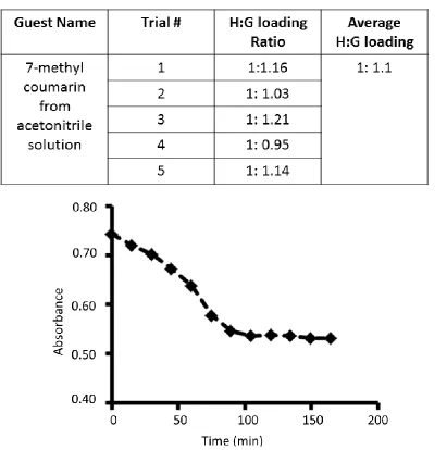
Table 2.4 Comparison of loading of 7-methyl coumarin from acetonitrile solution…….62
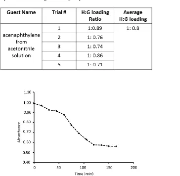
Table 2.5 Comparison of loading of acenaphthylene from acetonitrile solution………...63
Table 2.6 Comparison of loading of trans-stilbene from acetonitrile solution…………..64
Table 2.7 Literature reported and experimentally obtain PXRD data for host 1 • guest complexes and guest molecules……….70
Table 2.8 Photoreaction of 7-methyl coumarin inside host. ...81
Table 2.9 Moves and associated probability of Canonical Monte Carlo simulations for chemical potential calculations………..…83
Table 2.10 Moves and associated probability of Grand Canonical Monte Carlo
simulations……….84
Table 3.1 Guests absorbed by host 1………...112
Table 3.3 Moves and associated probabilities of Grand Canonical Monte Carlo
simulations………...134
Table 5.1 Electrochemical data for 1 in 0.1 M TBAPF6/DMF, GC as working electrode, Pt as counter electrode and scan rate of 100 mVs-1. Potentials reported versus the normal hydrogen electrode………...189
Table 5.2 Summary of Photocatalytic Studies ...192
Table 5.3 Photophysical properties of Ru(bpy)32+ and 1 in N2 deaerated acetonitrile at
room temperature (λex = 450 nm)………198
Table 5.4 Photocatalytic experiments in detail………202
Table 6.1 Crystal data and refinement results for compounds 1-6………..216
LIST OF FIGURES
Figure 1.1 Organic hosts that are used in solid-state host-guest chemistry discussed in this
chapter………..6
Figure 1.2 Structures of conventional urea host structure………...7
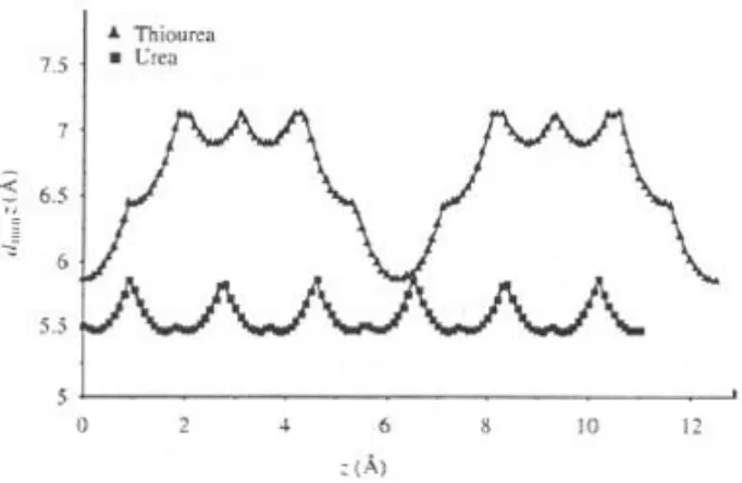
Figure 1.3 Compression of Minimum tunnel diameter (dmin) of urea and thiourea as a function of crystallographic axis z………...8
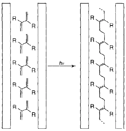
Figure 1.4 Packing of individual diene monomer molecules inside the channels formed by urea and thiourea. (R = H, 1,3-butadiene; R = CH3, 2,3-dimethylbutadiene; R = Cl, 2,3-dichlorobutadiene)………...10
Figure 1.5 Structure of the DCA. ...11
Figure 1.6 Schematic drawing of the trans-1,4-polybutadiene in the channels of its inclusion compound with PHTP. ...16
Figure 2.1 Columnar assembled host 1 forms porous crystals with accessible channels for binding guests………26
Figure 2.2 Views of host 1 and host 1 complexes……….28
Figure 2.3 Reversible absorption/desorption of guests ………..31
Figure 2.4 Absorption of guests by host 1 ...33
Figure 2.5 Formation of host•guest complexes ...34
Figure 2.6 Comparison of the observed PXRD of host 1 and its host•guest complexes ...37
Figure 2.7 Comparison of solid-state 13C{1H}CP-MAS NMR spectra ...39
Figure 2.8 GCMC simulations for the host 1•coumarin complex. ...47
Figure 2.9 GCMC simulation results for coumarin derivatives (Partial guest molecules omitted for clarity). ...50
Figure 2.11 Two bis-urea macrocycles studied using Hyperpolarized Xe-129 NMR with a side view of the packing arrangement of adjacent channels. ...53
Figure 2.12 Two bis-urea macrocycles studied using Hyperpolarized Xe-129 NMR with a side view of the packing arrangement of adjacent channels. ...54
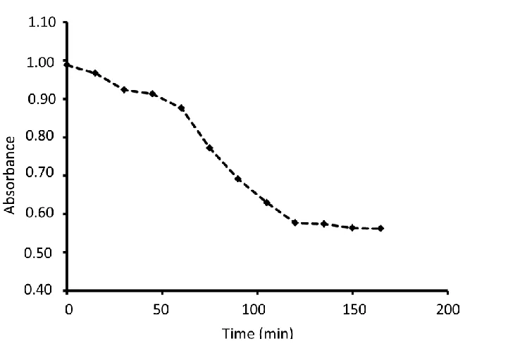
Figure 2.13 Depletion of 6-methyl coumarin concentration during introduction of this guest into the host 1 crystals with respect to time. Monitored by UV-vis spectroscopy at 273 nm………...59
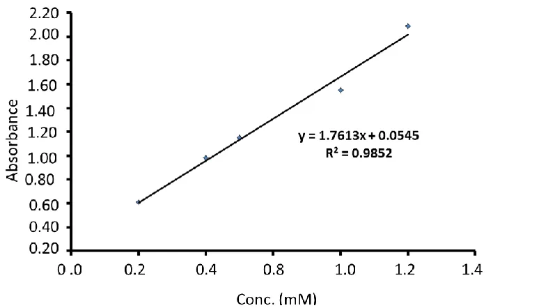
Figure 2.14 The Lambert-Beer analysis of 6-methyl coumarin solution in acetonitrile at increasing concentration monitored by UV-vis spectroscopy………..….60
Figure 2.15 Depletion of 7-methyl coumarin concentration during introduction of this guest into the host 1 crystals with respect to time as monitored by UV-vis spectroscopy at 276 nm. ...61
Figure 2.16 The Lambert-Beer analysis of 7-methyl coumarin solution in acetonitrile at increasing concentration monitored by UV-vis spectroscopy. ...61
Figure 2.17 Depletion of acenaphthylene concentration during introduction of this guest into the host 1 crystals with respect to time as monitored by UV-vis spectroscopy at 322 nm………..62
Figure 2.18 The Lambert-Beer analysis of acenaphthylene solution in acetonitrile at increasing concentration monitored by UV-vis spectroscopy………...63
Figure 2.19 Depletion of trans-stilbene concentration during introduction of this guest into the host 1 crystals with respect to time as monitored by UV-vis spectroscopy at 294 nm. ...63
Figure 2.20 The Lambert-Beer analysis of trans-stilbene solution in acetonitrile at increasing concentration monitored by UV-vis spectroscopy………...64
Figure 2.21 Desorption of cis-stilbene from host 1 as observed by TGA ...65
Figure 2.22 The PXRD analysis of host 1cis-stilbene complex compared with empty host crystals………...65
Figure 2.23 Desorption of trans-β-methyl styrene from host 1 as observed by TGA experiment………66
Figure 2.25 Depletion of 7-methoxy coumarin concentration during introduction of this guest into the host 1 crystals with respect to time as monitored by UV-vis spectroscopy at 315 nm. ...67
Figure 2.26 The Lambert-Beer analysis of 7-methoxy coumarin solution in acetonitrile at increasing concentration monitored by UV-vis spectroscopy………...67
Figure 2.27 Solid-state 13C{1H}CP-MAS NMR spectra for host 1. ...68
Figure 2.28 Solid-state 13C{1H}CP-MAS NMR for host 1•coumarin complex. ...68
Figure 2.29 Solid-state 13C{1H}CP-MAS NMR for host 1•6-methyl coumarin
complex………...68
Figure 2.30 Solid-state 13C{1H}CP-MAS NMR for host 1•6-methyl coumarin complex expanded to show the 20-60 ppm range. The arrow indicates a resonance with a reasonable shift for the 6-methyl group on coumarin………69
Figure 2.31 Solid-state 13C{1H}CP-MAS NMR for host 1• 7-methyl coumarin
complex………..69
Figure 2.32 Solid-state 13C{1H}CP-MAS NMR for host 1•7-methyl coumarin complex expanded to show the 20-60 ppm range. The arrow indicates a resonance with a reasonable shift for the 7-methyl group on coumarin ...69
Figure 2.33 Solid-state 13C{1H}CP-MAS NMR for host 1• 7-methoxy coumarin complex………..70
Figure 2.34 PXRD analysis of host 1• 6-methyl coumarin. ...71
Figure 2.35 PXRD analysis of host 1• 7-methyl coumarin complex. ...71
Figure 2.36 Predicted PXRD analysis of acenaphthylene crystals. The pattern was generated using the X-ray crystal data from reference 68……….72
Figure 2.37 Predicted PXRD analysis of only guest crystals trans- stilbene. The pattern
was generated using the X-ray crystal data from reference
69………...72
Figure 2.38 Predicted PXRD analysis of only guest crystals, another polymorph of 7-methyl coumarin. The pattern was generated using the X-ray crystal data from reference 70………...….73
Figure 2.39 The PXRD analysis of host 1trans-β-methyl styrene complex compared with
Figure 2.40 The PXRD analysis of host 1• acenaphthylene complex (top) and empty
crystals of host 1 (bottom)……….74
Figure 2.41 The PXRD analysis of host 1• trans-stilbene
complex………...74
Figure 2.42 The PXRD analysis of host 1•cis stilbene complex ...75
Figure 2.43 1H-NMR analysis of the product obtained from photoreaction of host 1 •6-methyl coumarin. The peaks correspond to the cyclobutyl region of the photodimers are shown. ………...75
Figure 2.44 1H-NMR analysis of the product obtained from photoreaction of host 1 •7-methyl coumarin. The peaks correspond to the cyclobutyl region of the photodimers are shown………...76
Figure 2.45 1H-NMR analysis of the product obtained from photoreaction of host 1 •7-methoxy coumarin……….76
Figure 2.46 1H NMR analysis of the reaction of the solid host 1•acenaphthylene complex under UV-irradiation for 12-96 h in an argon atmosphere. ...77
Figure 2.47 1H NMR spectra of anti-HH photodimer of 6-methyl coumarin (84%) and
syn-HH (~16%) dimer of 6-methyl coumarin. ...77
Figure 2.48 1H NMR spectra of anti-HH photodimer of 7-methyl coumarin. ...78
Figure 2.49 1H NMR spectra of syn photodimer of acenaphthylene. ...78
Figure 2.50 1H-NMR analysis of the product obtained from photoreaction of host 1 •6-methyl coumarin. The peaks correspond to the cyclobutyl region of the photodimers are shown……….50
Figure 2.51 1H-NMR analysis of the product obtained from photoreaction of host 1 •6-methyl coumarin (Ar atmosphere). The peaks correspond to the cyclobutyl region of the photodimers are shown………...…...51
Figure 2.52 1H-NMR analysis of the product obtained from photoreaction of host 1 •7-methyl coumarin. The peaks correspond to the cyclobutyl region of the photodimers are shown……….80
Figure 2.53 1H-NMR analysis of the product obtained from photoreaction of host 1 •7-methyl coumarin (Ar atmosphere). The peaks correspond to the cyclobutyl region of the photodimers are shown………..80
•β-Figure 2.55 GCMC simulation outcome of host 1• coumarin complex ...84
Figure 2.56 GCMC simulation outcome of host 1• 6-methycoumarin complex. ...85
Figure 2.57 GCMC simulation outcome of host 1• 7-methycoumarin complex. ...85
Figure 2.58 GCMC simulation outcome of host 1• 7-methoxy coumarin complex. ...86
Figure 2.59 GCMC simulation outcome of host 1• Acenaphthylene complex. ...86
Figure 3.1 Self-assembled phenylethynylene bis-urea macrocycles used as a confinement for conducting selective photodimerization of chromones ...99
Figure 3.2 Host 1 structure and schematic of guest exchange ...103
Figure 3.3 Analysis of chromone solid-state structures highlights the closest contact between potentially reactive alkenes (purple bonds). ...104
Figure 3.4 Results of GCMC modeling of host 1•guest complexes and analysis of the relative orientation of neighboring reactants. ...108
Figure 3.5 Loading of the guests and the depletion of each guest from the solution monitored by UV/Vis spectrophotometry. ...111
Figure 3.6 Monitoring the photoreaction of host 1•chromone and observed photoproducts. ...115
Figure 3.7 Monitoring the photoreaction of host 1•6-fluorochromone. ...116
Figure 3.8 Photoreaction of host 1· 6-bromochromone and observed photoproduct. ...117
Figure 3.9 1H NMR (DMSO-d6, 400 MHz) of 1. ...123
Figure 3.10 13C NMR spectra (DMSO-d6, 100 MHz) of 1. ...124
Figure 3.11 TGA profile of freshly crystallized host 1•DMSO………...125
Figure 3.12 One-dimensional chains of 7-hydroxy-4-chromone stack into layers with offset aryl stacking interactions………...125
Figure 3.13 1H NMR (300 MHz) of control photoreactions after 96 h under Ar (g) ...126
Figure 3.14 Loading of chromone into host 1 to form host 1•chromone complex. ...126
Figure 3.16 Loading of 6-bromochromone into host 1 to form host 1•6-bromochromone
complex. ...127
Figure 3.17 Loading of 7-hydroxy-4-chromone into host 1 to form host 1• 7-hydroxy-4-chromone complex. ...128
Figure 3.18 Loading of 3-cyanochromoneinto host 1. ...128
Figure 3.19 1H NMR (CDCl3, 300 MHz) of the chromone photodimer mixture in CDCl3 (anti-HT and anti-HH) after removal of the residual starting material………...129
Figure 3.20 GC trace of the chromone photodimer mixture (anti-HT and anti-HH) after 96 h UV-irradiation of host 1•chromone complex. Residual chromone was removed prior to GC by preparative TLC………...129
Figure 3.21 MS of the GC purified chromone photodimers anti-HT (top) and anti-HH (bottom) after 96 h UV-irradiation of host 1•chromone complex. ...130
Figure 3.22 1H NMR (CD2Cl2, 300 MHz) of the 6-fluorochromone anti-HH photodimer………...131
Figure 3.23 1H NMR (CDCl3, 300 MHz) of the 6-bromochromone aryl coupling adduct……….……..132
Figure 3.24 13C NMR (CDCl3, 300 MHz) of 6-bromochromone aryl coupling adduct..132
Figure 3.25 2D COSY NMR (CDCl3, 300 MHz) of 6-bromochromone aryl coupling adduct………...133
Figure 3.26 GCMC simulation outcome of host 1•chromone complex. ...135
Figure 3.27 GCMC simulation outcome of host 1•6-fluorochromone complex. ...135
Figure 3.28 GCMC simulation outcome of host 1•6-bromochromonecomplex. ...136
Figure 3.29 GCMC simulation outcome of host 1•7-hydroxy-4-chromonecomplex. ....136
Figure 4.1 Conventional synthesis compared to stereoselective polymerization of isoprene in the pyridyl phenylethynylene bis-urea ...144
Figure 4.2 Assembly of 1 and comparison with 2. (a) Chem draw structure of hosts 1 (X = N) and 2 (X= CH). ...147
Figure 4.3 Vapor loading of isoprene into the host 1 and PXRD analysis. ...149
Figure 4.6 13C-NMR (CDCl3, 100 MHz) of the diol compound. ...154
Figure 4.7 1H-NMR (CDCl 3, 400 MHz) of the dibromo compound. ...156
Figure 4.8 13C-NMR (CDCl3, 100 MHz) of the dibromo compound. ...156
Figure 4.9 1H-NMR (CDCl3, 300 MHz) of the protected macrocycle………....158
Figure 4.10 13C-NMR (CDCl3, 100 MHz) of the protected macrocycle……….158
Figure 4.11 1H-NMR (CDCl3, 400 MHz) of the bis-urea macrocycle………....160
Figure 4.12 13C-NMR (CDCl 3, 100 MHz) of the bis-urea macrocycle. ...160
Figure 4.13 1D channels extended along the crystallographic b axis. ...166
Figure 4.14 X-ray crystal structure of urea protected 1. ...167
Figure 4.15 Hirshfeld surface analysis of the macrocycle 1. ...170
Figure 4.16 Hirshfeld surface analysis of the macrocycle 2. ...171
Figure 4.17 Thermogravimetric analysis of host 1. ...172
Figure 4.18 Loading of isoprene, photo irradiation and polymer isolation. ...172
Figure 4.19 1H-NMR (CDCl3, 400 MHz) of trans-1, 4-polyisoprene. ...173
Figure 4.20 13C-NMR (CDCl3, 125 MHz) of trans-1,4-polyisoprene……….174
Figure 4.21 GPC trace of trans-1,4-polyisoprene. (Eluent: THF, calibrated to polystyrene standards)……….174
Figure 5.1 A conformationally mobile bipyridyl macrocycle was used as bridging ligand to complex two ruthenium bis(2,2'-bipyridine) units………..….183
Figure 5.2 Synthesis and the structure of [(bpy)2Ru(µ-L)Ru(bpy)2]Cl4•6H2O (1)……..185
Figure 5.3 Normalized absorption and emission spectra of 1 in N2 deaerated acetonitrile at room temperature (λex = 450 nm)……….187
Figure 5.4 DPV (top) and CV (below) of 1 complex in 0.1 M TBAPF6 in DMF...190
Figure 5.6 Overview of the Diels-Alder reaction between trans-anethole and isoprene.192
Figure 5.7 1H NMR spectrum of L in DMSO-d6………...194
Figure 5.8 1H NMR (400 MHz, CD3CN) spectrum of 1………...195
Figure 5.9 13C NMR (100 MHz, CD3CN) spectrum of 1………....196
Figure 5.10 Graphical plot ofCurrent vs. √𝜈 for first oxidation……….199
Figure 5.11 Comparison of oxidative currents of 10-3 M solution of 1(top) and 10-3 M solution of Ferrocene (below)………..200
Figure 5.12 Controlled potential (at 1.55V vs. NHE) electrolysis in 0.1 M TBAPF6/MeCN solution over the period of 60 minutes. ...200
Figure 5.13 CVs of complex 1 before (below) and after (top) 61 minutes of electrolysis in 0.1 M TBAPF6/MeCN solution. ...201
Figure 5.14 Absorption spectra of complex 1 in 0.1 M TBAPF6/MeCN solution before (red) and after (black) 61 mins of electrolysis………...201
Figure 5.15 1H NMR (300 MHz, CDCl3) spectrum of 4……….203
Figure 5.16 13C NMR (100 MHz, CDCl3) spectrum of 4………203
Figure 5.17 Hydrogen bonding network forms layers parallel to the crystallographic (bc) plane………...204
Figure 6.1 Probes for the effects of electron donating groups at the 6-position………..220
Figure 6.2 Crystal structure of chromones containing electron withdrawing groups at the 6- position. ...221
Figure 6.3 Fingerprint plots and surface maps for compound 1. ...223
Figure 6.4 Fingerprint plots and surface maps for compound 2. ...225
Figure 6.5 Fingerprint plots and surface maps for compounds 3. ...226
Figure 6.6 Fingerprint plots and surface maps for compounds 4. ...228
Figure 6.7 Fingerprint plots and surface maps for compounds 5. ...229
Figure 6.8 Fingerprint plots and surface maps for the 6-bromochromone 6. ...230
LIST OF SCHEMES
Scheme 1.1 Addition modes of isoprene during conventional polymerization leads to multiple isomers………...5
Scheme 1.2 Possible dimer products from the [2+2] photodimerization of chalcone and dibenzylidene………...13
Scheme 1.3 Possible dimer products from the [2+2] photodimerization
of coumarin.………...13
Scheme 2.1 Photolysis of Coumarin derivatives………...41
Scheme 4.1 Synthesis of the macrocycle……….…152
Scheme 4.2 Synthesis of the diol compound………..….153
Scheme 4.3 Synthesis of the dibromo compound………155
Scheme 4.4 Synthesis of the protected macrocycle ...157
CHAPTER I
1.1 Structure and reactivity of organic inclusion compounds: As reaction media for [2+2]
photodimerization and polymerization reactions.
Inclusion compounds have been demonstrated as powerful and fruitful media to
probe the solid-state host-guest chemistry. Early work by Sir Humphrey in 1811, reported
the first inclusion compound known as chlorine clathrate, which results from chlorine gas
trapped in water-ice sockets.The term inclusion compound was then introduced by W.
Schlenk to describe the crystalline adduct where the host molecule leads to isolation of
the guest molecule into well-defined cavities via the crystallization of host molecules in a
matrix.1 In 1945, H. M. Powell coined the synonym clathrate derived from the Latin word
clathratus which means “to fit with bars”.2 In addition, inclusion compounds with more
than one kind of discrete molecules in the crystal lattice have also been described using
the term “cocrystal”. Solid-state inclusion chemistry has proven useful for the separation
of mixtures,in the storage of gases and toxic substances, in the stabilization of reactive
compounds, in the control of release profile of a drugs under physiological conditions,
and for modulating reaction pathways by using as a molecular vessels.3 This chapter
focuses on solid inclusion in which guest molecule are embedded in the host lattice
structure.
The cavity free crystalline host is often referred to as the alpha phase. The empty
host (beta phase or apohost) is the host crystallized in a different crystal form that
contains cavities but is free of guest molecules. The cavities provide “inclusion space”
that can span a range of sizes and shapes. The interior geometry of inclusion spaces
include tunnels, isolated cages, inter-lamellar regions within layered hosts, interconnected
metastable form marked by its low density, which can be easily converted back to its
alpha phase. Guests can exist in the form of a solid, a liquid or even a gas. In contrast to
the solution state host-guest chemistry, in the solid-state, the aggregation of single
molecules builds up the host crystal. Here, the crystal itself is considered as the unit
entity. Therefore, the cavity for binding guest or guests does not need to be an intrinsic
property of the individual host molecules. When host molecules are crystalized with
suitable guest molecules, the guest may be trapped within the host.
Chemistry of solution state host-guest complexes, can be monitored in solution by
a range of techniques that are taught in the undergraduate level. Solid-state
characterization techniques are more typically seen in upper level courses. These
techniques range from the powerfully elucidating technique of single crystal X-ray,
which gives atomic resolution. In the absence of single crystals a number of solid state
material characterization material characterization methods must be applied to elucidate
information about the structure of the host-guest complex and to probe the key
interactions that occur to trap the guest within the host. Such characterization methods
include Single Crystal X-Ray Diffraction (SCXRD), Powder X-Ray Diffraction (PXRD),
solid state: NMR, IR, Raman spectroscopy, UV-vis, diffuse reflectance, X-ray
Photoelectron Spectroscopy (XPS), porosity analysis (BET), optical microscopy, and
thermochemical methods such as Thermogravimetric Analysis (TGA) and Differential
Scanning Calorimetry (DSC).
The host framework imposes structural and geometrical constraints on confined
guests within the inclusion compound, rendering the confined guests to display different
less mobile than in solution but may have sufficient mobility in order to undergo a
reaction with the nearest neighboring molecule. These reactions proceed according to the
“topochemical principle”5,6 where a minimum amount of molecular motion is required. In
other words, both regiochemistry and stereochemistry of the reaction product may be
governed by the relative position and the orientation of two reactant molecules within the
inclusion space. Therefore, the relative energies of the transition states within the solid
host could be very different from the relative energies for the corresponding reactions in
free state.4 Hence, the reactions occur in the inclusion space may favor a particular
reaction pathway. This affords more control over the reactivity and the reaction
selectivity by limiting the side reactions and often leads to the formation of one major
product.
Chapter 4 focuses on the polymerization of isoprene within confined channels of
assembled bis-urea macrocycles. Thus, it is expedient for us to consider the example of
polymerization of isoprene by conventional means.7 Free radical polymerization methods
yield polyisoprene that has multiple stereoisomers within its microstructure (Scheme 1.1).
Scheme 1.1. Addition modes of isoprene during conventional polymerization leads to multiple isomers.
In comparison, when isoprene is constrained into small one dimensional channels
inclusion polymerization can selectively yield the linear form trans-1,4-isomer.8 Also
advantageous, the use of host•isoprene inclusion complex as a medium for
polymerization removes the need for radical initiators, solvents, and specialized handling
procedures, which are necessary for conventional polymerization. This method employs
mild initiation techniques that are sufficient to generate the initial radicals needed for
polymerization. Polymers generated often are well defined and in some occasions have
low polydispersities. It is indicative of a controlled radical polymerization inside the one
dimensional channels of the host structure. Most importantly, after removing the resultant
polymer molecules the host materials can be recovered and reused. The sustainability of
the host crystals may make this approach more environmentally friendly.
This introductory chapter discusses the functionality of solid-state inclusion
compounds in terms of their ability to act as molecular scale vessels to carry out
photoreactions in high selectivity and conversion efficiency. We will limit our discussion
to [2+2] photodimerizations and polymerizations as model reactions to understand how
solid state host-guest complexes can alter the reactivity of guest and control the regio and
stereo selectivity of the reaction. The structural features of corresponding inclusion
Figure 1.1. Organic hosts that are used in solid-state host-guest chemistry discussed in this chapter
1.2 Urea and thiourea based inclusion compounds
Urea and its sulfur analogue thiourea (1 and 2 in Figure 1.1) are known to form
solid-state clathrates with variety of hydrocarbons. Both compounds form chiral helical
hollow structures, which are stabilized by intermolecular hydrogen bonds between NH2
protons and oxygen or sulphur atom of the adjacent molecule (Figure 1.2). Although they
have similar bonding pattern, the structures formed by both urea and thiourea has its own
subtle differences probably arising from the chemical nature of oxygen and sulphur. Urea
forms helical tunnel like hexagonal shaped channels also known as β-urea with channel
diameters around 5.5 Å whereas thiourea has a more cage like cavity with diameters
about 7 Å (Figure 1.2).3,4,9-12 Furthermore, Figure 1.3 illustrates a comparison of channel
Figure 1.2. Structures of conventional urea host structure. (a) Hexagonal channels parallel to the channel axis (b) Similar view showing van der Waals radii of the host molecules (c) Helical ribbon structure.
Urea tunnel structures are known to have smooth internal surfaces in comparison
to cavities formed by thiourea.3 The differences in the structure and nature of the cavities
dictate the binding of guests to form the corresponding clathrate. Urea tends to absorb
linear hydrocarbons where as thiourea has the ability to absorb branched hydrocarbons.
In host-guest chemistry studies, both urea and thiourea based clathrates have been widely
explored.13-17 Since urea does not have an auxiliary hydrogen bonding site for guest
molecules, the clathrates often show nonstoichiometric guest binding and substantial
Figure 1.3. Compression of Minimum tunnel diameter (dmin) of urea and thiourea as a function of crystallographic axis z.
Urea and based clathrates have been used to conduct inclusion polymerization
reactions that employ a range of monomers. The first polymerization was reported by
Clasen and coworkers in 1956.18 They observed that the inclusion compound formed
between thiourea and 2,3-dimethylbutadiene undergoes spontaneous polymerization
overtime without any initiation. Since then a number of groups have investigated the
polymerization reactions in urea and thiourea. These findings are summarized in the table
Table 1.1. Summary of the polymerization reactions done in urea and thiourea based inclusion compounds.
Entry Host Monomer Polymer Reference
1 Urea 1,3-butadiene trans-1,4-polybutadiene 19
2 Urea vinyl chloride syndiotactic
polyvinylchloride 19
3 Urea acrylonitrile polyacrylonitrile 20
4 Urea acrylonitrile isotactic polyacrylonitrile 20
5 Thiourea
2,3-dimethylbutadiene
trans
-1,4-polydimethylbutadiene 22
6 Thiourea
2,3-dichlorobutadiene
trans
-1,4-polydichlorobutadiene 22
7 Thiourea
1,3-cyclohexadiene
trans
-1,4-polycyclohexadiene 22
White and co-workers polymerized 1,3-butadiene and vinyl chloride in urea
inclusion complexes.19 Gamma irradiation of urea•1,3-butadiene selectively produced the
trans-1,4-polybutadiene (100%) (Table 1.1 entry 1) and urea•vinyl chloride yielded the
highly stereo regular syndiotactic polyvinylchloride (Table 1.1 entry 2). Tonelli and
coworkers have reported the polymerization of acrylonitrile in urea matrix under two
different conditions.20 At room temperature, photoirradiation of urea•acrylonitrile
complex yielded polyacrylonitrile (Table 1.1 entry 3) while polymerization at low
temperatures produced isotactic polyacrylonitrile with >80% m-diad content (Table 1.1
entry 4).19,21 Thiourea inclusion complexes have also been investigated to drive
polymerizations. Brown and White reported the polymerization of
2,3-dimethylbutadiene, 2,3-dichlorobutadiene, and 1,3-cyclohexadiene with thiourea to
selectively produce trans-1,4-polymer (Figure 1.4) in each case (Table 1.1 entry 5, 6, and
7 ).22 In 2008, Cataldo and coworkers analyzed the microstructure of
polymerization.23-25 The polymers from inclusion polymerizations showed high trans
content (97%) when compared to polymers obtained from bulk polymerization, which
yielded very low trans content and high percentages of 1,2-units.
Figure 1.4. Packing of individual diene monomer molecules inside the channels formed by urea and thiourea. (R = H, 1,3-butadiene; R = CH3, 2,3-dimethylbutadiene; R = Cl, 2,3-dichlorobutadiene)
1.3 Acid based inclusion compounds
Discovery of clathrates with fatty acid derivatives dates back to early 1910 when
Wieland discovered a series of crystalline compounds known as “choleric acids”.
Clathrates formed by fatty acid Deoxycholic Acid (DCA) (3 in Figure 1.1) have been
particularly well studied. In the solid state, DCA molecules assemble into bilayer helical
type structure held together by hydrogen bonding between two hydroxyl groups.26 These
lipophilic layer with the channel diameters of 2.6 x 7.0 Å (Figure 1.5).27,28 The first
account of solid state polymerization within DCA was reported by Miyata and
coworkers.29 They investigated the dimethyl butadiene and
DCA•2,3-dichlorobutadiene to form the corresponding well defined polymers with high trans
content.30 Since then a number of groups have investigated the polymerization in DCA.
These findings are summarized in the table 1.2.
Figure 1.5. Structure of the DCA. (a) Individual DCA molecules held together by hydrogen bonding to form chains. (b) Stacking pattern of chains to form layers. (c) Bilayers consist of alternating stacks of hydrophobic and lipophilic layers with the channel diameters of 2.6 x 7.0 Å.
Table 1.2. Summary of the polymerization reactions done in DCA based inclusion compounds.
Entry Monomer Polymer Reference
1 cis-1,3-pentadiene trans-1,4-polypentadiene 31
2 trans
-1,3-pentadiene trans-1,4-polypentadiene 31
3
2,3-dimethylbutadiene trans-1,4-polydimethylbutadiene 33
4
Audisio and coworkers discovered that in the presence of DCA polymerization of
cis-1,3-pentadiene and trans-1,3-pentadiene yield trans-1,4-polypentadiene.31 This
polymerization was induced by gamma irradiation and the cis-1,3-pentadiene shows the
highest stereospecificity. Further investigations showed that the stereospecificity is
controlled by the van der Waals interactions between the host tunnels and the monomer
molecules.32 Miyata and coworkers have reported the polymerization of
2,3-dimethylbutadiene in DCA and variety of DCA derivatives such as apocholic acid
(ACA), cholic acid (CA), and chenodeoxycholic acid (CDCA).33 The DCA and ACA
having the similar channel diameter gave the highest selectivity for trans
-1,4-polydimethylbutadiene (>99%). The CA and CDCA, which have larger channel
diameters compared to DCA and ACA produced polymers with less trans-content (54%)
with 38-42% cis-1,4- and 4-8% of 1,2- addition product.The polymerization of
3-methyl-1,4-pentadiene was reported by Cataldo and coworkers in 2010 using DCA. They were
able to synthesize trans-1,4-poly(3-methyl-1,4-pentadiene) in very high selectivity.34,35
Stereoregular polymers have a tendency to pack efficiently, rendering highly crystalline
materials with improved mechanical properties. Particularly stereoregular polymers
produced from diene monomers such as isoprene and 1,3-butadiene are heavily used in
the synthetic elastomer industry.
1.4 Diol based clathrates
Solid state inclusion compounds based on alcohol containing host molecules have
been investigated. Figure 1.1 illustrates the molecular structures of such host molecules
acceptor moieties. Therefore, the inclusion complex is held together by hydrogen
bonding interactions that organize and position the reactive alkenes in the solid-state.
This organization defines relative orientation of the nearest neighboring guest molecule
and controls the stereo and regiochemistry of the product.
Scheme 1.2. Possible dimer products from the [2+2] photodimerization of chalcone and dibenzylidene.
Photodimerization of chalcone and dibenzylidene acetone have been extensively
studied with the hosts 4 and 5. In solid state they both can form four possible dimer
products under UV- irradiation (Scheme 1.2).36-39 UV-irradiation of neat trans-chalcone
results in the formation of mixture of [2+2] dimer products in low product selectivity
(Table 1.3 entry 1). As reported by Kaftory and coworkers, the irradiation of trans
-Chalcone with host 4 produced product 3 exclusively (Table 1.3 entry 2).40
Dibenzylidene acetone on the other hand forms complex with host 5, which selectively
control experiments without the host molecules shows no product formation (Table 1.3
entry 4).40 Host molecules 6, 7, and 8 have been investigated in terms of facilitating the
[2+2] dimerization of coumarin. In the presence of UV light coumarin has the possibility
to form four different dimer products (Scheme 1.3).
Table 1.3. Summary of the [2+2] photodimerization of chalcone and dibenzylidene acetone using hots 4 and 5.
Entry Media Guest molecule Product 1 Product 2 Product 3 Product
4 Reference
1 Neat solid
trans
-Chalcone x x x x 36
2 4 trans
-Chalcone x 40
3 Neat solid
Dibenzylidene
acetone 37
4 5 Dibenzylidene
acetone x 40
Scheme 1.3. Possible dimer products from the [2+2] photodimerization of coumarin.
Reaction of host 6•coumarin selectively produces the syn-HH product exclusively
(Table 1.4 entry 2) while the reaction of host 7•coumarin yields a different dimer product
namely anti-HT (Table 1.4 entry 3).38 Toda and coworkers analyzed the effects of host 8
on the dimerization of coumarin.39 Interestingly the outcome of the reaction was
depended on the solvent used for the crystallization of the inclusion compound. The
complex host 8•coumarin crystallized from ethyl acetate/hexane gives the product anti
(Table 1.4 entry 5).39 In addition Venkatesan and coworkers have used host 6 to drive the
photodimerization of several coumarin derivatives including methylcoumarin,
7-methoxycoumarin, 4,7-dimethylcoumarin, 4,6-dimethylcoumarin, and 4-chlorocoumarin.
Photoirradiation of the inclusion compound, host 6•7-methylcoumarin yielded the syn
-HH in 90% and host 6•7-methoxycoumarin proceed to form syn-HH in 66%. No
reactions were observed with 4,7-dimethylcoumarin, 4,6-dimethylcoumarin, and
4-chlorocoumarin with the host 6.41
Table 1.4. Summary of the [2+2] photodimerization of coumarin done using hots 6, 7, and 8.
Entry Media syn-HH syn-HT anti-HH anti-HT Reference
1 Neat
solid x x x x
2 6 x 38
3 7 x 38
4 8 x 39
5 8 x 39
1.5 Perhydrotriphenylene (PHTP)
The host molecule perhydrotriphenylene was synthesized by Sohrauth and Gorig
in 1923.42 The compound exists in two stereoisomeric forms. The isomer 9 appears to be
the most stable form due to high symmetry (Figure 1.1). Isomer 9 can exist in optically
active enantiomers in spite of its high symmetry. PHTP forms channels that are nearly
cylindrical in shape with diameters in the range of 5.25-5.50 A.27,43,44 Their aliphatic
channels. Farina and co-workers have used the tunnel hosts to facilitate inclusion
polymerization of a wide range of monomers.45,46
Figure 1.6. Schematic drawing of the trans-1,4-polybutadiene in the channels of its inclusion compound with PHTP.
The monomers such as 1,3-butadiene, 2,3-dimethylbutadiene, trans-pentadiene,
and cis-pentadiene have been investigated as complexes with PHTP. All these monomers
form the trans-1,4 polymer exclusively upon irradiation of their PHTP inclusion
complexes. The radical molecule serving as the initiator needed for the polymerization
was produced using gamma rays and the polymer was extracted using a suitable solvent
under refluxing conditions. The same group reported the first example of asymmetric
(Figure 1.6). The investigations showed that the optical activity arises from the chiral
environment of the (-)(R)-PHTP host structure.43,47,48
The research thrust of Shimizu group lies on solid-state host-guest chemistry,
which is introduced in the above description. The solid host is formed by bis-urea
macrocycles. The succeeding chapters focus on three bis-urea macrocyclic systems,
namely phenylethynylene, pyridine-phenylethylene, and bipyridine with an emphasis on
self-assembly and their utility to complex guests and modulate the reactivity of the
included guests. The chapters 2 and 3 describe studies done on the scope and applications
of the self-assembled phenylethynylene bis-urea macrocycle as a nanoreactor for
selective [2+2] photodimerization reactions. The chapter 4 discusses the synthesis and
utility of pyridine-phenylethylene macrocycles as confined environment for producing
stereo-regular polymers. The chapter 5 provides a concise account of utility of bipyridine
bis-urea macrocycle as a candidate to study the architectures formed by metal ligand
coordination and hydrogen bonds in the self-assembled system.
1.6 References
(1) Schlenk, W. Liebigs Ann. Chem.1949, 564, 204-240.
(2) Palin, D. E., Powell, H. M. Nature1945, 156, 334-335.
(3) Steed, J. W.; Atwood, J. L.: Solid-State Inclusion Compounds. In Supramolecular
Chemistry; John Wiley & Sons, Ltd, 2009; pp 385-440.
(4) Hollingsworth, M. D.; Harris, K. D. M. Comprehensive Supramolecular Chemistry,
eds. Toda, F.; MacNicol, D. D.; Bishop, R. Pergamon Press, 1996, vol. 6, pp. 177.
(5) Elizabé, L.; Kariuki, B. M.; Harris, K. D. M.; Tremayne, M.; Epple, M.; Thomas, J.
Chloroacetate: Structure Determination from Powder Diffraction Data by the Monte
Carlo Method. The Journal of Physical Chemistry B1997, 101, 8827-8831.
(6) Kohlschütter, V.; Tüscher, J. L. Zur Kenntnis topochemischer Reaktionen. Über
Bildung und Verhalten von Kupferhydroxyd. Zeitschrift für anorganische und allgemeine
Chemie1920, 111, 193-236.
(7) Jitchum, V.; Perrier, S. Living Radical Polymerization of Isoprene via the RAFT
Process. Macromolecules2007, 40, 1408-1412.
(8) Allcock, H. R.; Silverberg, E. N.; Dudley, G. K.; Pucher, S. R. Inclusion
Polymerization within a Tris(2,3-naphthylenedioxy)cyclotriphosphazene Clathrate.
Macromolecules1994, 27, 7550-7555.
(9) Fetterly, L. C. Non-Stochiometric Compounds, ed. Mandelcorn, L. Academic Press,
New York, 1964, pp. 491.
(10) Harris, K. D. M. Structural and dynamic properties of urea and thiourea inclusion
compounds. Journal of Molecular Structure1996, 374, 241-250.
(11) Harris, K. D. M. Meldola Lecture: understanding the properties of urea and thiourea
inclusion compounds. Chemical Society Reviews1997, 26, 279-289.
(12) Harris, K. D. M.; Palmer, B. A.; Edwards-Gau, G. R.: Reactions in Solid-State
Inclusion Compounds. In Supramolecular Chemistry; John Wiley & Sons, Ltd, 2012.
(13) Chao, M.-H.; Kariuki, B. M.; Harris, K. D. M.; Collins, S. P.; Laundy, D. Design of
a Solid Inclusion Compound with Optimal Properties as a Linear Dichroic Filter for
X-ray Polarization Analysis. Angewandte Chemie2003, 115, 3090-3093.
(14) Guillaume, F. Des cristaux organiques d'intercroissance incommensurables 1-D : les
(15) Harris, K. D. M. Investigating the Structure and Dynamics of a Family of Organic
Solids: The Alkane/Urea Inclusion Compounds. Journal of Solid State Chemistry1993,
106, 83-98.
(16) Harris, K. D. M. Fundamental and Applied Aspects of Urea and Thiourea Inclusion
Compounds. Supramolecular Chemistry2007, 19, 47-53.
(17) Hollingsworth, M. D. Crystal Engineering: From Structure to Function. Science
2002, 295, 2410-2413.
(18) Clasen, H. Elektrochem. 1956, 60, 982-987.
(19) White, D. M. Stereospecific Polymerization in Urea Canal Complexes1. Journal of
the American Chemical Society1960, 82, 5678-5685.
(20) Yang, H.; El-Shafei, A.; Schilling, F. C.; Tonelli, A. E. Why is Stereoregular
Polyacrylonitrile Obtained by Polymerization in Urea Canals Isotactic? Macromolecular
Theory and Simulations2007, 16, 797-809.
(21) Minagawa, M.; Ute, K.; Kitayama, T.; Hatada, K. Determination of Stereoregularity
of .gamma.-Irradiation Canal Polymerized Polyacrylonitrile by 1H 2D J-Resolved NMR
Spectroscopy. Macromolecules1994, 27, 3669-3671.
(22) Brown, J. F.; White, D. M. Stereospecific Polymerization in Thiourea Canal
Complexes1. Journal of the American Chemical Society1960, 82, 5671-5678.
(23) Chatani, Y.; Kuwata, S. Structural Investigation of Radiation-Induced Urea Canal
Polymerization of 1,3-Butadiene. Macromolecules1975, 8, 12-18.
(24) Chatani, Y.; Nakatani, S. Structural Evidence of Radiation-Induced Thiourea Canal
(25) Chatani, Y.; Nakatani, S.; Tadokoro, H. Structural Study of the Polymerization of
2,3-Dichlorobutadiene in the Thiourea Canal Complex. Structural Change during Canal
Polymerization and the Crystal Structure of the Resultant Polymer. Macromolecules
1970, 3, 481-486.
(26) Kato, K.; Sugahara, M.; Tohnai, N.; Sada, K.; Miyata, M. Drastic Increase in the
Flexibility of Open Host Frameworks of a Steroidal Host Compound upon Shortening Its
Spacer. European Journal of Organic Chemistry2004, 2004, 981-994.
(27) Giglio, E. I. c., eds. Atwood, J. L.; Davies, J. E. D. MacNicol, Academic Press, New
York, 1984, 207.
(28) Miyata, M. S., K. Comprehensive Supramoleculer Chemistry, eds. Toda, F.;
MacNicol, D. D.; Bishop, R. Pergamon Press, 1996, vol. 6, pp. 147.
(29) Miyata, M.; Takemoto, K. Radiation-induced polymerization of 2,3-disubstituted
butadienes in deoxycholic acid inclusion compounds. Journal of Polymer Science:
Polymer Letters Edition1975, 13, 221-223.
(30) Miyata, M.; Takemoto, K. Radiation-induced polymerization of vinyl and diene
monomers in deoxycholic acid inclusion compounds. Journal of Polymer Science:
Polymer Symposia1976, 55, 279-285.
(31) Audisio, G.; Silvani, A. Inclusion asymmetric polymerization of penta-1,3-dienes in
deoxycholic acid. Journal of the Chemical Society, Chemical Communications 1976,
481-482.
(32) Audisio, G.; Silvani, A.; Zetta, L. Inclusion asymmetric polymerization in
deoxycholic acid by "through-space" asymmetric induction. Macromolecules 1984, 17,
(33) Nakano, K.; Sada, K.; Miyata, M. Inclusion Polymerization in Polymorphic Crystals
of Cholic Acid. Polym J2001, 33, 172-176.
(34) Cataldo, F.; Angelini, G.; Iglesias-Groth, S.; Manchado, A. Solid state radiolysis of
amino acids in an astrochemical perspective. Radiation Physics and Chemistry 2011, 80,
57-65.
(35) Cataldo, F.; Ursini, O.; Ragni, P.; Rosati, A. Radiation-induced polymerization of
2,3-dimethyl-1,3-butadiene clathrate in deoxycholic acid. Journal of Radioanalytical and
Nuclear Chemistry2009, 280, 99-106.
(36) Lewis, F. D.; Barancyk, S. V. Lewis acid catalysis of photochemical reactions. 8.
Photodimerization and cross-cycloaddition of coumarin. Journal of the American
Chemical Society1989, 111, 8653-8661.
(37) Moorthy, J. N.; Venkatesan, K.; Weiss, R. G. Photodimerization of coumarins in
solid cyclodextrin inclusion complexes. The Journal of Organic Chemistry 1992, 57,
3292-3297.
(38) Tanaka, K.; Mochizuki, E.; Yasui, N.; Kai, Y.; Miyahara, I.; Hirotsu, K.; Toda, F.
Single-Crystal-to-Single-Crystal Enantioselective [2+2] Photodimerization of Coumarin,
Thiocoumarin and Cyclohex-2-enone in the Inclusion Complexes with Chiral Host
Compounds. Tetrahedron2000, 56, 6853-6865.
(39) Tanaka, K.; Toda, F. Selective photodimerisations of coumarin in crystalline
inclusion compounds. Journal of the Chemical Society, Perkin Transactions 11992,
943-944.
(40) Kaftory, M. Reactions in the solid state : III. Structural aspects of photochemical
(41) Moorthy, J. N.; Venkatesan, K. Stereospecific photodimerization of coumarins in
crystalline inclusion complexes. Molecular and crystal structure of 1:2 complex of
(S,S)-(-)-1,6-bis(o-chlorophenyl)-1,6-diphenyl-hexa-2,4-diyne-1,6-diol and coumarin. The
Journal of Organic Chemistry1991, 56, 6957-6960.
(42) Sohrauth, W. G., K. Ber. 1923, 56, 2024.
(43) Farina, M.; Di Silvestro, G.; Sozzani, P. Optically active block copolymers by
inclusion polymerization: Evidence for ‘through-space’ asymmetric induction. Die
Makromolekulare Chemie, Rapid Communications1981, 2, 51-54.
(44) Farina, M. in 'Inclusion Compounds' (Eds J. L. Atwood et al.), Vol. 2, Academic,
New York, 1984, p. 69
(45) Farina, M.; Natta, G.; Allegua, G.; Löffelholz, M. Inclusion compounds of linear
polymers and polymerization of monomers included in perhydrotriphenylene. Journal of
Polymer Science Part C: Polymer Symposia1967, 16, 2517-2524.
(46) Farina, M.; Pedretti, U.; Gramegna, M. T.; Audisio, G. Stereospecific and
Asymmetric Inclusion Polymerization I. Polymerization of trans- and cis-1,3-Pentadiene
Included in Racemic Perhydrotriphenylene. Macromolecules1970, 3, 475-480.
(47) Colombo, A.; Allegra, G. Single Crystal to Single Crystal Transformation of
Perhydrotriphenylene Inclusion Compounds during Canal Polymerization of Butadiene.
Macromolecules1971, 4, 579-584.
(48) Farina, M.; Audisio, G.; Natta, G. Asymmetric synthesis. Radiation
polymerization of trans-1,3-pentadiene included in optically active perhydrotriphenylene.
CHAPTER II
APPLICATIONS OF A BIS-UREA PHENYLETHYNYLENE SELF-ASSEMBLED
NANOREACTOR FOR [2+2] PHOTODIMERIZATIONS*
2.1 Abstract
Confined environments can be used to alter the selectivity of a reaction by influencing the
organization of the reactants, altering the mobility of trapped molecules, facilitating one
reaction pathway or selectively stabilizing the products. This chapter utilizes a series of
potentially photoreactive guests to interrogate the utility of the one-dimensional
nanochannels of a porous host to absorb and to facilitate the reaction of encapsulated
guests. The host is a columnar self-assembled phenylethynylene bis-urea macrocycle,
which absorbs guests including coumarin, 6-methyl coumarin, methyl coumarin,
7-methoxy coumarin, acenaphthylene, cis-stilbene, trans-stilbene and trans-β-methyl
styrene to afford crystalline inclusion complexes. We examine the structure of the
host:guest complexes using powder X-ray diffractions, which suggests that they are
well-ordered highly crystalline materials. Investigations using solid state cross-polarized
magic angle spinning 13C{1H}CP-MAS NMR spectroscopy indicate that the guests are
mobile relative to the host. Upon UV-irradiation, we observed selective
photodimerization reactions for coumarin, 6-methyl coumarin, 7-methyl coumarin, and
acenaphthylene, while the other substrates were unreactive even under prolonged
UV-irradiation. Grand Canonical Monte Carlo simulations suggest that the reactive guests
were close paired and preorganized in configurations that facilitate the photodimerization
with high selectivity while the unreactive guests did not exhibit similar close pairing. A
greater understanding of the factors that control diffusion and reaction in confinement
2.2 Introduction
Confined environments can potentially be used to modulate the chemical reactivity of
encapsulated guests with the goal of controlling their reactions and inducing selectivity.1,2
A host that provides a confinement environment for reaction is popularly termed a
‘nanoreactor’.3 A few of the chemical processes that are facilitated within nanoreactors
include unimolecular aza-cope rearrangements,4,5 bimolecular Diels-Alder reactions,6,7
oxidations,8,9 and [2+2] photodimerization reactions.10,11 They have also been used to
stabilize reactive substances12,13 and intermediates.14-17 In many cases, the encapsulated
guest molecules interact both with the walls of the host and with each other and can be
constrained to adopt a particular orientation within these small spaces.18 The interactions
that orient these guests depend on their chemical nature and on the specific structure of
the hosts and occur between the host and guests and between neighboring guests. The
strength, directionality and reversibility of these interactions guide the structure of these
complexes both before and after reaction. A greater understanding of the factors that
control reaction in confinement could lead to the development of better catalysts.
Recently, we reported bis-urea phenylethynylene macrocycle 1 (Figure 2.1a), which
assembles into columnar structures from several solvents.19 These columns pack together
to afford micron sized porous crystals with nanometer range channels. The crystallization
solvent could be removed by heating, and the empty host displayed permanent porosity
by gas adsorption and showed a surface area of ~ 350 m2/g. From the X-ray structure of
host 1•nitrobenzene (Figure 2.1c), one can see that the accessible columns are lined with
ureas and aryl groups. This manuscript explores the absorption of a series of guests
porous crystals. We examine the structure of these crystalline inclusion complexes by
both solid-state and computational studies using Grand Canonical Monte Carlo (GCMC)
simulations. The simulations were able to differentiate between the guests that undergo
reactions within the columnar channels (coumarin, 6-methyl coumarin, 7-methyl
coumarin and acenaphthylene) versus guests that were unreactive within the channels
(7-methoxy coumarin, stilbenes and styrene). Guests that were reactive were bound in close
proximity within the channels in relative geometries that were close to those required for
photoreaction. In contrast, unreactive guests were not close paired but were randomly
distributed within the tubes and displayed few contacts with neighboring guests.
The uptake of reactants into open cavities, pores, and channels or the formation of
co-crystals results in complexes where the guests display restricted motion, altered mobility
or preorganization that can lead to selective conversions or facilitate pathways and
products that are not observed in solution.20 It is the organization of reactants within this
confined space or ‘reaction cavity’21-23 which are key to understanding the product
distribution for a given transformation. Imagination and synthetic accessibility are a few
of the limits when it comes to designing a confined space. The confined space may
consist of a discrete cavity or pore in a small molecular host in solution such as
cyclodextrins,24 calixarenes,25 or cucurbiturils.26 It could be the larger interiors of small to
medium sized assembled structures, such as cavitands27 or Gibbs Octa acids,28 as well as
nanoscale structures such as coordination spheres,1 proteins and polymers.29 Reaction
cavities are not limited to soluble hosts in solution, but can also be voids in solids or
templated and preorganized asssemblies in co-crystals such as the innovative work from
Toda,30 and MacGillivray.31
In comparison, host 1 presents a high density of aligned, one-dimensional channels
with ~ 9 Å diameter (Figure 2.1a), which are accessible to guests. Previously, we have
loaded coumarin into these channels. Figure 2.2a illustrates a view of half of the channel
from the X-ray structure to highlight the aryl, ethynylene, and urea groups that line the
channels. Our hypothesis is the ureas are unable to participate in further hydrogen
bonding interactions with the guests as the three centered urea hydrogen bonding motif is
used to construct the columnar framework. Molecular modeling with Scienomics
MAPS32 of the host 1•coumarin complex suggests that the encapsulated guests form aryl
channel as well as dipole interactions between the coumarin oxygen and the urea groups
(Figure 2.2b).
Figure 2.2. Views of host 1 and host 1 complexes: a) View of half of the channel illustrating the aryl, ethynyl and hydrogen bonded urea groups that line the interior. b) Molecular models of the host 1•coumarin inclusion complexes generated with Scienomic’s MAPS illustrate the aryl stacking interactions that can occur between the neighboring coumarins as well as the dipole-dipole interactions between coumarin and the column walls. c) Aryl stacking and CH-л aid in binding of 6-methyl coumarin in the channel’s interior.
We chose to test seven different of guests: those that undergo [2+2]
photocycloadditions (6-methyl and 7-methyl coumarin, 7-methoxy coumarin and
-stilbene and trans-β-methyl styrene) (Table 3.1). In addition, trans-β-methyl styrene can
also act as a probe to test if the host itself can be a photosensitizer, as it will only undergo
isomerization in the presence of a medium energy sensitizer, such as chrysene or
1-acetylnaphthalene.33 We evaluated the absorption of these guests by host 1 and
characterized the structure of their inclusion complexes by solid-state methods. We then
investigated if these encapsulated guests would undergo photochemical reactions upon
UV-irradiation. Some of the guests underwent photochemical reactions within the solid
complex in moderate to good yields with high selectivity while others were unreactive
within these solid inclusion complexes. Molecular modeling studies allowed us to probe
the fit of the guests inside the channel of the host. These studies were directed at
understanding the following questions: Are certain orientations of the guest molecules
stabilized by the confinement? Are they appropriately oriented to undergo
photodimerization or photoisomerization reaction?
2.3 Results and discussion
In our previous work, we reported the X-ray structure of host 1 from
DMSO/nitrobenzene (host 1•nitrobenzene) and demonstrated that the structure of the host
is similar when crystallized from DMSO (host 1•DMSO).The encapsulated solvent can
be removed from the channels of each of these complexes to give the same empty host as
indicated by their identical PXRD pattern. The channels can subsequently be reloaded
with either solvent or alternatively with a different guest. Figure 2.1c highlights the
channel of this host, which is approximately ~ 9 Å in diameter. The channel is lined with
polar urea groups that are occupied in the hydrogen bonding scheme that runs along the