International Journal of Nanomedicine
Evaluation of the increase in permeability
of the blood–brain barrier during tumor
progression after pulsed focused ultrasound
Feng-Yi Yang1,* Hsin-Ell Wang1,* Guan-Liang Lin1 Hui-Hsien Lin1 Tai-Tong Wong2
1Department of Biomedical Imaging
and Radiological Sciences, School of Biomedical Science and Engineering, National Yang-Ming University,
2Department of Neurosurgery,
Neurological Institute, Taipei Veterans General Hospital, Taipei, Taiwan
*These authors contributed equally to this work
Correspondence: Feng-Yi Yang Department of Biomedical Imaging and Radiological Sciences, School of Biomedical Science and Engineering, National Yang-Ming University, Taipei, Taiwan
Tel +886 2 2826 7281 Fax +886 2 2820 1095 Email fyyang@ym.edu.tw
Purpose: The purpose of this study was to evaluate the permeability of the blood–brain barrier after sonication by pulsed high-intensity focused ultrasound and to determine if such an approach increases the tumor:ipsilateral brain permeability ratio.
Materials and methods: F98 glioma-bearing Fischer 344 rats were injected intravenously with Evans blue with or without blood–tumor barrier disruption induced by transcranial pulsed high-intensity focused ultrasound. Sonication was applied at a frequency of 1 MHz with a 5% duty cycle and a repetition frequency of 1 Hz. The permeability of the blood–brain bar-rier was assessed by the extravasation of Evans blue. Contrast-enhanced magnetic resonance images were used to monitor the gadolinium deposition path associated with transcranial pulsed high-intensity focused ultrasound, and the influencing size and location was also investigated. In addition, whole brain histological analysis was performed. The results were compared by two-tailed unpaired t-test.
Results: The accumulation of Evans blue in brains and the tumor:ipsilateral brain permeability ratio of Evans blue were significantly increased after pulsed high-intensity focused ultrasound exposure. Evans blue injection followed by sonication showed an increase in the tumor:ipsilateral brain ratio of the target tumors (9.14:1) of about 2.23-fold compared with the control tumors (x4.09) on day 6 after tumor implantation. Magnetic resonance images showed that pulsed high-intensity focused ultrasound locally enhances the permeability of the blood–tumor barrier in the glioma-bearing rats.
Conclusion: This method could allow enhanced synergistic effects with respect to other brain tumor treatment regimens.
Keywords: focused ultrasound, blood–brain barrier, permeability, brain tumor, glioma
Introduction
A significant number of advances have recently been made in agents for the chemo-therapeutic treatment of brain tumors, but the blood–brain barrier (BBB) often prevents cytotoxic levels being achieved. The survival of patients with malignant glioma is on average less than 2 years, even after surgical resection and extensive treatment using chemotherapy and high-dose irradiation.1,2 New therapeutic modalities that improve
the life expectancy of these patients are critically needed. Although the blood–tumor barrier (BTB) is more permeable than the BBB, malignancies of the brain remain hard to treat with chemotherapy because the selective permeability of the BTB still blocks many agents from reaching their target.3 Therefore, the efficacy of therapeutic
agents will be improved and any side effects minimized if an approach is available that allows the efficient delivery of a therapeutic dose to a planned target volume without harming the brain.
Dove
press
O R I G I N A L R E S E A R C H open access to scientific and medical research
Open Access Full Text Article
International Journal of Nanomedicine downloaded from https://www.dovepress.com/ by 118.70.13.36 on 23-Aug-2020
For personal use only.
Number of times this article has been viewed
This article was published in the following Dove Press journal: International Journal of Nanomedicine
Despite the fact that the integrity of the BBB is often reduced near a brain tumor, antitumor agents are rarely effec-tive in patients with brain tumors because of the BTB.4 The
regions of BBB disruption that occur in tumors, if they occur at all, are generally near the tumor center; this region has been found to generally have a higher permeability than normal brain. The BBB in the tissues surrounding brain tumors and at the peripheral margin of the brain tumor are usually intact. However, malignant cells may have already invaded these areas. Therefore, in order to improve the efficacy of treatments of malignant glioma, drugs need to be delivered not only to tumor cells in the central region where the BTB has broken down but also where the migrating tumor cells are infiltrating the intact regions.
Many approaches have been tried to enhance the delivery of drugs to brain tumors but these have met with limited suc-cess. Chemotherapy accompanied by biochemical or osmotic BBB disruption may moderately augment the delivery of drugs to the brain.5,6 A solution of mannitol produces a
hyper-osmotic environment in which the BBB endothelial cells are temporarily dehydrated and therefore shrink, which opens the tight junctions and so temporarily disrupts the BBB. Our previous study has demonstrated that after BBB disruption by injection of mannitol the concentration of boron in tumors and the tumor:normal brain ratio of boron are significantly higher than that without BBB disruption.7 However, these
methods require intra-arterial catheterization and produce nonfocal BBB disruption within the entire tissue volume supplied by the injected artery branch.8,9
Pulsed high-intensity focused ultrasound (HIFU) has been shown to locally and reversibly increase the permeability of the BBB, and these changes to the BBB are affected by the applied pressure amplitude and dose of ultrasound contrast agent (UCA).10–12 Pulsed HIFU can produce mechanical
effects that increase the permeability of the sonicated tis-sue in a noninvasive manner. Previous studies have shown that enhanced delivery of various chemotherapeutic agents to tumors occurs after pulsed HIFU and that this improves their antitumor effects.13 The purpose of this study was to
investigate the use of pulsed HIFU to enhance delivery of a relatively large amount of drugs to gliomas during tumor progression and determine if such an approach increases the tumor:ipsilateral brain ratio.
Materials and methods
F98 glioma brain tumor model
All animal experiments were performed according to the appropriate guidelines and approved by the National
Yang-Ming University’s Animal Care and Use Committee. Male Fischer 344 rats (9–12 weeks, approximately 290–340 g) were anesthetized by an intraperitoneal admin-istration of pentobarbital at a dose of 40 mg/kg of body weight. Then 1 × 10⁵ F98 rat glioma cells in 10 µL Hanks’
balanced salt solution without Mg2+ and Ca2+ were injected
into the brains of the rats. The glioma cells were stereotacti-cally injected into one location in each hemisphere (5.0 mm posterior and 3.0 mm lateral to the bregma) of each rat’s brain at a depth of 5.0 mm from the brain surface. Next, the holes in the skull were sealed with bone wax and the wound was flushed with iodinated alcohol. The twenty-seven glioma-bearing rats were randomly divided into 3 groups. All rats in groups 1 and 2 were sonicated, group 1 was used for Evans blue (EB), and groups 2 and 3 were used for magnetic resonance (MR) imaging and histology.
Pulsed HIFU system and sonication
The pulsed HIFU was generated by a 1 MHz single-element focused transducer (A392S, Panametrics, Waltham, MA) with a diameter of 38 mm and a radius of curvature of 63.5 mm. The half-maximum of the pressure amplitude of the focal zone had a diameter and length of 3 mm and 26 mm, respectively. The whole system has been described in detail in our previous report.14
The rat’s head was mounted on the stereotaxic apparatus with the nose bar positioned 3.3 mm below the interaural line. UCA (SonoVue, Bracco International, Amsterdam, The Netherlands) was injected into the femoral vein of the rats approximately 15 seconds before each sonication. The UCA contains phospholipid-coated microbubbles with a mean diameter of 2.5 µm, and at a concentration of 1 × 10⁸ to 5 × 10⁸
bubbles/ml. Sonication was pulsed with a burst length of 50 ms at a 5% duty cycle and a repetition frequency of 1 Hz. The duration of the sonication was 60 seconds. The pulsed HIFU was delivered to one location in the right hemisphere of the brain at the location of tumor cell implantation. For all of the animal experiments, the rats were sonicated after an injec-tion of 300 µL/kg UCA at an acoustic power of 5.72 W.
Assessment of BBB disruption
The permeability of the BBB can be quantified based on the extravasation of EB, which acts as a marker of albumin extravasation.11,14,15 EB (Sigma, St Louis, MO) (100 mg/kg)
was injected intravenously approximately 5 minutes before pulsed HIFU exposure. The animals were sacrificed approximately 30 minutes after the sonication (Figure 1). The rats were perfused with saline via the left ventricle until
Dovepress
Yang et al
International Journal of Nanomedicine downloaded from https://www.dovepress.com/ by 118.70.13.36 on 23-Aug-2020
colorless perfusion fluid appeared from the right atrium. After perfusion and brain removal, the hemispheres of the brain were dissected into tumor tissue and normal brain tissue before mea-suring the amount of EB extravasation. The left unsonicated brains acted as the controls. Samples were weighed and then soaked in 50% trichloroacetic acid solution. After homogeni-zation and centrifugation, the extracted dye was diluted with ethanol (1:3), and the amount of dye present measured using a spectrophotometer (PowerWave 340, BioTek, Winooski, VT) at 620 nm. The EB present in the tissue samples was quantified using a linear regression standard curve derived from seven concentrations of the dye; the amount of dye was denoted in absorbance per gram of tissue. Results are typically expressed as means ± standard error of the mean. Any differ-ences in EB concentration were analyzed by t-test. Statistical significance was defined as a P value # 0.05.
MR imaging
Magnetic resonance imaging (MRI) of the glioma-bearing rats was performed using a 3T MRI system (TRIO 3-T MRI, Siemens AG MAGNETOM, Erlangen, Germany). A loop coil (Loop Flex Coil, small, approximately 4 cm in diameter; Siemens AG) for radio frequency reception was used. Each rat was injected intravenously with 1 mmol/kg of gadolinium (Gd-DTPA, Omniscan, GE Healthcare, Cork, Ireland) immediately after sonication. The rats were anesthe-tized with 1.5% isoflurane mixed with O2, and maintained on 1% isoflurane during the imaging procedure. A multislice spin echo sequence was performed to obtain 20 slices of the T1-weighted MR image covering the whole brain in order to monitor BBB disruption. The imaging plane was located across the tumor at the depth of the tumor center. In addition, tumor volumes were assessed from T2-weighted images by
adding the tumor area measured from each slice and multi-plying by the slice thickness (1 mm). The parameters for the MRI scans are listed in Table 1. MRI contrast enhancement was analyzed 30 minutes after the gadolinium injection. The contour maps describing the spatial distribution of the contrast enhancement were quantified in a second group of experiments. For each rat, the regions of contrast enhance-ment above four, eight, twelve, and 16 standard deviations of the averaged spatial normal brain regions were color-coded, allowing the distinguishable features to be easily observed.
Histological observations
After the MRI scanning, the second group of rats was prepared for histological evaluation. The rat was perfused with saline and 10% neutral buffered formalin. The brain was removed and embedded in paraffin and then serially sectioned into 6 µm slices. The slices were stained with hematoxylin-eosin (H&E) in order to confirm tumor progression. Terminal deoxynucle-otidyl transferase dUTP nick end labeling (TUNEL) staining (DeadEnd Colorimetric TUNEL system, G7130, Promega, Madison, WI) was used for the detection of DNA fragmenta-tion and apoptotic bodies in the cells. The histological evalu-ation was carried out by light microscopy (Olympus BX61, Olympus, Shinjuku-Ku, Tokyo, Japan).
Results
Body weight and tumor progression
The rats’ mean body weight was greatest immediately after tumor implantation and decreased as a function of time due to growth of the tumor. Compared to the first day following tumor implantation, the mean body showed no obvious dif-ference on day 6. However, it was significantly decreased on day 8 and had declined rapidly by day 12 (Figure 2).
Evans blue Microbubble
Sonication Heart perfusion
5 min 15 sec 60 sec 30 min
Figure 1 Experimental time line for BTB disruption. EB was injected intravenously several minutes before sonication. Pulsed HIFU was applied 15 seconds after UCA administration in the right hemispheres. Rats were perfused 30 minutes after sonication to allow EB quantification.
Abbreviations: EB, Evans blue; HIFU, high-intensity focused ultrasound; UCA, ultrasound contrast agent.
Table 1 MRI parameters used in this study
Sequence Purpose Repetition time (ms) Echo time (ms) Matrix Slice thickness (mm)
SE T1-W Contrast enhancement 500 13 243 × 512 1
T2-W Tumor volume 6690 104 243 × 512 1
Abbreviation: MRI, magnetic resonance imaging.
Dovepress Permeability of BTB after pulsed HIFU
International Journal of Nanomedicine downloaded from https://www.dovepress.com/ by 118.70.13.36 on 23-Aug-2020
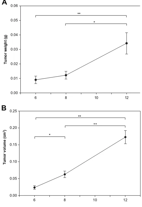
The permeability of the BTBs was therefore assessed in rat brains on days 6, 8, and 12 after tumor cell implantation. Tumor progression was evaluated in terms of weight and volume (Figure 3A and B). The mean tumor weight and tumor volume at day 12 were significantly greater than on day 6 or day 8. The mean tumor volume of the day 8 group was significantly greater than those of the day 6 group, but no significant difference in mean tumor weight was evident between the day 6 and day 8 groups.
Assessment of EB extravasation
The BBB disrupted region was observed to occur in the focal zone of the ultrasound beam as assessed by EB extravasation. Figure 4 illustrates the degree of EB staining in the right and left hemispheres with and without sonication at the three time points after tumor implantation. Both the size and color intensity of the EB staining increased with tumor progres-sion and that of the sonicated right hemispheres was greater than the nonsonicated left hemispheres for the three chosen days following tumor implantation. Figure 5 shows the mean extravasation of EB per unit mass (in micrograms per gram of tissue) for the brain tumors and the neighboring normal brain tissues with or without sonication at three time points after tumor implantation. EB extravasation was quantified in each tumor-implanted hemisphere brain; both the sonicated tumor and contralateral unsonicated control tumor were examined. Not only was the permeability of the control tumor BBB significantly greater than that of the adjacent normal brain region, but it was also found that the BTB disruption was significantly greater at the tumor site after sonication than in the control tumor for all three days. Pulsed HIFU exposure administered after EB introduction increased
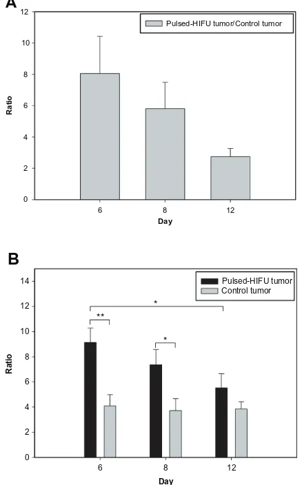
the EB concentration in the tumor by 805%, 580%, and 274% on days 6, 8, and 12, respectively (Figure 6A). There was no obvious difference in the derived tumor:ipsilateral brain ratios in the control tumor across the three days. Importantly, however, the derived tumor:ipsilateral brain ratios were greater after sonication than without sonication for all days; this change was significant on day 6 and day 8 (Figure 6B). The detailed ratios are listed in Table 2.
0 250
Body weight (g
)
260 270 280 290 300 310 320 330
2 4 6
Day
8 10
** ** ** *
12
Figure 2 Mean body weight in grams of glioma-bearing rats during tumor progression. Notes: *P, 0.05; **P, 0.01.
Figure 3 Mean tumor weight (A) (0.009 ± 0.003, 0.012 ± 0.003, 0.034 ± 0.007 g) and tumor volume (B) (0.02 ± 0.01, 0.06 ± 0.01, 0.17 ± 0.02 cm3) in glioma-bearing rats on days 6, 8, and 12 after tumor implantation.
Notes: *P, 0.05; **P, 0.01.
0.00
6 8
Day
Tumor weight (g
)
10
* **
12 0.01
0.02 0.03 0.04 0.05 0.06
A
6 0.00 0.05 0.10 0.15 0.20 0.25
B
8
Tumor volume (cm
3)
10 12
** **
*
Figure 4 Distribution of BBB disruption for brain tumors as evaluated by extravasation of EB into the brain. Right brain: tumor with pulsed HIFU exposure. Left brain: control tumor without pulsed HIFU exposure. The tumor area and sonication path are consistent with the MR images in Figure 7A.
Abbreviations: BBB, blood–brain barrier; EB, Evans blue; HIFU, high-intensity focused ultrasound; MR, magnetic resonance.
Dovepress
Yang et al
International Journal of Nanomedicine downloaded from https://www.dovepress.com/ by 118.70.13.36 on 23-Aug-2020
0
6
* *
***
***
*** **
*
*
8
Day
Pulsed-HIFU tumor Control tumor Pulsed-HIFU normal brain Control normal brain
Evans blue extravasation
(
µ
g/g tissue)
12 50
100 150 200 250 300
Figure 5 Concentration of EB in the tumor and neighboring normal brain regions with and without sonication. The EB extravasation in the brain tumor with sonication was significantly higher than in brain tumor without sonication. Compared with normal tissues neighboring the control tumors, there was a significant difference for the control tumors and for the neighboring normal tissues of sonicated tumors on all days and on days 6 and 8, respectively.
Notes: *P, 0.05; **P, 0.01; ***P, 0.001. Abbreviation: EB, Evans blue.
Figure 6 The ratio of EB concentration between sonicated tumor and control tumor (A) and the derived tumor:ipsilateral brain ratios after sonication and without sonication (B) during tumor progression.
Abbreviation: EB, Evans blue. 6 0
2 4 6
Ratio
8 10 12 A
8
Day
Pulsed-HIFU tumor/Control tumor
12
6 0
2 4 6
Rati
o 8 10 12 14
B
8
Day
12
Pulsed-HIFU tumor Control tumor *
* **
Table 2 Tumor:ipsilateral normal brain ratios
Day after implantation Ratio of tumor to brain Pulsed HIFU Control Mean SEM Mean SEM
6 9.14* 1.13 4.09 0.89
8 7.36* 1.21 3.72 0.96
12 5.52† 1.12 3.86 0.57
Notes: *Significant (P, 0.05) difference compared with control group; †significant (P, 0.05) difference compared with pulsed HIFU tumor on day 6.
Abbreviations: HIFU, high-intensity focused ultrasound; SEM, standard error of the mean.
MRI analysis and histology
MRI studies were carried out in a subset of animals to noninvasively quantify tumor growth as a function of time; furthermore, the sonication pathway can be monitored using MR images in the right sonicated hemispheres (Figure 7A). In addition, the corresponding H&E and TUNEL-stained sections were observed for tumor progression and apoptotic evaluation (Figure 7B and C). Based on the histological observations, tumor progression was consistent with the MR images and no significant difference in apoptosis was found between the sonicated and control tumors. To better understand the extent of deposition of gadolinium, the con-tour maps of the spatial distribution of gadolinium for tumors with and without sonication are presented in Figure 8. The contrast-enhanced regions in the right sonicated tumor were greater than in the left control tumor, especially for tumors on day 6 and day 8. During histological analysis of the soni-cated and control tumors, local displacement and increased
Figure 7 Images of the tumors with and without pulsed HIFU exposure by (A) T1-weighted MR images, (B) hematoxylin-eosin-stained sections, and (C) TUNEL staining on days 6, 8, and 12 after implantation. The tumor progression and sonication pathway can be monitored in the MR images. H&E and TUNEL stained sections were observed for histology and apoptotic evaluation.
Abbreviations: H&E, hematoxylin-eosin; HIFU, high-intensity focused ultrasound; MR, magnetic resonance; TUNEL, terminal deoxynucleotidyl transferase dUTP nick end labeling.
Dovepress Permeability of BTB after pulsed HIFU
International Journal of Nanomedicine downloaded from https://www.dovepress.com/ by 118.70.13.36 on 23-Aug-2020
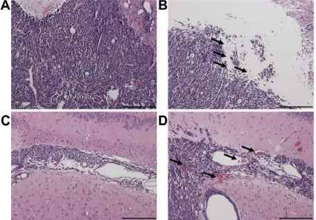
extravasation of red blood cells were seen in the sonicated tumor tissues in and around the focal region (Figure 9).
Discussion
This study demonstrates that pulsed HIFU can not only signif-icantly increase the permeability of the BTB in brain tumors, but also significantly elevated the tumor:brain drug ratio in the focal region that was elicited by an ultrasound beam passing through the intact skull. The brain entry of drugs is impeded by the BBB, even though the permeability of this barrier may be partially increased due to the presence of a tumor.
However, selective delivery of chemotherapeutic agent to brain tumor cells across the BTB remains a major obstacle to many approaches to brain tumor treatment. Our previous studies have shown that injecting an appropriate quantity of UCA effectively increases and localizes the BBB disruption that is induced by pulsed HIFU exposure.11,14,15 In this study, a
combination of pulsed HIFU and microbubbles increased the permeability of the BTB as measured by EB extravasation. The use of MRI contrast enhancement also revealed that this approach increased the level of gadolinium entering the brain tumor tissue.
The main limitation of this study was the size of the ultra-sound beam focal zone produced by our device, which was not large enough to produce delivery enhancement for the whole tumor region on day 8 and day 12. This may be why the enhancement induced by pulsed HIFU decreased from day 6 to day 12 after implantation. This phenomenon suggests that the permeability of the BTB is increased only locally by pulsed HIFU. In addition, pulsed HIFU also enhanced permeability in the normal brain tissue surrounding the sonicated tumor on day 6 and day 8. Nevertheless, the enhanced uptake in the tumor tissue would be advantageous when treating larger tumors in humans using a phased array transducer for multiple focal sonications. Figures 5 and 6B reveal that there were no differences in EB extravasation per gram of control tumor tissue or in the control tumor:ipsilateral brain ratio as the tumors progressed. Moreover, pulsed HIFU was able to significantly increase the permeability of the BTB and the ratio of tumor to brain, especially for the smaller tumors on day 6 and day 8; this was when the focal zone almost covered the whole tumor region.
Gadolinium deposition and the pattern of contrast enhancement were monitored by signal intensity level. Figure 8 shows that these are larger at high intensity level sites in the sonicated tumor on all days, especially on day 6 and day 8. This is consistent with the EB extravasa-tion results. The sonicaextravasa-tion pathway can be observed from the brain surface to the bottom of the brain (Figure 8A). Additionally, the pattern of contrast enhancement while the pulsed HIFU beam is targeted over a non-homogeneous tumor tissue does not correspond to the circular pattern of the ultrasound beam on the cross section (Figure 8B).
One recent study has demonstrated that sonication after EB injection can lead to nearly a fivefold increase in EB extravasation in target hepatocellular carcinoma compared with contralateral controls. However, this effect is reduced while EB is administered after sonication.16 If the previous
hypothesis is correct, then pulsed HIFU should be able to be used for the enhancement of local drug delivery when there
Figure 8 In vivo magnetic resonance images of rats bearing F98 gliomas in the (A) coronal view and (B) axial view. The spatial distribution of brain tumor BBB disruption with and without sonication in the right and left hemispheres, respectively, is shown. The rat brains were analyzed 30 minutes following gadolinium administration. Regions of contrast enhancement .4, .8, .12, and .16 standard deviations above the average MRI signal intensity of the normal brain tissue are shown in green, yellow, blue, and red respectively .
Abbreviations: BBB, blood–brain barrier; MRI, magnetic resonance imaging.
Figure 9 The structure of the contralateral brain tumor without sonication (A and C) and of the brain tumor tissue with sonication (B and D) by H&E staining (original magnification × 100 from Figure 7B; scale bar = 200 µm). Local displacement and increased extravasation of red blood cells were indicated by arrows in the sonicated tumor tissues in and around the focal region.
Abbreviation: H&E, hematoxylin-eosin.
Dovepress
Yang et al
International Journal of Nanomedicine downloaded from https://www.dovepress.com/ by 118.70.13.36 on 23-Aug-2020
is a transient increase in capillary permeability. Therefore, the present study was performed using pulsed HIFU after EB injec-tion in order to elevate extravasainjec-tion of EB in the brain tumor tissue as much as possible. Furthermore, both focused and unfocused ultrasound exposure have previously been shown to produce a widening of intercellular gaps.17,18 Compared with
the control tumors (Figure 9A and C), H&E staining showed pulsed HIFU exposure led to a widening of intercellular gaps and an increase in red blood cell extravasation (Figure 9B and D). Thus, the driving force behind the induction of BTB disruption by sonication may be increased drug extravasation due to presence of large gaps in the endothelium.19
Many methods having been tested to facilitate drug deliv-ery through the disruption of the BTB, but none have been practical when applied clinically. For instance, the BTB has been shown to be disrupted after an intracarotid artery injec-tion of a hyperosmotic soluinjec-tion such as mannitol using boron neutron capture therapy. Nevertheless, due to restrictions on drug brain entry by the BTB and BBB, the selective delivery of a drug to individual brain tumor cells or the tissues sur-rounding brain tumors remains one of the major challenges to the boron neutron capture therapy of brain tumors, and many approaches have been reported.20 Another study
dem-onstrated that pulsed HIFU increased the uptake of antibody into surrounding tissue, but the net increase was marginal.21
The current research has shown that completely noninvasive focal disruption of the BTB in the tumor and BBB in the adjacent normal brain tissue is possible through pulsed HIFU. Figure 6B indicates that BTB disruption induced by sonica-tion causes a 2.23-fold increase in the tumor:ipsilateral brain ratio for EB in the target tumors compared with the control tumors. The results of this pilot study therefore suggest that the further evaluation of other treatment strategies is warranted – this may include multiple sonications to increase delivery of chemotherapeutic agents to larger brain tumors.
Acknowledgments
This study was supported by grants from the National Science Council of Taiwan (no NSC 100-2321-B-010-010 and NSC 99-2321-B-010-017), Cheng Hsin General Hospital Founda-tion (no 100F117CY25), Veterans General Hospitals University System of Taiwan Joint Research Program (#VGHUST100-G1-3-3 and V100E6-007), Yen Tjing Ling Medical Foun-dation (grant CI-100-17), Department of Health of Taiwan (DOH101-TD-PB-111-TM012 and DOH100-TD-C-111-007).
Disclosure
The authors report no conflicts of interest in this work.
References
1. Brem H, Mahaley MS Jr, Vick NA, et al. Interstitial chemotherapy with drug polymer implants for the treatment of recurrent gliomas.
J Neurosurg. 1991;74(3):441–446.
2. Florell RC, Macdonald DR, Irish WD, et al. Selection bias,
survival, and brachytherapy for glioma. J Neurosurg. 1992;76(2):
179–183.
3. Black KL, Ningaraj NS. Modulation of brain tumor capillaries for
enhanced drug delivery selectively to brain tumor. Cancer Control.
2004;11(3):165–173.
4. Neuwelt EA. Implications of the Blood-Brain Barrier and its
Manipulation. New York, NY: Plenum, 1989.
5. Haluska M, Anthony ML. Osmotic blood-brain barrier
modifica-tion for the treatment of malignant brain tumors. Clin J Oncol Nurs.
2004;8(3):263–267.
6. Kemper EM, Boogerd W, Thuis I, Beijnen JH, van Tellingen O. Modulation of the blood-brain barrier in oncology: therapeutic
opportunities for the treatment of brain tumours? Cancer Treat Rev.
2004;30(5):415–423.
7. Hsieh C, Chen Y, Chen F, et al. Evaluation of pharmacokinetics of 4-borono-2-18F-fluoro-L-phenylalanine for boron neutron capture therapy in a glioma-bearing rat model with hyperosmolar blood–brain
barrier disruption. J Nucl Med. 2005;46(11):1858–1865.
8. Abbott NJ, Romero IA. Transporting therapeutics across the blood-brain
barrier. Mol Med Today. 1996;2(3):106–113.
9. Kroll RA, Neuwelt EA. Outwitting the blood-brain barrier for
thera-peutic purposes: osmotic opening and other means. Neurosurgery.
1998;42(5):1083–1099; discussion 1099–1100.
10. Hynynen K, McDannold N, Sheikov NA, Jolesz FA, Vykhodtseva N. Local and reversible blood-brain barrier disruption by noninvasive focused ultrasound at frequencies suitable for trans-skull sonications.
Neuroimage. 2005;24(1):12–20.
11. Yang F, Fu W, Chen W, Yeh W, Lin W. Quantitative evaluation of the use of microbubbles with transcranial focused ultrasound on
blood-brain-barrier disruption. Ultrason Sonochem. 2008;15(4):636–643.
12. Treat LH, McDannold N, Vykhodtseva N, Zhang Y, Tam K, Hynynen K. Targeted delivery of doxorubicin to the rat brain at
therapeutic levels using MRI-guided focused ultrasound. Int J Cancer.
2007;121(4):901–907.
13. Frenkel V, Li KC. Potential role of pulsed-high intensity focused
ultrasound in gene therapy. Future Oncol. 2006;2(1):111–119.
14. Yang FY, Liu SH, Ho FM, Chang CH. Effect of ultrasound contrast agent dose on the duration of focused-ultrasound-induced blood-brain
barrier disruption. J Acoust Soc Am. 2009;126(6):3344–3349.
15. Yang F, Fu W, Yang R, Liou H, Kang K, Lin W. Quantitative evalua-tion of focused ultrasound with a contrast agent on blood-brain barrier
disruption. Ultrasound Med Biol. 2007;33(9):1421–1427.
16. Bekeredjian R, Kroll R, Fein E, et al. Ultrasound targeted microbubble
destruction increases capillary permeability in hepatomas. Ultrasound
Med Biol. 2007;33(10):1592–1598.
17. Frenkel V, Kimmel E, Iger Y. Ultrasound-induced intercellular space
widening in fish epidermis. Ultrasound Med Biol. 2000;26(3):
473–480.
18. Mesiwala AH, Farrell L, Wenzel HJ, et al. High-intensity focused
ultra-sound selectively disrupts the blood-brain barrier in vivo. Ultrasound
Med Biol. 2002;28(3):389–400.
19. Boucher Y, Baxter LT, Jain RK. Interstitial pressure gradients in
tissue-isolated and subcutaneous tumors: implications for therapy. Cancer
Res. 1990;50(15):4478–4484.
20. Chen W, Mehta S, Lu D. Selective boron drug delivery to brain
tumors for boron neutron capture therapy. Adv Drug Deliv Rev.
1997;26(2–3):231–247.
21. Khaibullina A, Jang BS, Sun H, et al. Pulsed high-intensity focused ultrasound enhances uptake of radiolabeled monoclonal antibody
to human epidermoid tumor in nude mice. J Nucl Med. 2008;49(2):
295–302.
Dovepress Permeability of BTB after pulsed HIFU
International Journal of Nanomedicine downloaded from https://www.dovepress.com/ by 118.70.13.36 on 23-Aug-2020
International Journal of Nanomedicine
Publish your work in this journal
Submit your manuscript here: http://www.dovepress.com/international-journal-of-nanomedicine-journal
The International Journal of Nanomedicine is an international, peer-reviewed journal focusing on the application of nanotechnology in diagnostics, therapeutics, and drug delivery systems throughout the biomedical field. This journal is indexed on PubMed Central, MedLine, CAS, SciSearch®, Current Contents®/Clinical Medicine,
Journal Citation Reports/Science Edition, EMBase, Scopus and the Elsevier Bibliographic databases. The manuscript management system is completely online and includes a very quick and fair peer-review system, which is all easy to use. Visit http://www.dovepress.com/ testimonials.php to read real quotes from published authors.
Dovepress
Dove
press
Yang et al
International Journal of Nanomedicine downloaded from https://www.dovepress.com/ by 118.70.13.36 on 23-Aug-2020