Paper on Segmentation of Color Image using
Morphological Processing
Komal Jain, Prof. Anshul Awasthi, Dr. Manish Gurjar
M. Tech. Scholar, Technocrats Institute of Technology- Advance, Bhopal (M.P.), India Professor, Technocrats Institute of Technology- Advance, Bhopal (M.P.), India Head of Department, Technocrats Institute of Technology- Advance, Bhopal (M.P.), India
ABSTRACT: Image Segmentation plays vital role in Computer Vision and Digital Image Processing. It is the process of separating the digital image into distinct region(s) possessing homogeneous properties. The main objective of image segmentation is to extract various features of the image that are used for analyzing, interpretation and understanding of images. Image segmentation is applied in various applications like medical imaging, shape detection, content-based image retrieval, robot vision, etc. Several techniques have been developed for image segmentation such as pixel-based segmentation, edge based segmentation and region based segmentation. In this paper, segmentation technique is defined using the edge detection and morphological operations. Edge detection is done using Fuzzy Canny method for better output. After detecting the edges of image, segmentation is done using morphological operation. This gives better results.
KEYWORDS: - Dilation, Morphology, Erosion
I.INTRODUCTION
An image is basically two dimensional signal defined by mathematical function, F(x, y) where x and y gives value of horizontal and vertical co-ordinates. Digital image processing[1] deals with system that perform various operation on digital image to improve the quality of the image by removing noise and unwanted pixels and to obtain intentional information from an image. Image segmentation is a key step in digital image processing that subdivides an image into its constituent region or object that share homogeneous attributes [2]. The main purpose of the segmentation process is to get more information in the region of interest in an image which helps in annotation of the object scene [3].
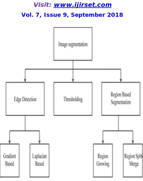
Image segmentation fundamentally works on two properties [2]:
Discontinuity: Division of the digital image predicated due to sudden changes on intensity. For example, edge detection, point detection and line detection.
Figure 1: Image Segmentation Methods
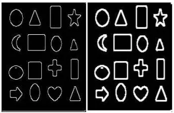
An edge is defined as boundaries of objects or sudden change in an image which is not in a continuous form that helps to detect and identify the objects in a given image [5]. The main aim behind edge detection [6, 7] method is to identify and locate the points in a digital image at which intensity of the image changes. Among various technique of various edge detection technique Canny [8] operator gives better output than Sobel [9], Prewitt [10], and Laplacian method. In Thresholding method grayscale value of the image is checked out with predefined value of the threshold. If grayscale value of the input pixel is large then output value of that pixel becomes 1 or else 0.
There are two types of thresholding:
Global Thresholding: Segment out the whole image with a unique threshold value.
Local Thresholding: Segment out the image into sub-images and each sub-image has an individual threshold value. Region Based segmentation methods divide an image into region having similar characteristic like color, texture etc. Region Growing algorithm performs a segmentation of an image with examine the neighboring pixels of a set of points, known as seed points, and determine whether the pixels could be classified to the cluster of seed point or not [11]. In method of region Split-merge whole Image which is considered as a seed region splitting out into quadrant until the homogenous sub region is obtained, after the process of Splitting Merging process merge two adjacent regions according to similar characteristic.
II. LITERATUREREVIEW
complexity of the studied objects. So there is the necessity of having a 3D computer representation of many real objects. In this paper we are presenting the denoising and segmentation techniques developed for 3D MRI based on fractal and morphological approaches.
D. Chudasama et al. [2], the vast improvements in information technology have led to significant enhancements in the field of medical imaging. Computed tomography, positron emission tomography, magnetic resonance imaging and ultrasound imaging have been adopted for diagnostic of many types of diseases and choosing of the most adequate therapy. These methods are considered as the most accurate imaging modalities available for early detection and diagnosis of different diseases.
Maini, Raman et al. [3], however, the presence of noise and blur, the variability of tissues and biological shapes make the analysis of medical images a very hard task. Moreover the presence of masking structures lead to superposing of images of different organs but, as a rule, the radiologist (or machine) inspects only one image. Thus and so, the potential for artifacts and the demands on image processing are considerably big. Furthermore, a simple method of analysis of 2D tomographic data can lead to false conclusions. The classic example is a conic. Indeed, even for this simple object a cross-section may be a parabola, circle, ellipse, one or two lines or one point. One of the ways for tackling of these disadvantages is 3-dimentional imaging, in particular the 3D denoising and segmentation.
Er. Komal Sharma et al. [4], image segmentation is a process of decomposition of the images into a set of homogeneous non-overlapping regions which union is the entire image. Its role is to locate the volume of interest. It is one of the first and most difficult tasks of any image analysis process. In a medical context, segmentation means that the set of pixels representing the same tissue type have to be grouped to form a single region. Namely this region offers major interest for the practitioner, e.g., a specific human organ. In order to distinguish the objects in image are used many different features: brightness, boundaries, specific shapes, textures.
III.MORPHOLOGICALOPERATIONSBASEDSEGMENTATION
Binary images may contain countless defects. In some circumstances binary regions constructed by simple thresholding are buckled by noise and textures. Morphology is a vast extent of image processing operations that modifies the images based on shapes. It is considered to be one of the data processing methods useful in image processing. It has many applications like texture analysis, noise elimination, boundary extraction etc. Morphological image processing follows the goal of eliminating all these defects and maintaining structure of image. Morphological operations are confident only on the associated ordering of pixel values, rather than their numerical values, so they are focused more on binary images, but it can also be applied to grayscale images such that their light transfer functions are unknown and thus their absolute pixel values are not taken into consideration. Morphological techniques verify the image with a small template called structuring element. This structuring element is applied to all possible locations of the input image and generates the same size output. In this technique the output image pixel values are based on similar pixels of input image with is neighbors. This operation produces a new binary image in which if test is successful it will have non-zero pixel value at that location in the input image. There are various structuring element like diamond shaped, square shaped, cross shaped etc. The base of the morphological operation is dilation, erosion, opening, closing expressed in logical AND, OR notation and described by set analysis. Among them in this paper only two operations are used dilation and erosion. Dilation adds pixels while erosion removes the pixels at boundaries of the objects. This removal or adding of pixels depends on the structuring element used for processing the image.
Dilation
of dilation can be as follows [1]: Suppose A be a set of input image coordinates and B be a set of structuring element coordinates and Bx is a translation of B so that its origin is at x. Thus dilation of A by B is set of all points of x such that intersection of Bx with A is not null. In terms of set operations dilation of A by B is defined as [7]:
Figure 2: Dilation image
Filling the region
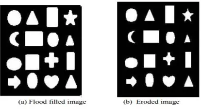
Dilation operation makes the boundaries of the object thick so for segmenting the object the next step is to fill the holes. The flood fill operation is most commonly known to fill the holes in the given input image. For binary images, it basically changes the background pixels to foreground pixels until it reaches the object boundaries and for grayscale images it makes the intensity level same i.e. it makes the dark areas surrounded by lighter areas to same intensity levels [2]. In binary images and gray-scale images the boundaries of the objects need to be specified by connectivity. In binary images the starting point for filling can also be specified. If we specify holes as an argument then it is of no need to specify any starting points [2]. In this paper fill operation is used on binary image with arguments holes so it automatically fills the holes of different objects in image. Below image shows the flood fill image on diluted image output:
Erosion
Erosion is also one of the basic operators in mathematical morphology.
Figure 3: Flood fill image
boundaries erosion is applied so as to make the boundaries of the objects thinner for better output. Erosion like same dilation takes two parts as data. First one is the input image to be eroded and second is the structuring element. With the help of this structuring element only it determines how much the image is to be eroded. The mathematical definition of erosion can be as follows [1]: Suppose A be a set of input image coordinates and B be a set of structuring element coordinates and Bx is a translation of B so that its origin is at x. Thus dilation of A by B is set of all points of x such that Bx is a subset of A. In terms of set operations erosion of A by B is defined as [9]:
Figure 4: Erosion Image
IV.EDGEDETECTION
Edge detection includes a variety of mathematical methods that aim at identifying points in a digital image at which the image brightness changes sharply or, more formally, has discontinuities. The points at which image brightness changes sharply are typically organized into a set of curved line segments termed edges. The same problem of finding discontinuities in one-dimensional signals is known as step detection and the problem of finding signal discontinuities over time is known as change detection. Edge detection is a fundamental tool in image processing, machine vision and computer vision, particularly in the areas of feature detection and feature extraction
The purpose of detecting sharp changes in image brightness is to capture important events and changes in properties of the world. It can be shown that under rather general assumptions for an image formation model, discontinuities in image brightness are likely to correspond to:
Discontinuities in depth
Discontinuities in surface orientation,
Changes in material properties and
Variations in scene illumination.
In the ideal case, the result of applying an edge detector to an image may lead to a set of connected curves that indicate the boundaries of objects, the boundaries of surface markings as well as curves that correspond to discontinuities in surface orientation.
V. CONCLUSION
REFERENCES
[1]Jianbing Shen, Senior Member, IEEE, Jianteng Peng, Xingping Dong, Ling Shao, and Fatih Porikli, “Higher-Order Energies for Image Segmentation”, IEEE Transactions on Image Processing, Volume: 26, Issue: 10, Oct. 2017
[2]A.G. Rudnitskii, M.A. Rudnytska, “Segmentation and Denoising of Phase Contrast MRI Image of the Aortic Lumen Via Fractal and Morphological Processing”, 37th International Conference on Electronics and Nanotechnology (ELNANO), 2017 IEEE.
[3]D. Chudasama, T. Patel, S. Joshi, G. Prajapati “Survey on Various Edge Detection Techniques on Noisy Images” , IJERT International Journal of Engineering Research & Technology ISSN: 2278-0181 Vol. 3 Issue 10, October- 2014.
[4]Maini, Raman, and Himanshu Aggarwal, "Study and comparison of various image edge detection techniques", International Journal of Image Processing (IJIP), Issue 3, no. 1, Pp. 1-11, 2009.
[5]Er. Komal Sharma, Er. Navneet Kaur, “Comparative Analysis of Various Edge Detection Techniques”, International Journal of Advanced Research in Computer Science and Software Engineering, Volume 3, Issue 12, December 2013
[6]Ur Rehman Khan, K. Thakur “An Efficient Fuzzy Logic Based Edge Detection Algorithm for Gray Scale Image”, International Journal of Emerging Technology and Advanced Engineering Website: www.ijetae.com (ISSN 2250-2459, Volume 2, Issue 8, August 2012).
[7]S. Patel, P.Trivedi, V. Gandhi and G. Prajapati, “2D Basic Shape Detection Using Region Properties” IJERT International Journal of Engineering Research & Technology, Vol. 2 Issue 5, May-2013.
[8]Mrs. A. Borkar, Mr. M.Atulkumar “Detection of Edges Using Fuzzy Inference System”, International Journal of Innovative Research in Computer and Communication Engineering, Vol. 1, Issue 1, March 2013.
[9]T. Gajpal, Mr. S. Meshram “Edge Detection Technique Using Hybrid Fuzzy logic Method”, IJERT International Journal of Engineering Research & Technology, Vol. 2 Issue 2, Febuary-2013.
[10]M. L Comer, E. J. Delp “Morphological operations for color image processing” electronic imaging spiedigitallibrary.