DOI: 10.1534/genetics.106.058362
The Conserved ATPase Get3/Arr4 Modulates the Activity of
Membrane-Associated Proteins in
Saccharomyces cerevisiae
Kathryn L. Auld,* Amy L. Hitchcock,*
,1Hugh K. Doherty,
†Seth Frietze,*
Linda S. Huang
†and Pamela A. Silver*
,2*Department of Systems Biology, Harvard Medical School, Boston, Massachusetts 02115 and†Department of Biology, University of Massachusetts, Boston, Massachusetts 02125
Manuscript received March 17, 2006 Accepted for publication June 19, 2006
ABSTRACT
The regulation of cellular membrane dynamics is crucial for maintaining proper cell growth and division. The Cdc48-Npl4-Ufd1 complex is required for several regulated membrane-associated processes as part of the ubiquitin–proteasome system, including ER-associated degradation and the control of lipid composition in yeast. In this study we report the results of a genetic screen inSaccharomyces cerevisiaefor extragenic suppressors of a temperature-sensitive npl4 allele and the subsequent analysis of one sup-pressor,GET3/ARR4. TheGET3 gene encodes an ATPase with homology to the regulatory component of the bacterial arsenic pump. Mutants of GET3 rescue several phenotypes of the npl4 mutant and transcription ofGET3is coregulated with the proteasome, illustrating a functional relationship between GET3 and NPL4 in the ubiquitin–proteasome system. We have further found that Get3 biochemically interacts with thetrans-membrane domain proteins Get1/Mdm39 and Get2/Rmd7 and thatDget3is able to suppress phenotypes of get1 and get2 mutants, including sporulation defects. In combination, our characterization ofGET3 genetic and biochemical interactions withNPL4,GET1, andGET2implicates Get3 in multiple membrane-dependent pathways.
I
NTRACELLULAR membranes such as the nuclear envelope and the compartments of the secretory pathway are the key feature that distinguishes the eukaryotic cell from bacteria. Along with the evolution of membrane-bound organelles came the need for in-tricate and highly regulated mechanisms to control the composition and movements of these membranes in re-sponse to environmental changes and during complex cellular events such as the cell cycle or meiosis (Howe and McMaster2001; reviewed in Albertsonet al.2005). The highly conserved Cdc48-Npl4-Ufd1 complex is an important regulator of several membrane-associated cellular processes in eukaryotic cells. In one critical role, the Cdc48-Npl4-Ufd1 complex participates in the clear-ing of aberrantly folded proteins from the endoplas-mic reticulum (ER) through ER-associated degradation (ERAD) (Bays et al. 2001). During ERAD, misfolded ER proteins are retrotranslocated to the cytosol where they are ubiquitinated and degraded by the proteasome (reviewed in Romisch 2005). The increased level of certain ER proteins that occurs if this process is pre-vented can cause drastic alterations in the organization of the ER compartment (Wright et al. 2003). TheCdc48-Npl4-Ufd1 complex also functions to regulate intracellular membranes in yeast through a pathway that controls production of unsaturated fatty acids. Specifi-cally, the Cdc48-Npl4-Ufd1 complex directs the ubiquitin– proteasome-dependent cleavage and activation of two ER-membrane anchored transcription factor precur-sors, Spt23 and Mga2 (Hoppeet al. 2000; Hitchcock
et al. 2001). Once released from the membrane, Spt23 and Mga2 activate transcription of the OLE1 gene (Zhanget al.1999; Chellappaet al.2001), which en-codes a fatty acid desaturase enzyme (Stukey et al. 1990). Thus, not only is the Cdc48-Npl4-Ufd1 complex important for the proteasome-dependent degradation and processing of protein substrates at the ER mem-brane, but it is also required for the regulation of cel-lular unsaturated fatty acid (UFA) content, and in turn membrane fluidity, through transcriptional control of OLE1. A further requirement has been shown for Cdc48-Npl4-Ufd1 in postmitotic nuclear membrane assembly in higher eukaryotes (Hetzeret al.2001).
Forward genetic screens in the yeast Saccharomyces cerevisiaehave been critical in elucidatingNPL4function in ERAD (Bays et al. 2001) and in OLE1 regulation (Hoppeet al.2000; Hitchcocket al.2001). As such, we sought to learn more about NPL4function and mem-brane dynamics by performing a screen for extragenic suppressors of a temperature-sensitivenpl4mutant. In this study, we present the full panel of genes that we iden-tified as extragenic suppressors of annpl4mutation. In
1Present address: Department of Molecular and Cellular Biology,
Harvard University, Cambridge, MA 02138.
2Corresponding author:Department of Systems Biology, Harvard Medical
School, WAB536, 200 Longwood Ave., Boston, MA 02115. E-mail: [email protected]
addition, we present extensive characterization of one gene identified as annpl4suppressor, theGET3/ARR4 gene. GET3 encodes a highly conserved ATPase with homology to ArsA, the regulatory component of the bacterial arsenic export pump (Boskovicet al. 1996), and to a human protein of unknown function, hASNA-I (Kurdi-Haidaret al.1996). Get3 has been suggested to have roles in cellular resistance to stress (Shen et al. 2003), in metal ion homeostasis (Metzet al.2006), and, in complex with Get1 and Get2, in protein sorting via the secretory pathway (Schuldineret al.2005).
Here we present evidence that GET3 displays func-tional interactions with NPL4 in the context of the ubiquitin–proteasome pathway. We further illustrate interactions ofGET3withGET1andGET2during spor-ulation, a process in which dynamic new growth of cellular membranes is required for proper spore and spore wall formation. In particular, we have demon-strated that Get3 can play an antagonistic role in both
settings, leading to the proposal that Get3 is a functionally conserved regulator of membrane-associated proteins.
MATERIALS AND METHODS
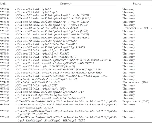
Yeast strains and manipulations: Standard yeast methods and media were used (Guthrie and Fink 1991). The genotypes of all strains used in this study are provided in Table 1. Thenpl4-1mutant strains are FY23-backcrossed strains derived from PSY825 and PSY826, which were previously described (DeHoratiusand Silver1996). Null alleles and C-terminal tags were integrated using PCR-based techniques (Baudinet al.1993; Knopet al.1999).CPY* was integrated into wild-type (WT),npl4-1, andDget3 npl4-1strains using two-step gene replacement (Adamset al.1997) of plasmid pRS306-prc1-1as described (Knopet al.1996).
Screens for extragenic suppressors of npl4-1:npl4-1cells were mutagenized with either ethyl methanesulfonate (EMS) or an mTn-lacZ/LEU2transposon library (Burnset al.1994). Methods for the EMS screen and cloning ofUBC7andCUE1 have been described (Hitchcocket al.2003). Suppressing TABLE 1
Saccharomyces cerevisiaestrains used in this study
Strain Genotype Source
PSY3322 MATaura3-52 leu2D1 trp1D63 This study
PSY3323 MATaura3-52 leu2D1 trp1D63 npl4-1 This study PSY3383 MATaura3-52 leu2D1 his3D200 trp1D63 npl4-1 cue1:Tn [LEU2] This study PSY3384 MATaura3-52 leu2D1 his3D200 trp1D63 npl4-1 vps27:Tn [LEU2] This study PSY3385 MATaura3-52 leu2D1 his3D200 trp1D63 npl4-1 srn2:Tn [LEU2] This study PSY3386 MATaura3-52 leu2D1 his3D200 trp1D63 npl4-1 get3:Tn [LEU2] This study
PSY3091 MATaura3-52 leu2D1 trp1D63 npl4-1 spt23:Tn [LEU2] Hitchcocket al.(2001) PSY3387 MATaura3-52 leu2D1 his3D200 trp1D63 npl4-1 ifh1:Tn [LEU2] This study
PSY3388 MATaura3-52 leu2D1 his3D200 trp1D63 npl4-1 prp6:Tn [LEU2] This study PSY3389 MATaura3-52 leu2D1 his3D200 trp1D63 npl4-1 cbp80:Tn [LEU2] This study PSY3390 MATaura3-52 leu2D1 his3D200 trp1D63Dget3THIS3 This study
PSY3391 MATaura3-52 leu2D1 trp1D63 Get3tn-3HA [KanMX] This study PSY3392 MATaura3-52 leu2D1 his3D200 trp1D63 npl4-1Dget3THIS3 This study
PSY3393 MATaura3-52 leu2D1 trp1D63Dget1TKanMX This study
PSY3394 MATaura3-52 leu2D1 trp1D63Dget2TKanMX This study
PSY3395 MATaura3-52 leu2D1 trp1D63Dget1TKanMX npl4-1 This study
PSY3396 MATaura3-52 leu2D1 his3D200 npl4DTNPL4-sGFP [URA3] Get3-tevProA [KanMX] This study
PSY3066 MATaura3-52 leu2D1 his3D200 trp1D63 npl4DTNPL4-sGFPTURA3 This study
PSY3397 MATaura3-52 leu2D1 trp1D63 Get3-EGFP [KanMX] This study PSY3398 MATaura3-52 leu2D1 his3D200 trp1D63 Get3-EGFP [KanMX]Dget1TLEU2 This study
PSY3399 MATaura3-52 leu2D1 his3D200 trp1D63 Get3-EGFP [KanMX]Dget2THIS3 This study
PSY3400 MATaura3-52 leu2D1 his3D200 Get3-EGFP [KanMX]Dget1TLEU2Dget2THIS3 This study
PSY3164 MATahis3D1 leu2D0 met15D0 ura3D0Dget3TKanMX This study
PSY1930 MATahis3D1 leu2D0 met15D0 ura3D0 Winzeleret al.(1999) PSY3402 MATaura3-52 leu2D1 trp1D63 CPY* This study
PSY3403 MATaura3-52 leu2D1 trp1D63 npl4-1 CPY* This study PSY3404 MATaura3-52 leu2D1 his3D200 trp1D63Dget3THIS3 CPY* This study
PSY3405 MATaura3Dleu2D Dget3TLEU2Dget2TKanMX This study
PSY3406 MATaura3-52 leu2D1 his3D200Dget3THIS3Dget1TKanMX This study
PSY3407 MATa/MATahoThisG/hoThisG lys2/lys2 ura3/ura3 leu2/leu2 his3/his3 trp1DFA/trp1DFA Benjaminet al.(2003) PSY3408 MATa/MATahoThisG/hoThisG lys2/lys2 ura3/ura3 leu2/leu2 his3/his3 trp1DFA/trp1DFA
Dget3TKanMX/Dget3TKanMX
This study
PSY3409 MATa/MATahoThisG/hoThisG lys2/lys2 ura3/ura3 leu2/leu2 his3/his3 trp1DFA/trp1DFA
Dget2TTRP1/Dget2TTRP1
This study
PSY3410 MATa/MATahoThisG/hoThisG lys2/lys2 ura3/ura3 leu2/leu2 his3/his3 trp1DFA/trp1DFA
Dget3TKanMX/Dget3TKanMXDget2TTRP1/Dget2TTRP1
mutations inDOA4andUBP3were identified by transforma-tion with a CEN-based URA3-marked plasmid library con-taining yeast genomic DNA fragments (Rose et al. 1987) and subsequent analysis of the rescuing plasmids pPS2915, pPS2918, and pPS2931. Transposon mutagenesis was per-formed essentially as described (Seifert et al. 1986). Two micrograms ofNotI-digested DNA were transformed intonpl4-1 cultures, and colonies able to grow at the nonpremissive temperature of 30°were observed at the rate of 53105. The
location of insertion was identified for nine of these colonies by vectorette PCR, as described (Kumarand Snyder2000).
ERAD assay:Cells were grown to OD600¼0.5–1.0, pelleted, and resuspended in 3 ml YPD containing 100 ug/ml cyclo-hexamide. Samples were removed at each time point and prepared as described ( Johnsonet al.1995) for immunoblot-ting with anti-CPY (Molecular Probes, Eugene, OR). Equal protein loading was verified by subsequent staining with amido black.
Northern blot analysis:Cultures were grown in YPD at 25°to log phase and then split such that half continued growth at 25° while half were shifted to 37°for 2 hr. All samples were frozen and total RNA was prepared in parallel. Northern blotting and analysis were performed as described (Hitchcocket al.2001). Cell fractionation: Get3-EGFP cells were grown at 30° to logarithmic phase in YPD. Subcellular fractionation and solu-bilization of the P13 fraction were performed essentially as described (Munoz-Centenoet al. 1999). To determine the fractionation profile of Get3, equal cell volumes were analyzed by Western blot with antibodies to GFP (Seedorfet al.1999) and Sec62.
Affinity purification and mass spectrometry: Get3-TEV-protein A and control cells expressing Get3-TEV-protein A from plasmid pPS1973 were grown in media lacking leucine to logarithmic phase. Microsomes were prepared essentially as described (Bakeret al.1990). After homogenization of spheroplasts, the membrane fraction was isolated by centrifugation at 13,000 rpm for 15 min. The pellet was resuspended and washed in B88 buffer (20 mm HEPES, pH 6.8, 150 mmKOAc, 5 mm Mg(OAc)2, 250 mmsorbitol). Equal protein amounts were resuspended in IPPT-150 buffer (10 mmTris pH 8.0, 150 mm NaCl, 0.5% Triton X-100) supplemented with 0.5 mmDTT, 0.5 mm PMSF, and protease inhibitors, and proteins were extracted by rocking at 4°for 1–5 hr.
Affinity purification was performed overnight at 4° using 30ml of IgG Sepharose bead slurry (Pharmacia/Amersham, Piscataway, NJ), 50ml of 80% glycerol, and 500ml of extracted proteins. After washing with IPPT-150 buffer (as above except 0.1% Triton), cleavage with TEV protease (GIBCO, Grand Island, NY) was performed in IPPT-150 supplemented with 0.5 mmEDTA and 1 mmDTT at 14°. After SDS–PAGE, silver-stained bands were excised from the gel and analyzed by MALDI-TOF mass spectrometry at the Southern Alberta Mass Spectrometry Centre (Calgary, AB, Canada).
Transcriptional profiling and genomic analysis:Expression profiling was carried out in triplicate, with swapping of fluor orientation, fromDget3and WT cells grown at 30°in YPD to early logarithmic phase. Total RNA preparation, cDNA prep-aration, and hybridization were performed as described (Casolariet al.2004). Up- and downregulated genes were defined as genes with fold change$1.5 andP-value#0.05 as determined by Rosetta Resolver. Analysis for enrichment of functional classes was performed using FuncAssociate (Berriz et al. 2003). Data mining for GET3 transcription was per-formed using Cluster and TreeView software from Eisenet al. (1998) and tools available on the Saccharomyces Genome Database (Dolinskiet al.2006).
Sporulation experiments: Synchronous sporulation of ho-mozygous SK1 diploid cells was performed using previously
described methods (Padmoreet al.1991; Huanget al.2005). Samples were monitored throughout sporulation to ensure good synchrony and efficiency. Each sample was fixed and stained with DAPI before observation by light and fluores-cence microscopy (Grether and Herskowitz 1999). For quantification, images were recorded of 36-hr samples using a Nikon microscope equipped with a DAPI filter (Chroma Technology, Brattleboro, VT) and a 1003 DIC (Nomarski) objective. At least 200 cells were scored for each strain. Spore viability was determined by tetrad dissection after sporulation for 2–4 days on 1% potassium-acetate plates.
RESULTS
Identification of npl4-1 extragenic suppressors: Sev-eral phenotypes affecting cellular membranes have been observed in the npl4-1 mutant allele, including defects in ERAD, and in nuclear envelope structure and nucleocytoplasmic transport likely due to the misregu-lation of OLE1(DeHoratius and Silver 1996; Bays
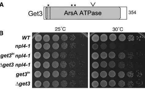
et al. 2001). To learn more about these effects, we conducted a genetic screen for extragenic suppressors of the temperature-sensitive lethality of this npl4 mu-tant at 30°.npl4-1 cells were mutagenized with either EMS or transposon-mutagenized yeast genomic DNA, and mutants capable of growth at 30° were isolated, characterized, and cloned. This analysis resulted in the identification of 11 genes whose mutation was capable of rescuing growth at the nonpermissive temperature (Table 2). Suppressing mutations isolated by EMS mutagenesis fell into five distinct complementation groups, four of which were identified by various techni-ques (materials and methods). An additional 7 genes with suppressing mutations were isolated as transposon insertions and were cloned by vectorette PCR. One gene,CUE1, was isolated by both techniques. None of the isolated suppressors restored wild-type growth to the npl4-1mutant or were able to rescue at higher temper-atures (Figure 1B and data not shown), suggesting that in each case the deficits of this mutation were only partially corrected.
Mutations in genes affecting several pathways, in-cluding ERAD, secretion, and gene expression, were able to ameliorate the temperature-sensitive lethality of npl4-1 (Table 2). Two of the suppressors are active in ERAD (CUE1 and UBC7) (Biederer et al. 1997) and have already been reported by our lab as suppressors of npl4-1(Hitchcocket al.2003). All four suppressors in the secretory pathway class have also been implicated in ubiquitin-dependent processes: DOA4 and UBP3 have direct roles in deubiquitination at secretory vesicles (Ameriket al.2000; Cohenet al.2003a), whereasVPS27 and SRN2/VPS37 are involved in the sorting and deg-radation of ubiquitinated proteins via the multivesicular body (MVB) pathway (Katzmannet al.2001; Bilodeau
et al.2002). Genes in the third class ofnpl4-1extragenic suppressors affect transcription (SPT23,IFH1) (Zhang
2004; Wadeet al.2004) or have roles in RNA processing (PRP6,STO1) (Abovichet al.1990; Daset al.2000). The rescue ofOLE1transcription in npl4-1cells by trunca-tions ofSPT23andMGA2has been reported previously (Hitchcock et al. 2001); however, the links between the remaining members of class 3 and Npl4 and/or ubiquitin–proteasome function are unclear.
In addition to these genes with known functions, we identified a suppressing mutation in theYDL100Copen reading frame (ORF), which had no reported functions
at the time of the screen. YDL100C has since been named ARR4 for its homology to the bacterial ArsA protein (Shen et al. 2003) and, more recently, GET3 (Schuldineret al.2005). We chose to pursue the char-acterization of this gene as a means to gain insight into both Get3 and Npl4 function.
get3 mutants suppress npl4-1-associated phenotypes:
The get3mutant isolated as a suppressor of npl4-1is a truncation produced by transposon insertion and is designated get3tn
. The transposon insertion adds two amino acids and a premature stop codon after K215 of Get3, generating a truncated protein product consist-ing of the first 60% of Get3 (Figure 1A). This region contains the P-loop ATP-binding site and several pre-dicted myristoylation sites. We observed that bothget3tn
and the null alleleDget3 were able to suppress npl4-1 temperature-sensitive lethality at 30°(Figure 1B). How-ever, phenotypic differences between the two alleles suggested that the isolated transposon insertion does not generate a null allele; specifically,Dget3is a weaker suppressor of npl4-1 and has a more marked growth defect in cells that are wild type forNPL4than does the get3tnallele (Figure 1B).
We next tested whether mutants ofget3rescuednpl4-1 phenotypes in addition to temperature-sensitive lethal-ity, including a diminished capacity for ERAD and misregulation ofOLE1transcription (Figure 2). To test for function of the ERAD pathway inget3 npl4-1cells, a mutant allele of carboxypeptidase Y (CPY*) that is sub-ject to rapid degradation via the ERAD system was in-tegrated into the npl4-1andget3 mutant strains. Cells TABLE 2
Suppressors ofnpl4-1
Gene Cellular activity Allele(s)
ERAD
YMR264W CUE1a,c Docking of Ubc7 in ERAD Transposon after AA72, EMS (5) YMR022W QRI8/UBC7a,c Ubiquitin-conjugation to ERAD substrates EMS (16)
Secretion
YNR006W VPS27 Sorting of ubiquitinated proteins in MVB pathway Transposon after AA260 YLR119W SRN2/VPS37 Sorting of ubiquitinated proteins in MVB pathway Transposon after AA44 YDR069C DOA4c Deubiquitination in endosome to vacuole transport EMS (8)
YER151C UBP3c Deubiquitination in vesicle transport EMS (2)
Transcription/RNA modulation
YKL020C SPT23b Activation ofOLE1transcription Transposon after AA710 YLR223C IFH1 Chromatin assembly and silencing Transposon after AA155 YBR055C PRP6 Pre-mRNA splicing (U4/U6-U5 snRNP) Transposon after AA793
YMR125W STO1/CBP80 Nuclear mRNA cap-binding protein Transposons (2) in noncoding regions
YDL100C GET3c Homolog of bacterial arsenite transporter Transposon after AA215
Suppressors generated by EMS or transposon mutagenesis are listed by functional class. Several were isolated multiple times, as indicated. For mutants isolated from the transposon screen, the integration site is listed.
aPreviously published in Hitchcocket al.(2003). b
Previously published in Hitchcocket al.(2001). c
Null allele was tested and also found to rescue growth ofnpl4-1.
expressing CPY* were treated with cyclohexamide to halt protein synthesis, and levels of this protein remaining at various time points after treatment were determined by Western blot analysis with anti-CPY antibodies. As ex-pected, CPY* was degraded rapidly in wild-type cells with a half-life of30 min (Figure 2A). In contrast, the half-life of CPY* was significantly extended innpl4-1cells, as has been previously reported (Bays et al. 2001). In-terestingly, the get3 null and transposon alleles both displayed partial rescue of the npl4-1 ERAD defect (Figure 2A and data not shown), decreasing the half-life of the CPY* protein in npl4-1 cells to ,1 hr. We found that theDget3allele does not have a strong defect or enhancement of ERAD in cells that are wild type for NPL4 (supplemental material at http://www.genetics. org/supplemental/).
To determine whether get3 mutation was able to rescueOLE1transcription innpl4-1cells, we performed Northern blot analysis using probes forOLE1, and for ACT1(actin) as a loading control. Total RNA was ana-lyzed from npl4-1 and get3 single- and double-mutant cells, shifted to the nonpermissive temperature of 37° for 2 hr. After shift to 37°,OLE1transcript levels were observed to increase in wild-type cells by 40% and decrease in thenpl4-1mutant by50% compared to the unshifted samples (Figure 2B; Hitchcocket al.2001).
We found that the transposon allele ofget3was able to rescueOLE1transcription to wild-type levels (Figure 2B). It appears that this effect is specific to theget3tnallele,
sinceDget3does not restoreOLE1transcription innpl4-1 cells; however, Dget3does block the elevation ofOLE1 transcript levels seen at 37°inNPL4cells (supplemental material at http://www.genetics.org/supplemental/). We then compared the ability of get3tn
to rescueOLE1 transcription with that of a dominant activating trunca-tion of Spt23 (spt23tn
), which encodes a transcription factor that activatesOLE1gene expression (Zhanget al. 1999). Surprisingly, we found that the rescue ofOLE1 transcript level in npl4-1 is more robust in the get3 mutant than in thespt23mutant.
GET3 is coregulated with the proteasome and the Cdc48-Npl4-Ufd1 complex: Given that genes with sim-ilar function often exhibit transcriptional coregulation, we used genomic analyses of available transcriptional profiling data to examineGET3expression and coregu-lation with other yeast genes under various growth conditions. First, the transcriptional profiles of all yeast genes under conditions including specific cell cycle stage, diauxic shift, and sporulation were clustered as described (materials and methods; Eisenet al.1998). The genes showing the most similar expression with GET3 under these conditions, shown in Figure 3, in-clude a significant number of genes encoding protea-somal components (P , 0.001 by FuncAssociate) and those encoding the Cdc48-Npl4-Ufd1 complex. Sup-porting this result, we found that GET3also clustered with proteasome-encoding genes using a different computational method ( Jelinsky et al. 2000). These genomic data fit well with the genetic interaction we found between GET3andNPL4and provide evidence indicating functional connections between GET3 and the ubiquitin–proteasome pathway.
The strongest coregulation observed betweenGET3, NPL4, and the proteasome components occurs under conditions relating to sporulation (Figure 3). Expres-sion of nearly all the genes in this cluster is elevated early in sporulation and under altered expression ofNDT80 (Figure 3), which encodes a master regulator for transcriptional activation of middle sporulation-specific genes (Xu et al. 1995; Chu and Herskowitz 1998; Hepworthet al.1998). Looking more specifically at the regulation ofGET3during sporulation in existing data sets for genomewide expression during sporulation in SK1 cells (Chuet al.1998; Primiget al.2000), we found thatGET3mRNA expression is slightly elevated shortly after induction of sporulation. The set of 20 genes with the most similar expression pattern to GET3 during sporulation, according to the Saccharomyces Genome Database (Dolinskiet al.2006), includes genes encod-ing several proteasome subunits (data not shown) and is significantly enriched for genes involved in ubiquitin-dependent protein catabolism (P , 0.001 by Func-Associate).
Get3 membrane localization depends on Get1 and Get2: We next examined the subcellular localization and biochemical characteristics of the Get3 protein. For these analyses, DNA sequence encoding an EGFP tag was integrated in frame at the 39 end (translated C terminus) of the genomic locus ofGET3; the resulting GET3-EGFPstrain exhibits no growth defect (data not shown), indicating that the EGFP tag does not disrupt Get3 function. Upon examination by live-cell fluores-cence microscopy, Get3-EGFP was observed at the ER/ nuclear membrane and in the cytoplasm (Figure 4A). This finding was consistent with published reports for Get3 localization in rich media (Huh et al. 2003; Schuldineret al.2005).
To biochemically confirm that a population of Get3 associates with cellular membranes, cells expressing Get3-EGFP were lysed under gentle, nondetergent conditions and fractionated by two successive rounds of centrifugation (materials and methods). Proteins copurifying with the isolated membrane and soluble fractions were separated by SDS–PAGE and probed with anti-GFP antibodies to visualize Get3-EGFP and with antibodies to Sec62, an integral ER-membrane protein. This analysis revealed that the majority of Get3 protein sedimented with the nuclei and ER membranes in the P13 membrane fraction (Figure 4B), consistent with our
observation of a membrane-associated population of Get3-EGFP by fluorescence microscopy. We also ob-served a population of Get3 that remained soluble, even upon ultracentrifugation (S100 fraction), confirming our visualization of a cytoplasmic pool of Get3 by microscopy (Figure 4B).
On the basis of sequence predictions and its homol-ogy to ArsA (Boskovicet al.1996), the Get3 protein is not expected to contain a trans-membrane domain, raising the question of how Get3 associates with cellular membranes. To assess the nature and extent of Get3 membrane association, cellular membranes purified from Get3-EGFP-expressing cells (P13 fraction) were resuspended in various buffers, incubated for 10 min on ice, and repelleted. Proteins released into the soluble phase (S) as well as those remaining in the membrane pellet (P) were analyzed by Western blot as above. We observed that Get3-EGFP remained tightly membrane associated in the presence of high salt and high pH; only treatment with a detergent was able to solubilize Get3 to the supernatant (Figure 4C).
On the basis of our microscopic and biochemical analysis of Get3 subcellular localization, we hypothe-sized that Get3 was likely to interact with one or more proteins embedded in the ER/nuclear membrane. To identify these potential proteins, we affinity purified
protein A-tagged Get3 (Get3-ProA) from solubilized cell membranes by binding to IgG Sepharose. Proteins copurifying with Get3-ProA were eluted by TEV pro-tease cleavage, separated by SDS–PAGE, and detected by silver staining. Two protein bands that were specifically observed in the Get3-ProA purification, and not in a negative control purification from cells expressing protein A alone, were present in similar quantities as Get3-ProA itself (Figure 4D). These were identified by MALDI-TOF mass spectrometry to be Get1 and Get2. A fainter band running at 35 kDa was also observed, but unfortunately this interactor was of insufficient quantity to identify. Further increasing our confidence that Get1 and Get2 represent true Get3-binding partners, these two proteins were also found among the proteins pu-rified with Get3 in a large-scale study (Hoet al.2002). Despite their colocalization at the ER/nuclear membrane (Hitchcock et al. 2001; Huh et al. 2003), no physical interactions were observed between Get3 and members of the Npl4 complex by Western blot (data not shown).
To test the possibility that Get3 membrane locali-zation is mediated by interaction with thetrans-membrane domain-containing proteins Get1 and Get2, we mon-itored Get3-EGFP localization in Dget1 and Dget2 single- and double-mutant cells. Strikingly, Get3-EGFP was completely absent from the ER membrane in deletions of eitherGET1orGET2(Figure 4E). Instead it was found in punctate sites in the cytoplasm, which are likely Golgi compartments as suggested by Schuldiner
et al.(2005). Conversely, we found that the localization of Get1-EGFP and Get2-EGFP was unaffected by the absence of Get3 (supplemental material at http://www. genetics.org/supplemental/). Thus, under these con-ditions the ER-membrane localization of Get3 is de-pendent upon the presence of both Get2 and Get1; however, even in the absence of Get1 and Get2, Get3 appears to retain some capacity to interact with other cellular membranes.
Sporulation genes are misregulated in aget3mutant:
As a complementary approach to identify pathways
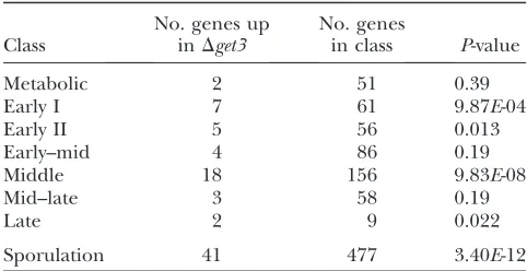
affected by GET3, transcriptional profiling of Dget3 cells was performed. Total RNA was purified from WT and Dget3 cells, differentially labeled, and competi-tively hybridized to microarrays spotted with cDNAs representing 6200 predicted yeast ORFs. Statistical analysis of WT vs. Dget3 signal for each ORF led to the identification of 265 genes whose transcription is upregulated and 345 genes whose transcription is downregulated inDget3relative to WT cells, as defined in materials and methods (complete data set avail-able in supplemental material at http://www.genetics. org/supplemental/). The most striking finding from this analysis was that the set of genes upregulated in
Dget3is significantly enriched for genes whose expres-sion increases during sporulation (P ¼3.4 31012, by
Fisher’s exact test). Table 3 shows the breakdown of these sporulation- andDget3-induced genes into stages of transcriptional activation during sporulation, on the basis of the classifications made by Chu et al.(1998). Although a large fraction of genes involved in all stages of sporulation was induced inDget3cells, enrichment of genes involved in the middle and early I stages was the most significant (P¼9.833108andP¼9.873104,
respectively; Table 3).
Given the genetic interactions between GET3 and NPL4, we sought to compare the set of genes with altered transcription in Dget3 mutant cells with those whose transcription is altered in npl4-1 mutant cells (Auldet al.2006). Surprisingly, the genes with altered transcription in Dget3 and npl4-1 strains showed no significant similarity, except in the activation of genes encoding heat-shock and other stress response proteins (data not shown). Interestingly, however, the transcrip-tion ofGET3 was significantly induced in npl4-1(P ¼ 4.333103). In all, this transcriptional profiling analysis
supports the existence of a functional connection betweenGET3andNPL4, but also suggests thatGET3 may function independently ofNPL4 in affecting the expression of sporulation-specific genes.
Sporulation phenotypes of GET complex andnpl4-1 mutants: We found the misregulation of sporulation genes inDget3to be of particular interest givenGET3’s strong transcriptional coregulation with NPL4 and other ubiquitin–proteasome system genes during this process (see above and Figure 3). Furthermore, both the Get3 interactors Get1 and Get2 (Figure 4 and Schuldiner et al. 2005) have been implicated in sporulation by a large-scale study (Enyenihi and Saunders2003). Taken together, these data suggested that the GET complex may have a role, perhaps in conjunction with Npl4 and the ubiquitin–proteasome system, in sporulation. To investigate this model, dip-loid yeast cells homozygous fornpl4-1,Dget3,Dget2, or
Dget1 (in the synchronously sporulating SK1 strain background) were induced to sporulate, and meiotic divisions were assayed by microscopic analysis of DAPI-stained samples throughout the time course. The terminal phenotype was then recorded by fluorescence and Nomarski microscopy 36 hr after induction. We found that the timing and occurrence of meiotic divisions in the GET complex deletion strains were not significantly altered compared to those in a wild-type strain (supplemental material at http://www.genetics. org/supplemental/). In fact, no strong sporulation defect was apparent in theDget3andnpl4-1SK1 strains, even after characterization of the terminal sporulation phenotype (Figure 5B and data not shown).
In contrast toDget3 andnpl4-1 cells, we observed a striking defect in the terminal sporulation phenotype of Dget2 and Dget1mutant cells (Figure 5, A and B). Whereas the majority of wild-type cells generated tet-rads with four spores ‘‘impenetrable’’ to DAPI when stained using the conditions described (materials and methods), only 20% ofDget2cells exhibited this phe-notype. Instead, the majority ofDget2diploid cells pro-duced ‘‘partial penetrable’’ tetrads, which contain one or more spores that are refractile, but immature looking and penetrable to DAPI (Figure 5, A and B). TheDget2 strain also exhibited a mild increase in ‘‘multinucleate’’ cells, those with multiple or fragmented nuclei but with no refractile spores. A similar, though more severe, phenotype was observed in theDget1strain (Figure 5A): only 10% of Dget1 cells produced complete tetrads after 36 hr in sporulation medium, whereas40% dis-played the multinucleate phenotype. A significant num-ber (30%) of Dget1 diploids also formed abnormal refractile spores similar to those seen inDget2, suggest-ing that these genes function within the same process. All strains tested had a similar percentage of ‘‘single-nucleus’’ cells that have not undergone sporulation, indicating that the fraction of cells initiating sporula-tion was not significantly different in the various strains (Figure 5B and data not shown). Tetrad dissection was performed to analyze the viability of spores produced in
Dget1andDget2strains. Overall, 79.7% of Dget2spores (287/360 spores) and 86.3% ofDget1spores (335/388) TABLE 3
Misregulation of sporulation genes inDget3
Class
No. genes up inDget3
No. genes
in class P-value
Metabolic 2 51 0.39
Early I 7 61 9.87E-04
Early II 5 56 0.013
Early–mid 4 86 0.19
Middle 18 156 9.83E-08
Mid–late 3 58 0.19
Late 2 9 0.022
Sporulation 41 477 3.40E-12
from tetrads with normal appearance were viable, compared with 99.5% viability of WT spores (199/200). These observed phenotypes ofDget1andDget2spores (Figure 5A) are similar to those previously described for spo11-1 mutant diploids, which undergo the meiotic divisions but fail to package all of their spores correctly (Klapholz et al. 1985). Since Spo11 is important in catalyzing double-strand breaks necessary for recombi-nation during meiosis (Keeneyet al.1997), this similar-ity suggested that meiotic recombination may be affected in GET complex mutants. To test this hypoth-esis, the frequency of heteroallele recombination in
Dget1andDget2was analyzed by a return-to-growth time-course experiment during the first 8 hr after induction of sporulation. Interestingly, we observed no significant defect in either mutant (data not shown). An alternative explanation for these phenotypes, in keeping with previous proposals of spore wall defects in Dget1 (Enyenihi and Saunders 2003), is that the GET complex is important for spore packaging. For further characterization of the mutant phenotypes, we also examined the localization of Don1-GFP, a marker for the leading edge of the prospore membrane (Knopand Strasser2000), in our mutant strains. Prospore mem-brane growth appeared normal as well, suggesting that GET1 and GET2 function at a later step in spore development.
Dget3 suppresses phenotypes of Dget2 and Dget1:
Given that get3 mutants are capable of suppressing phenotypes of npl4-1 cells, we asked whether Dget3 might also be able to suppress the sporulation defect displayed by Dget2 cells. To this end, we generated a homozygous diploidDget2Dget3double mutant in the SK1 strain background and analyzed its terminal spor-ulation phenotype as described above. Strikingly, we
found that the deletion ofget3completely rescued the significant sporulation defect ofDget2(Figure 5B). The sporulation defect of Dget1 was also rescued by Dget3 (data not shown).
To investigate the extent of these epistatic interac-tions, we sought to determine whether Dget3 could rescue other defects displayed by Dget2 and/orDget1. Previous studies have demonstrated thatDget2is sensi-tive to the DNA-damaging agents hydroxyurea (HU) and methyl methanesulfonate (MMS) (Zewail et al. 2003). To test whether Dget3is able to rescue the HU sensitivity of Dget2 cells, we compared the growth of single- and double-deletion strains of the GET complex on rich media plates containing HU. As expected,Dget2 cells were sensitive to HU, showing a growth defect of at least two orders of magnitude as compared to wild-type cells (Figure 5C). We found that Dget1 cells were similarly sensitive to HU, whereasDget3cells were only slightly more sensitive than wild-type cells (less than one order of magnitude, Figure 5C). Interestingly, the HU sensitivity of bothDget2andDget1cells was rescued by
Dget3, such that the double mutants displayed theDget3 phenotype (mild HU sensitivity, Figure 5C). In all, this genetic analysis has revealed thatDget3suppresses both the terminal sporulation phenotype of Dget2cells and the HU sensitivity ofDget2andDget1cells.
DISCUSSION
In this study we have characterized the yeast GET3/ ARR4, a highly conserved gene encoding an ATPase whose bacterial homolog regulates arsenic transport (Rosenet al.1995). We isolated a truncation ofget3in a genetic screen for suppressors of an npl4 mutant, along with mutations in several other genes involved in
Figure5.—Dget3rescues pheno-types ofDget2 andDget1 cells. (A) Representative sporulated cells displaying wild-type or defective terminal sporulation phenotypes. Synchronously sporulated SK1 cells were fixed 36 hr after shift to spor-ulation medium (SPM) and nuclei were stained with DAPI. Nomarski (left) and DAPI (right) images representative of the indicated ter-minal (36 hr) sporulation pheno-types are shown. (B)Dget3rescues terminal sporulation defects dis-played by Dget2 cells. Synchro-nously sporulated wild-type (WT),
ubiquitin-dependent events in protein trafficking through the secretory system. We have demonstrated that mutants ofget3rescue multiple defects in thisnpl4 mutant and thatGET3exhibits transcriptional coregu-lation with genes encoding proteasome components. These results combine to suggest that GET3 can antagonize the pathways of Cdc48-Npl4-Ufd1 complex activity in the ubiquitin–proteasome system. Further characterization ofGET3, independent of its interaction with NPL4, included biochemical and localization studies demonstrating that the ER-membrane localiza-tion of Get3 depends on the presence of both Get1 and Get2. Transcriptional profiling and phenotypic analyses then illustrated a role for the GET complex in sporula-tion. Intriguingly, Get3 appears to have a negative role in this process, as evidenced by our discovery thatDget3 can reverse the sporulation phenotypes ofget1andget2 mutants. In combination, we have demonstrated that GET3can modulate pathways requiring either the Npl4 complex or the GET complex.
Several lines of evidence from this and other studies suggest that Get3 may act in a regulatory capacity in these pathways. First, the Get3 protein itself is not required for the processes of sporulation or ERAD (Figure 5B and supplemental Figure S2 at http:// www.genetics.org/supplemental/), yet it modulates these pathways in mutants of the GET complex genes andnpl4, respectively (Figures 5B and 2A). This type of interaction implies a regulatory relationship that could
be mediated by the ATPase domain of Get3
(Schuldineret al.2005; Shenet al.2003). The physical interaction between Get3, Get2, and Get1 (Figure 4D) supports the possibility of a direct action of Get3 on these proteins. Finally, the homology of Get3 to a bacterial ATPase that regulates the channel for trans-port of arsenic across the plasma membrane (Boskovic
et al.1996) supports the idea that the yeast GET3may have a conserved function with its bacterial homolog in the regulation of membrane-associated proteins.
A screen for suppressors of npl4: The set of genes isolated as suppressors ofnpl4-1reveals several insights into Npl4 function and membrane dynamics (Table 2). First,CUE1andUBC7are important for the ubiquitina-tion step of ERAD (Biedereret al.1997) and deletions in these genes reverse the accumulation of ubiquiti-nated proteins at the ER membrane in npl4-1 cells (Hitchcock 2003); thus our isolation of these partic-ular ERAD components suggests this accumulation as one reason for temperature-sensitive lethality innpl4-1 cells. Additional suppression likely occurs in these mutants through increased half-life of the Ole1 protein (Hitchcock 2003), which is a documented ERAD substrate (Braunet al.2002). Second, the isolation of a class of secretory pathway genes suggests intimate connections between the phenotypes of thenpl4mutant and secretory function. The mechanism of rescue by these suppressors is not clear but could be caused by a
reduction in ER protein load or by the diversion of more proteins to alternative degradation systems such as the vacuole (Spearand Ng2003). Finally, the involvement of many of these secretory pathway suppressors in ubiquitin-dependent events illustrates the importance of degradation pathways in maintaining proper func-tion of the membranes of the secretory system.
It is more difficult to interpret potential mechanisms ofnpl4 phenotype suppression by the RNA-processing genesPRP6andCBP80. Thenpl4mutants were initially isolated on the basis of their ability to block nuclear transport (DeHoratiusand Silver1996). Thus these RNA-processing mutants may alleviate an RNA trans-port defect, now presumed to be caused by the membrane defects in npl4-1 cells; alternatively, they may increase the half-life ofOLE1ornpl4-1transcripts.
GET3 and the ubiquitin–proteasome system:
Several plausible explanations can be envisioned for get3-mediated suppression of npl4-1 phenotypes. One possibility is that this suppression is due to activity of the GET complex in retrograde transport from the Golgi to the ER (Schuldineret al.2005) as part of the secretory pathway. The identification in our screen of other genes affecting secretion, including one (UBP3) that also participates in this type of retrograde transport (Cohen
et al. 2003b), supports this model. However, our data suggest that Get3 has a more specific function relating to the ubiquitin–proteasome system and that it is this function that explains the ability of get3 mutants to partially rescuenpl4-1cells. First, given our understand-ing of the transcriptional regulation of OLE1, it is difficult to explain how an ER–Golgi transport defect would rescue the OLE1 transcription defect of npl4-1 cells more potently than a dominant activating trunca-tion of theOLE1transcription factor Spt23 (Figure 2B). Second, the coregulation ofGET3with genes encoding the Npl4 complex and components of the proteasome (Figure 3) suggests a functional interaction between GET3and this pathway. Finally—and most convincingly— GET1andGET2, which are required along with GET3 for retrograde transport, are not coregulated with the proteasome (Figure 3 and data not shown) and are unable, when deleted, to suppressnpl4-1temperature sensitivity (data not shown). If the ability of Dget3 to suppressnpl4-1were due to its activity in the secretory pathway, it would be expected that GET1 and GET2 would display identical genetic interactions withNPL4. Thus, our finding that GET3 has a unique ability to antagonize NPL4 activities indicates a functional con-nection between Get3 and the ubiquitin–proteasome system, independent of its interactions and functions with the GET complex.
Dget2/rmd7(Enyenihiand Saunders2003). This func-tion of Get3 is likely distinct from its interacfunc-tions with the ubiquitin–proteasome system, as discussed above.
Several hypotheses can be proposed for the mecha-nism of GET complex function in sporulation. The first possibility is that the sporulation defects we observed in Dget1 and Dget2 mutants are a result of defects in retrograde protein transport in the secretory pathway. Given the requirement of the GET complex for proper localization of proteins within the ER and Golgi (Schuldineret al.2005), and the importance of these compartments to prospore membrane and spore wall formation (Neiman 2005), these structures could be affected in the get1 and get2 mutant strains. The in-creased HU sensitivity and DAPI penetrability ofDget2 spores may reflect membrane or spore wall defects, supporting this model.
The sporulation phenotypes we observed are some-what different from the previously described defects in spore wall formation (Dget1) and meiotic division (Dget2) (Enyenihi and Saunders 2003). While pre-liminary data suggest that there may be spore wall defects, we observed no defect in meiotic divisions (supplemental material at http://www.genetics.org/ supplemental/). It is possible that this discrepancy is due to strain differences, particularly in the timing and efficiency of sporulation. Our detailed analysis ofDget1 and Dget2 mutants in synchronously sporulated SK1 cells, especially the observed increases in partial pene-trable tetrads, fragmented nuclei, and nonviable spores, suggests that the GET complex may act at a postmeiosis step to allow proper spore packaging (Figure 5 and results).
Our genetic analysis of the sporulation phenotypes of GET complex mutants provides evidence that the sporulation defects of Dget2 may not be due to the secretory activity of the complex. In the secretory path-way, the presence of GET3 is required for the retro-grade transport of proteins meant to reside in the ER (Schuldiner et al. 2005). In contrast, GET3 itself is not required for sporulation, but this process cannot occur correctly when the complex is disrupted by the absence of GET2 (Figure 5B). The rescue of sporula-tion and HU-sensitive phenotypes ofDget1andDget2in
Dget3 illustrates that in addition to cooperating with Get2 and Get1 in the secretory pathway, in other cases Get3 is capable of antagonizing the function of Get1 and Get2.
The evolution of GET3 function: The bacterial homolog ofGET3, ArsA, is the cytoplasmic regulatory subunit of the pump required to export arsenic through the plasma membrane (Rosenet al.1995). One poten-tial hypothesis suggested by our work is that this activity as a regulatory ATPase for membrane-associated pro-teins is conserved in the yeast protein Get3. Through our analysis of theGET3gene as a suppressor ofnpl4-1 we have shown that it can antagonize the pathways of
Npl4 complex activity, while experiments independent of this interaction have shown a similar capacity for Get3 with Get1 and Get2. Thus, in combination, our studies ofGET3have illustrated its role in two different cellular pathways: proteasome-dependent events at the ER membrane through Cdc48-Npl4-Ufd1 and sporulation through Get1 and Get2. Interestingly, a recent study has shown a similar interaction between Get3 and the intracellular CLC chloride-transport protein, Gef1 (Metz
et al.2006), suggesting metal ion homeostasis as yet a third pathway subject to potential regulation by Get3.
In summary, Get3 has been implicated, through this and other studies, in such seemingly disparate cellular pathways as the ubiquitin–proteasome system, secre-tion, and sporulation. We propose the regulation of intracellular membrane composition and organization to be the fundamental connection among these activi-ties, and future studies of Get3 will undoubtedly further our understanding of its role in the membrane dynam-ics of the eukaryotic cell.
The authors gratefully acknowledge Y. Ye and T. Rapaport for plasmids and antibodies and M. Snyder for the transposon library. We thank D. Drubin, S. Komili, and O. Johnstone for critical reading of the manuscript and the members of the Silver laboratory for stimulating discussion. This work was supported by a training grant from the National Cancer Institute to K.L.A. and A.L.H., a grant from the National Institutes of Health (NIH) to L.S.H., and grants from the NIH to P.A.S.
LITERATURE CITED
Abovich, N., P. Legrainand M. Rosbash, 1990 The yeast PRP6
gene encodes a U4/U6 small nuclear ribonucleoprotein particle (snRNP) protein, and the PRP9 gene encodes a protein required for U2 snRNP binding. Mol. Cell. Biol.10:6417–6425. Adams, A., D. E. Gottschling, C. A. Kaiser and T. Stearns,
1997 Methods in Yeast Genetics.Cold Spring Harbor Laboratory Press, Plainview, NY.
Albertson, R., B. Riggsand W. Sullivan, 2005 Membrane traffic:
a driving force in cytokinesis. Trends Cell Biol.15:92–101. Amerik, A. Y., J. Nowak, S. Swaminathanand M. Hochstrasser,
2000 The Doa4 deubiquitinating enzyme is functionally linked to the vacuolar protein-sorting and endocytic pathways. Mol. Biol. Cell11:3365–3380.
Auld, K. L., C. R. Brown, J. M. Casolari, S. Komiliand P. A. Silver,
2006 Genomic association of the proteasome demonstrates overlapping gene regulatory activity with transcription factor sub-strates. Mol. Cell21:861–871.
Baker, D., L. Wuestehube, R. Schekman, D. Botsteinand N. Segev,
1990 GTP-binding Ypt1 protein and Ca21function indepen-dently in a cell-free protein transport reaction. Proc. Natl. Acad. Sci. USA87:355–359.
Baudin, A., O. Ozier-Kalogeropoulos, A. Denouel, F. Lacroute
and C. Cullin, 1993 A simple and efficient method for direct
gene deletion in Saccharomyces cerevisiae. Nucleic Acids Res.21:
3329–3330.
Bays, N. W., S. K. Wilhovsky, A. Goradia, K. Hodgkiss-Harlow
and R. Y. Hampton, 2001 HRD4/NPL4 is required for the
pro-teasomal processing of ubiquitinated ER proteins. Mol. Biol. Cell
12:4114–4128.
Benjamin, K. R., C. Zhang, K. M. Shokat and I. Herskowitz,
2003 Control of landmark events in meiosis by the CDK Cdc28 and the meiosis-specific kinase Ime2. Genes Dev.17:1524–1539. Berriz, G. F., O. D. King, B. Bryant, C. Sanderand F. P. Roth,
Biederer, T., C. Volkweinand T. Sommer, 1997 Role of Cue1p in
ubiquitination and degradation at the ER surface. Science278:
1806–1809.
Bilodeau, P. S., J. L. Urbanowski, S. C. Winistorferand R. C. Piper,
2002 The Vps27p Hse1p complex binds ubiquitin and mediates endosomal protein sorting. Nat. Cell Biol.4:534–539. Boskovic, J., A. Soler-Mira, J. M. Garcia-Cantalejo, J. P. Ballesta,
A. Jimenezet al., 1996 The sequence of a 16,691 bp segment of
Saccharomyces cerevisiae chromosome IV identifies the DUN1, PMT1, PMT5, SRP14 and DPR1 genes, and five new open read-ing frames. Yeast12:1377–1384.
Braun, S., K. Matuschewski, M. Rape, S. Thomsand S. Jentsch,
2002 Role of the ubiquitin-selective CDC48(UFD1/NPL4)cha-perone (segregase) in ERAD of OLE1 and other substrates. EMBO J.21:615–621.
Burns, N., B. Grimwade, P. B. Ross-Macdonald, E. Y. Choi,
K. Finberget al., 1994 Large-scale analysis of gene expression,
protein localization, and gene disruption in Saccharomyces cer-evisiae. Genes Dev.8:1087–1105.
Casolari, J. M., C. R. Brown, S. Komili, J. West, H. Hieronymus
et al., 2004 Genome-wide localization of the nuclear transport machinery couples transcriptional status and nuclear organiza-tion. Cell117:427–439.
Chellappa, R., P. Kandasamy, C. S. Oh, Y. Jiang, M. Vemulaet al.,
2001 The membrane proteins, Spt23p and Mga2p, play distinct roles in the activation of Saccharomyces cerevisiae OLE1 gene expression. Fatty acid-mediated regulation of Mga2p activity is in-dependent of its proteolytic processing into a soluble transcrip-tion activator. J. Biol. Chem.276:43548–43556.
Chu, S., and I. Herskowitz, 1998 Gametogenesis in yeast is
regu-lated by a transcriptional cascade dependent on Ndt80. Mol. Cell
1:685–696.
Chu, S., J. DeRisi, M. Eisen, J. Mulholland, D. Botstein et al.,
1998 The transcriptional program of sporulation in budding yeast. Science282:699–705.
Cohen, M., F. Stutz, N. Belgareh, R. Haguenauer-Tsapisand
C. Dargemont, 2003a Ubp3 requires a cofactor, Bre5, to
spe-cifically de-ubiquitinate the COPII protein, Sec23. Nat. Cell Biol.
5:661–667.
Cohen, M., F. Stutzand C. Dargemont, 2003b Deubiquitination, a
new player in Golgi to endoplasmic reticulum retrograde trans-port. J. Biol. Chem.278:51989–51992.
Das, B., Z. Guo, P. Russo, P. Chartrandand F. Sherman, 2000 The
role of nuclear cap binding protein Cbc1p of yeast in mRNA termination and degradation. Mol. Cell. Biol.20:2827–2838. DeHoratius, C., and P. A. Silver, 1996 Nuclear transport defects and
nuclear envelope alterations are associated with mutation of the Saccharomyces cerevisiae NPL4 gene. Mol. Biol. Cell7:1835–1855. Dolinski, K., R. Balakrishnan, K. R. Christie, M. C. Costanzo,
S. S. Dwight et al., 2006 Saccharomyces Genome Database
(http://www.yeastgenome.org/).
Dula, M. L., and S. G. Holmes, 2000 MGA2 and SPT23 are
modi-fiers of transcriptional silencing in yeast. Genetics156:933–941. Eisen, M. B., P. T. Spellman, P. O. Brown and D. Botstein,
1998 Cluster analysis and display of genome-wide expression patterns. Proc. Natl. Acad. Sci. USA95:14863–14868.
Enyenihi, A. H., and W. S. Saunders, 2003 Large-scale functional
genomic analysis of sporulation and meiosis inSaccharomyces cer-evisiae.Genetics163:47–54.
Grether, M. E., and I. Herskowitz, 1999 Genetic and biochemical
characterization of the yeast spo12 protein. Mol. Biol. Cell10:
3689–3703.
Guthrie, C., and G. R. Fink, 1991 Guide to Yeast Genetics and
Molec-ular Biology.Academic Press, San Diego.
Hepworth, S. R., H. Friesenand J. Segall, 1998 NDT80 and the
meiotic recombination checkpoint regulate expression of mid-dle sporulation-specific genes in Saccharomyces cerevisiae. Mol. Cell. Biol.18:5750–5761.
Hetzer, M., H. H. Meyer, T. C. Walther, D. Bilbao-Cortes, G.
Warrenet al., 2001 Distinct AAA-ATPase p97 complexes function
in discrete steps of nuclear assembly. Nat. Cell Biol.3:1086–1091. Hitchcock, A. L., 2003 Regulation of ubiquitinated membrane
proteins. Ph.D. Thesis, Harvard University, Cambridge, MA. Hitchcock, A. L., H. Krebber, S. Frietze, A. Lin, M. Latterich
et al., 2001 The conserved npl4 protein complex mediates
proteasome-dependent membrane-bound transcription factor activation. Mol. Biol. Cell12:3226–3241.
Hitchcock, A. L., K. Auld, S. P. Gygiand P. A. Silver, 2003 A
sub-set of membrane-associated proteins is ubiquitinated in response to mutations in the endoplasmic reticulum degradation machin-ery. Proc. Natl. Acad. Sci. USA100:12735–12740.
Ho, Y., A. Gruhler, A. Heilbut, G. D. Bader, L. Moore et al.,
2002 Systematic identification of protein complexes in Saccha-romyces cerevisiae by mass spectrometry. Nature415:180–183. Hoppe, T., K. Matuschewski, M. Rape, S. Schlenker, H. D. Ulrich
et al., 2000 Activation of a membrane-bound transcription fac-tor by regulated ubiquitin/proteasome-dependent processing. Cell102:577–586.
Howe, A. G., and C. R. McMaster, 2001 Regulation of vesicle
traf-ficking, transcription, and meiosis: lessons learned from yeast re-garding the disparate biologies of phosphatidylcholine. Biochim. Biophys. Acta1534:65–77.
Huang, L. S., H. K. Dohertyand I. Herskowitz, 2005 The Smk1p
MAP kinase negatively regulates Gsc2p, a 1,3-beta-glucan syn-thase, during spore wall morphogenesis in Saccharomyces cere-visiae. Proc. Natl. Acad. Sci. USA102:12431–12436.
Huh, W. K., J. V. Falvo, L. C. Gerke, A. S. Carroll, R. W. Howson
et al., 2003 Global analysis of protein localization in budding yeast. Nature425:686–691.
Jelinsky, S. A., P. Estep, G. M. Church and L. D. Samson,
2000 Regulatory networks revealed by transcriptional profiling of damaged Saccharomyces cerevisiae cells: Rpn4 links base ex-cision repair with proteasomes. Mol. Cell. Biol.20:8157–8167. Johnson, E. S., P. C. Ma, I. M. Otaand A. Varshavsky, 1995 A
pro-teolytic pathway that recognizes ubiquitin as a degradation sig-nal. J. Biol. Chem.270:17442–17456.
Katzmann, D. J., M. Babstand S. D. Emr, 2001
Ubiquitin-depen-dent sorting into the multivesicular body pathway requires the function of a conserved endosomal protein sorting complex, ESCRT-I. Cell106:145–155.
Keeney, S., C. N. Girouxand N. Kleckner, 1997 Meiosis-specific
DNA double-strand breaks are catalyzed by Spo11, a member of a widely conserved protein family. Cell88(3): 375–384. Klapholz, S., C. S. Waddelland R. E. Esposito, 1985 The role of
theSPO11gene in meiotic recombination in yeast. Genetics110:
187–216.
Knop, M., and K. Strasser, 2000 Role of the spindle pole body of
yeast in mediating assembly of the prospore membrane during meiosis. EMBO J.19:3657–3667.
Knop, M., A. Finger, T. Braun, K. Hellmuth and D. H. Wolf,
1996 Der1, a novel protein specifically required for endoplas-mic reticulum degradation in yeast. EMBO J.15:753–763. Knop, M., K. Siegers, G. Pereira, W. Zachariae, B. Winsor
et al., 1999 Epitope tagging of yeast genes using a PCR-based strategy: more tags and improved practical routines. Yeast15:
963–972.
Kumar, A., and M. Snyder, 2000 Genome-wide transposon
muta-genesis in yeast, pp. 13.3.1–13.3.15 inCurrent Protocols in Molecular Biology, edited by F. M. Ausubel, R. Brent, R. E. Kingston, D. D.
Moore, J. G. Seidmanet al.John Wiley & Sons, New York.
Kurdi-Haidar, B., S. Aebi, D. Heath, R. E. Enns, P. Narediet al.,
1996 Isolation of the ATP-binding human homolog of the arsA component of the bacterial arsenite transporter. Genomics36:
486–491.
Metz, J., A. Wachter, B. Schmidt, J. M. Bujnickiand B. S chwap-pach, 2006 The yeast Arr4p ATPase binds the chloride
trans-porter Gef1p when copper is available in the cytosol. J. Biol. Chem.281:410–417.
Munoz-Centeno, M. C., S. McBratney, A. Monterrosa, B. Byers,
C. Mannet al., 1999 Saccharomyces cerevisiae MPS2 encodes
a membrane protein localized at the spindle pole body and the nuclear envelope. Mol. Biol. Cell10:2393–2406.
Neiman, A. M., 2005 Ascospore formation in the yeast
Saccharomy-ces cerevisiae. Microbiol. Mol. Biol. Rev.69:565–584.
Padmore, R., L. Caoand N. Kleckner, 1991 Temporal comparison
of recombination and synaptonemal complex formation during meiosis in S. cerevisiae. Cell66:1239–1256.
Primig, M., R. M. Williams, E. A. Winzeler, G. G. Tevzadze, A. R.
Conwayet al., 2000 The core meiotic transcriptome in
Romisch, K., 2005 Endoplasmic reticulum-associated degradation.
Annu. Rev. Cell Dev. Biol.21:435–456.
Rose, M. D., P. Novick, J. H. Thomas, D. Botsteinand G. R. Fink,
1987 A Saccharomyces cerevisiae genomic plasmid bank based on a centromere-containing shuttle vector. Gene60:237–243. Rosen, B. P., H. Bhattacharjeeand W. Shi, 1995 Mechanisms of
metalloregulation of an anion-translocating ATPase. J. Bioenerg. Biomembr.27:85–91.
Schawalder, S. B., M. Kabani, I. Howald, U. Choudhury,
M. Werneret al., 2004 Growth-regulated recruitment of the
es-sential yeast ribosomal protein gene activator Ifh1. Nature432:
1058–1061.
Schuldiner, M., S. R. Collins, N. J. Thompson, V. Denic,
A. Bhamidipati et al., 2005 Exploration of the function and
organization of the yeast early secretory pathway through an epistatic miniarray profile. Cell123:507–519.
Seedorf, M., M. Damelin, J. Kahana, T. Tauraand P. A. Silver,
1999 Interactions between a nuclear transporter and a subset of nuclear pore complex proteins depend on Ran GTPase. Mol. Cell. Biol.19:1547–1557.
Seifert, H. S., E. Y. Chen, M. Soand F. Heffron, 1986 Shuttle
mu-tagenesis: a method of transposon mutagenesis for Saccharomy-ces cerevisiae. Proc. Natl. Acad. Sci. USA83:735–739. Shen, J., C. M. Hsu, B. K. Kang, B. P. Rosenand H. Bhattacharjee,
2003 The Saccharomyces cerevisiae Arr4p is involved in metal and heat tolerance. Biometals16:369–378.
Spear, E. D., and D. T. Ng, 2003 Stress tolerance of misfolded
car-boxypeptidase Y requires maintenance of protein trafficking and degradative pathways. Mol. Biol. Cell14:2756–2767.
Stukey, J. E., V. M. McDonoughand C. E. Martin, 1990 The OLE1
gene of Saccharomyces cerevisiae encodes the delta 9 fatty acid desaturase and can be functionally replaced by the rat stearoyl-CoA desaturase gene. J. Biol. Chem.265:20144–20149. Wade, J. T., D. B. Halland K. Struhl, 2004 The transcription
factor Ifh1 is a key regulator of yeast ribosomal protein genes. Nature432:1054–1058.
Winzeler, E. A., D. D. Shoemaker, A. Astromoff, H. Liang,
K. Anderson et al., 1999 Functional characterization of the
S. cerevisiae genome by gene deletion and parallel analysis. Sci-ence285:901–906.
Wright, R., M. L. Parrish, E. Cadera, L. Larson, C. K. Matson
et al., 2003 Parallel analysis of tagged deletion mutants effi-ciently identifies genes involved in endoplasmic reticulum bio-genesis. Yeast20:881–892.
Xu, L., M. Ajimura, R. Padmore, C. Klein and N. Kleckner,
1995 NDT80, a meiosis-specific gene required for exit from pachytene in Saccharomyces cerevisiae. Mol. Cell. Biol. 15:
6572–6581.
Zewail, A., M. W. Xie, Y. Xing, L. Lin, P. F. Zhanget al., 2003 Novel
functions of the phosphatidylinositol metabolic pathway discov-ered by a chemical genomics screen with wortmannin. Proc. Natl. Acad. Sci. USA100:3345–3350.
Zhang, S., Y. Skalskyand D. J. Garfinkel, 1999 MGA2 or SPT23
is required for transcription of the delta9 fatty acid desaturase gene, OLE1, and nuclear membrane integrity inSaccharomyces cer-evisiae.Genetics151:473–483.