Optimization of Bilateral Filter for DICOM
Images
Karanvijay Bhullar1, Dr. Neelu Jain2
P.G. Scholar, Department of ECE, PEC University of Technology, Chandigarh, India1
Professor, Department of ECE, PEC University of Technology, Chandigarh, India2
ABSTRACT: DICOM (Digital Imaging and Communication in Medicine) is the standard for storing, handling, printing and transmitting information in Medical Imaging. DICOM Image files are also known as ‘.dcm’ format files. DICOM Images are prone to noise and get corrupted due to reasons like presence of ambient noise from environment, acquisition and transmission noise from the equipment etc which can considerably affect the diagnosis and treatment. Presence of noise is a major concern which calls for efficient smoothing and filtering of DICOM Images. Bilateral Filter has gained a lot of importance in Medical Imaging as it preserves fine details, edges and textures of an image. In this paper, a technique using Bilateral Filter and Genetic Algorithm is proposed to denoise and to smooth the textures of DICOM Images. The database used is of 20 MRI Images and 20 Ultrasound Images in ‘.dcm’ format. The results are analyzed on the basis of parameters MSE and PSNR. The accuracy of filter is analyzed using Kappa statistic. For the collected DICOM database, implementation has also been done for existing technique which is Lemma distributions based Fast Bilateral Filter and is compared with the proposed technique. The results show that the proposed technique is better in terms of MSE, PSNR and Kappa Coefficient. The proposed technique is found to be very effective in filtering and smoothing the textures of DICOM Images and can be deployed where smoothing and filtering of DICOM Images are required for proper diagnosis and treatment.
KEYWORDS: Bilateral Filter, DICOM, Genetic Algorithm, Image Filtering, Image Smoothing, Medical Images, MSE, Optimization and PSNR.
I. INTRODUCTION
implementation of communication between medical image equipments [4]. All equipments that accord with DICOM format can connect with PACS and transport and exchange information with other equipments directly. DICOM Images get corrupted in many ways and are prone to ambient noise from environment, acquisition and transmission noise from the equipment etc which can considerably affect the diagnosis and treatment. A technique using Bilateral Filter and Genetic Algorithm has been proposed to efficiently denoise and to smooth and filter DICOM Images. The rationale of bilateral filtering is that two pixels are close to each other not only if they occupy nearby spatial locations but also if they have some similarity in the photometric range. The Bilateral Filter [5] BF [I]p is defined by:
[ ] = ∑ (‖ − ‖) − (1)
Where normalization factor makes certain sum of weights of pixels to 1.0:
=∑ ∈ (‖ − ‖) − (2)
Parameters σ and σ depict the amount of filtering for the image I. Equation of BF[I] is a normalized weighted mean where Gσ is a spatial Gaussian weighting that diminishes the impact of pixels p that are away, Gσ is a range Gaussian
that diminishes the impact of pixels q when their intensity values differ from I . [6]
The rest of the paper is organized as follows: Section II gives brief idea about the related work in the domain of Bilateral Filter and DICOM Images. Section III presents methodology in detail. Section IV shows analysis and results of the proposed method. Section V gives conclusion and References are in Section VI.
II. RELATED WORK
A lot of work has been done in the domain of medical images, Bilateral Filter and DICOM Images.
Schaefer et al. (2006) tried to investigate how DICOM standard in its current form can be used to store and communicate medical infrared images. Most of the medical areas that employ digital imaging have subscribed to DICOM standard as a common standard for storing and retrieving medical images. A conversion program will convert CTHERM image to a DICOM file. [7]
Arif et al. (2011) used two popular methods: Bilateral Filter and Anisotropic-Diffusion Filtering to denoise medical images. MSE, PSNR evaluation was done for 23 MRI Images in JPEG format. The proposed method Bilat_Aniso gained PSNR and CR (Compression Ratio) performance when compared with traditional methods by 8%. [8]
Tang et al. (2012) investigated reduction of speckle noise in 3D Ultrasound images using Adaptive 3D Bilateral Filter for cattle follicle segmentation and volume estimation. The quantitative analysis verified the effectiveness of the proposed method. [9]
Sumalatha et al. (2013) proposed a technique which used salt and pepper noise corrupted MRI and Ultrasound images. Experimental results show that the proposed filter outperforms the mean, median and Adaptive median filters in terms of MSE and PSNR. [10]
Mahesh Mohan et al. (2013) proposed contourlet transform as a step which will be executed as a preprocessing step on Bilateral Filtering or on NLM filtering. It utilized CT image of abdomen and MRI image of brain. It is superior approach when it comes to calculating PSNR and perceptual quality in comparison to that of Bilateral Filtering or NLM denoising performed alone. [11]
Kalavathi et al. (2016) proposed Histogram-based Localized wiener filter in order to remove impulse noise from MR brain images and the proposed method gives better results on denoising of impulse noises in medical images compared to other existing methods. [12]
Kaur et al. (2016) used Multifarious denoising filters and proposed a technique which includes Genetic Algorithm to denoise medical images. The results used Ultrasound images and are compared with spatial domain based denoising filtering techniques. Experimental results show that the proposed technique gives enhanced visual clarity for diagnosing the medical images. [13]
Muthuswamy (2017) proposed a new edge preserving hybrid method which is a blend of two filters (Bilateral Filter and Wiener Filter) to denoise liver CT images and abdominal CT images. The technique was found efficient in removing noise from liver CT images. [15]
III.METHODOLOGY
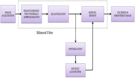
The proposed work has been motivated by the fact that Bilateral Filter is very efficient at smoothing the textures of an image while preserving edges and fine details. In this paper, the performance of Bilateral Filter has been improved for DICOM Images. The algorithm used for optimization is Genetic Algorithm. The first and foremost step is converting ‘.dcm’ format files to ‘.bmp’ format files for visualization and processing for experimental purpose. After that, pre-processing of noise present in images is done using ‘imfilter function’ in MATLAB. Now, instead of pre-processing complete image, an image is segmented into various segments i.e. sets of pixels to make the processing easy and this is done using Trigonometric Polynomials approximation. Then, Quantization is performed and the quantized values are then given as population to Genetic Algorithm. Genetic Algorithm with the help of its genetic operators performs selection, crossover and mutation using fitness function and yields optimized values as filtered image pixels. The output optimized values provided by Genetic Algorithm are then fed to Kernel which will help in the reconstruction of an image and a smoothed and filtered output image is obtained as result. The Block Diagram of the Proposed Method is shown in Fig. 1.
Figure 1: Block diagram of the proposed method
The steps involved in the implementation are:
1) Image Acquisition
images using ‘imfilter function’ in MATLAB. The acquired images are then used as input images for simulation purpose.
2) Trigonometric Polynomials
Bilateral Filter comprises three parts: Trigonometric Polynomials approximation, Quantization and Kernel Design. The parameters for Bilateral Filter are shown in Table 1.
Table 1: Parameters of Bilateral Filter
Symbol Parameter Value
w Window size 9
Spatial domain standard deviation 1 Intensity domain standard deviation 20
Segmentation is basically done to divide an image into different parts for better analysis. Cluster based segmentation approach is followed using trigonometric polynomials. A Trigonometric Polynomial is a finite linear combination of functions sin( ) and cos( ) i.e.
F(x) = cos( ) and sin( ) for k= 0………n.
The pixels in each row and column are processed by means of trigonometric polynomials of type as defined by T(x). T(x) stands for a Trigonometric Polynomial function of degree n.
( ) = + ∑ ( cos + sin ) (3) Where ∈ , ≠0 or ≠0.
k= total number of pixels a= total number of rows b= total number of columns.
3) Quantization
The result obtained by Trigonometric Polynomials Approximation is then used for quantization to produce Quantized values. These Quantized values then act as Population for Genetic Algorithm. Quantization is achieved by compressing a range of values to a single quantum value. It has a relation with the number of gray levels as it determines the number of gray levels that an image can have. Quantization used is expressed as below:
( ) = (1 + + + + ) (4) Where z= Total Polynomials and e= error.
The supposed values are:
= (3.6) = (2.916) = (4.80) = (0.6561)
4) Genetic Algorithm
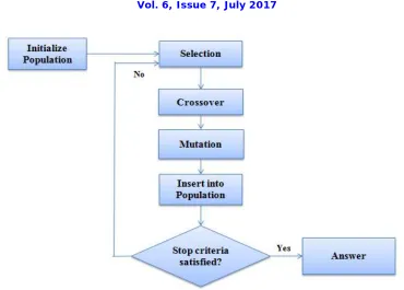
Figure 2: Flowchart of simple Genetic Algorithm
The steps involved are:
i) Initialization of Population
For the Algorithm to run, population is required. From the population, Parents are selected and then crossover and mutation takes place [17]. After terminating condition has been satisfied, the algorithm comes out of the loop and gives the result. Here, the quantized values obtained after Quantization step act as population to GA. With the initialization of the algorithm, the first generation starts and the algorithm runs till the maximum number of generations defined. ii) Evaluation of Population
In this step, every pixel value is taken under consideration. The loop runs as shown below: for i= 1 to N do
for j= 1 to L do Evaluate P[i] and P[j].
Where N shows the pixel values are evaluated row wise, L shows the pixel values are evaluated column wise. P[i] and P[j] represent the pixel values being taken under consideration.
iii) Selection
The selection operator is based on two functions: Fitness function and Roulette function. Parents are selected according to their fitness and using Roulette wheel selection function.
Fitness function is an objective function which describes how close a solution to the problem can be achieved according to the set aims.
The fitness function is described as:
If (x≥roulettefunctionvalue) or If (x>0)
f(x) = ( ) ∗0.9 (5) Else
Where x= [length of chromosome, blocks, pixel value, roulette function value]
Length of chromosome= 5, number of pixels taken under consideration at a time= 10, block size= 2*5.
Roulette-wheel selection is the most common used selection mechanism for the selection of Parents to produce Offspring. [17]
Roulette wheel model selects Parents according to their fitness. Roulette-wheel Parent selection follows the steps as shown below:
Sum the fitness of all the population members
Generate a random number (n) between 0 and total fitness value of all the population members. Return the first population member whose fitness added to the fitness of the preceding
population members is greater than or equal to n.
Using these two functions, Selection operator selects the Parents and the loop that runs for this operator in Algorithm is for i= 1 to N do
for j= 1 to L do
Parent[i] = fitness (P[i], roulette function) Parent[j] = fitness (P[j], roulette function)
The two Parent matrices selected in this step are then subjected to crossover to produce an offspring matrix. iv) Crossover
Crossover indicates exchange of genetic material between two Parent matrices and the resultant matrix is termed as Offspring. Crossover occurs according to the user-definable Crossover Probability [18]. The steps for uniform crossover to take place are:
for i= 1 to N step2 do for j= 1 to L do If (j≤ crossover_point)
Offspring [i] [j] = Parents [i] [j] Offspring [i+1] [j] = Parents [i+1] [j] Else
Offspring [i] [j] = Parents [i+1] [j] Offspring [i+1] [j] = Parents [i] [j]. v) Mutation
Mutation is a genetic operator that alters some gene values in Offspring from its initial state. Mutation occurs according to the user-definable Mutation Probability [18]. This probability should be set low. If it is too high, the search will turn into primitive random search. The steps for mutation to take place are:
for i= 1 to N do for j= 1 to L do
If (random_number<mutation rate) Offspring [i][j] = random (cell(i,j)) Else
Offspring [i][j] = Offspring [j][i] vi) Reinsertion into Population
The resultant Offspring after mutation is again inserted into population and there is a start of new generation. Again, there will be selection of fittest individuals to be Parents. In the new generation, again Parents undergo crossover to produce Offspring. The process continues till the termination condition has been satisfied.
vii) Termination Condition
The terminating condition says if current generation is less than maximum number of generations defined for the process, algorithm rums and jumps to selection step again. Otherwise it comes out of the loop and gives the result. The Parameters of GA taken under consideration are: Maximum population size= 100, Crossover probability= 0.6, Mutation probability= 0.2 and Maximum number of generations= 200.
5) Kernel Design
Reconstruction of image is the last task done by Bilateral Filter and is done with the use of Kernel. Kernel helps in the reconstruction of image using the result provided by GA. It helps in parallel processing. The kernel designed is:
V = v = ∑ ∑ (7)
Where
f Represent the coefficient of convolution values for kernel at position in the kernel (i,j). Represent the data value of the pixel that corresponds to f
F= either the sum of the coefficients of the kernel or, 1 if the sum of the coefficients is equal to zero. represent the output pixel value. In case it is less than zero, it is clipped to zero.
V= the output image.
A smoothed and filtered Image is produced as output.
IV.EXPERIMENTAL RESULTS
Database of 20 MRI and 20 Ultrasound images has been collected from UCI Machine Learning Repository. Images of collected database have been resized to 256*256 dimensions for further processing in MATLAB. The performance analysis has been done on the basis of parameters MSE and PSNR. The accuracy of the filter is analyzed using kappa coefficient.
i)Mean Square Error (MSE): It represents the cumulative squared error that occurs between denoised and original form of the image. Its mathematical representation is:
MSE= ∑ ∑ [ ( , )− ( , )] (8)
Where m represents number of rows in an image, n represents number of columns in an image. ( , ) Is noisy image and ( , ) is the denoised image.
ii) Peak signal-to-noise Ratio (PSNR): It computesPeak signal-to-noise Ratio between images. It is mathematically expressed as:
PSNR= 10. log ( ) (9)
Where, MAXI is the maximum possible pixel value of the image or is also known as maximum fluctuation in the input image data type. When the pixels are represented using 8 bits per sample, this is 255 and if the input image has a double-precision floating-point data type, MAXI= 1. The PSNR value approaches infinity as the MSE approaches zero. Higher value of PSNR of denoised and original image implies that the performance of the denoising filter method and visual quality of the denoised image is good [19].
iii) Kappa Coefficient: Kappa coefficient is used to measure the agreement between two processes. Here, the filter accuracy has been measured using kappa coefficient for both existing and proposed technique. It is defined as:
kappa s coef icient= (10)
(a) (b)
(c) (d)
Figure 3: (a) Input MRI image 1. (b) Filtered MRI image 1. (c) Input MRI image 2. (d) Filtered MRI image 2.
20 Ultrasound images were used for the work and the filtered output image corresponding to two sample input Ultrasound images by proposed method is shown in Fig. 4.
(c) (d)
Figure 4:a) Input Ultrasound image 1. (b) Filtered Ultrasound image 1. (c) Input Ultrasound image 2. (d) Filtered Ultrasound image 2.
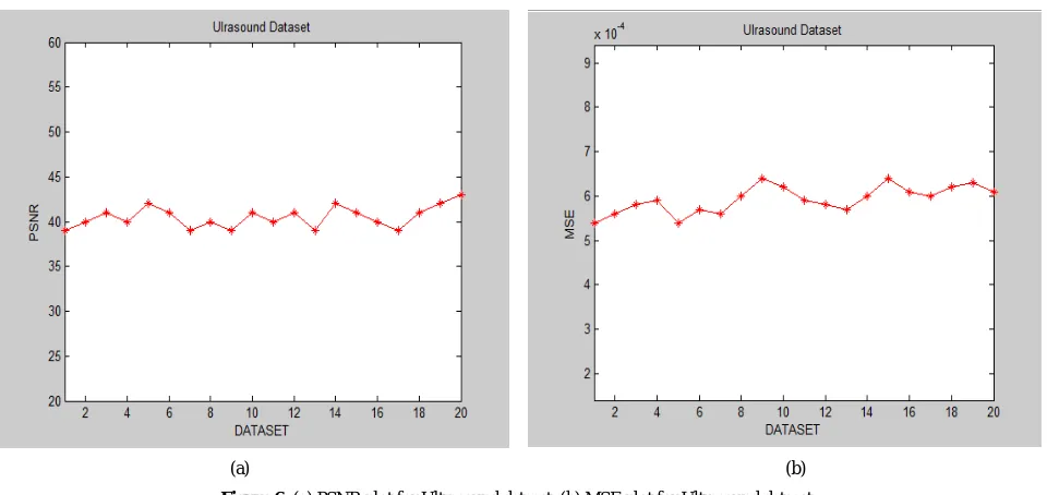
PSNR and MSE parameters depict the visual quality of an image. Fig. 5 shows PSNR and MSE plot for MRI Dataset (20 images) and Fig. 6 shows PSNR and MSE plot for Ultrasound Dataset (20 images).
(a) (b)
(a) (b)
Figure 6: (a) PSNR plot for Ultrasound dataset. (b) MSE plot for Ultrasound dataset.
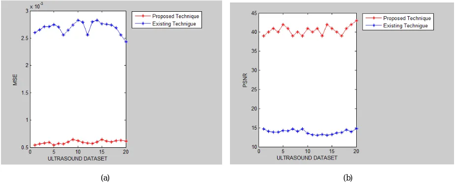
The comparison of existing technique [14] and proposed technique has been done on the basis of parameters Mean Square Error, Peak Signal-to-noise Ratio, and Kappa coefficient. Fig. 7 shows MSE plot and PSNR plot for comparative analysis done on MRI dataset. Fig. 8 shows MSE plot and PSNR plot for comparative analysis done on Ultrasound dataset.
(a) (b)
(a) (b)
Figure 8: (a) MSE plot for comparative analysis on Ultrasound dataset. (b) PSNR plot for comparative analysis on Ultrasound dataset.
Another parameter used for comparative analysis is Kappa’s Coefficient. The value of kappa coefficient can be at the most equal to 1 or can be described in percentage. On MRI dataset, Kappa’s value for existing technique is 78.9 (%) and for proposed technique is 89.1. On Ultrasound dataset, Kappa’s value for existing technique is 80.9 and for proposed technique is 91.1.
From the performance analysis and comparative analysis done on both the datasets, it can said that the proposed technique is efficient in smoothing and filtering DICOM Images as it yields higher PSNR values and lower MSE values for both datasets (MRI and Ultrasound) used for experimental purpose.
V. CONCLUSION
Medical Imaging is one of the most important techniques which play a major role in diagnosis of diseases present in body. There has been an enormous increase in the use of digital systems in medical field over last two decades. DICOM (Digital Imaging and Communications in Medicine) is a standard for handling, storing, printing and transmitting information in Medical Imaging. DICOM Images are vulnerable to noise and efficient filtering technique is required for these images. In this paper, a technique using Bilateral Filter and Genetic Algorithm has been proposed to smooth and filter DICOM Images. The implementation of Genetic Algorithm was done in MATLAB. Bilateral Filter has three main aspects: Trigonometric Polynomials approximation, Quantization and Reconstruction of image using Kernel. Quantized values are fed as population to GA and GA gives optimized values as output. Kernel designed helps in reconstruction of images to give smooth and filtered images as output. The database used is of 20 MRI Images and 20 Ultrasound Images. For the database collected, implementation was also done for existing technique which is Lemma distributions based Fast Bilateral Filter. The analysis done on database is based on the parameters MSE and PSNR. The filter accuracy is analyzed using Kappa statistic. Results show proposed technique gives lower MSE and higher PSNR values and is very efficient in smoothing the textures of DICOM Images and hence can be employed where smoothing and filtering of DICOM Images are required.
REFERENCES
[1] Goel N., Yadav A. and Singh B. M., “Medical image processing: A review”, Second International Innovative Applications of Computational Intelligence on Power, Energy and Controls with their Impact on Humanity (CIPECH), Ghaziabad, pp. 57-62, 2016.
[2] Dandu R. V., “Managing DICOM images: Tips and tricks for the Radiologist”, Indian Journal of Radiology & Imaging, Vol. 22 Issue 1, pp. 4-13, January 2012.
[3] Bhagat A. P., Atique M., “Medical Images: Formats, Compression techniques and DICOM Image retrieval- A Survey”, IEEE International conference on Devices, Circuits and Systems, Coimbatore, India, 15-16 March 2012.
[4] Liu B., Zhu M., Zhang Z., Yin C., Liu Z. and Gu J., “Medical Image Conversion with DICOM”, IEEE Canadian Conference on Electrical and Computer Engineering, Vancouver, BC, pp. 36-39, 2007.
[5] Tomasi C. and Manduchi R., "Bilateral filtering for gray and color images”, IEEE Sixth International Conference on Computer Vision,Bombay, pp. 839-846, 1998.
[6] Paris S., Korprobst P., Tumblin J. and Durand F., “Bilateral Filter: Theory and Applications”, Foundations and Trends in Computer Graphics and Vision, Vol. 4, Issue1, 2009.
[7] Schaefer G., Huguet J., Zhu S. Y., Plassmann P. and Ring F., “Adopting the DICOM standard for medical infrared images”, IEEE International Conference of Engineering in Medicine and Biology Society, New York, NY, pp. 236-239, 2006.
[8] Arif A. S., Mansor S. and Logeswaran R., “Combined Bilateral and Anisotropic-Diffusion Filters for Medical Image De-noising”, IEEE Student conference on Research and Development, Cyberjaya, Malaysia, 19-20 December 2011, pp. 420-424, 2011.
[9] Tang J. and Guo S., “A 3-D bilateral filter for speckle reduction in 3-D ultrasound images for cattle follicle segmentation”, IEEE International Conference on Systems, Man, and Cybernetics (SMC), Seoul, pp. 2485-2488, 2012.
[10] Sumalatha R. and Subramanyam M. V., “Image denoising using spatial adaptive mask filter for medical images”, IEEE International Conference on Electrical, Electronics, Signals, Communication and Optimization (EESCO), Visakhapatnam, pp. 1-4, 2015.
[11] Mahesh Mohan M.R. and Sheeba V. S., “A novel method of medical image de-noising using bilateral and NLM De-noising”, IEEE Third international conference on advances in computing and communications, Cochin, August 2013, pp. 186-191, 2013.
[12] Kalavathi P. and Priya T., “Removal of impulse noise using Histogram-based Localized Wiener Filter for MR brain image restoration”, IEEE International Conference on Advances in Computer Applications (ICACA), Coimbatore, pp. 4-8, 2016.
[13] Kaur P., Singh G. and Kaur P., “Image enhancement of ultrasound images using multifarious denoising filters and GA”, IEEE International Conference on Advances in Computing, Communications and Informatics (ICACCI), Jaipur, pp. 2375-2384, 2016.
[14] Papari G., Idowu N. and Varslot T., “Fast Bilateral Filtering for Denoising Large 3D Images”, IEEE Transactions on Image Processing, vol. 26, Issue 1, pp. 251-261, January 2017.
[15] Muthuswamy J., “New Edge Preserving Hybrid Method for better enhancement of Liver CT Images”, International Journal of Science and Technology, Volume 10, Issue 10, pp. 1-7, March 2017.
[16] Holland J. H., “Adaptation in Natural and Artificial systems”, Cambridge, MA: MIT Press, 1975.
[17] Man K F., Tang K S. and Kwong S, “Genetic Algorithms: Concepts and applications”, IEEE Transactions on Industrial electronics, Vol. 43, Issue 5, pp. 519-534, October 1996.
[18] Hole K. R., Gulhane V. S. and Shellokar N. D., “Application of Genetic Algorithm for Image Enhancement and Image Segmentation”, International Journal of Advanced Research in Computer Engineering & Technology, Vol. 2, Issue 4, pp. 1342-1346, April 2013.