Aurora-A: an expedition to the pole of the spindle
in Xenopus egg extracts
JACEK Z. KUBIAK
1,2,3and CLAUDE PRIGENT*
,1,21CNRS, Unité Mixte de Recherche UMR6290, 2Université de Rennes 1, Institut de Génétique et Développement de Rennes, UEB, 35000 Rennes, France and 3Department of Hygiene and Physiology,
Military Institute of Hygiene and Epidemiology, Warsaw, Poland.
ABSTRACT The aim of this short review is to describe the contribution of Xenopus laevis egg ex-tracts to the discovery and understanding of the regulation and function of the serine/threonine kinase Aurora-A. The power of these extracts to recapitulate cell cycle events makes them a precious tool to decipher complex biological processes at the molecular level, including the mechanisms that affect Aurora-A (post-translational modifications) and mechanisms in which Aurora-A plays a crucial role (bipolar spindle assembly). We focus on the results obtained in cell-free extracts, but we also give an updated overview of Aurora A functions found in other systems.
KEY WORDS:
Aurora-A, Eg2, Ipl1, cell-free extract, Xenopus laevis, TPX2, spindle assembly
Xenopus oocytes and eggs
Amphibian oocytes and eggs have been used to study the regula-tion of cell cycle controls at the biochemical level since the seventies. Oocytes and eggs are naturally arrested at particular stages of cell cycle. Xenopus laevis oocytes are arrested in prophase of meiosis I while unfertilized eggs (or matured oocytes) in metaphase of meiosis II through the action of CSF (cytostatic factor). The transition from prophase I to metaphase II in oocytes can easily be induced in vitro by using progesterone or insulin (Fortune et al., 1975) (El-Etr et al., 1979). When fertilized, the egg enters intense phases of cell divi-sions with a succession of S and M phases without any G phases. All these divisions occur without transcription and without any gain of volume of the embryo. It is only at stage 12 that the G phases appear together with transcription and the differentiation of future tissues begins. To ensure all these rapid divisions the unfertilized egg (or mature oocyte) has stored during oogenesis a large amount of proteins involved in cell cycle regulation. To make a rough estimate, one female Xenopus laevis can lay several thousand eggs, and one egg contains the same amount of proteins as 106 HeLa cells. For
these reasons Xenopus laevis eggs have become a suitable and affordable model to study the biochemistry of cell division. In addi-tion, protein extracts prepared from oocytes and eggs recapitulate many biological events and in particular those of the cell cycle, such as oocyte maturation, DNA repair, DNA replication, spindle
as-Int. J. Dev. Biol. 60: 255-261 (2016)
doi: 10.1387/ijdb.160189jk
www.intjdevbiol.com
*Address correspondence to: Claude Prigent. Institut de Génétique et Développement de Rennes, IGDR, CNRS UMR6290 Université de Rennes 1, 2 avenue du Pr
Léon Bernard, 35043 Rennes cedex, France. E-mail: [email protected]
Accepted: 13 June, 2016.
ISSN: Online 1696-3547, Print 0214-6282 © 2016 UPV/EHU Press
Printed in Spain
Abbreviations used in this paper: CSF, cytostatic factor; EDEN, embryo-deadenylation elemeNt; MPF, M-phase promoting factor; MTOC, microtubule organizing center. sembly, signal transduction etc. (Maller, 2012). Extracts are easy to manipulate for structure/function studies. For instance, proteins can be depleted using antibodies, and mutated protein added to assess their function. Several protocols have been published to facilitate extracts’ use and most of them are available on the Xenopus web site (http://www.xenbase.org) (James-Zom et al., 2015)
Eg2: the Xenopus Aurora-A
this kinase rapidly increased when it was found overexpressed in breast cancer cell lines (Sen et al., 1997) and demonstrated to be an oncogene (Bishoff et al., 1998) (Zhou et al., 1998)
The first Aurora-A function discovered in Xenopus
cell-free extract
The function of the kinase was identified using Xenopus leavis egg extracts. Protein extracts were prepared from metaphase-arrested oocytes (unfertilized egg), from which the kinase was eliminated by immunoprecipitation using a specific monoclonal antibody. The extract was incubated with demembraned sperm nuclei (source of DNA) and rhodamine-labelled tubulin (to visualize the microtubules) in the presence of calcium to mimic fertilization and trigger exit from metaphase II. The extract replicated DNA and formed nuclei. At this point fresh extract from UFE was added to arrest the extract at metaphase. While bipolar mitotic spindles were assembled by the control extract, only abnormal spindles (monopolar) were observed in extracts lacking Eg2 (Aurora-A) (Roghi et al., 1998). This study reported that Eg2 was a centrosome protein that binds to microtubules and its kinase activity was required to assemble bipolar mitotic spindle. This was further confirmed by others showing that in Xenopus laevis egg extracts Aurora-A belongs to a protein complex that participates in nucleation of microtubules dedicated to mitotic spindle assembly (Koffa et al., 2006). More precise function of the kinase was demonstrated when beads coated with Aurora-A and incubated in Xenopus laevis egg extract were found to behave just like centrosome’s MTOCs (Microtubule Organizing Center). These beads assembled bipolar spindles in the presence of RanGTP in an Aurora-A kinase activity dependent manner (Tsai and Zheng, 2005). Thus, Aurora-A kinase is not only required for bipolar spindle assembly, but is directly involved in the regulation of microtubule nucleation (Sardon et al., 2008).
Aurora-A mRNA regulation via polyadenylation and
deadenylation
As mentioned above, the Eg2, later named Aurora-A, was dis-covered due to the particular behaviour of its mRNA in oocytes and embryos of Xenopus laevis (Paris et al., 1990). In short, this maternal mRNA was polyadenylated and translated in eggs and deadenyl-ated after fertilisation. In the same screen other maternal mRNAs showing the same behaviour were identified and named Eg1, 2, etc. (for Eg as “egg”). Many of these clones were further identified as important genes regulating cell cycle or embryo development. Eg1 is CDK2, Eg3 MELK kinase, Eg5 a microtubule-associated kinesin, Eg7 a condensing complex partner protein, a homologue to human hCAP-D2. In contrast, identified by this screen Cl clones (Cl1 and Cl2) were polyadenylated following fertilisation. Cl2 was later identified as Saccharomyces cerevisiae SUP45 (also called SUP1 or SAL4) gene (Tassan et al., 1993), and Cl2 as a homo-logue of Limb-bud and Heart (LBH) protein (Takada et al., 2005).
The regulation of thee maternal mRNAs was also studied in cell-free extract of Xenopus oocytes and embryos. The deadenylation of Eg-type mRNAs including Eg2/Aurora-A mRNA depended on a short sequence element in their 3’ untranslated region (3’-UTR) named EDEN (Embryo-Deadenylation ElemeNt; Bouvet et al., 1994). A protein called EDEN-BP (for EDEN-Binding Protein, further renamed for Celf1) was identified to bind to EDEN and to
be necessary for these mRNAs deadenylation (Legagneux et al., 1992). Short polyadenylated plasmids containing EDEN sequence placed in cell-free extracts of embryos were efficiently deadenylated when EDEN-BP was present. The regulation of EDEN-BP was also tested in cell-free extracts. Calcium addition to CSF extract triggered activation of the extract, entering the embryo interphase, deadenylation of Eg2 3’UTR-containing sequences mRNA and the rapid dephosphorylation of EDEN-BP (Detivaud et al., 2003). Phosphorylation of EDEN-BP and its deadenylation activity toward Eg2 mRNA was found to be mediated by CDK1 (MPF for M-phase Promoting Factor) and ERK2 MAP kinase. Accordingly, EDEN-BP was highly phosphorylated and inactive when the activities of these kinases were high during oocyte maturation, and was dephos-phorylated and active when CDK1 and ERK2 were inactivated following fertilisation (Detivaud et al., 2003). These results showed an important interrelationship between the cell cycle regulation and maternal mRNAs regulation including Eg2/Aurora-A mRNA during Xenopus laevis oocyte maturation and early development.
Aurora-A function during oocyte maturation
Stage VI oocytes in Xenopus laevis are arrested in G2 waiting for an outside signal triggering meiotic maturation. This signal is provided by progesterone that through binding to a receptor will trigger the decrease of PKA activity and the activation of signal transduction leading to oocyte maturation. Several cascades of kinase activation eventually lead to the production of c-mos and the activation of MAPK and MPF. A screen designed to search for proteins activated early after progesterone binding identified Aurora-A as one of the first kinase to be phosphorylated and activated (Andrésson and Ruderman, 1998). Aurora-A was found to induce the translation of c-mos mRNA by phosphorylating and regulating the cytoplasmic polyadenylation element binding factor (CPEB) (Mendez et al., 2000). Aurora-A was then found to be involved in the regulation of mRNA translation.
Regarding the activation of Aurora-A early after progesterone treatment, other groups reported that Aurora-A activity was not detectable in prophase oocytes, but activated in parallel to MPF (Frank-Vaillant et al., 2000). However, progesterone does trigger Aurora-A biosynthesis and accumulation during oocyte maturation, but its phosphorylation and activation depends on MPF activation and not on c-mos/MAPK pathway activation (Maton et al., 2003). This was also confirmed by the study showing that Aurora-A activation followed two M-phases during oocyte maturation at metaphase of meiosis I and at metaphase of meiosis II, exactly like MPF activity (Ma et al., 2003). This biphasic activation of Aurora-A was later explained by finding that Aurora-A was phosphorylated on Ser349 between meiosis I and meiosis II and that this phosphorylation down regulated its kinase activity (Pascreau et al., 2008).
Aurora-A in Xenopus egg extracts 257
meiosis I–II transition has also been reported (Castro et al., 2003). Phosphorylation of TACC3/Maskin by Aurora-A was identified during mitosis and was related to centrosome localisation of TACC3 and bipolar spindle assembly (Giet et al., 2002) (Barros et al., 2005).
Regulation of Aurora-A by degradation
The first studies reported either an absence of destruction box (D-box) in Aurora-A or the presence of potential destruction box and a potential KEN-box (Honda et al., 2000) (Walter et al., 2000). The demonstration of the presence of a fully functional destruction box in Aurora-A was done using Xenopus leavis XL2 cell extracts and recombinant Xenopus laevis Aurora-A kinase (Arlot-Bonnemains et al., 2001). The D-box was identified at position 378-381 in the C-terminal end of the protein. The D-box was found conserved not only in Aurora-A from other species, but also in all Aurora-A, -B and -C kinases (Arlot-Bonnemains et al., 2001).
The D-box of Aurora-A was further studied in Xenopus laevis egg extract. This study showed that the D-box dependent degradation of Aurora-A was driven by the anaphase-promoting complex/cyclo-some (APC/C) activated by Fizzy-related (Cdh1 in human) (Castro et al., 2002a). The putative KEN-box of Aurora-A was found to be nonfunctional in the same study. However a new sequence in the N-terminal end of the in Xenopus laevis Aurora-A was discovered by the same authors and called DAD for D-box-activating domain (44VSAQRILGPSNV55). Both Aurora-A and Aurora-B proteins
pos-sess a D-box, but only Aurora-A and not Aurora-B was degraded in an APC/C dependent manner. The authors demonstrated that the functionality of the D-box of Aurora-A depended on the presence of the DAD that was absent in Aurora-B (Castro et al., 2002b). The group of Joan Ruderman reported concomitantly the same discovery, but named the sequence the A-box because of its pres-ence only in Aurora-A and not in Aurora-B or -C (Littlepage and Ruderman, 2002). The same group also showed that Xenopus laevis Aurora-A contains Serine 53, which phosphorylation regu-lates functionality of the A-box. Phosphorylation of S53 prevents degradation of Aurora-A protein (Littlepage et al., 2002; Fig. 1)
Regulation of Aurora-A by phosphorylation
Aurora-A belongs to the family of serine/threonine kinases ac-tivated by phosphorylation of a threonine residue in the activation loop of the kinase (Walter et al., 2000). The exact mechanism of activation was deciphered in Xenopus laevis egg extract (Tsai et al., 2003) (Eyers et al., 2003). Binding of Aurora-A to its activator TPX2 (Targeting for Xklp2) triggers intermolecular autophosphorylation of T295 (T288 in human) (Eyers et al., 2003) and protects T295 from being dephosphorylated by the phosphatase PP1 (Tsai et al., 2003). This has been observed when (His)6-tagged Aurora-A
prepared from insect cells (Sf9) or from bacteria (E. coli) were used for kinase assay. Aurora-A purified from Sf9 had to be treated with okadaic acid to inhibit the phosphatase PP1 that co-purified with the kinase, while it was not necessary when Aurora-A was puri-fied from bacteria. While TPX2 binds to and activates Aurora-A, the INCENP (Inner Centromere Protein) plays the same role for Aurora-B (Adams et al., 2000). The way the TPX2 differentiates between Aurora-A and –B has been identified at the structural level (Bayliss et al., 2004). A single amino acid change G205N in Xenopus laevis Aurora-A is sufficient to abolish activation by TPX2 (Eyers et al., 2005), and the same mutation in human (G198N) transforms the Aurora-A kinase into an “Aurora-B like kinase” that can be activated by INCENP but not by TPX2 (Hans et al., 2009). Although phosphorylation of the threonine in the activation loop is essential to obtain an active Aurora-A kinase, the protein is sub-jected to other phosphorylation events that can either up-regulate or down regulate its kinase activity. In Xenopus laevis egg extract Aurora-A is phosphorylated on Serine 349 and the mutation S349D completely abolished the kinase activity (Littlepage et al., 2002) (Haydon et al., 2003). The purpose of this phosphorylation in vivo is to down-regulate Aurora-A kinase activity during oocyte maturation between meiosis I and meiosis II. Indeed, during this period Aurora-A kinase activity decreases by approximately 50% (Pascreau et al., 2008). Phosphorylation of S349 is not due to autophosphoryla-tion per se, because active Aurora-A can not phosphorylate S349 (Pascreau et al., 2008). However, when Aurora-A is primed through phosphorylation of S290/S291 by the glycogen synthase kinase 3 (GSK-3), then Aurora-A S290p/S291p does autophosphorylate its
S449 (Sarkissian et al., 2004). Phosphorylation of S349 can also be performed in vitro by the kinase xPAK1 (p21-activated kinases 1) that is required for spindle assembly (Vadlamudi et al., 2000). In vitro phosphorylation of Aurora-A leads also to a 50% decrease of its kinase activity (Pascreau et al., 2008). In human cells, the same serine S342 has been found phosphorylated in G2 arrested cells when the DNA damage checkpoint is activated. It also down regulates Aurora-A activity (Krystyniak et al., 2006).
Another site has been identified in Aurora-A, that serves as an up-regulation site, but only in human kinase (S89) that had been phosphorylated on T288. This serine is an autophosphorylation site that becomes phosphorylated at the centrosome in the pres-ence of the chaperone protein nucleophosmine (Reboutier et al., 2012). Although S89 is conserved in Xenopus laevis Aurora-A, its phosphorylation has not been proven yet to stimulate its kinase activity. For details see Fig. 2.
Aurora-A substrates identified in Xenopus
Many substrates of Aurora-A have been identified using Xeno-pus laevis egg extracts, not only at the biochemical level through
378-381
protein phosphorylation, but also at the functional level. Indeed, the egg extracts are used to assemble bipolar mitotic spindle that can be visualized by fluorescence microscopy (Sawin and Mitchison, 1991). Because the protein of interest can be depleted from the extract and replaced by a mutated version of the same protein (Walczakat al., 1996) and because Aurora-A is involved in bipolar spindle assembly (Roghi et al., 1998) Xenopus egg extracts have been extensively used to study Aurora-A substrates.
Eg5 mRNA was identified in the same screen as Eg2 mRNA in Xenopus laevis (Paris et al., 1990). Eg5 mRNA that encodes a molecular motor (Le Guellec et al., 1991) had been renamed kine-sin-5. (Lawrence et al., 2004). Because the depletion of Aurora-A (Eg2) or kinesin-5 (Eg5) from egg extracts show the same phe-notype during spindle assembly, i.e. the formation of monopolar spindles, we asked whether the kinesin-5 could be a substrate of Aurora-A. Indeed, Aurora-A phosphorylates kinesin-5 in its stalk domain, however, the functional role of this phosphorylation still remains unclear (Giet et al., 1999).
Using Xenopus egg extract we also demonstrated that Aurora-A (but not Aurora-B) phosphorylates the tail of histone H3 on Serine 10. This phosphorylation event is required for chromosome assem-bly (Scrittori et al., 2001). Since the discovery that Aurora kinase
substrate in human cells where phosphorylation of its serine 315 was found to lead to a destabilisation of the protein, while phos-phorylation of serine 215 inhibited DNA binding properties of p53 (Katayama et al., 2004)(Liu et al., 2004). In Xenopus laevis egg extracts, p53 inhibits Aurora-A activity unless Aurora-A was previ-ously bound to TPX2 (Eyers and Maller, 2004). In this last case Aurora-A phosphorylates p53 on serines 129 and 190 (equivalent of human S215), which abrogates p53 transactivation activity (Pascreau et al., 2009).
Spindle assembly
As mentioned at the beginning of this article, Aurora-A is involved in bipolar spindle assembly (Roghi et al., 1998). The detailed mechanism of its action became clear after understanding of the role of its substrates and activators TPX2, HURP and MCAK. The kinase is activated by TPX2 in a Ran dependant manner (Tsai et al., 2003). Ran is a GTPase protein (ras-related nuclear protein). Its guanine-nucleotide-exchange factor is the protein RCC1 (Regula-tor of chromosome condensation 1) that loads the GTP on Ran. In G2 phase RCC1 localises on chromosomes with Ran-GTP and TPX2 being sequestered by importin-b in the cytoplasm. Upon
*
S89
pUp-regulation
Serine 89 Threonine 295 Serine 349
Serine 89 Threonine 288 Serine 342
Xenopus laevis
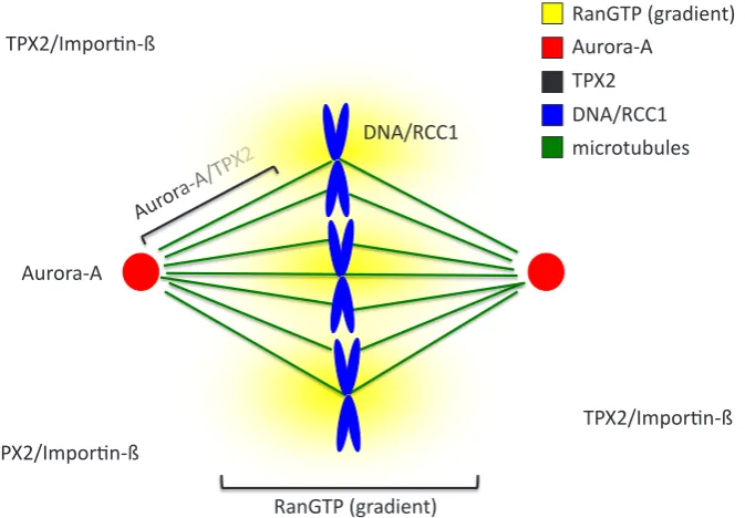
Fig. 3. Regulation of Aurora-A by Tpx2 in RanGTP dependent manner. In interphase, Aurora-A local-izes at the centrosomes and TPX2 in the cytoplasm sequestered by importin-b. RanGTP locallocal-izes as a gradient around RCC1 that is located on chromosome. During bipolar spindle assembly in mitosis, TPX2/ importin-b meets the RanGTP gradient, which releases TPX2 that associates with Aurora-A, activates its autophosphorylation on T288 and relocalizes the kinase to the spindle poles along microtubules.
phosphorylates H3 serine 10, a peptide corresponding to the tail of H3 tagged with GST is currently used together with an antiphosphoserine 10 antibody as an in vitro substrate to assay the Aurora kinases activity (Hsu et al., 2000).
Maskin (TACC3 in human) was identified as a Xenopus laevis Aurora-A partner in a two-hybrid screen (Pascreau et al., 2005). Phosphorylation of maskin seems to participate in the inhibition of the translation of some mRNA (masked mRNA). However, it is not the only role of this phosphorylation because it was also demonstrated in Xenopus egg extracts that maskin localisation and function in spindle assembly depends on Aurora-A activity (Peset et al., 2005; Kinoshita et al., 2005). An interaction between D-TACC (Drosophila maskin) has previously been reported. Aurora-A is required for D-TACC localisation and function in the nucleation of microtubules from the centrosomes (Giet et et al., 2002). The serine residue in maskin phosphorylated by Aurora-A is conserved in Xenopus, human and Drosophila.
The p53 was first identified as Aurora-A
Aurora-A in Xenopus egg extracts 259
nuclear envelope breakdown, Ran-GTP forms a gradient around the chromosomes, TPX2 is released from importin-b and translocates to the centrosome where it activates Aurora-A and localises it at the pole of the spindle (Kufer et al., 2002) (Tsai et al., 2003). The discovery that importin-b sequestered factors that were released during mitosis by Ran-GTP was an important breakthrough, thanks to Xenopus egg extracts, in the understanding of mechanisms involved in bipolar spindle assembly (Kalab et al., 1999) (Wilde et al., 1999) (Zhang et al., 1999) (Wiese et al., 2001) (Nachury et al., 2001) (Gruss et al., 2001; Fig. 3).
TPX2 and maskin are two of these importin-b sequestered fac-tors (Albee et al., 2006). Although, as described above, maskin is an Aurora-A substrate which phosphorylation regulates mRNA translation maskin is also directly involved in bipolar spindle as-sembly. Depletion of maskin from Xenopus laevis egg extracts leads to short microtubule asters and abnormal spindle formation. Interestingly enough, rescue experiments revealed that maskin is required for at least two steps in spindle assembly (O’Brien et al., 2005) (Kinoshita et al., 2005) (Peset et al., 2005).
The hepatoma up-regulated protein (HURP), is another protein sequestered by importin-b (Silljé et al., 2006). It is also a substrate of Aurora-A. In human cells the HURP is phosphorylated on S627, S-S725, S-S757 and S-S830 by Aurora-A, which contributes to the stabilization of the protein (Yu et al., 2005).
To build a bipolar spindle in Xenopus laevis egg extract, mi-crotubules must be first nucleated from MicroTubule Organising Centre (MTOC) to form asters that will further organise bipolar spindle. The Mitotic Centromere-Associated Kinesin (MCAK) is a key player in these events and is also phosphorylated by Aurora-A. MCAK localises at the centre of MTOC where it participates in spindle pole focusing required to form spindles in an Aurora-A dependent manner. Aurora-A also regulates MCAK localisation at spindle poles by phosphorylating S719 (Zhang et al., 2008). Beads coated with Aurora-A kinase can organise MTOC in the presence of RanGTP. These MTOC not only nucleate microtubule but also recruit factors essential to build a spindle (Tsai and Zheng, 2005). Thus, Aurora-A plays a central role in bipolar spindle assembly in Xenopus laevis egg extract.
Conclusion
Since its identification in 1995, Aurora-A kinase has become one of the major protein kinase regulating cell cycle progression. Very early, Aurora-A attracted a lot of attention because its kinase activity is directly linked to cancer. Thus, the novel chemotherapeutic approaches in cancer treatment may potentially involve inhibitors of Aurora-A (D’assoro et al., 2005)(Marugán et al., 2016). Since 1998, Xenopus egg extracts have been crucial for understanding the function and regulation of Aurora-A, and we do not take too much risk to say that they will continue to help us to better under-stand the roles of this kinase in the future.
Reference
ADAMS RR, WHEATLEY SP, GOULDSWORTHY AM, KANDELS-LEWIS SE, CAR-MENA M, SMYTHE C, GERLOFF DL, EARNSHAW WC. (2000). INCENP binds the Aurora-related kinase AIRK2 and is required to target it to chromosomes, the central spindle and cleavage furrow. Curr Biol. 10: 1075-1078.
ALBEE AJ, TAO W, WIESE C. (2006). Phosphorylation of maskin by Aurora-A is regulated by RanGTP and importin beta. J Biol Chem. 281: 38293-38301.
ANDRÉSSON T, RUDERMAN JV. (1998). The kinase Eg2 is a component of the
Xeno-pus oocyte progesterone-activated signaling pathway. EMBO J. 17: 5627-5637.
ARLOT-BONNEMAINS Y, KLOTZBUCHER A, GIET R, UZBEKOV R, BIHAN R, PRIGENT C. (2001). Identification of a functional destruction box in the Xenopus
laevis aurora-A kinase pEg2. FEBS Lett. 508: 149-152.
BARROS TP, KINOSHITA K, HYMAN AA, RAFF JW. (2005). Aurora A activates D-TACC-Msps complexes exclusively at centrosomes to stabilize centrosomal microtubules. J Cell Biol. 170: 1039–1046.
BISCHOFF, J. R., ANDERSON, L., ZHU, Y., MOSSIE, K., NG, L., SOUZA, B., SCHRYVER, B., FLANAGAN, P., CLAIRVOYANT, F., GINTHER, C., CHAN, C. S., NOVOTNY, M., SLAMON, D. J. AND PLOWMAN, G. D. (1998). A homologue of
Drosophila aurora kinase is oncogenic and amplified in human colorectal cancers. EMBO J. 17: 3052-3065.
BOUVET P, OMILLI F, ARLOT-BONNEMAINS Y, LEGAGNEUX V, ROGHI C, BASSEZ T, OSBORNE HB. (1994). The deadenylation conferred by the 3’ untranslated region of a developmentally controlled mRNA in Xenopus embryos is switched to polyadenylation by deletion of a short sequence element. Mol Cell Biol. 14: 1893-1900.
CASTRO A, ARLOT-BONNEMAINS Y, VIGNERON S, LABBÉ JC, PRIGENT C, LORCA T. (2002). APC/Fizzy-Related targets Aurora-A kinase for proteolysis.
EMBO Rep. 3: 457-462.
CASTRO A, VIGNERON S, BERNIS C, LABBÉ JC, PRIGENT C, LORCA T. (2002). The D-Box-activating domain (DAD) is a new proteolysis signal that stimulates the silent D-Box sequence of Aurora-A. EMBO Rep. 3: 1209-1214.
CASTRO A, MANDART E, LORCA T, GALAS S. (2003). Involvement of Aurora A kinase during meiosis I-II transition in Xenopus oocytes. J Biol Chem. 278: 2236-2241. CHAN CS, BOTSTEIN D. (1993). Isolation and characterization of chromosome-gain
and increase-in-ploidy mutants in yeast. Genetics 135: 677-691.
D’ASSORO AB, HADDAD T, GALANIS E. (2016). Aurora-A Kinase as a Promising Therapeutic Target in Cancer. Front Oncol. 5: 295.
DETIVAUD L, PASCREAU G, KARAISKOU A, OSBORNE HB, KUBIAK JZ. (2003). Regulation of EDEN-dependent deadenylation of Aurora A/Eg2-derived mRNA via phosphorylation and dephosphorylation in Xenopus laevis egg extracts. J
Cell Sci. 116: 2697-2705.
EL-ETR M, SCHORDERET-SLATKINE S, BAULIEU EE. (1979). Meiotic maturation in Xenopus laevis oocytes initiated by insulin. Science 205: 1397-1399. EYERS PA, ERIKSON E, CHEN LG, MALLER JL. (2003). A novel mechanism for
activation of the protein kinase Aurora A. Curr Biol. 13: 691-697.
EYERS PA, CHURCHILL ME, MALLER JL. (2005). The Aurora A and Aurora B protein kinases: a single amino acid difference controls intrinsic activity and activation by TPX2. Cell Cycle 4: 784-789.
EYERS PA, MALLER JL. (2004). Regulation of Xenopus Aurora A activation by TPX2.
J Biol Chem. 279: 9008-9015.
FORTUNE JE, CONCANNON PW, HANSEL W. (1975). Ovarian progesterone levels during in vitro oocyte maturation and ovulation in Xenopus laevis. Biol Reprod. 13:561-567.
FRANK-VAILLANT M, HACCARD O, THIBIER C, OZON R, ARLOT-BONNEMAINS Y, PRIGENT C, JESSUS C. (2000). Progesterone regulates the accumulation and the activation of Eg2 kinase in Xenopus oocytes. J Cell Sci. 113: 1127-1138. GIET R, MCLEAN D, DESCAMPS S, LEE MJ, RAFF JW, PRIGENT C, GLOVER DM. (2002). Drosophila Aurora A kinase is required to localize D-TACC to centrosomes and to regulate astral microtubules. J Cell Biol 156: 437–451.
GLOVER DM, LEIBOWITZ MH, MCLEAN DA, PARRY H. (1995). Mutations in aurora prevent centrosome separation leading to the formation of monopolar spindles.
Cell 81: 95-105.
GRUSS OJ, CARAZO-SALAS RE, SCHATZ CA, GUARGUAGLINI G, KAST J, WILM M, LE BOT N, VERNOS I, KARSENTI E, MATTAJ IW. (2001). Ran induces spindle assembly by reversing the inhibitory effect of importin alpha on TPX2 activity. Cell 104: 83-93.
HANS F, SKOUFIAS DA, DIMITROV S, MARGOLIS RL. (2009). Molecular distinc-tions between Aurora A and B: a single residue change transforms Aurora A into correctly localized and functional Aurora B. Mol Biol Cell. 20: 3491-3502. HAYDON CE, EYERS PA, AVELINE-WOLF LD, RESING KA, MALLER JL, AHN NG.
HONDA K, MIHARA H, KATO Y, YAMAGUCHI A, TANAKA H, YASUDA H, FURU-KAWA K, URANO T. (2000). Degradation of human Aurora2 protein kinase by the anaphase-promoting complex-ubiquitin-proteasome pathway. Oncogene 19: 2812-2819.
HSU JY, SUN ZW, LI X, REUBEN M, TATCHELL K, BISHOP DK, GRUSHCOW JM, BRAME CJ, CALDWELL JA, HUNT DF, LIN R, SMITH MM, ALLIS CD. (2000). Mitotic phosphorylation of histone H3 is governed by Ipl1/aurora kinase and Glc7/ PP1 phosphatase in budding yeast and nematodes. Cell 102: 279-291. JAMES-ZORN C, PONFERRADA VG, BURNS KA, FORTRIEDE JD, LOTAY VS, LIU
Y, BRAD KARPINKA J, KARIMI K, ZORN AM, VIZE PD. (2015). Xenbase: Core features, data acquisition, and data processing. Genesis 53: 486-497. KALAB P, PU RT, DASSO M. (1999). The ran GTPase regulates mitotic spindle
as-sembly. Curr Biol. 9: 481-484.
KATAYAMA H, SASAI K, KAWAI H, YUAN ZM, BONDARUK J, SUZUKI F, FUJII S, ARLINGHAUS RB, CZERNIAK BA, SEN S. (2004). Phosphorylation by aurora kinase A induces Mdm2-mediated destabilization and inhibition of p53. Nat Genet. 36: 55-62.
KINOSHITA K, NOETZEL TL, PELLETIER L, MECHTLER K, DRECHSEL DN, SCHWAGER A, LEE M, RAFF JW, HYMAN AA. (2005). Aurora A phosphorylation of TACC3/maskin is required for centrosome-dependent microtubule assembly in mitosis. J Cell Biol. 170: 1047-1055.
KOMRSKOVA P, SUSOR A, MALIK R, PROCHAZKOVA B, LISKOVA L, SUPOLIKOVA J, HLADKY S, KUBELKA M. (2014). Aurora kinase A is not involved in CPEB1 phosphorylation and cyclin B1 mRNA polyadenylation during meiotic maturation of porcine oocytes. PLoS One. 9: e101222.
KRYSTYNIAK A, GARCIA-ECHEVERRIA C, PRIGENT C, FERRARI S. (2006). Inhibition of Aurora A in response to DNA damage. Oncogene. 25: 338-348. KUFER TA, SILLJÉ HH, KÖRNER R, GRUSS OJ, MERALDI P, NIGG EA. (2002).
Human TPX2 is required for targeting Aurora-A kinase to the spindle. J Cell Biol. 158: 617-623.
KOFFA MD, CASANOVA CM, SANTARELLA R, KÖCHER T, WILM M, MATTAJ IW. (2006). HURP is part of a Ran-dependent complex involved in spindle formation.
Curr Biol. 16: 743-754.
LAWRENCE CJ, DAWE RK, CHRISTIE KR, CLEVELAND DW, DAWSON SC, ENDOW SA, GOLDSTEIN LS, GOODSON HV, HIROKAWA N, HOWARD J, MALMBERG RL, MCINTOSH JR, MIKI H, MITCHISON TJ, OKADA Y, REDDY AS, SAXTON WM, SCHLIWA M, SCHOLEY JM, VALE RD, WALCZAK CE, WORDEMAN L. (2004). A standardized kinesin nomenclature. J Cell Biol. 167: 19-22.
LEGAGNEUX V, BOUVET P, OMILLI F, CHEVALIER S, OSBORNE HB. (1992). Identification of RNA-binding proteins specific to Xenopus Eg maternal mRNAs: association with the portion of Eg2 mRNA that promotes deadenylation in embryos.
Development 116: 1193-1202.
LE GUELLEC R, PARIS J, COUTURIER A, ROGHI C, PHILIPPE M. (1991). Clon-ing by differential screenClon-ing of a Xenopus cDNA that encodes a kinesin-related protein. Mol Cell Biol. 11: 3395-3398.
LIU Q, KANEKO S, YANG L, FELDMAN RI, NICOSIA SV, CHEN J, CHENG JQ. (2004). Aurora-A abrogation of p53 DNA binding and transactivation activity by phosphorylation of serine 215. J Biol Chem. 279: 52175-5282.
LITTLEPAGE LE, RUDERMAN JV. (2002). Identification of a new APC/C recognition domain, the A box, which is required for the Cdh1-dependent destruction of the kinase Aurora-A during mitotic exit. Genes Dev. 16: 2274-2285.
LITTLEPAGE LE, WU H, ANDRESSON T, DEANEHAN JK, AMUNDADOTTIR LT, RUDERMAN JV. (2002). Identification of phosphorylated residues that affect the activity of the mitotic kinase Aurora-A. Proc Natl Acad Sci USA. 99: 15440-15445. MA C, CUMMINGS C, LIU XJ. (2003). Biphasic activation of Aurora-A kinase during the meiosis I- meiosis II transition in Xenopus oocytes. Mol Cell Biol. 23: 1703-1716. MALLER JL. (2012). Pioneering the Xenopus oocyte and egg extract system. J Biol
Chem. 287: 21640-21653.
MARUGÁN C, TORRES R, LALLENA MJ. (2016). Phenotypic Screening Approaches to Develop Aurora Kinase Inhibitors: Drug Discovery Perspectives. Front Oncol. 5: 299. MATON G, THIBIER C, CASTRO A, LORCA T, PRIGENT C, JESSUS C. (2003).
Cdc2-cyclin B triggers H3 kinase activation of Aurora-A in Xenopus oocytes. J
Biol Chem. 278: 21439-21449.
MENDEZ R, HAKE LE, ANDRESSON T, LITTLEPAGE LE, RUDERMAN JV, RICHTER JD. (2000). Phosphorylation of CPE binding factor by Eg2 regulates translation of c-mos mRNA. Nature 404: 302-307.
NACHURY MV, MARESCA TJ, SALMON WC, WATERMAN-STORER CM, HEALD R, WEIS K. (2001). Importin beta is a mitotic target of the small GTPase Ran in spindle assembly. Cell 104: 95-106.
O’BRIEN LL, ALBEE AJ, LIU L, TAO W, DOBRZYN P, LIZARRAGA SB, WIESE C. (2005). The Xenopus TACC homologue, maskin, functions in mitotic spindle as-sembly. Mol BiolCell. 16: 2836-2847.
PARIS J, PHILIPPE M. (1990). Poly(A) metabolism and polysomal recruitment of maternal mRNAs during early Xenopus development. Dev Biol. 140: 221-224. PASCREAU G, DELCROS JG, CREMET JY, PRIGENT C, ARLOT-BONNEMAINS
Y. (2005). Phosphorylation of maskin by Aurora-A participates in the control of sequential protein synthesis during Xenopus laevis oocyte maturation. J Biol
Chem. 280: 13415-13423.
PASCREAU G, DELCROS JG, MORIN N, PRIGENT C, ARLOT-BONNEMAINS Y. (2008). Aurora-A kinase Ser349 phosphorylation is required during Xenopus laevis oocyte maturation. Dev Biol. 317: 523-530.
REBOUTIER D, TROADEC MB, CREMET JY, FUKASAWA K, PRIGENT C. (2012). Nucleophosmin/B23 activates Aurora A at the centrosome through phosphoryla-tion of serine 89. J Cell Biol. 197: 19-26.
ROGHI C, GIET R, UZBEKOV R, MORIN N, CHARTRAIN I, LE GUELLEC R, COU-TURIER A, DORÉE M, PHILIPPE M, PRIGENT C. (1998). The Xenopus protein kinase pEg2 associates with the centrosome in a cell cycle-dependent manner, binds to the spindle microtubules and is involved in bipolar mitotic spindle as-sembly. J Cell Sci. 111: 557-572.
SARKISSIAN M, MENDEZ R, RICHTER JD. (2004). Progesterone and insulin stimula-tion of CPEB-dependent polyadenylastimula-tion is regulated by Aurora A and glycogen synthase kinase-3. Genes Dev. 18: 48-61.
SEN S, ZHOU H, WHITE RA. (1997). A putative serine/threonine kinase encoding gene BTAK on chromosome 20q13 is amplified and overexpressed in human breast cancer cell lines. Oncogene 14: 2195-2200.
SARDON T, PESET I, PETROVA B, VERNOS I. (2008). Dissecting the role of Aurora A during spindle assembly. EMBO J. 27: 2567-2579.
SAWIN KE, MITCHISON TJ. (1991). Mitotic spindle assembly by two different path-ways in vitro. J Cell Biol. 112: 925-940.
SILLJÉ HH, NAGEL S, KÖRNER R, NIGG EA. (2006). HURP is a Ran-importin beta-regulated protein that stabilizes kinetochore microtubules in the vicinity of chromosomes. Curr Biol. 16: 731-742.
STILL IH, VINCE P, COWELL JK. (1999). The third member of the transforming acidic coiled coil-containing gene family, TACC3, maps in 4p16, close to translocation breakpoints in multiple myeloma, and is upregulated in various cancer cell lines.
Genomics 58: 165-170.
TAKADA H, HATTORI D, KITAYAMA A, UENO N AND TAIRA M. (2005). Identification of target genes for the Xenopus Hes-related protein XHR1, a prepattern factor specifying the midbrain-hindbrain boundary. Dev. Biol. 283: 253-267.
TASSAN JP, LE GUELLEC K, KRESS M, FAURE M, CAMONIS J, JACQUET M, PHILIPPE M. (1993). In Xenopus laevis, the product of a developmentally regulated mRNA is structurally and functionally homologous to a Saccharomyces cerevisiae protein involved in translation fidelity. Mol Cell Biol. 13: 2815-2821.
TSAI MY, WIESE C, CAO K, MARTIN O, DONOVAN P, RUDERMAN J, PRIGENT C, ZHENG Y. (2003). A Ran signalling pathway mediated by the mitotic kinase Aurora A in spindle assembly. Nat Cell Biol. 5: 242-248.
TSAI MY, ZHENG Y. (2005). Aurora A kinase-coated beads function as microtubule-organizing centers and enhance RanGTP-induced spindle assembly. Curr Biol. 15: 2156-2163.
VADLAMUDI RK, ADAM L, WANG RA, MANDAL M, NGUYEN D, SAHIN A, CHERNOFF J, HUNG MC, KUMAR R. (2000). Regulatable expression of p21-activated kinase-1 promotes anchorage-independent growth and abnormal organization of mitotic spindles in human epithelial breast cancer cells. J Biol Chem. 275: 36238-36244. WALCZAK CE, MITCHISON TJ, DESAI A. (1996). XKCM1: a Xenopus kinesin-related protein that regulates microtubule dynamics during mitotic spindle assembly.
Cell 84: 37-47.
WALTER AO, SEGHEZZI W, KORVER W, SHEUNG J, LEES E. (2000). The mitotic serine/threonine kinase Aurora2/AIK is regulated by phosphorylation and degra-dation. Oncogene 19: 4906-4916.
WIESE C, WILDE A, MOORE MS, ADAM SA, MERDES A, ZHENG Y. (2001). Role of importin-beta in coupling Ran to downstream targets in microtubule assembly.
Aurora-A in Xenopus egg extracts 261
WILDE A, ZHENG Y. (1999). Stimulation of microtubule aster formation and spindle assembly by the small GTPase Ran. Science 284: 1359-1362.
YU CT, HSU JM, LEE YC, TSOU AP, CHOU CK, HUANG CY. (2005). Phosphorylation and stabilization of HURP by Aurora-A: implication of HURP as a transforming target of Aurora-A. Mol Cell Biol. 25: 5789-5800.
ZHANG C, HUGHES M, CLARKE PR. (1999). Ran-GTP stabilises microtubule asters
and inhibits nuclear assembly in Xenopus egg extracts. J Cell Sci. 112: 2453-2461. ZHANG X, EMS-MCCLUNG SC, WALCZAK CE. (2008). Aurora A phosphorylates
Control of timing of embryonic M-phase entry and exit is differentially sensitive to CDK1 and PP2A balance
Mohammed El Dika, Damian Dudka, Claude Prigent, Jean-Pierre Tassan, Malgorzata Kloc and Jacek Z. Kubiak Int. J. Dev. Biol. (2014) 58: 767-774
Identification of the sperm motility-initiating substance in the newt, Cynops pyrrhogaster, and its possible relationship with the acrosome reaction during internal fertilization
Toshihiko Watanabe, Hideo Kubo, Shinya Takeshima, Mami Nakagawa, Manami Ohta, Saori Kamimura, Eriko Takayama-Watanabe, Akihiko Watanabe, and Kazuo Onitake
Int. J. Dev. Biol. (2010) 54: 591-597
Signalling molecules involved in mouse bladder smooth muscle cellular differentiation
Benchun Liu, Dongxiao Feng, Guiting Lin, Mei Cao, Yuet Wai Kan, Gerald R. Cunha and Laurence S. Baskin Int. J. Dev. Biol. (2010) 54: 175-180
Nuclear reprogramming in zygotes
Chanchao Lorthongpanich, Davor Solter and Chin Yan Lim Int. J. Dev. Biol. (2010) 54: 1631-1640
Faithful reprogramming to pluripotency in mammals - what does nuclear transfer teach us?
Julien Maruotti, Alice Jouneau and Jean-Paul Renard Int. J. Dev. Biol. (2010) 54: 1609-1621
Gonad-stimulating substance-like molecule from the radial nerve of the sea cucumber
Hideki Katow, Tomoko Katow and Akihiko Moriyama Int. J. Dev. Biol. (2009) 53: 483-491
Cyclin B2/cycldependent kinase1 dissociation precedes CDK1 Thr-161 dephosphorylation upon M-phase promoting factor in-activation in Xenopus laevis cell-free extract
Franck Chesnel, Franck Bazile, Aude Pascal and Jacek Z. Kubiak Int. J. Dev. Biol. (2007) 51: 297-305
Remodeling of sperm chromatin induced in egg extracts of amphibians.
C Katagiri and K Ohsumi
Int. J. Dev. Biol. (1994) 38: 209-216
The influence of mouse sera, regenerating liver extracts and bacterial products on the abilities of different cells in vitro.
N Zarkovic, M Osmak, D Novak, N Lers and M Jurin Int. J. Dev. Biol. (1991) 35: 239-249
Hormonal factors from the mammalian pineal gland interfere with cell development in Hydra.