Gdf11/Smad signalling and Cdx proteins cooperate
to activate the Hoxc8 early enhancer in HepG2 cells
STEPHEN J. GAUNT*
Department of Zoology, University of Cambridge, Cambridge, U.K.
ABSTRACT Developing anatomy along the head-tail axis of bilaterian embryos is specified, to a large extent, by the overlapping patterns of expression of the Hox genes. Hox gene enhancers re-spond to a variety of signals in order to regulate these discreet domains of expression. For mouse Hoxc8, the 399bp “early enhancer” plays a major role. Activation of this enhancer is now examined using luciferase expression constructs transfected into HepG2 cells. Constructs are activated by the combined actions of Gdf11/Smad and Cdx protein signalling pathways, both of which are functional in early embryos. Each of these pathways alone has little stimulatory effect. Stimulation by the two pathways together exceeds the sum of the effects of each pathway alone, indicating synergistic activity. By mutation analysis, two Smad binding motifs are identified as mediators of the Gdf11 effect and two Cdx binding motifs mediate the Cdx effect. The two Smad motifs and one of the Cdx sites are conserved from fish to mammals. Gdf11 stimulation is partially inhibited by Specific Inhibitor of Smad3, suggesting that Smad3 plays a part in signal transduction. Fgf2 increases luciferase activation by the Hoxc8 enhancer, but not, apparently, by specific interactions with either Gdf11 or Cdx effects.
KEY WORDS:
Gdf11, Smad, Hox, embryo, cell culture
Enhancer elements regulate the position-specific expression of developmental genes, including Hox genes (Gaunt and Paul, 2012). Mouse Hox gene enhancers have typically been identified as short sequences of DNA that, when placed upstream of a mini-mal promoter and lacZ gene, can activate lacZ reporter transgene expression in mouse embryos in a Hox-like pattern. Examples include the ‘early enhancer’ of Hoxc8 (Shashikant et al., 2007, Shashikant and Ruddle, 1996, Wang et al., 2004) and the region VIII enhancer of Hoxd11 (Gaunt et al., 2013, Gerard et al., 1993). The location of chromosomal integration is apparently not critical for this particular pattern of transgene expression. Analysis of the enhancer sequence can lead to the identification of transcription factor binding sites and their activators, and thereby provide in-formation about regulation of the gene.
The Hoxc8 early enhancer (399bp fragment) activates lacZ expression in transgenic mouse embryos with anterior boundaries in mesoderm and neurectoderm that are similar to the expression of endogenous Hoxc8 (Shashikant and Ruddle, 1996). The follow-ing studies indicate the importance of Cdx proteins in regulation of
Hoxc8. 1) Cdx1-/- mouse embryos show a one-segment posterior
shift in endogenous Hoxc8 expression within mesoderm (Subra-manian et al., 1995). 2) Mouse Hoxc8 early enhancer/lacZ reporter
www.intjdevbiol.com
*Address correspondence to: Stephen J. Gaunt. Department of Zoology, University of Cambridge, Downing Street, Cambridge, U.K., CB2 3EJ.
Tel: +44-1223-768917. Fax: +44-1223-336676. Email: sg397@cam.ac.uk http://orcid.org/0000-0001-6038-2272
Submitted: 16 March, 2017; Accepted: 7 June, 2017.
ISSN: Online 1696-3547, Print 0214-6282 © 2017 UPV/EHU Press
Printed in Spain
Abbreviations used in this paper: FGF, fibroblast growth factor; GDF, growth diffe-rentiation factor.
expression is activated by Cdx1 in Xenopus embryos (Schyr et
al., 2012). 3) Hoxc8 early enhancer/lacZ transgene expression
boundaries in mouse embryos are disrupted by mutations within two enhancer Cdx binding sites (Shashikant et al., 2007, Shashikant and Ruddle, 1996). 4) EMSA studies reveal binding of Cdx2 to both of these Cdx binding sites (Taylor et al., 1997). Cdx proteins bind to the motif [A/T] [T] [A/T] [A] [T] [A/G] (Margalit et al., 1993).
Gain-of-function studies indicate Gdf11 protein as another activator of Hoxc8, although it is not established that this acts via the early enhancer. Thus, expressions of Hoxc6 to Hoxc10 genes, including Hoxc8, are shifted forward in chick neural tube following over-expression of Gdf11, with accompanying rostralized neural identity (Liu, 2006). Gdf11 is a member of the TGF-b family of
enhancers. The Smad3 and Smad4 DNA binding motif contains a repeated AGAC sequence or its reverse complement GTCT (Dennler et al., 1998). The optimal binding motif is the palindrome GTCTAGAC (Zawel et al., 1998). Smad2 may bind to this motif via its complex with Smad4, but Smad2 does not itself have DNA binding activity (Feng and Derynck, 2005).
All three Cdx genes (Gaunt et al., 2005) and Gdf11 (Gamer
et al., 1999, McPherron et al., 1999, Nakashima et al., 1999) are
expressed in the embryo tailbud, mesoderm and neural tissues at the time of Hoxc8 activation. Both Smad2 and Smad3 are ex-pressed together in most of the tissues of the embryo (Tremblay
et al., 2000).
Reporter transgenes expressed in mouse embryos provide a reductionist approach to the analysis of Hox gene expression. Any given enhancer is likely to be only one of multiple sites that affect expression of the endogenous Hox gene, even in control-ling its expression up to its given anterior boundary. For example, deletions of the Hoxc8 early enhancer (Juan and Ruddle, 2003) or the Hoxd11 region VIII enhancer (Zakany et al., 1997) each produce early posterior shifts in Hox expressions, but these later revert to normal. A further reductionist approach is now presented in the present paper where conditions are described for activa-tion of mouse Hoxc8 early enhancer/reporter constructs in cell culture. HepG2 cells are used since they respond to Gdf11 with activation of the Smad2/3 signalling pathway (Andersson et al.,
A B
C
D E
Tetraodon CG--CTCTGCTGGGACAAAAATGCCAGTTTTACAGCCCCTCTTGGAGTTCGGCTG-TTTGT Stickleback CG--CTCAACTGGGACAAAAATGCCAGTTTTACAGCCCTGGTTGGACCTCGACTA-TTTGT Cod CG--CACTGCAAGCACCAAAATGCTAGTTTTACAGCCC-TGTTGGAGCACGGCTG-TTTGT Zebrafish CG--CCCTAG---AACTAAAATGCCAGTTTTACAGCCC-TGTTGGAGCTTGGTGT-TGTTT Xenopus CTCCCAGA---GAAATGCCATCTTTTACAGCCCAGTTTGTCTCACTCATGTGTGTG Anole Lizard CGCAGCCC---AAAAATGCCAGTTTTACAGCTCTGTTTGTCTCA--CTGC-TGAGG Turtle CGCAGAGGGAGAGAAAAAAAAAGCCCGTTTTACAACTCTGTC----TCC--CTGC-TATGC Duck CGCAGCCAAAA---AAATGCCACTTTTACAACTCTGTTTGTCTCA--CTGCTATGCG Tasmanian Devil CGCAGCTC---CAAAATGCCACTTTTATGACGCTGTTTGTCTCC--CTGA-GCTGG Mouse CGTAGCCC---AGAAATGCCACTTTTATGGCCCTGTTTGTCTCC--CTGC-TCTAG * ** * ***** * *
Tetraodon CTCAAATGCAATGACGAACAAAGCAAACTAGACTAACTGGCTAGACGTCTGGGCTAAATG Stickleback CTCAAATGCA--GACGAACAAAGCAAACTCGACTAACTGGCTAGACGTCTGGGCTAAATT Cod CTCAAATGCA--GACGAACAAAGCAAACTCGACTAACCGGCTAGACGTCTGGGCTAAATT Zebrafish GTCTCTGATGCAGACGAACAAAGCCAACCTGGCTAACTGGCTAGACGTCTGGGCTAAATT Xenopus TCTTTGAATGGATGTGAACAAAACAAGACCATAGGACTGGCTAGACGTCTGGCTTTAATT Anole Lizard GTGCTGAATAGGATGGAACAAAACAGGACCCCAGAACCGGCTAGACGTCTGGCCTTAATT Turtle GCACTGAATAGAACTGAACAAAACAGGACCACAGAACTGGCTAGACGTCTGGGTTTAATT Duck GGGCTGAATAGGAGCGAACAAAACAGGACCCTAGATCTGGCTAGACGTCTGGCTTTAATT Tasmanian Devil GTCCTGAATAGGACTGAACAAAACAGCATCGCTGAGCTGGCTAGACGTCTGGGTTTAATT Mouse GTTCTGAATGGGGCTGAACAAAACAGCAGTGCAGAGCTGGCTAGACGTCTGGGCTTAATT ******* * * ************** * *** Tetraodon ACTTTATGGTTTTAATGGACGGTGGTGTTGGTGGTGGACTCGTTCAAAGGGGAACTCGGG Stickleback ACTTTATGGTTTTAATGGATTGTGTTGGTGGACTCCTTCAAAGGAGAGCTCAGTTGCGGG Cod ACTTTATGGTTTTAATGGACGCTATTGGTGCACTGGTTCAAAGCAGAACCATATAGGTTT Zebrafish ACTTTATGGTTTAATGGACGTTGTTGGTGGACCTTAAAGTGTTCTGCAAAGCCATTTAGT Xenopus GTTTTATGGTTTAAATAAGGTGCATACTCTGCTCTTTGAAACGGAATTATTGGAATGTTT Anole Lizard GTTTTATGGTTTAAATAAGGTGGGTGCTCTTCTCTTTGAAACCGGATTATTGGAATGTTT Turtle GTTTTATGGTTTAAATAAGGTTTGTGCTCTTCTCTTTGAAACCGGATTATTGGAATGTTT Duck GCTTTATGGTTTAAATAAGGTGGGTGCTCTTCTCTTTGAAACCGGATTATTGGAATGTTT Tasmanian Devil GTTTTATGGTTTAAATAAGGTGGACGTTCTTTCCTTTGAAATCGGATTATAGGAGTGTTT Mouse GTTTTATGGTTTAAATAAGGTGGACACTCTTTCCTTTGAAATCGGATTATAGGAATGTTT ********** *
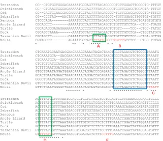
Fig. 1. Conservation between species in the Hoxc8 early enhancer sequence. Align-ments are shown only within the essential ca. 200bp enhancer region, as earlier identified in transgenic mouse embryos (Shashikant et al., 2007, Wang et al., 2004). Blue box, the pair of putative Smad binding elements. Green boxes, Cdx binding motifs. Elements A to E, shown red on the mouse sequence, are motifs that when mutated, alone or in combina-tion, can disrupt Hoxc8 reporter expression in mouse embryos (Shashikant et al., 2007). The sequences, from Ensembl, are aligned by Clustal Omega.
expression of Hoxc8/lacZ reporter in the mouse embryo (Shashikant et al., 2007, Shashikant and Ruddle, 1996), and to bind Cdx2 protein (Taylor
et al., 1997).
Gdf11 and Cdx proteins cooperate to activate
Hoxc8 enhancer in HepG2 cells
In Fig. 2A, all cultures were transfected with
Hoxc8 reporter construct #1 (shown boxed), and
all received 50ng/ml Gdf11. Cultures shown at bar on right were co-transfected with doxycycline-inducible Cdx1 expression construct and show 10-fold stimulation in response to doxycycline. In control experiments: (i) cultures given no Cdx expression construct are not stimulated by doxycycline (left), and (ii) cultures co-transfected with a modified expression construct in which the Cdx1 coding sequence truncates at the start of the homeobox show little or no stimulation by doxycycline (middle). In this latter case, the induced Cdx1 protein lacks the DNA binding homeodomain. Fig. 2B shows a Gdf11dose response curve. All subsequent experiments were performed at 50ng/ml Gdf11.
As shown in Fig. 2C, Cdx1 (doxycycline) and Gdf11 alone each show little or no activation of
Hoxc8 reporter but in combination they produce
about 8-fold stimulation. This suggests that Cdx1 and Gdf11 may exert a cooperative (synergistic) effect upon activation of Hoxc8. This is supported by plotting raw luminescence values (Fig. 2D), where activity in monolayers exposed to both Cdx1 (doxycycline) and Gdf11 is found greater than the sum of the values for cultures exposed 2006, Reissmann et al., 2001). This is sufficient for activation of the Hoxd11 region VIII enhancer (Gaunt et al., 2013). The new findings for Hoxc8 early enhancer show that Gdf11 and Cdx proteins each, alone, provide only weak activation in HepG2 cells. However, Gdf11 and Cdx proteins given together provide strong activation. The relevant binding motifs are identified in mutagenesis studies.
Results
Identification of candidate Smad and Cdx binding motifs in
Hoxc8 early enhancer
Fig. 1 shows that the mouse Hoxc8 early enhancer contains the motif GGCTAGACGTCTGGGC which is highly conserved from fish to mammals (blue box). This contains two putative variants of the optimal Smad binding motif with one inverted (on the opposite DNA strand) relative to the other. In comparison to the optimal motif, GTCTGGGC is 76.7% as effective in binding to Smad3, and GTCTAGCC is 79.6% as effective in binding to Smad4 (Zawel et al., 1998).
to either Cdx1 or Gdf11 alone. A similar result was obtained for Cdx4 plus Gdf11 (Fig. 2F). The results for Cdx2 plus Gdf11 are less indicative of a synergistic effect (Fig. 2E) although, for equal cell numbers, the added luminescence for Cdx alone and Gdf11 alone should ideally be compared with double the value shown for Cdx plus Gdf11 treated cultures.
The positive response to Gdf11 in cultures co-transfected with the un-induced Cdx2 construct (Fig. 2E) are likely due to leakage in Cdx2 transcription in absence of doxycycline, since no activa-tion occurs when the Hoxc8 enhancer includes mutaactiva-tions in its Cdx binding motifs (Fig. 3D). The reduced effects of un-induced
Cdx1 and Cdx4 expression vectors upon response to Gdf11 (Fig.
2 D,F) may indicate either less transcriptional leakage from these constructs, or reduced sensitivity of the Hoxc8 enhancer to Cdx1 and Cdx4 proteins relative to Cdx2.
Smad and Cdx response motifs identified in vitro by mutagenesis
Mutations were introduced into each or both of the putative Smad binding motifs (labelled SmadA and SmadB in Fig. 3A). Mutations within either of the two conserved Smad sites (constructs #2 and #3) result in a reduction of response to Gdf11, and response is reduced further by mutation of both sites (construct # 4) (Fig. 3B). This result was observed irrespective of whether Cdx1 or Cdx4
Fig. 2. Gdf11 and Cdx proteins activate Hoxc8 syner-gistically. (A) All cultures were transfected with construct #1 (shown boxed: yellow bar is Hoxc8 early enhancer; blue bar is SV40 minimal promoter/luciferase/SV40 polyA), and received Gdf11 at 50ng/ml. Cultures shown at right were co-transfected with doxycycline-inducible Cdx1 expression construct; cultures at middle were co-transfected with inducible Cdx1 construct lacking the homeobox; cultures at left were not co-transfected with Cdx construct. Fold stimulation shows effect of doxycycline relative to replicate cultures, similarly treated, but not given doxycycline (dot-ted baseline). (B) Gdf11 dose response curve. All cultures received Hoxc8 construct #1, Cdx1 expression construct, and doxycycline. Data are plotted as fold stimulation relative to no Gdf11. (C-F) All cultures co-transfected with construct #1 and with a doxycycline inducible Cdx expression con-struct. In (C), data are plotted as fold stimulation relative to replicate cultures given no Gdf11 or doxycycline (dotted baseline). In (D-F) data are shown as raw luminometry readings. Throughout, each bar shows average values for three replicate cultures, and range bars are shown. luc, luciferase; Dox, doxycycline.
nomenclature of other researchers (Fig. 1) (Shashikant et al., 2007). Dashes indicate identity with the wild-type sequence of construct #1. (B) Both Smad motifs contribute to Gdf11 stimulation in pres-ence of Cdx. (C) Both Cdx motifs contribute to Cdx1 stimulation in presence of Gdf11. (D) Mutations in the Cdx motifs inhibits response to Gdf11. Cdx proteins were provided, as indicated, from co-transfected doxycycline-inducible expression constructs. In (B,D), bars show fold stimulation by Gdf11 relative to results for replicate cultures given doxycycline but no Gdf11 (shown as dotted baseline). In (C), bars show fold stimulation by doxycycline relative to results for replicates given Gdf11 but no doxycycline (shown as dotted baseline). Each bar shows average values for three replicate cultures, and range bars are shown. Dox, doxycycline.
Fig. 3. Gdf11 and Cdx stimulatory effects inhibited by mutations in the putative Smad and Cdx binding motifs. (A) Muta-tions introduced (constructs #2 to #6) to putative Smad motifs (blue underline) and Cdx motifs (green underline). Cdx sites are designated A and D in accordance with the
0 CdxA →54N←SmadA SmadB CdxD Construct
#1
CdxA →54N←SmadA SmadB CdxD Construct
B
C
D
of Smad3 with Smad4. SIS3 does not affect phosphorylation of Smad2 or the expression of Smad4 (Jinnin et al., 2006). In HepG2 cells, Gdf11 plus Cdx1 activation of Hoxc8 reporter construct #1 is inhibited by SIS3 in a dose-dependent manner (Fig. 4A). This suggests that Smad3 mediates, at least in part, the effect of Gdf11 upon Hoxc8.
Effect of Fgf2
The effect of Fgf2 was examined since this is proposed to act cooperatively with Cdx2 and/or Gdf11 in activation of the endog-enous Hoxc8 gene (Bel-Vialar et al., 2002, Liu, 2006, Mazzoni
et al., 2013). Fgf2 was given at 100ng/ml because this is the
concentration used in the earlier, in vitro study (Mazzoni et al., 2013). In the HepG2 assay Fgf2 increased luciferase activation by the Hoxc8 enhancer under all conditions tested, including in cells grown without Gdf11 or Cdx proteins (Fig. 4 B,C). There is no clear evidence that Fgf2 specifically operates synergistically with either Gdf11 or Cdx signalling. Notably, synergy between Gdf11 and Cdx on the Hoxc8 early enhancer is clearly greater than is any possible synergy between either Fgf2 and Cdx, or Fgf2 and Gdf11 (Fig. 4 B,C).
Discussion
Identification of factors that activate an enhancer element in vitro does not prove that the same mechanism operates in the early embryo. The chances of this being so are, however, increased if, as here, the enhancer investigated is known to regulate a Hox-like reporter expression pattern in transgenic embryos, and the acti-vating transcription factors identified are known to be functional in the embryo. Indeed, an in vitro system offers certain advantages over in vivo studies since large numbers of assays can readily be conducted, allowing activators, inhibitors and co-factors to be more easily, and quantitatively, tested.
Both of the Cdx binding motifs identified have already been shown to be essential for the mouse embryo expression patterns of Hoxc8 early enhancer transgenes (Shashikant et al., 2007, Sha-shikant and Ruddle, 1996). The principal new finding now made in HepG2 cells is that effective Cdx activation requires cooperation by
Gdf11/ Smad signalling. Two putative Smad binding motifs located in the enhancer near to the Cdx motifs are shown by mutagenesis to be essential for this synergistic effect. This is in-keeping with a common finding that enhancer activation by Smad2/3 requires the nearby binding of a major transcription factor (Mullen et al., 2011). Hoxc8 early enhancer regulates mid to posterior thoracic vertebral patterning and neural control in limbs (Juan and Ruddle, 2003). Evolutionary change in either of these functions could, in an early mammalian ancestor, potentially have been facilitated by acquisition of the additional Cdx binding site.
The questions now arise as to whether Smad signalling is also required during embryonic activation of the endogenous
Hoxc8 gene and, if so, whether this pathway is activated by
Gdf11. Overexpression of Gdf11 in the chick neural tube induces increase in phosphorylated Smad2/3 proteins and accompanying forward shifts in the expression boundary of Hoxc8 (Liu, 2006). Conversely, neural overexpression of follistatin, an inhibitor of endogenous Gdf11, produces posterior shift in Hoxc8 expression (Liu, 2006). These results indicate that the Smad2/3 signalling pathway activates Hoxc8 in vivo. However, they do not prove that Gdf11 is the primary signal since other TGFb ligands, including
Gdf8, activin and nodal, also bind to the AcvrIIB receptor to activate Smad2/3, and are also inhibited by follistatin. AcvrIIB-/- embryos show posterior shifts in the paraxial mesoderm expressions of a variety of more posteriorly-active Hox genes including Hoxc8 (Oh and Li, 1997) but, again, this does not specifically identify Gdf11 as the primary ligand.
In spite of the positive evidence that Gdf11 may be a physiological activator of Hoxc8 in the embryo (Liu, 2006) there are apparently contradictory findings. Gdf11-/- mouse embryos at 9.5 to 12.5 days show normal anterior boundaries of Hoxc8 expression in mesoderm and neural tissues, but with caudally-extended posterior boundar-ies (Jurberg et al., 2013, Liu, 2006, McPherron et al., 1999). This may indicate that Gdf11/Smad signalling represses, rather than activates, embryo expression of Hoxc8. More posterior genes
Hoxc10 and Hoxc11 show caudal shifts of their entire expression
domains (Jurberg et al., 2013, McPherron et al., 1999). The role of Gdf11/Smad signalling in embryonic Hoxc8 expression is, therefore, far from clear but the following possibilities are suggested. 1) The
Fig. 4. SIS3 and Fgf2 effects upon Gdf11 and Cdx activation of mouse Hoxc8 reporter. (A) SIS3, an inhibitor of Smad3 activities. (B,C) Fgf2. Hoxc8 reporter construct #1 was co-transfected with Cdx1 (A,B) or Cdx2 (C) doxycycline-inducible expression construct. Each graph bar shows average values for three replicate cultures, and range bars are shown. Each culture in (A) was exposed to 1% DMSO solvent. Dox, doxycycline.
was provided as co-activator.
As shown in Fig. 3C, mutation of the downstream CdxD binding motif (construct #5) results in reduced response to Cdx1 when given in presence of Gdf11, and this is reduced further by the additional mutation of the upstream CdxA binding motif (construct #6). The double Cdx mutant (construct #6) is also severely impaired in its response to Gdf11 when given in presence of either Cdx1, Cdx2 or Cdx4 (Fig. 3D). This provides further evi-dence that response to Gdf11 depends upon synergistic action of Cdx protein binding, and both Cdx binding sites appear to contribute to this function.
Effect of SIS3, an inhibitor of Smad3
SIS3, Specific Inhibitor of Smad3, is a dose-dependent inhibitor of Smad3 phosphoryla-tion, Smad3-DNA binding, and interaction
observations so far made upon Hoxc8 expression in Gdf11-/- em-bryos are likely, at 9.5 to 12.5 days, to be too late to detect a caudal shift in anterior expression limits. Mouse embryos deleted for the
Hoxc8 early enhancer show posterior shift in Hoxc8 expression
at 8 days, but not at 8.5 days or later (Juan and Ruddle, 2003). 2) Gdf11 may be partially redundant with other TGFb proteins in its
role as a Hox gene activator. If Hoxc8 is more sensitive to Smad signalling than Hoxc10 and Hoxc11, as proposed in a morpho-gen gradient hypothesis (Liu, 2006), then this might explain why caudal shifts in anterior expression boundaries are more readily detected for the more posterior genes. Notably, Gdf8 function and expression in embryonic mesoderm is known to overlap with that of Gdf11 (Amthor et al., 2002, Lee et al., 2005, McPherron et al., 2009). 3) Separate regulatory elements, lying outside the early enhancer, may be inhibited by Gdf11/Smad signalling to explain the extended posterior boundary of endogenous Hoxc8 expression in Gdf11-/- embryos.
In the Hoxc8 reporter assays of the present paper Fgf2 provided a general stimulatory effect upon luciferase activity but this was not apparently due to specific synergistic actions with either Cdx or Gdf11/Smad signalling. This conclusion is, however, limited to activity within the Hoxc8 early enhancer. An earlier report that Fgf2 acts synergistically with Cdx2 to activate endogenous Hox genes, including Hoxc8, did not determine whether Fgf2 acts directly upon the Hoxc8 early enhancer (Mazzoni et al., 2013).
Materials and Methods
Expression constructs
The mouse Hoxc8 early enhancer was cloned as a 399bp DNA fragment. This is the same fragment that, in lacZ transgenes, has been shown to give a Hoxc8-like pattern of expression in both embryonic neurectoderm and mesoderm (Shashikant and Ruddle, 1996). The regulatory elements in the enhancer are thought to be located within 200bp located at the 3′ end of the 399bp fragment (Fig. 1) (Shashikant et al., 2007, Wang et al., 2004). For use in luciferase reporter assays, the 399bp fragment was inserted, in 5′ to 3′ orientation, upstream of the minimal promoter in pGL3-promoter (Promega) (construct #1; Fig. 2A). Various mutations were introduced by PCR into the putative Smad and Cdx binding motifs of the Hoxc8 enhancer (constructs #2 to #6), as shown in Fig. 3A.
Cdx expression constructs were prepared by cloning full-length coding sequences of mouse Cdx1, Cdx2 or Cdx4, with Kozak motif upstream of ATG, into the pTRE3G-IRES vector (Clontech). A Cdx1-minus-homeobox/
pTRE3G-IRES plasmid was also prepared for use as a control.
Cell culture and luminometry
Cells were the HepG2 Tet-On Advanced transgenic cell line (Clontech, cat. 631150) designed for use with the doxycycline-inducible pTRE3G-IRES plasmids. Cell culture in gelatin-coated 24-well plates, transfections using Lipofectamine 2000 (Invitrogen), and luciferase assays (Promega, cat. E1500) were carried out in accordance with manufacturers’ instructions and as described earlier (Gaunt and Paul, 2011), but with use of a Biotech Synergy HT luminometer. Cultures were maintained in DMEM supplemented with tetracycline-free foetal bovine serum (Clontech).
All data shown within any one bar chart were obtained in the same experiment. Each bar on each chart shows the mean value obtained from three replicate cultures (n=3). Range bars show the values obtained from the highest and lowest of these three biological replicates. Range bars are preferred to statistical error bars where n is small, including n=3 (Krzywinski and Altman, 2013).
Gdf11 and Fgf2 were from R&D systems; SIS3 from Calbiochem; and doxycycline hyclate from Sigma. Unless otherwise indicated Gdf11 was
given at 50ng/ml; Fgf2 at 100ng/ml; and doxycycline at 10mM. Exposure
of transfected cultures to these reagents was for 18 hours, prior to lysis for luciferase assay.
Acknowledgements
I thank Rob Asher for the provision of laboratory space, and Adrian Kelly for use of the luminometer.
References
AMTHOR, H., HUANG, R., MCKINNELL, I., CHRIST, B., KAMBADUR, R., SHARMA, M. and PATEL, K. (2002). The regulation and action of myostatin as a negative regula-tor of muscle development during avian embryogenesis. Dev Biol 251: 241-257. ANDERSSON, O., REISSMANN, E. and IBANEZ, C.F. (2006). Growth differentiation
factor 11 signals through the transforming growth factor-beta receptor ALK5 to regionalize the anterior-posterior axis. EMBO Rep 7: 831-837.
BEL-VIALAR, S., ITASAKI, N. and KRUMLAUF, R. (2002). Initiating Hox gene expres-sion: in the early chick neural tube differential sensitivity to FGF and RA signaling subdivides the HoxB genes in two distinct groups. Development 129: 5103-5115. DENNLER, S., ITOH, S., VIVIEN, D., TEN DIJKE, P., HUET, S. and GAUTHIER,
J.M. (1998). Direct binding of Smad3 and Smad4 to critical TGF beta-inducible elements in the promoter of human plasminogen activator inhibitor-type 1 gene. EMBO J 17: 3091-3100.
FENG, X.H. and DERYNCK, R. (2005). Specificity and versatility in tgf-beta signaling through Smads. Annu Rev Cell Dev Biol 21: 659-693.
GAMER, L.W., WOLFMAN, N.M., CELESTE, A.J., HATTERSLEY, G., HEWICK, R. and ROSEN, V. (1999). A novel BMP expressed in developing mouse limb, spinal cord, and tail bud is a potent mesoderm inducer in Xenopus embryos. Dev Biol 208: 222-232.
GAUNT, S.J., DRAGE, D. and TRUBSHAW, R.C. (2005). cdx4/lacZ and cdx2/lacZ protein gradients formed by decay during gastrulation in the mouse. Int J Dev Biol 49: 901-908.
GAUNT, S.J., GEORGE, M. and PAUL, Y.L. (2013). Direct activation of a mouse Hoxd11 axial expression enhancer by Gdf11/Smad signalling. Dev Biol 383: 52-60. GAUNT, S.J. and PAUL, Y.-L. (2011). Origins of Cdx1 regulatory elements suggest
roles in vertebrate evolution. Int J Dev Biol 55: 93-98.
GAUNT, S.J. and PAUL, Y.-L. (2012). Changes in Cis-regulatory Elements during Morphological Evolution. Biology 1: 557-574.
GERARD, M., DUBOULE, D. and ZAKANY, J. (1993). Structure and activity of regulatory elements involved in the activation of the Hoxd-11 gene during late gastrulation. EMBO J 12: 3539-3550.
JINNIN, M., IHN, H. and TAMAKI, K. (2006). Characterization of SIS3, a novel specific inhibitor of Smad3, and its effect on transforming growth factor-beta1-induced extracellular matrix expression. Mol Pharmacol 69: 597-607.
JUAN, A.H. and RUDDLE, F.H. (2003). Enhancer timing of Hox gene expression: deletion of the endogenous Hoxc8 early enhancer. Development 130: 4823-4834. JURBERG, A.D., AIRES, R., VARELA-LASHERAS, I., NOVOA, A. and MALLO, M.
(2013). Switching axial progenitors from producing trunk to tail tissues in vertebrate embryos. Dev Cell 25: 451-462.
KRZYWINSKI, M. and ALTMAN, N. (2013). Points of significance: error bars. Nat Methods 10: 921-922.
LEE, S.J., REED, L.A., DAVIES, M.V., GIRGENRATH, S., GOAD, M.E., TOMKINSON, K.N., WRIGHT, J.F., BARKER, C., EHRMANTRAUT, G., HOLMSTROM, J. et al., (2005). Regulation of muscle growth by multiple ligands signaling through activin type II receptors. Proc Natl Acad Sci USA 102: 18117-18122.
LIU, J.P. (2006). The function of growth/differentiation factor 11 (Gdf11) in rostrocaudal patterning of the developing spinal cord. Development 133: 2865-2874. MARGALIT, Y., YARUS, S., SHAPIRA, E., GRUENBAUM, Y. and FAINSOD, A. (1993).
Isolation and characterization of target sequences of the chicken CdxA homeobox gene. Nucleic Acids Res 21: 4915-4922.
MASSAGUE, J., SEOANE, J. and WOTTON, D. (2005). Smad transcription factors. Genes Dev 19: 2783-2810.
MCPHERRON, A.C., HUYNH, T.V. and LEE, S.J. (2009). Redundancy of myostatin and growth/differentiation factor 11 function. BMC Dev Biol 9: 24.
MCPHERRON, A.C., LAWLER, A.M. and LEE, S.J. (1999). Regulation of anterior/ posterior patterning of the axial skeleton by growth/differentiation factor 11. Nat Genet 22: 260-264.
MULLEN, A.C., ORLANDO, D.A., NEWMAN, J.J., LOVEN, J., KUMAR, R.M., BILODEAU, S., REDDY, J., GUENTHER, M.G., DEKOTER, R.P. and YOUNG, R.A. (2011). Master transcription factors determine cell-type-specific responses to TGF-beta signaling. Cell 147: 565-576.
NAKASHIMA, M., TOYONO, T., AKAMINE, A. and JOYNER, A. (1999). Expression of growth/differentiation factor 11, a new member of the BMP/TGFbeta superfamily during mouse embryogenesis. Mech Dev 80: 185-189.
OH, S.P. and LI, E. (1997). The signaling pathway mediated by the type IIB activin receptor controls axial patterning and lateral asymmetry in the mouse. Genes Dev 11: 1812-1826.
REISSMANN, E., JORNVALL, H., BLOKZIJL, A., ANDERSSON, O., CHANG, C., MINCHIOTTI, G., PERSICO, M.G., IBANEZ, C.F. and BRIVANLOU, A.H. (2001). The orphan receptor ALK7 and the Activin receptor ALK4 mediate signaling by Nodal proteins during vertebrate development. Genes Dev 15: 2010-2022. SCHYR, R.B., SHABTAI, Y., SHASHIKANT, C.S. and FAINSOD, A. (2012). Cdx1
is essential for the initiation of HoxC8 expression during early embryogenesis. FASEB J 26: 2674-2684.
SHASHIKANT, C.S., BOLANOWSKY, S.A., ANAND, S. and ANDERSON, S.M. (2007). Comparison of diverged Hoxc8 early enhancer activities reveals modification of
regulatory interactions at conserved cis-acting elements. J Exp Zool B Mol Dev Evol 308: 242-249.
SHASHIKANT, C.S. and RUDDLE, F.H. (1996). Combinations of closely situated cis-acting elements determine tissue-specific patterns and anterior extent of early Hoxc8 expression. Proc Natl Acad Sci USA 93: 12364-12369.
SUBRAMANIAN, V., MEYER, B.I. and GRUSS, P. (1995). Disruption of the murine homeobox gene Cdx1 affects axial skeletal identities by altering the mesodermal expression domains of Hox genes. Cell 83: 641-653.
TAYLOR, J.K., LEVY, T., SUH, E.R. and TRABER, P.G. (1997). Activation of enhancer elements by the homeobox gene Cdx2 is cell line specific. Nucleic Acids Res 25: 2293-2300.
TREMBLAY, K.D., HOODLESS, P.A., BIKOFF, E.K. and ROBERTSON, E.J. (2000). Formation of the definitive endoderm in mouse is a Smad2-dependent process. Development 127: 3079-3090.
WANG, W.C., ANAND, S., POWELL, D.R., PAWASHE, A.B., AMEMIYA, C.T. and SHA-SHIKANT, C.S. (2004). Comparative cis-regulatory analyses identify new elements of the mouse Hoxc8 early enhancer. J Exp Zool B Mol Dev Evol 302: 436-445. ZAKANY, J., GERARD, M., FAVIER, B. and DUBOULE, D. (1997). Deletion of a
HoxD enhancer induces transcriptional heterochrony leading to transposition of the sacrum. EMBO J 16: 4393-4402.
The significance of Hox gene collinearity
Stephen J. Gaunt
Int. J. Dev. Biol. (2015) 59: 159-170 https://doi.org/10.1387/ijdb.150223sg
Synergistic action in P19 pluripotential cells of retinoic acid and Wnt3a on Cdx1 enhancer elements
Stephen J. Gaunt and Yu-Lee Paul Int. J. Dev. Biol. (2014) 58: 307-314 https://doi.org/10.1387/ijdb.140003sg
5 yr ISI Impact Factor (2013) = 2.879
The road to the vertebral formula
Moises Mallo, Tania Vinagre and Marta Carapuҫo Int. J. Dev. Biol. (2009) 53: 1469-1481
https://doi.org/10.1387/ijdb.072276mm
cdx4/lacZ and cdx2/lacZ protein gradients formed by decay during gastrulation in the mouse
Stephen J. Gaunt, Deborah Drage and Richard C. Trubshaw Int. J. Dev. Biol. (2005) 49: 901-908
http://doi.org/10.1387/ijdb.052021sg
Additional enhancer copies, with intact cdx binding sites, anteriorize Hoxa-7/lacZ expression in mouse embryos: evidence in keeping with an instructional cdx gradient
Stephen J. Gaunt, Adam Cockley and Deborah Drage Int. J. Dev. Biol. (2004) 48: 613-622
https://doi.org/10.1387/ijdb.041829sg
Initiation, establishment and maintenance of Hox gene expression patterns in the mouse
Jacqueline Deschamps, Eric van den Akker, Sylvia Forlani, Wim de Graaff, Tony Oosterveen, Bernard Roelen and Jeroen Roelfsema