Early embryonic lethality in gene trap mice
with disruption of the Arfgef2 gene
PAWEL GRZMIL*
,1,2, ZANABAZAR ENKHBAATAR
1, BATJARGAL GUNDSAMBUU
1,
ODGEREL OIDOVSAMBUU
1, SAFAK YALCIN
1, STEPHAN WOLF
1,
WOLFGANG ENGEL
1and JÜRGEN NEESEN
#,1 1Institute of Human Genetics, University of Göttingen, Göttingen, Germany and2Department of Genetics and Evolution, Institute of Zoology, Jagiellonian University, Kraków, Poland
ABSTRACT The switching of ADP-ribosylation factors from the inactive form to the active form is catalyzed by ARF-GEF (ADP ribosylation factor - guanine nucleotide exchange protein) proteins containing a Sec7 domain. The murine Arfgef2 gene encoding the BIG2 protein belongs to the class of high molecular mass (>100 kDa) ARF-GEF proteins. BIG2 is believed to be associated with the trans-Golgi network and the recycling endosomes. In humans, mutations in the ARFGEF2 gene cause autosomal recessive periventricular heterotopia with microcephaly. To elucidate the function of BIG2 in mouse we studied a gene-trap mouse line with a functional disruption of the Arfgef2 gene. Heterozygous mutants did not reveal phenotypic abnormalities and were fertile. However, no homozygous embryos were obtained from breeding heterozygous females and males. To explore the reason for embryonic lethality, we analysed the pattern of expression of Arfgef2. Arfgef2 transcripts were detected in several adult tissues. Interestingly, Arfgef2 under-goes alternative splicing and the splicing pattern differs among tissues from adult animals. Moreover, the LacZ reporter gene of the gene-trap construct was used to reveal the expression of Arfgef2 during embryonic development. Here, we show that Arfgef2 mRNA is stored in the oocyte and is likely translated during the first embryonic divisions. SNP (Single Nucleotide Polymor-phism) markers were used to demonstrate that the embryonic Arfgef2 gene is activated first at the 4-cell stage, suggesting an important role for embryonic development. This assumption is supported by the failure of Arfgef2-deficient oocytes fertilized with Arfgef2-deficient sperm to develop into 4-cell stage embryos. Our results indicate that murine BIG2 is essential for early embryonic development.
KEY WORDS: gene-trap, embryonic development, zygotic gene activation, BFA, ADP-ribozylation
Introduction
The recruitment of coat proteins to membrane for the genera-tion of transporting vesicles at intracellular sites between Golgi, ER and plasma membrane is controlled by the ADP ribosylation factors, small GTPases that cycle between GTP-bound active form and GDP-bound inactive form. The activation of ARFs requires the activity of guanine nucleotide exchange proteins (GEF, Moss and Vaughan, 1998; Kirchhausen, 2000). All ARF– GEFs identified so far possess a Sec7 domain composed of 200 amino acids as a minimum unit for the catalysis of replacement of
BIOLOGY
www.intjdevbiol.com*Address correspondence to: Pawel Grzmil. Institute of Human Genetics, University of Göttingen, Heinrich-Düker-Weg 12, 37073 Göttingen, Germany. e-mail: pgrzmil@gwdg.de
#Present address: Department of Medical Genetics, Medical University of Vienna, 1090 Vienna, Austria.
Supplementary Material (a table) for this paper is available at: http://dx.doi.org/10.1387/ijdb.092959pg
Accepted: 22 September 2009. Final author corrected PDF published online: 20 August 2010.
ISSN: Online 1696-3547, Print 0214-6282 © 2010 UBC Press
Printed in Spain
Abbreviations used in this paper: ARF, ADP ribosylation factor; BFA, brefeldin A; GEF, guanine nucleotide exchange proteins; LacZ, β-galactosidase; TGN, trans-Golgi network.
(Ramaen et al. 2007; Ishizaki et al. 2008). So far in mammals only
one member of the Gea/GBF group, the GBF1 has been de-scribed, that primarily functions in the trafficking between the cis-Golgi and ER-cis-Golgi intermediate compartment (Claude et al.
1999; Kawamoto et al. 2002; Zhao et al. 2002; 2006; Garcia-Mata et al. 2003). From the Sec7/BIG group BIG1 and BIG2 proteins
were purified from bovine brain together with macromolecular complexes (>670 kDa) (Morinaga et al. 1996). Several studies
reported the association of BIG proteins with the trans-Golgi network (TGN) (Mansour et al. 1999; Yamaji et al. 2000; Shinotsuka et al. 2002a,b; Zhao et al. 2002), in addition, the association with
recycling endosomes was also demonstrated (Shin et al. 2004;
Shen et al. 2006). BIG1 and BIG2 proteins revealed considerable
similarities to each other in the sequence and domain organiza-tion (Mouratou et al. 2005), however, it is still not clear whether
both proteins play redundant or distinct role in the control of membrane trafficking. BIG1 but not BIG2 is necessary for β1 integrin glycosylation by Golgi enzymes (Shen et al., 2007). On
the other hand the knockdown of BIG1 and BIG2 by RNAi in cell culture indicated that BIG1 and BIG2 play redundant role in trafficking between trans-Golgi network (TGN) and endosomes
(Ishizaki et al. 2008). In mouse BIG1 and BIG2 are encoded by Arfgef1 and Arfgef2 genes, located at chromosome 1 and 2,
respectively. The Arfgef2 gene is brightly expressed in different
tissues (Togawa et al. 1999)
To analyse the role of this gene in mouse embryonic develop-ment we have identified a gene-trap line with vector integration in the Arfgef2 gene. Our data indicate that murine zygotic copies of
the Arfgef2 gene are activated in 4-cell stage embryos and that
this activation is essential for early embryonic development. Moreover, our results suggest that Arfgef2 function can not be
compensated by Arfgef1.
Results
Characterization of the Arfgef2 gene-trap mouse line Here we report the characterization of a gene-trap mouse line in which the gene trap vector was integrated in mouse Arfgef2
gene. To identify the trapped gene a 5’ rapid amplification of cDNA ends (RACE) analysis from known vector sequences was per-formed. A 900 bp fusion transcript was amplified in brain RNA isolated from a heterozygous male. Sequence analysis of the RACE PCR product revealed that flanking sequences of the gene trap vector derived from the mouse Arfgef2 gene (Gene ID:
99371). Sequence alignment suggested that the integration of the gene-trap construct occurred downstream of exon 7 of the Arfgef2
gene. To prove the integration on genomic level a genomic phage library of a heterozygous mouse was generated and screened using a β-galactosidase gene specific probe. Sequence analysis of identified clones demonstrated that the gene-trap vector was integrated in intron 7 of the Arfgef2 gene (Fig. 1A).
The gene trap vector contains a polyadenylation recognition motif just after the LacZ reporter gene indicating that a putative fusion transcript lacks part of the Arfgef2 gene, including
se-quences encoding for the sec7 domain (Fig. 1B). It can be assumed that such truncated transcripts do not encode for func-tional ARFGEF2 polypeptides. The deduced protein (if any pro-duced) encoded by the trapped transcript will be truncated
up-LacRn RTFP
E 6 E 7 LacZ pA
AAAAAAAA
GT RT-PCR Product size 200 bp
GT mRNA
WT RT-PCR Product size 520 bp LacRn LacR gration site. Gene-trap vec-tor integration had occurred after the exon 7 of the mouse Arfgef2 gene (A), thus the resulting fusion transcript (B) lacks se-quences encoding the sec7 domain (resulted protein is truncated after amino acid in position 309). Positions of primers used for expres-sion analyses are indicated. (C) In RT-PCR analysis a 200 bp fusion transcript was detected in all tissues iso-lated from heterozygous animal (+/-). In addition two approximately 500 bp am-plification products were observed, derived from the wild type Arfgef2 allele. In wild type control RNA no
stream the sec7 domain, after the amino acid 309. Therefore, we analysed the expression pattern of the Arfgef2-gene in wild type
and heterozygous animals by RT-PCR. The primer combination RTFP and RTRP (Fig. 1A) was used to amplify a 520 bp product representing the wild type allele. For transcripts derived from the trapped allele the primer pair RTFP and LacRn (Fig. 1B) was utilized to amplify a 200 bp product. This analysis revealed that transcripts of the wild type allele as well as of the trapped allele could be amplified in RNA from kidney, brain, liver, lung and heart tissue isolated from a heterozygous mouse while in the RNA from a wild type control only the 520 bp fragment was detected (Fig. 1C). Moreover, RT-PCR analysis demonstrated alternative splic-ing of the‘Arfgef2 gene. In addition to the 520 bp PCR product a
second Arfgef2 cDNA fragment was amplified lacking sequences
of exon 7 (Fig. 1C lower WT band).
Expression of the trapped gene was also investigated using β -galactosidase staining. Reporter gene activity could be detected in adult tissues (data not shown) and in different embryonic developmental stages. In blastocysts inner cell mass as well as trophoectoderm cells were stained (Fig. 1D). No staining was observed in wild type control (Fig. 1E).
RT-PCR results proved that integration of the gene trap vector construct into intron 7 of the Arfgef2 gene generated a fusion
transcript and resulted in inactivation of the function of the trapped
Arfgef2 allele. Animals heterozygous for the integration of the
gene trap vector were fertile and did not demonstrate any obvious malformation. However, genotype analyses of more than 200 offspring from heterozygous breeding detected no mice homozy-gous for the integration. This result indicates that homozyhomozy-gous inactivation of the Arfgef2 gene leads to embryonic lethality. To
determine the time of embryonic lethality embryos were genotyped by PCR. First we analysed embryos after the implantation stage, however, we could not detect any Arfgef2 deficient embryos (data
not shown). Then fertilized oocytes were isolated and cultured in vitro up to the blastocyst stage. Also no Arfgef2 deficient embryos
could be detected in 4 cell, morula or blastocyte stages. In
contrast, in the syngamy and 2-cell stages homozygous Arfgef2
deficient embryos were detected, although some of them demon-strated progressive fragmentation of the cytoplasm (Fig. 1F, Table 1). It should be pointed that the ratio of homozygous, heterozygous and wild type embryos did not correspond to expected Mendelian ratio because PCR genotyping failed by some embryos.
This finding indicates that disruption of the Arfgef2 gene
function by the gene-trap approach causes embryonic lethality at first stages of embryonic development.
Brefeldin A treatment inhibits embryonic cell division It has been demonstrated that ARFGEF2 is inhibited by brefeldin A (BFA) (Togawa et al. 1999), therefore we tested whether BFA
treatment inhibits early embryonic development in mouse. Wild type female mice were superovulated and mated with wild type males. Females positive for a vaginal plug were sacrificed and fertilized oocytes were isolated. Using light microscopy isolated fertilized oocytes were tested for the presence of two pronuclei and then cultured in the medium containing BFA. As controls fertilized oocytes were cultured in medium alone and in medium added with methanol because BFA was solved in methanol. At BFA concentration of 10 μg/ml (35 μM) we observed an inhibition
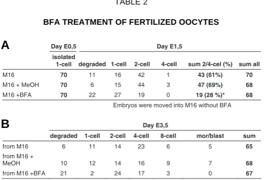
of embryonic cell division, but embryos were still alive, because after removing BFA the effect was reversible. A total of 70 fertilized oocytes were used in each group. After 24 hours of incubation with BFA embryos were washed, counted and moved to the medium without BFA. As demonstrated in table 2 BFA inhibited significantly (p<0.01) the first embryonic division and no embryo reached the 4-cell stage. After transferring oocytes to culture medium (without BFA) almost all embryos restored the ability to divide (44 out of 46) but they were delayed in embryonic development by 1 day as compared to the control embryos (incubated from the beginning without BFA). By using higher BFA concentration complete inhibition of embryonic division was in-duced, however, this effect was not reversible and most of embryos died (data not shown). These results support the as-sumption that disruption of the function of Arfgef2 (BIG2) leads to
an arrest of the embryonic development at the very early stage.
Arfgef2 undergoes alternative splicing
RT-PCR analyses of the Arfgef2 in heterozygous gene-trap or
wild type mice have identified two Arfgef2 transcript variants (Fig.
1C). This prompted us to analyze putative alternative splicing of this gene. The genomic organization of the mouse Arfgef2 gene
was obtained from the ENSEMBL database (www.ensembl.org) and series of primers were designed to cover the complete mRNA of this gene (supplementary table 1D). Besides the splicing variant without exon 7 no additional alternative splicing products were observed using RNA extracted from adult brain (Fig. 2A) or kidney (data not shown). Complete transcript was designated as
Arfgef2a while transcript missing exon 7 was denoted as Arfgef2b.
The exon 7 spans 69 bp and encodes for 23 amino acids, thus alternative splicing of exon 7 preserves the reading frame in the
Arfgef2b transcript variant. Using different computer programs
(http://smart.embl-heidelberg.de, Schultz et al., 1998) no known
domain could be predicted within the 23 amino acids encoded by exon 7.
Next, the alternative splicing of exon 7 was analysed in differ-ent tissues. Primers located in exons 6 and 10 were used to amplify both transcript variants. In all analysed tissues both transcript variants could be detected. However, different intensi-ties of the amplification products of Arfgef2a and Arfgef2b were
observed (Fig. 2B). In RNA isolated from eye and brain both variants displayed approximately same intensity. In contrast, in kidney, liver, spleen, ovary and testis Arfgef2a seemed to be more
Embryo's genotype
Development WT/WT WT/GT GT/GT unknown Total
Syngamy degraded 0 1 8 28 37
Syngamy 21 23 5 42 91
2-cell degraded 4 2 3 8 17
2-cell 85 107 7 35 234
4-cell degraded 1 0 2 2 5
4-cell 93 101 0 42 236
8-cell 10 18 0 3 31
Morula 22 34 0 23 79
Blastocyst 31 62 0 23 116
TABLE 1
abundant, whereas in RNA from heart, lung and muscle tissues preferentially the Arfgef2b was detected. The splicing pattern was
also analysed during embryonic development (Fig. 2C). Both transcript variants were detected from unfertilized oocyte up to embryonic day 18.5. At 4-cell stage both variants were of the same intensity, whereas in more advance stages of embryonic develop-ment as well as in embryonic stem cells (ESC) the Arfgef2a variant
was more abundant. The expression in testis was analysed in more detail to clarify whether stronger Arfgef2a expression in the testis
is due to germ cells or somatic tissue. RNA isolated from adult testes of mutant mouse strains with spermatogenesis arrested at different stages was analyzed by RT-PCR: W/Wv mutants have no germ cells in the testis, while in Tfm mice primary spermatocytes
can be detected. In the olt/olt mutant spermiogenesis is affected
after step 13 and in qk/qk mice elongated spermatids can be found
(step 16, Lyon and Searle, 1989; Chubb, 1992). Moreover,
Arfgef2 expression was analyzed in germ stem cells (maGSCs,
Guan et al., 2006). Similar expression patterns were observed in
maGSCs and in W/Wv mutants indicating that the stronger ex-pression of the Arfgef2a variant in the testis is due to somatic as
well as germ cells (Fig. 2C).
Zygotic Arfgef2 gene is activated at 4-cell stage
The lethality of Arfgef2-deficient embryos during the first steps
of development indicated that the ARFGEF2 protein encoded by the embryonic genome acts very early in mouse embryonic development. Arfgef2 mRNA can be detected in unfertilized
oocyte (Fig. 2C) and in different embryonic stages. To determine the stage of embryonic development in which the embryonic copy of the Arfgef2 gene is activated, the SNP data base (http://
www.ncbi.nlm.nih.gov/SNP) was explored to indentify polymor-phic markers within the Arfgef2 cDNA sequence. Two SNPs
differentiating 129/Sv and C57Bl strains were identified in exon 20 and 21 of Arfgef2 (Fig. 3A). For the test five 129/Sv females were
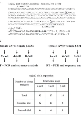
mated with five C57Bl males and vice versa (Fig. 3B). Two-cell embryos were isolated from vaginal plug positive females. From approximately 40 embryos the mRNA was directly prepared, while the rest of the 2-cell embryos were cultured in M16 medium until 4-cell and 8-cell stages. Approximately thirty 4-cell stage and thirty 8-cell stage embryos were collected for RNA isolation. After reverse transcription the cDNA was used as a template in a PCR reaction with primers L16AGCTFP and L16AGCTRP (Fig. 3A, supplementary table 1E). PCR products were purified and cloned into pGEM-Teasy vector and analysed by sequencing. Because it was not possible to discriminate between maternal RNA storage and Arfgef2 transcripts derived from the maternal allele, only the
activation of the paternal Arfgef2 allele could be determined.
Thirty-two clones were sequenced derived from the 2-cell stage embryos and all represented the maternal SNP variant. In con-trast, out of 17 clones from 4-cell stage embryos 13 represented the maternal SNP transcript and 4 clones revealed the paternal SNP sequence. From the 8-cell stage 14 sequences were analysed and 6 clones represented the maternal transcript while 8 se-quences harbour the paternal SNP (Fig. 3C). This result indicates Fig. 2. Analyses of alternative splicing of the Arfgef2 gene. Series of
primers were designed to cover whole mRNA of the Arfgef2 gene (accession number in the ENSEMBL data base ENSMUST00000099078). RNA was isolated from tissues of 129Sv mice. RT-PCR analysis revealed that two alternative spliced products of the Arfgef2 gene could be amplified with primers located in exons 5 and 11 (A). Sequence analysis revealed that the larger transcript (Arfgef2a) contains exons 5 to 11 while the shorter transcript (Arfgef2b) lacks sequences derived from exon 7. (B) RT-PCR was used to investigate alternative splicing in different tissues. Results suggest a tissue specific expression pattern of both transcript variants. In contrast to brain and eye where similar intensity of both variants were observed, in kidney, liver, spleen, ovary and testis the Arfgef2a is predominantly expressed whereas in heart, lung and muscle Arfgef2b is expressed at higher level. (C) The alternative splicing could also be visualized during mouse embryonic development, in germ cells (maGSC), embryonic stem cells (ESC) and in the testis from different mutants. Abbreviations: maGSC, multipotent adult germline stem cells; ESC, embryonic stem cells; Tfm, testicular feminization; olt/olt, oligotriche, qk/qk, quaking; W/Wv, white spotting.
M O Exon
M O Brain E10.5E15.5 E18.5 ESC ma GS
(A) Fertilized oocytes were cultured in medium containing BFA (35 mM, row M16+BFA). As controls fertilized oocytes were cultured in medium alone (row M16) and in medium added with MeOH (row M16+MeOH). After 24 hours of incubation with BFA embryos were washed, counted, moved to the medium without BFA and cultured for further two days (B). *- the number of 2/4-cell stage embryos cultured in medium with BFA was significantly reduced as compare with those cultured in M16 or M16 + MeOH, p<0.01.
Day E0,5 Day E1,5
Embryos were moved into M16 without BFA
Day E3,5
BFA TREATMENT OF FERTILIZED OOCYTES
that the activation of the paternal Arfgef2 allele occurs at the 4-cell
stage in the mouse.
Discussion
We have analysed a mouse gene trap line in which the function of the Arfgef2 gene encoding BIG2 was affected. The insertion of
the gene trap vector occurred in intron 7 of the Arfgef2 gene
resulting in a putative truncated gene product missing essential domains of the encoded BIG2 polypeptide. Arfgef2 transcripts
were detected in all investigated tissues of wild type mouse suggesting a ubiquitous expression of this gene. Moreover, alternative Arfgef2 transcripts missing exon 7 were identified,
although the physiological relevance of the splice variant is
unknown. Also in human alternatively spliced variants were re-ported for ARFGEF1, ARFGEF2 and GBF1 (Claude et al. 2003;
Mouratou et al. 2005). However, for the ARFGEF2 gene the
alternative splicing affects exon 35 (Mouratou et al. 2005). Arfgef2
transcripts were detected in oocytes indicating maternal storage and putative function of Arfgef2 in first steps of zygotic
develop-ment. In mammals maternal mRNA deposited in the oocyte are degraded shortly after fertilization and can control only first few cell divisions (Thompson et al. 1998). Zygotic genome activation
(ZGA) occurs very early during embryonic development. In the mouse, a minor burst of ZGA toward the end of the one-cell stage is followed by a major burst during the two-cell to four-cell stages and then the mid-preimplantation gene activation (MGA) occurs (Latham et al. 1992; Vernet et al. 1992; Aoki et al. 1997; Thompson
Fig. 3. Arfgef2 expression during early embryonic development. To ascer-tain the onset of Arfgef2 gene expression during embryonic development two single nucleotide polymorphisms (SNPs): rs29577446 and rs27295574 were identified in ENSEMBL database for differentiation between Arfgef2 transcripts derived from C57Bl6 and 129/Sv mouse strains (A). C57Bl females were bred with 129/Sv males and vice versa 129/Sv females with C57Bl males (B). Two-cell, 4-cell and 8-cell embryos were isolated. Arfgef2 transcripts were amplified by RT-PCR with primers L16AGCTFP and L16AGCTRP (marked as underlined sequence), cloned and sequenced. (C) In 2-cell embryos no paternal allele expression could be identified, whereas in RNA from 4-cell and 8-cell embryos Arfgef2 transcripts derived fromboth, maternal and paternal alleles were found.
et al. 1998; Schultz, 2002; Hamatani et al. 2006). The Arfgef2
zygotic activation belongs to the major ZGA at 4-cell stage. The Arfgef2 mRNA stockpile deposited in oocyte might
explain the first embryonic division in some Arfgef2GT/GT homozygous embryos. However, the lack of 4-cell mutant embryos indicates fast wastage of the stored Arfgef2 mRNA.
In contrast to the mouse phenotype, mutations in either of two genes, Filamin A (FLNA) or ADP-ribosylation factor guanine exchange factor 2 (ARFGEF2) were described as
the underlying causes of periventricular heterotopia in human (Fox et al. 1998; Sheen et al. 2004; de Wit et al.
2009). In three families autosomal recessive inherited mu-tations have been identified in the ARFGEF2 gene. In one
family a child with inherited double frame shift mutation followed by premature stop codon was reported. The dupli-cation c.2031-2038 was located within the Sec7 domain and the deletion c.3798-3802 after the sequence encoding for this domain (de Wit et al. 2009). In one pedigree reported
by Sheen et al. (2004) the 625G-A transition in exon 6 was
found, which resulted in amino acid substitution E209K. Interestingly, in another pedigree from the same report a complex mutation in exon 3 with two nucleotide substitu-tions and homozygous single base deletion was found in an affected patient resulting in a predicted premature protein truncation upstream of the Sec7 domain (Sheen et al.
2004). Although, the predicted truncated protein lacks the Sec7 domain, similar as it could be suggested for the gene trap insertion, different phenotypes resulted in human and mouse. This observation could reflect different mecha-nisms controlling the GDP to GTP exchange in human and rodents. It has been demonstrated that Tyrphostin AG1478 disperses cis-Golgi network by inhibiting GBF1 in different
human cells whereas rodent cells seems to be resistant to AG1478 (Pan et al. 2008). Moreover, activities of other
proteins that are controlling the GDP to GTP exchange may be regulated in different ways in human and rodents. This can explain the distinct consequences of BIG2 inactivation in human and mouse. It should be also noted that in human the molecular characterization of ARFGEF2 function is
widely restricted to its role in neuron migration (Guerrini et al. 2008; Spalice et al. 2009) while BIG2 role in early human
embryonic development still remains unknown. The gene trap mutation shows an autosomal recessive mode of inheritance. The heterozygous animals appeared normal therefore, the semi-dominant model seems to be unlikely, Arfgef2 part of cDNA sequence (position 2891-3168):
L16AGCTFP
ATTGGCCTGCAGAACTGTGATGACACGGAGGTGGCCTCCCTGTGTTTG
GAAGGCATCAGGTGTGCAGTCCGCATTGCCTGCATCTTTGG
R
ATGCA GCTGGAACGAGATGCCTATGTTCAGGCCCTTGCTCGCTTTTCCCTGCTG ACAGCCAGCTCCAGCATCACAGAAATGAAGCAGAAAAACATCGACACCATAAAGACACTCATCACTGTGGCTCACAC
Y
GATGGCAACTACCTTG GCAACTCCTGGCATGAGATCTTGAAGTGCATCAGCCAGCTL16AGCTRP
Arfgef2SNPs:
rs29577446 Chr2:166554884f R=A/G C57Bl – A, 129/Sv – G rs27295574 Chr2:166556023f Y=C/T C57Bl – C, 129/Sv – T
female C57Bl x male 129/Sv
4-cell 8-cell 2-cell
RT – PCR and sequence analysis
female 129/Sv x male C57Bl
4-cell 8-cell 2-cell
RT – PCR and sequence analysis
Arfgef2allele expression
Number of clones analyzed
Embryonic stage
2-cell 4-cell 8-cell
Total 32 17 14
Maternal allel 32 13 6
Paternal allel 0 4 8
Arfgef2allele expression
Number of clones analyzed
Embryonic stage
2-cell 4-cell 8-cell
Total 32 17 14
Maternal allel 32 13 6
Paternal allel 0 4 8
B
although we can not exclude it completely. Embryonic lethality of homozygous mice suggests a los-of-function mode of action by the gene trap insertion.
Brefeldin A (BFA) has been used as an inhibitor of BIG proteins (Mansour et al. 1999; Togawa et al. 1999; Shin and Nakayama
2004). Interestingly, in molluscs the inhibition of protein process-ing and secretion by BFA treatment resulted in abnormal bilateral cleavage of 3D blastomere (Gonzales et al. 2007). The
subse-quent body plan of BFA-treated mollusc embryos was radial symmetric. In mice, blastocysts incubated with BFA resulted in reduced outgrowth and reduced fibronectin binding activity of trophoblast cells (Schultz et al. 1997). Even BFA concentration of
400 μM did not compromise blastocyst viability, since embryos exposed to this inhibitor retained their ability to outgrowth. In contrast, we have observed that in BFA concentrations more than 40 μM fertilized oocytes or 2-cell stage embryos died after 24 h of incubation (data not shown). This observation probably indicates that the very early embryonic development is particularly sensi-tive to the inhibition of protein trafficking. The cytotoxic character of BFA was also demonstrated on human Jurkat-T cell line (Guo
et al. 1998) and PC 12 cells incubated with BFA over 24 hrs (Chen
and Gao 2002). In C. elegans BFA disrupted the terminal phase
of cytokinesis of the EMS blastomere (Skop et al. 2001).
Blas-tomere cells start to divide symmetrically instead of asymmetri-cally and the furrow regresses. As a result of this event the EMS cell had two nuclei. The other blastomere P2 attempted to divide but has regressed (Skop et al. 2001). We could demonstrate that
BFA can partially resemble the phenotype of Arfgef2GT/GT mutant embryonic development. One can argue that BFA inhibits not only BIG2 but also GBF1 and BIG1, however, it has been demonstrated that overexpression of GBF1 and BIGs have opposite effects on the sensitivity to BFA (Claude et al. 1999;
Shinotsuka et al. 2002b; Manolea et al. 2008). Moreover, the
phenotype of GBF1-depleted cells by using siRNA differed significantly from those observed in cells incubated with BFA (Szul et al. 2007). On the other hand the expression of a
catalytically inactive mutant of BIG2 induces membrane tubulation similar to the effect of BFA treatment (Shin et al.
2004). BIG1 and BIG2 form heterodimers (Yamaji et al. 2000)
therefore, we can not exclude that BFA inhibition of BIG1 might also influences cell division in early embryonic development, however, the observations that homozygous Arfgef2GT/GT em-bryos died at the 2/4 cell stage clearly indicates that BIG1 can not compensate the lack of BIG2 in mouse.
Taken together, we conclude that Arfgef2 (BIG2) function is
essential for early steps in mouse embryonic development and this function can not be compensated by Arfgef1 (BIG1).
Materials and Methods
Generation of the Arfgef2 gene trap mouse
The gene trap line was generated in a large-scale gene trap-screening programme as described before (Adham et al. 2008). Briefly, the gene trap vector consists of a splice acceptor (SA) sequence, the encephalomyocarditis virus internal ribosome entry site (IRES) that directs the translation of a fusion protein with β-galactosidase followed by the simian virus 40 late poly (A) signal, next the beta-actin promoter was located controlling the activity of the neomycin-resistance gene. The generation of recombinant ES-cell line 2C98 was performed as described previously (Salminen et al. 1998). From chimeric males
mated with NMRI females heterozygous progeny were obtained and genotyped by PCR using LacF, LacR primers localized in βGeo cas-sette (Chowdhury et al. 1997, Supplementary table 1A). To generate F2 animals heterozygous males and females were crossbred. To distinguish between wild type and gene trap allele a PCR reaction with primers L16F, L16R and LacR (Supplementary table 1A) was estab-lished. All animal experimentations were reviewed and approved by the Institutional Animal Care and Use Committee of the University of Goettingen.
Embryo collection and genotyping
Heterozygous males and females were mated. The day of vaginal plug was counted as day E0.5. Fertilized oocytes (syngamy stage) were collected at day E0.5 and tested for pronuclear stages using light microscopy. Pronuclear stage embryos were incubated for 4-6 hours in M16 (Sigma) medium to allow the pronuclei to fuse (syngamy) and were than frozen until further analysis. Embryos at 2-cell stage were flushed out from oviduct at day E1.5. To avoid contamination with maternal cells, 2-cell stage embryos were cultivated in M16 medium (Sigma) at 37°C and 5% CO2 until they developed to 4-cell, 8-cell, morula or blastocyst stages. In order to remove polar bodies, zona pellucida was dissolved in a drop of Tyrode’s acid solution and zona-free embryos were transferred into the drop of M2 medium (Sigma). Next, embryos were incubated in the drop of trypsin (Trypsin/EDTA, PAN Biotech Germany) and the dissociation of polar bodies was controlled in light microscope. All embryos were collected individually in a PCR tube with 4 μl of water, boiled at 95°C for 10 min and subsequently frozen at -80°C for 15 min. This boiling/freezing step was repeated again and the DNA obtained from lysed embryos was used for PCR based genotyping. For the first PCR reaction 16 cycles for amplification were done and then 5 μl were used as template in a nested PCR with following primers: L16Fnest, L16Rnest and LacRn (Supplementary table 1A). The size of the amplified fragment was 167 bp for the wild type allele and 325 bp for the gene trap allele.
LacZ staining of blastocysts
Blastocysts were flushed out from the uterus at the day E4.5 with M2 medium (Sigma), rinsed with PBS and fixed for 10 min at 4°C with 0.25 % glutaraldehyde in PBS. After four washes in PBS, the embryos were incubated in the staining solution containing 0.04% 5-bromo-4-chloro-3-indolyl-β-D-galactopyranoside (X-gal; Sigma Chemical), 1mM MgCl2, 10 mM potassium ferricyanide and 10 mM potassium ferrocyanide in PBS. Incubation was carried out for 12h at 37°C. Stained embryos were analysed using an Olympus BX60 light microscope with software equipment.
5’RACE PCR
Cloning of the gene-trap fusion transcript was performed by using 5’RACE System (Version 2.0, GibcoBRL, Karlsruhe, Germany) ac-cording to the manufacturer’s instruction. The cDNA was synthesized using the GSP1 primer and 1 μg of total brain RNA isolated from a heterozygous mouse. After the TdT tailing the cDNA was used as a template in PCR amplification with GSP2 and AAP (5’RACE System) primers. The first PCR product was diluted 10 times and used in second PCR amplification using GSP3 and AUAP (5’RACE system) primers. Primer’s sequences are given in supplementary table 1B. The resulting PCR product was purified, cloned into the pGEM-Teasy vector (Promega, Mannheim, Germany) and sequenced.
Construction of genomic phage library
trap vector insertion were identified using the BLAST program (Altschul et al. 1990).
RT-PCR analysis
Total RNA (1μg) was reverse-transcribed with oligo(dT)-primers in a final volume of 20 μl using 200 units of Superscript reverse transcriptase (Invitrogen, Karlsruhe, Germany). PCR was carried out with 2 μl of cDNA, 10 pmol of forward and reverse primers each and 3 units of Taq polymerase. The primer combination RTFP and RTRP (Supplementary table 1C) generated two products (520 and 451 bp) representing the two wild type splice variants whereas by using primers RTFP and LacRn a 200 bp gene trap fusion transcript was amplified. Alternative splicing of the Arfgef2 gene was determined by primers sets located in different exons of the gene (Supplementary table 1D) and RT-PCR condition as de-scribed above.
Brefeldin A treatment
Female mice were superovulated and mated with males. Vaginal plug positive females were sacrificed and oocytes were isolated from oviduct. Oocytes were monitored for the presence of two pronuclei under the light microscope and fertilized oocytes were cultivated in M16 medium (Sigma) containing brefeldin A (BFA, Sigma-Aldrich, Steinheim, Germany) solved in methanol. The cell culture was performed in 5% CO2 at 37°C, as described previously (Schultz et al. 1997). As a control fertilized oocytes were cultivated in normal medium and in medium containing methanol. After 24 hours of BFA incubation embryos were washed, counted and transferred into the M16 medium without BFA and cultivated for further 48 hrs. Finally, embryos were analysed under the Olympus BX60 micro-scope. Results were pooled for embryos at 2 and 4 cell stage and 2 x 2 chi-square statistic was calculated to test the differences in frequency of 2/4 cell stage embryos in the group treated with BFA against frequency of 2/4 stage cell embryos from the group cultured in M16 or M16 with methanol. A p value <0.01 was considered as significant.
Acknowledgment
The authors would like to thank B. Meyer for help by generating the gene-trap line. We thank J. Ulrich and Ch. Müller for excellent technical assistance by embryo isolation and culturing.
References
ADHAM IM, KHULAN J, HELD T, SCHMIDT B, MEYER BI, MEINHARDT A, ENGEL W (2008). Fas-associated factor (FAF1) is required for the early cleavage-stages of mouse embryo. Mol Hum Reprod 14: 207-213.
ALTSCHUL SF, GISH W, MILLER W, MYERS EW, LIPMAN DJ (1990). Basic local alignment search tool. J Mol Biol 215: 403-410
AOKI F, WORRAD DM, SCHULTZ RM (1997). Regulation of transcriptional activity during the first and second cell cycles in the preimplantation mouse embryo. Dev Biol 181: 296–307.
CHEN L, GAO X (2002). Neuronal apoptosis induced by endoplasmic reticulum stress. Neurochem Res 27: 891-898
CHOWDHURY K, BONALDO P, TORRES M, STOYKOVA A, GRUSS P (1997). Evidence for the stochastic integration of gene trap vectors into the mouse germline. Nucleic Acids Res 25: 1531–1536.
CHUBB C (1992). Oligotriche and quaking gene mutations. Phenotypic effects on mouse spermatogenesis and testicular steroidogenesis. J Androl 13: 312-317.
CLAUDE A, ZHAO BP, KUZIEMSKY CE, DAHAN S, BERGER SJ, YAN JP, ARMOLD AD, SULLIVAN EM, MELANCON P (1999). GBF1: A novel Golgi-associated BFA-resistant guanine nucleotide exchange factor that displays specificity for ADP-ribosylation factor 5. J Cell Biol 146: 71-84.
CLAUDE A, ZHAO BP, MELANCON P (2003). Characterization of alternatively spliced and truncated forms of the Arf guanine nucleotide exchange factor GBF1 defines regions important for activity. Biochem Biophys Res Commun 303: 160–169.
DONALDSON JG, JACKSON CL (2000). Regulators and effectors of the ARF
GTPases, Curr Opin Cell Biol 12: 475–482.
FOX JW, LAMPERTI ED, EKSIOGLU YZ, HONG SE, FENG Y, GRAHAM DA, SCHEFFER IE, DOBYNS WB, HIRSCH BA, RADTKE RA, BERKOVIC SF, HUTTENLOCHER PR, WALSH CA (1998). Mutations in filamin 1 prevent migration of cerebral cortical neurons in human periventricular heterotopia. Neuron 21: 1315-1325.
GARCIA-MATA R, SZUL T, ALVAREZ C, SZTUL E (2003). ADP-ribosylation factor/ COPI-dependent events at the endoplasmic reticulum-Golgi interface are regulated by the guanine nucleotide exchange factor GBF1. Mol Biol Cell 14: 2250-2261.
GONZALES EE, VAN DER ZEE M, DICTUS WJ, VAN DEN BIGGELAAR J (2007). Brefeldin A or monensin inhibits the 3D organizer in gastropod, polyplacophoran, and scaphopod molluscs. Dev Genes Evol 217: 105-118.
GUAN K, NAYERNIA K, MAIER LS, WAGNER S, DRESSEL R, LEE JH, NOLTE J, WOLF F, LI M, ENGEL W, HASENFUSS G (2006). Pluripotency of spermatogo-nial stem cells from adult mouse testis. Nature 440: 1199-1203.
GUERRINI R, DOBYNS WB, BARKOVICH AJ (2008). Abnormal development of the human cerebral cortex: genetics, functional consequences and treatment options. Trends Neurosci 31: 154–162.
GUO H, TITTLE TV, ALLEN H, MAZIARZ RT (1998). Brefeldin A-mediated apoptosis requires the activation of caspases and is inhibited by Bcl-2. Exp Cell Res. 245: 57-68.
HAMATANI T, KO MSH, YAMADA M, KUJI N, MIZUSAWA Y, SHOJI M, HADA T, ASADA H, MARUYAMA T, YOSHIMURA Y (2006). Global gene expression profiling of preimplantation embryos. Hum Cell 19: 98-117.
ISHIZAKI R, SHIN HW, MITSUHASHI H, NAKAYAMA K (2008). Redundant Roles of BIG2 and BIG1, Guanine-Nucleotide Exchange Factors for ADP-Ribosylation Factors in Membrane Traffic between the trans-Golgi Network and Endosomes. Mol Biol Cell 19: 2650-2660.
JACKSON CL, CASANOVA JE (2000). Turning on ARF: The Sec7 family of guanine-nucleotideexchange factors. Trends Cell Biol 10: 60–67.
KAWAMOTO K, YOSHIDA Y, TAMAKI H, TORII S, SHINOTSUKA C, YAMASHINA S, NAKAYAMA K (2002). GBF1, a guanine nucleotide exchange factor for ADP-ribosylation factors, is localized to the cis-Golgi and involved in membrane association of the COPI coat. Traffic 3: 483–495.
KIRCHHAUSEN T (2000). Three ways to make a vesicle. Nat Rev Mol Cell Biol 1: 187–198.
LATHAM KE, SOLTER D, SCHULTZ RM (1992). Acquisition of a transcriptionally permissive state during the 1-cell stage of mouse embryogenesis. Dev Biol 149: 457–462.
LYON MF, SEARLE AG (1989). Genetic variants and strains of the laboratory mouse. Oxford University Press, Sec. Edition
MANOLEA F, CLAUDE A, CHUN J, ROSAS J, MELANCON P (2008). Distinct functions for Arf guanine nucleotide exchange factors at the Golgi complex: GBF1 and BIGs are required for assembly and maintenance of the Golgi stack and trans-Golgi network, respectively. Mol Biol Cell 19: 523-535
MANSOUR SJ, SKAUG J, ZHAO XH, GIORDANO J, SCHERER SW, MELANCON P (1999). p200 ARF-GEP1: a Golgi-localized guanine nucleotide exchange protein whose Sec7 domain is targeted by the drug brefeldin A. Proc Natl Acad Sci USA 96: 7968-7973.
MORINAGA N, TSAI SC, MOSS J, VAUGHAN M (1996). Isolation of a brefeldin A-inhibited guanine nucleotide-exchange protein for ADP ribosylation factor (ARF) 1 and ARF3 that contains a Sec7-like domain. Proc Natl Acad Sci USA 93: 12856–12860.
MOSS J, VAUGHAN M (1998). Molecules in the ARF orbit. J Biol Chem 273: 21431– 21434.
MOURATOU B, BIOU V, JOUBERT A, COHEN J, SHIELDS DJ, GELDNER N, JURGENS G, MELANCON P, CHEFILS J (2005). The domain architecture of large guanine nucleotide exchange factors for the small GTP-binding protein Arf. BMC Genomics 6: 20
NAYERNIA K, VAUTI F, MEINHARDT A, CADENAS C, SCHWEYER S, MEYER BI, SCHWANDT I, CHOWDHURY K, ENGEL W, ARNOLD HH (2003). Inactivation of a testis-specific Lis1 transcript in mice prevents spermatid differentiation and causes male infertility. J Biol Chem 278: 48377-48385.
ADP-ribosylation factor and golgi membrane trafficking. J Biol Chem 283: 31087-31096.
RAMAEN O, JOUBERT A, SIMISTER P, BELGAREH-TOUZE N, OLIVARES-SANCHEZ MC, ZEEH JC, CHANTALAT S, GOLINELLI-COHEN MP, JACK-SON CL, BIOU V, CHERFILS J (2007). Interactions between conserved domains within homodimers in the BIG1, BIG2, and GBF1 Arf guanine nucle-otide exchange factors. J Biol Chem 282: 28834-28842.
SALMINEN M, MEYER BI, GRUSS P (1998). Efficient polyA trap approach allows the capture of genes specifically active in differentiated embryonic stem cells and in mouse embryos. Dev Dyn 212: 323–333.
SCHULTZ J, MILPETZ F, BORK P, PONTING CP (1998). SMART, a simple modular architecture research tool: identification of signaling domains. Proc Natl Acad Sci USA 95: 5857-5864.
SCHULTZ JF, MAYERNIK L, ROUT UK, ARMANT DR (1997). Integrin trafficking regulates adhesion to fibronectin during differentiation of mouse peri-implanta-tion blastocysts. Dev Genet 21: 31-43.
SCHULTZ RM (2002). The molecular foundations of the maternal to zygotic transition in the preimplantation embryo. Hum Reprod Update 8:323–331.
SHEEN VL, GANESH VS, TOPCU M, SEBIRE G, BODELL A, HILL RS, GRANT PE, SHUGART YY, IMITOLA J, KHOURY SJ, GUERRINI R, WALSH CA (2004). Mutations in ARFGEF2 implicate vesicle trafficking in neural progenitor prolif-eration and migration in the human cerebral cortex. Nat Genet 36: 69-76.
SHEN X, HONG MS, MOSS J, VAUGHAN M (2007). BIG1, a brefeldin A-inhibited guanine nucleotide-exchange protein, is required for correct glycosylation and function of integrin beta1. Proc Natl Acad Sci USA 104: 1230-1235.
SHEN X, XU KF, FAN Q, PACHECO-RODRIGUEZ G, MOSS J, VAUGHAN M (2006). Association of brefeldin A-inhibited guanine nucleotide-exchange pro-tein 2 (BIG2) with recycling endosomes during transferrin uptake. Proc Natl Acad Sci USA 103: 2635–2640.
SHIN HW, NAKAYAMA K (2004). Guanine nucleotide-exchange factors for arf GTPases: their diverse functions in membrane traffic. J Biochem 136: 761-767
SHIN HW, MORINAGA N, NODA M, NAKAYAMA K (2004). BIG2, a guanine nucleotide exchange factor for ADP-ribosylation factors: its localization to recycling endosomes and implication in the endosome integrity. Mol Biol Cell 15: 5283–5294.
SHINOTSUKA C, WAGURI S, WAKASUGI M, UCHIYAMA Y, NAKAYAMA K (2002a). Dominant-negative mutant of BIG2, an ARF-guanine nucleotide ex-change factor, specifically affects membrane trafficking from the trans-Golgi network through inhibiting membrane association of AP-1 and GGA coat
proteins. Biochem Biophys Res Commun 294: 254–260.
SHINOTSUKA C, YOSHIDA Y, KAWAMOTO K, TAKATSU H, NAKAYAMA K (2002b). Overexpression of an ADP-ribosylation factor-guanine nucleotide exchange factor, BIG2, uncouples brefeldin A-induced adaptor protein-1 coat dissociation and membrane tubulation. J Biol Chem 277: 9468–9473.
SKOP AR, BERGMANN D, MOHLER WA, WHITE JG (2001). Completion of cytokinesis in C. elegans requires a brefeldin A-sensitive membrane accumu-lation at the cleavage furrow apex. Curr Biol 11: 735-746
SPALICE A, PARISI P, NICITA F, PIZZARDI G, DEL BALZO F, IANNETTI P (2009). Neuronal migration disorders: clinical, neuroradiologic and genetics aspects. Acta Paediatr 98: 421-433
SZUL T, GRABSKI R, LYONS S, MOROHASHI Y, SHESTOPAL S, LOWE M, SZTUL E (2007). Dissecting the role of the ARF guanine nucleotide exchange factor GBF1 in Golgi biogenesis and protein trafficking. J Cell Sci 120: 3929-3940
THOMPSON EM, LEGOUY E, RENARD JP (1998). Mouse embryos do not wait for the MBT: chromatin and RNA polymerase remodeling in genome activation at the onset of development. Dev Genet 22: 31–42.
TOGAWA A, MORINAGA N, OGASAWARA M, MOSS J, VAUGHAN M (1999). Purification and cloning of a brefeldin A-inhibited guanine nucleotide-exchange protein for ADP-ribosylation factors, J Biol Chem 274: 12308–12315.
VERNET M, BONNEROT C, BRIAND P, NICOLAS JF (1992). Changes in permis-siveness for the expression of microinjected DNA during the first cleavages of mouse embryos. Mech Dev 36: 129–139.
DE WIT MC, DE COO IF, HALLEY DJ, LEQUIN MH, MANCINI GM (2009, in press). Movement disorder and neuronal migration disorder due to ARFGEF2 mutation. Neurogenetics. 10: 333-336 (doi: 10.1007/s10048-009-0192-2)
YAMAJI R, ADAMIK R, TAKEDA K, TOGAWA A, PACHECO-RODRIGUEZ G, FERRANS VJ, MOSS J, VAUGHAN M (2000). Identification and localization of two brefeldin A-inhibited guanine nucleotide-exchange proteins for ADPribosylation factors in a macromolecular complex. Proc Natl Acad Sci USA 97: 2567–2572.
ZHAO X, CLAUDE A, CHUN J, SHIELDS DJ, PRESLEY JF, MELANCON P (2006). GBF1, a cis-Golgi and VTCs-localized ARF-GEF, is implicated in ER-to-Golgi protein traffic. J Cell Sci 119: 3743–3753.
Further Related Reading, published previously in the Int. J. Dev. Biol.
See Special Issue Pattern Formation edited by Michael K. Richardson and Cheng-Ming Chuong at: http://www.ijdb.ehu.es/web/contents.php?vol=53&issue=5-6
Dicer is required for Sertoli cell function and survival
Gwang-Jin Kim, Ina Georg, Harry Scherthan, Matthias Merkenschlager, Florian Guillou, Gerd Scherer and Francisco Barrionuevo Int. J. Dev. Biol. (2010) 54: 867-875 (doi: 10.1387/ijdb.092874gk)
The effect of superovulation on the contributions of individual blastomeres from 2-cell stage CF1 mouse embryos to the blastocyst
Mika Katayama and R. Michael Roberts Int. J. Dev. Biol. (2010) 54: 675-681
Essential validation of gene trap mouse ES cell lines: a test case with the gene Ttrap
Liesbeth Vermeire, Abdelilah Ibrahimi, Thierry Voet, Lieve Umans, Kathleen Coddens, Annick Francis, Tom Van de Putte, Leo A. van Grunsven, An Zwijsen, Danny Huylebroeck and Luc Nelles
Int. J. Dev. Biol. (2009) 53: 1045-1051
Early mammalian embryo: my love. An interview with Andrzej K. Tarkowski
Marek Maleszewski and Andrzej K. Tarkowski Int. J. Dev. Biol. (2008) 52: 163-169
Expression of ADP-ribosylation factor (ARF)-like protein 6 during mouse embryonic development
Tatsuyuki Takada, Keiko Iida, Hiroshi Sasaki, Masanori Taira and Hiroshi Kimura Int. J. Dev. Biol. (2005) 49: 891-894