Laparoscopic pyeloplasty practice patterns in Canada
Thomas A. A. Skinner, MD, FRCSC1; Luke Witherspoon, MD, MSc1; Ali Dergham, BASc2;
Jeffery E. Warren, MD, FRCSC1; James Watterson, MD, FRCSC1; Brian Blew, MD, FRCSC1
1Division of Urology, Department of Surgery, The Ottawa Hospital and University of Ottawa, Ottawa, ON,
Canada; 2School of Medicine, Faculty of Health Sciences, Queen’s University, Kingston, ON, Canada
Cite as:Can Urol Assoc J 2019 January 21; Epub ahead of print. http://dx.doi.org/10.5489/cuaj.5675
Published online January 21, 2019
***
Abstract
Introduction: Ureteropelvic junction obstruction (UPJO) is a condition characterized by partial or complete obstruction of urine transport from the renal pelvis to the ureter and can present with intermittent flank pain, recurrent urinary tract infections, renal stones, or renal dysfunction. While historically, open pyeloplasty was the gold standard for surgical management, laparoscopic methods to repair UPJO have largely taken over as the preferred approach for adolescent and adult patients. Despite near universal adoption of laparoscopic pyeloplasty among Canadian urologists, it remains a technically complex procedure and considerable variability exists in the procedural steps performed.
Methods: An online survey was distributed to all urologists registered with the Canadian Urology Association (CUA). Participants were asked to describe their training background, comfort level with laparoscopic pyeloplasty, positioning preferences, procedural steps, and stenting practices.
Results: A total of 100 board-certified urologists completed our survey, with approximately half from a community setting and half with academic affiliations (56% and 43%, respectively). The vast majority (98%) reported preferring the Anderson-Hynes (dismembered) pyeloplasty
technique. Other technical steps of the procedure were variable among respondents, with no discernable pattern. Those who felt most comfortable with the procedure tended to perform a larger volume of laparoscopic pyeloplasties annually or work at higher-volume institutions.
Introduction
Ureteropelvic Junction Obstruction (UPJO) is a condition characterized by the partial or
complete obstruction of urine transport from the renal pelvis to the ureter.1–3 UPJO is commonly
a congenital abnormality, with an incidence of 1 in every 1000-2000 live births. UPJO can also occur idiopathically, or secondary to renal calculi, trauma from instrumentation, urothelial
neoplasms, or fibro-epithelial polyps, with an estimated incidence of 1 in every 1500 adults.4
Typical UPJO presentations involve symptoms of abdominal or lower back pain, recurrent urinary tract infections, or signs of loin pain, kidney stones, pyelonephritis, impaired
renal function, and haematuria.5,6 Intervention is indicated for symptomatic cases that might
involve overall renal function impairment, or progressive impairment of ipsilateral function,
recurrent stones, infections, or causal hypertension.4 Intervention is aimed at symptom relief and
preservation of renal function. Untreated UPJO can lead to hydronephrosis, and ultimately interstitial fibrosis, loss of nephrons, and renal failure.7
Historically, the gold standard intervention has been open surgery; in particular, the
Anderson-Hynes dismembered pyeloplasty.8 Open pyeloplasty has a high success rate and allows
treatment of all types of obstructions and removal of coexisting kidney stones.9 However, the
innovation of laparoscopic pyeloplasty has allowed this procedure to be performed with minimal morbidity.10 Initially introduced in 1993, poor visualization due to early fibre-optic probe
technology and limited experience led to challenges in intracorporeal suturing and lengthy
operative times.4,11,12 However, advances in instruments and greater surgical experience have led
to minimally invasive approaches rapidly becoming the first-line treatment option.13,14 Despite
many advances, laparoscopic pyeloplasty remains a technically challenging procedure that many urologists avoid or simply do not feel comfortable with.
Various laparoscopic pyeloplasty techniques have been described. Unlike open surgery there lacks consensus on a clearly superior option; and the technique used is often based on particular patient characteristics as well as surgeon preference. Some of the laparoscopic pyeloplasty techniques described in the literature include dismembered Anderson-Hynes pyeloplasty, Foley Y-V plasty, Culp-DeWeerd spiral flap pyeloplasty, Scardino-Prince vertical
flap pyeloplasty, and Heineke-Mikulicz ureteroplasty.9,15–17 While most of these techniques were
pioneered with an open approach, many of their steps are emulated laparoscopically to achieve the same result. While the laparoscopic approach is now widely used, it remains technically challenging and some centers/surgeons have adopted a robotic approach, sharing many of the steps used laparoscopically. However, robotic surgery is a limited option in Canada, due to cost and availability.18
Another parameter that varies with surgeon preference is the use of, and method, of placing a ureteral stent. Although internal stents facilitate drainage and provide support and
alignment for the healing tissue, some surgeons opt for stentless laparoscopic pyeloplasty.14
or post-operative). Intra-operative parameters such as patient positioning, extent of ureterolysis, and ureter spatulation are also surgeon-specific.
We set out to survey the practice patterns of Canadian urologists in regard to laparoscopic pyeloplasty. This study aimed to determine what variations exist and what surgical techniques and practices are preferred, in order to create a discussion and compile practice pearls for a technically difficult procedure.
Methods
Ethics approval was obtained from the Ottawa Hospital Research Ethics Board. English surveys were sent out to 942 active members of the Canadian Urological Association, comprising
attending urologists, and urology trainees. In addition, attendees at the 2018 Canadian Urological Association’s annual meeting (Halifax, NS) were invited to complete the survey. A single
reminder email was sent out 1 week following the initial email. The surveys were voluntary and completely confidential and were administered through an online survey platform (Google Forms). The survey presented a fictional case involving a thin, healthy 28 y.o. female with no operative history, a left-sided UPJO confirmed with CT and renal scan, 35% differential function on the affected side, and crossing vessels noted on CT. A copy of the survey can be found in the supplementary section. Although urology residents were surveyed, their responses were excluded from analysis. Descriptive statistics were used to analyze the responses. Demographic
information was collected from participants including their surgical and training background. Participants were then asked to report their comfort level with performing laparoscopic pyeloplasty, their preferred surgical technique and how they manage critical steps of the
procedure. Various subgroups were compared and analyzed using logistical regression, Pearson’s Chi-squared test, or Fisher’s exact test, as applicable.
Results
The survey was conducted between June 1st and July 31st of 2018, with 102 responses collected,
resulting in a 10.2% response rate. 2 responses came from residents/junior trainees and were excluded, leaving 100 analyzed responses. Respondent’s demographics are summarized in Table 1. The majority of responses (97%) came from attending urologists, while clinical fellows constituted the remainder. Roughly half of staff urologists specified working in a community setting (56%), while 43% indicated an academic practice (1% opted not to specify).
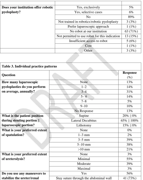
Respondents were surveyed on perceived institutional practice patterns, with results summarised in Table 2. The number of laparoscopic pyeloplasties performed at each institution annually varied greatly, with one third (31%) reporting 10-20 surgeries annually, and another third reporting 5-10 (37%). The number of surgeons performing laparoscopic pyeloplasty at a given institution also varied, with over half of respondents (62%) indicating only 1-2 surgeons at their institution perform the procedure. The survey found that 27% of respondents still offer open pyeloplasty as an option for UPJO, whereas only 11% of the respondents offer robotic
of robotic surgery at their institution and unavailability for this indication (71% and 15%, respectively).
Table 3 summarizes the individual practice patterns of respondents. Most respondents perform 3-4 pyeloplasties annually (36% of those that answered the question). In addition, the majority of respondents reported spending 2-3 hrs to complete a laparoscopic pyeloplasty (39%), with the average time being 2.6±1 hrs. All respondents indicated that they prefer the patient in a lateral decubitus position for the laparoscopic portion of the operation. On the other hand, 35% of respondents prefer to position the patient supine or in lithotomy for stent placement; of those, 30 (86%) re-prep and drape prior to the laparoscopic portion.
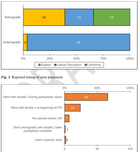
Urologists were also polled regarding their preferred method of stenting (Fig. 1).
Although double J stents are placed over a guide wire, our data does not capture the direction of wire placement, and rather focuses on double J stent placement - the majority of respondents preferred an antegrade stent approach (57%). Surgeons were also questioned regarding when they place stents (Fig. 2), with the majority stenting with a double J stent during pyeloplasty repair (65%), rather than before beginning procedure or after completing the anastomosis.
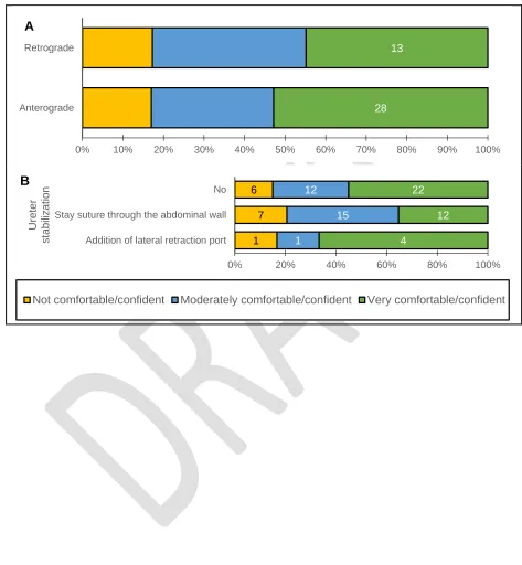
The majority of respondents (56%) routinely stabilize the ureter/renal pelvis, with most surgeons (73%) preferring the use of a stay suture through the abdominal wall (Table 3). The other technique used was the addition of a lateral retraction port. Respondents were also asked to report their spatulation and ureterolysis preferences - variability was seen in both habits, with about 39% of respondents preferring a spatulation of 3-5 mm and 55% preferring minimal ureterolysis.
The survey presented surgeons with a case involving crossing vessels and asked for their preferred laparoscopic pyeloplasty technique, and how their surgical approach would change if no crossing vessel was present (Table 3). While the vast majority of respondents (98%) agreed on the Anderson-Hynes (dismembered) pyeloplasty as an approach for a crossing vessel presentation, a presentation with no crossing vessels led 13 respondents to change their
approach. A third of respondents (34%) relied solely on visual inspection to confirm proper stent placement, followed by 25% who relied on a multi-modal approach; usually involving visual inspection and one of the other listed modalities. Overall, only 47% of respondents reported using X-ray (either intra or post-operative) to confirm stent placement.
Discussion
We set out to survey Canadian attending urologists and senior urology trainees about their laparoscopic pyeloplasty practices. In our centre alone, we have noticed great variability in many of the procedural steps including patient positioning, draping, direction and timing of stent placement, and equipment preferences between the surgeons who perform this procedure. The 100 responses collected comprised at least three graduates from every residency program in Canada, and at least one response from 9 of the 10 Canadian provinces. Although a response rate of 10.2% might appear low, the 942 active members of the CUA that were contacted include not only attending urologists, but also urology trainees, non-urologist physicians, non-physician healthcare workers, and researchers. The inability to filter such members out resulted in an artificially deflated response rate. Laparoscopic pyeloplasty is the definitive treatment option for ureteropelvic junction obstruction, however the operation remains a very low volume procedure in most centres; 94% of respondents indicated that less than 20 laparoscopic pyeloplasties are performed annually at their institution, and about 70% of institutions have 3 surgeons or fewer performing this procedure. As expected, the low volume adds to the perceived difficulty of the operation, where only half of respondents reported feeling very confident performing it.
This study highlights the great variability in approaches amongst Canadian urologists. The only things agreed upon unanimously are the superiority of the Anderson-Hynes
dismembered technique, regardless of whether vessels cross or not, and positioning the patient in lateral decubitus for the laparoscopic portion. Perhaps most surprisingly, the majority of
respondents opted for an antegrade stenting approach, even though retrograde stenting has been reported to be superior due to the ability rule out distal ureteral obstruction prior to pyeloplasty. An explanation for this preference could be the one offered by El-Shazly et al., who suggest that an antegrade approach offers easier suture placement and knot tying, and because it can avoid
having to re-position the patient which is a time consuming portion of the procedure.9
In addition to identifying practice patterns, another aim of this study was to identify factors, techniques, and practices that precipitate greater comfort or confidence. As expected, there is a slight, yet statistically significant increase in comfort levels as a result of greater
number of pyeloplasties performed by each surgeon (OR=1.04, p<0.001). A similar effect is seen with the total number of pyeloplasties performed at an institution and the number of surgeons performing pyeloplasties at each institution (OR=1.01, p<0.01, and OR=1.05, p<0.05,
respectively).
Interestingly, completing a fellowship led to a statistically significant increase in comfort level (OR=3.5, p=0.047). However, confidence levels were similar among academic and
This is the first study to investigate the technical preferences and comfort level of Canadian urologists in performing laparoscopic pyeloplasty. Our data is limited by the self-reported nature, and relatively small sample size. Moreover, confidence and comfort performing an operation are subjective parameters that are difficult to quantify, and suffer greatly from recall bias, especially in the case of very low volume procedures. Within our study 13% of respondents indicated they performed no pyeloplasty’s and yet completed the survey. It is unclear if this response indicated that the surgeons were performing less than one procedure per year,
answering based on their colleagues’ practices, their training or previous experience, answered incorrectly, or misunderstood the question. However, it is possible that the answers from these surgeons skewed the results. Further studies with a larger number of surgeons are warranted to better determine successful techniques and approaches but we hope this study will serve as a launching point for urologists to review their own practices and a discussion piece for training programs.
Conclusion
Laparoscopic pyeloplasty remains a technically challenging procedure. Although comfort levels have been shown to increase with volume and fellowship training, still many Canadian urologists remain uncomfortable performing this procedure. We hope to create discussion amongst
References
1. Whitaker, R. H. Some observations and theories on the wide ureter and hydronephrosis.
Br. J. Urol. 47, 377–385 (1975).
2. Koff, S. A., Hayden, L. J., Cirulli, C. & Shore, R. Pathophysiology of Ureteropelvic
Junction Obstruction: Experimental and Clinical Observations. J. Urol. 136, 336–338
(1986).
3. Koff, S. A. Pathophysiology of ureteropelvic junction obstruction. Clinical and
experimental observations. Urol. Clin. North Am. 17, 263–272 (1990).
4. Khan, F. et al. Management of ureteropelvic junction obstruction in adults. Nat. Rev.
Urol. 11, 629–638 (2014).
5. Riehle, R. A. & Vaughan, E. D. Renin Participation in Hypertension Associated with
Unilateral Hydronephrosis. J. Urol. 126, 243–246 (1981).
6. Jacobs, J. A., Berger, B. W., Goldman, S. M., Robbins, M. A. & Young, J. D.
Ureteropelvic Obstruction in Adults with Previously Normal Pyelograms: A Report of 5
Cases. J. Urol. 121, 242–244 (1979).
7. Sohn, B., Kim, M.-J., Han, S. W., Im, Y. J. & Lee, M.-J. Shear wave velocity
measurements using acoustic radiation force impulse in young children with normal
kidneys versus hydronephrotic kidneys. Ultrason. Ultrason. 33, 116–121 (2014).
8. Reddy, M. N. & Nerli, R. B. The Laparoscopic Pyeloplasty. Urol. Clin. North Am. 42,
43–52 (2015).
9. El-Shazly, M. A., Moon, D. A. & Eden, C. G. Laparoscopic Pyeloplasty: Status and
Review of Literature. J. Endourol. 21, 673–678 (2007).
10.Schuessler, W. W., Grune, M. T., Tecuanhuey, L. V. & Preminger, G. M. Laparoscopic
Dismembered Pyeloplasty. J. Urol. 150, 1795–1799 (1993).
11.Türk, I. A. et al. Laparoscopic Dismembered Pyeloplasty—The Method of Choice in the
Presence of an Enlarged Renal Pelvis and Crossing Vessels. Eur. Urol. 42, 268–275
(2002).
12.Calvert, R. C., Morsy, M. M., Zelhof, B., Rhodes, M. & Burgess, N. A. Comparison of
laparoscopic and open pyeloplasty in 100 patients with pelvi-ureteric junction
obstruction. Surg. Endosc. 22, 411–414 (2008).
13.Ahmed, K. et al. Effectiveness of Procedural Simulation in Urology: A Systematic
Review. J. Urol. 186, 26–34 (2011).
14.Bilen, C. Y. et al. Laparoscopic Pyeloplasty in Adults: Stented Versus Stentless. J. Endourol. 25, 645–650 (2011).
15.Anderson, J. C. & Hynes, W. Retrocaval ureter; a case diagnosed pre-operatively and
treated successfully by a plastic operation. Br. J. Urol. 21, 209–214 (1949).
16.Gulp, O. S. & DeWeerd, J. H. A Pelvic Flap Operation for Certain Types of
Ureteropelvic Obstruction: Observations After two Years’ Experience. J. Urol. 71, 523–
529 (1954).
17.Scardino, P. L. & Prince, C. L. Vertical flap ureteropelvioplasty. South. Med. J. 46, 325– 331 (1953).
Figures and Tables
Fig. 1. Preferred stenting approach by patient positioning.
Fig. 2. Reported timing of stent placement.
16
2
11
52
14
0% 25% 50% 75% 100%
Retrograde
Anterograde
Supine Lateral Decubitus Lithotomy
2 1 7
24
66
0% 50% 100%
0 50 100
I don't routinely stent Stent (retrograde) with double J after
pyeloplasty complete
Fig. 3.Effect of age (A), number of surgeons performing pyeloplasties (B), and volume of
pyeloplasties performed (C, D) on respondents’ comfort levels.
6
Not comfortable/confident Moderately comfortable/confident Very comfortable/confident
Fig. 4. Effect of stenting approach (A) and ureter stabilization practices (B) on respondents’ comfort levels.
13
28
0% 10% 20% 30% 40% 50% 60% 70% 80% 90% 100%
Retrograde
Anterograde
A
6
7
1
12
15
1
22
12
4
0% 20% 40% 60% 80% 100%
No
Stay suture through the abdominal wall
Addition of lateral retraction port
U
ret
er
s
ta
b
iliz
a
tio
n
Table 1. Demographics of survey respondents
Question Response
(%)
Age 20–29 0%
30–39 43%
40–49 40%
50–59 11%
60+ 6%
Current level of training Resident 0%
Fellow 3%
Attending 97%
Practice type (attending only) Academic 42 (43%)
Community 54 (56%)
No response 1 (1%)
Table 2. Institutional practice patterns Question
Response
(%)
How many surgeons perform laparoscopic pyeloplasty at your institution?
0–2 62%
3–4 22%
5–6 14%
7–8 1%
9–10 1%
How many laparoscopic pyeloplasties are performed at your institution, annually?
None 5%
<5 21%
5–10 37%
10–20 31%
20–30 4%
>30 2%
Does your institution still offer open pyeloplasty?
Yes 27%
Does your institution offer robotic pyeloplasty?
Yes, exclusively 5%
Yes, selective cases 6%
No 89%
Not trained in robotics/robotic pyeloplasty 3 (3%)
Prefer laparoscopic approach 1 (1%)
No robot at our institution 63 (71%)
Not permitted to use robot for this indication 13 (15%)
Insufficient access to robot 5 (6%)
Cost 1 (1%)
Other 3 (3%)
Table 3. Individual practice patterns
Question Response
(%)
How many laparoscopic pyeloplasties do you perform on average, annually?
None 13%
What is the patient position during stenting portion || laparoscopic portion?
Supine 20% || 0%
Lateral Decubitus 65% || 100%
Lithotomy 15% || 0%
What is your preferred extent of spatulation?
What is your preferred extent of ureterolysis?
None 3%
Minimal 55%
Moderate 39%
Maximal 3%
Do you use any maneuvers to stabilize the ureter/renal
Yes 56%
pelvis? Addition of lateral retraction port 8 (14%)
Other 7 (12%)
No 44%
What is your preferred approach for the case given?
(with|| without crossing vessels)
Anderson-Hynes (dismembered) pyeloplasty 98% || 85%
Scardino Prince vertical flap 0% || 2%
Culp-DeWeerd spiral flap 1% || 2%
Foley Y-V plasty 1% || 5%
Heineke-Mikulicz ureteroplasty 0% || 1%
Other 0% || 5%
How do you confirm stent position?
Intra-op fluoroscopy/X-ray 9%
Post-op X-ray 19%
Intra-op retrograde methylene blue 13%
Visually 34%
1. Your Age *
Mark only one oval.
20 - 29 30 - 39 40 - 49 50 - 59 60+
2. Gender *
Mark only one oval.
Prefer not to say Male Female Other:
3. Current Level of Training *
Mark only one oval.
PGY1 PGY2 PGY3 PGY4 PGY5 Fellow Attending
4. Number of Fellowships Currently In/Completed *
Mark only one oval.
0 1 2 3 4 5 6+
5. Type of Fellowship:
Check all that apply.
Transplant
Endo-urology MIS Oncology Reconstruction Pediatrics Female Infertility Other Fellowship 1
Fellowship 1
Mark only one oval.
BC
8. Practice type (attending surgeons):
Mark only one oval.
Academic Community
9. Optional: Practice Location
10. Number of people performing laparoscopic pyeloplasty at your institution? *
Mark only one oval.
0 1 2 3 4 5 6 7 8 9 10
11. Number of laparoscopic pyeloplasty performed at your institution per year? *
Mark only one oval.
None
12. On average, what is the number of laparoscopic pyeloplasty performed by YOU at your institution per year? *
Mark only one oval.
0 1 2 3 4 5 6 7 8 9 10
13. How comfortable/confident are you at performing laparoscopic pyeloplasty? *
Mark only one oval.
N/A
15. Do you perform robotic pyeloplasty? *
Mark only one oval.
Yes, exclusively Yes, selective cases
No (please indicate why in the question below)
16. If you answered no to the above question, why?
Mark only one oval.
Not permitted to use robot for this indication No robot at our institution
Prefer laparoscopic approach
Not trained in robotics/robotic pyeloplasty Insufficient access to robot
Cost Other:
17. How long on average does a laparoscopic pyeloplasty take you? *
Example: 4:03:32 (4 hours, 3 minutes, 32 seconds)
You are referred the following case:
18. Based on the above case, what would be your preferred lap. pyeloplasty technique? *
Mark only one oval.
Scardino Prince Vertical Flap Foley Y-V Plasty
Anderson-Hynes (Dismembered) Pyeloplasty Culp-DeWeerd Spiral Flap
Heineke-Mikulicz Ureteroplasty Other:
19. Do you reposition the patient before/after stent placement? *
Mark only one oval.
Yes No
20. If you reposition patient, do you re-prep and drape?
Mark only one oval.
Yes No
21. Patient Positioning: *
Mark only one oval per row.
Supine Lateral Decubitus Lithotomy During wire/stent placement
During laparoscopic portion
22. Method of stenting:
Check all that apply.
Ureteral catheter/wire Double J stent Anterograde
Double J stent None of the above
24. Timing of stent placement: *
Mark only one oval.
Pre-stented before OR
Stent with double J at beginning of OR Stent with double J during pyeloplasty repair
Stent (retrograde) with double J after pyeloplasty complete I don't routinely stent
25. Extent of spatulation: *
Mark only one oval.
None 1 - 3 mm 3 - 5 mm 5 - 10 mm >10 mm
26. Do you excise stenotic segments? *
Mark only one oval.
Yes No
27. Extent of ureterolysis *
Mark only one oval.
None Minimal Moderate Maximal
28. Do you perform any maneuvers to stabilize the ureter/renal pelvis during reconstruction? *
Mark only one oval.
Stay suture through the abdominal wall No
Addition of lateral retraction port Other:
29. How do you confirm stent position? *
Check all that apply.
Visually
Intra-op retrograde methylene blue Intra-op fluoroscopy/X-ray Post-op X-ray
Other:
30. If a similar patient presented with confirmed UPJO, a large redundant renal pelvis, and no crossing vessel, would this change your surgical approach? *
Mark only one oval.
Yes No
31. If yes, select preferred lap. technique:
Mark only one oval.
Culp-DeWeerd Spiral Flap
Anderson-Hynes (Dismembered) Pyeloplasty Foley Y-V Plasty