Noggin4 expression during chick embryonic development
ALEXANDER V. BORODULIN
#, FEDOR M. EROSHKIN
#, ANDREY V. BAYRAMOV and ANDREY G. ZARAISKY*
Shemyakin-Ovchinnikkov Institute of Bioorganic Chemistry, Russian Academy of Sciences, Moscow, Russia
ABSTRACT We describe here the expression pattern of Noggin4 during the early development of the chick embryo (Gallus gallus). The expression of this gene starts with the onset of gastrulation (stage HH4), in two bilateral bands along the primitive streak, with a local maximum around Hensen’s node. By the end of gastrulation, Noggin4 transcripts are distributed diffusely throughout the epi-blast, with the highest concentration in the head ectoderm. Interestingly, the expression of Noggin4 during the first half of gastrulation demonstrates a clear left-right asymmetry in Hensen’s node, being much more intensive in its right anterior portion. During neurulation, Noggin4 is expressed mainly in the neuroectoderm, with the most intensive expression in the head and lateral neural folds. In mesoderm derivatives, expression is seen in somites but not in the notochord. In general, primarily ectodermal and diffusive expression of Noggin4 in chick embryo, with a maximum in the anterior neurectoderm, resembles that of its ortholog in Xenopus, which indicates a conservative function of this gene in evolution.
KEY WORDS:
Noggin, Noggin4, chick embryo
During the last several years, at least three sub-families of
Nog-gin genes, including NogNog-gin (Smith and Harland, 1992), NogNog-gin2
(Fletcher et al., 2004) and Noggin4 (Eroshkin et al., 2006), were identified in vertebrates. Among them, proteins of Noggin4 sub-family have primary structures very different from those of Noggin1 and Noggin2. This indicates that molecular functions of Noggin4 could be different from those of other Noggins. In support of this, the Xenopus laevis Noggin4, whose probable homologs were identified recently in planarians and named Noggin-like genes (Nlg) (Molina et al., 2009), has demonstrated no ability to induce neurogenesis in the Xenopus laevis embryonic ectoderm (Molina
et al., 2011). It should be noted, however, that in contrast to the
planarian Nlgs, vertebrates Noggin4 proteins have no long (50-60 amino acids) and quite conservative fragments between their 5th and 6th cysteine residues, the feature that sharply distinguishes planarian Nlgs from other Noggin family proteins (Eroshkin et al., 2006; Molina et al., 2009).
Since the expression of Noggin4 was studied hitherto only in one vertebrate species (Xenopus laevis), it would be important to investigate expression of this gene in other organisms, preferably with maximally different type of embryogenesis. Such data could allow one to speculate on conservation or, conversely, variability, of Noggin4 functions during evolution. As no functional orthologs of
Noggin4 were found in mammals, we investigate now the
expres-www.intjdevbiol.com
*Address correspondence to: Andrey G. Zaraisky. Shemyakin-Ovchinnikov Institute of Bioorganic Chemistry, Russian Academy of Sciences, Moscow, 117997,
Russia. Tel: +7-495-336-3622. e-mail: [email protected]
#Note: These authors have contributed equally to this work.
Accepted: 3 May 2012. Final, author-corrected PDF published online: 27 June 2012.
ISSN: Online 1696-3547, Print 0214-6282
© 2012 UBC Press Printed in Spain
Abbreviations used in this paper: F, foregut; HF, head fold; HH#, a developmental stage
as it determined in [HAMBURGER, V. and HAMILTON, H.L. (1992); HN, Hensen’s node; HP, head process; NF, neural folds; PMZ, posterior marginal zone; PN, presumptive neuroectoderm; PS, primitive treak; S, somites.
sion pattern of Noggin4 in chick (Gallus gallus), a traditional model of developmental biology, with early course of embryogenesis sharply different from amphibian but resembling that of mammals.
Results
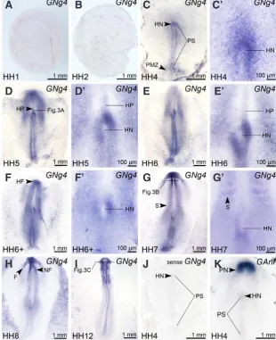
The developmental stages of embryos were defined as de-scribed (Hamburger and Hamilton, 1992). The expression pattern of Noggin4 was analyzed by RT-PCR and the whole mount in situ hybridization, beginning from Hamilton-Hamburger stage 1 (HH1) and up to HH12 stage.
404 A.V. Borodulin et al.
the expression was detected at stage HH4 in HN of some embryos (Fig. 2C’). During HH5-HH6, this asymmetry can be clearly seen in all embryos (Fig. 2 D’,E’,F’).
In the mesoderm, a weak expression of Noggin4 was observed at stages HH5-HH6 in cells of the head process (HP). However, the expression is disappeared from HP by the time of the head
and later in the neuroectoderm. From the very beginning, the expression is somewhat blur and tends towards wide spreading within the neuroectoderm, forming the anterior to posterior gradi-ent. Moreover, in the mesoderm of both species Noggin4 begins to be intensively expressed only in the forming somites, but no expression of this gene can be seen in the notochord. Thus, all this implies that functions of Noggin4 have been retained in frog as well as chick.
Remarkably, the expression of Noggin4 in early development appeared to be sharply different from that of two other representa-tives of Noggin family, Noggin1 and Noggin2. In contrast to
Nog-gin4, Noggin1 is expressed during gastrulation exclusively in the
presumptive chordamesoderm, and later on, together with Noggin2, in a thin stripe of cells corresponding to the presumptive dorsal forebrain (see Eroshkin et al., 2006 for expression of Noggin1 and 2 in Xenopus laevis and Connolly et al., 1997; Streit and Stern, 1999; Chapman et al., 2002 and GEISHA database, University of Arizona, Tucson, AZ 85724; URL: http://geisha.arizona.edu/
Fig. 1. Noggin 4 PCR with or without reverse transcription (+RT or –RT respectively) of total RNA from embryos at the indicated stages with primers for Noggin4 and ODC as the loading control.
Fig. 2. Expression of Noggin4 in chick embryos at stages HH1-HH12. Enlarged images of Hensen’s node region of embryos shown in (C-G) are presented in (C’-G’.) All embryos are shown from the dorsal side, anterior to the top. Abbreviations: F, foregut; HF, head fold; HN, Hensen’s node; HP, head process; NF, neural folds; PMZ, posterior marginal zone; PS, primitive streak; S, somite.
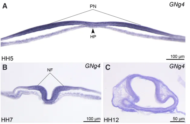
fold (HF) formation at HH6+ (Fig. 2F). Beginning from this stage, Noggin4 expression is localized primarily in the presumptive neuroectoderm (PN) (Fig. 3A). No expression exceeding the background level was detected at this time in the notochord and in cells of surrounding mesoderm.
At HH7, the highest expression level was detected in the ectoderm of HF, and in the neural folds (NF) (Fig. 2G). By this stage, a weak expression of
Nog-gin4 can be also seen in the forming first somites,
as well as in the un-segmented paraxial mesoderm. Simultaneously, the left-right asymmetry of the expres-sion, which was seen before in HN, is disappeared by this stage (Fig. 2G’).
As it may be seen on the transverse sections at the level of NF, the expression in the head region of embryo is localized almost exclusively in the neuroectoderm, rather than the underlying tissues (Fig. 3B). Similar localization of the expression was observed in this region at later stages (Fig. 3C). Beginning from this stage, the expression is characterized by a smooth anterior-posterior gradient, having the highest level at the rostral end of the embryonic body axis
Importantly, control hybridization of HH4 embryos with the sense Noggin4 probe gave no signal at all (Fig. 2J). On the other hand, treatment of embryos with the probe to the homeobox gene Ganf, resulted in the distribution of hybridization signal strongly dif-ferent from that generated by the anti-sense Noggin4 probe (Fig. 2K). Obviously, both these results confirm specificity of the results obtained with the latter probe.
Discussion
The expression pattern of Noggin4 in the early development of chick embryo is generally similar to that of its Xenopus laevis homolog (Eroshkin et al., 2006). In particular, in both species, the expression is revealed by the whole-mount in situ hybridization at the comparable stages (from stage HH4 and 10 in chick and Xenopus, respectively) primarily in PN
geisha/search.jsp?entrez_gene=459378 for expression of Noggin1 in chick). This indicates that physiological, as well as molecular, function of Noggin4 might be also different from those of Noggin1 and Noggin2. In support of this, no ability to inhibit BMP signaling was reported recently for Xenopus laevis Noggin4 (Molina et al., 2011). Instead, the authors of this paper have found out that this protein can slightly ventralize the Xenopus embryo by an unknown mechanism. Given all these, it would be important to test whether Noggin4 retains two other functions recently demonstrated for Noggin1 and Noggin2, namely, the ability to bind and antagonize non-BMP TGFbeta proteins and Wnt (Bayramov et al., 2011).
An intriguing feature of the Noggin4 expression pattern in chicks, which was not reported for Noggin4 in Xenopus, is its asymmetri-cal expression in HN. Interestingly, the expression of Noggin4 in HN at HH5 in chicks corresponds to that of two right-expressing genes – cAct-RIIa (Levin et al., 1995) and MID1 (Granata and Quaderi, 2003). As protein products of the latter two genes are involved in the early patterning along the left-right body axis, one may suppose that Noggin4 may also participate in the process of the left-right asymmetry establishment.
Materials and Methods
Cloning of Noggin4 cDNA and RT-PCR
Using the fact that all Noggin family genes contain no introns, we obtained the Noggin4 coding sequence by PCR from Gallus gallus genomic DNA with the following primers designed on the base of a sequence deposited in Gene Bank (AY779059):
forward: 5’- GCACCGCTCGCCATGCAGGACCCCT;
reverse: 5’- CCCCCTCAGCGGCAGGA. The purified Noggin4 cDNA was cloned into the pGEM-T vector (Promega) and sequenced.
Samples of the total RNA were prepared from the whole embryos at the appropriate stages (5 embryos for each sample) as described (Zaraisky et
al., 1992). PCR was performed with the following primers: 5′-
GGGATG-GAGCTGCCCCCTGA and 5′- CCAGCAGCTTGAGGTGAGCGA. The 25 PCR cycles were made. As the internal control the expression of ODC was monitored in the same samples with primers: 5′-CGGCGGAGGGTTTC-GGGGTT and 5′-AGGCCCCGGACCCAGGTTACT for 18 cycles of PCR.
replaced by 300 ml of digoxygenin-labeled RNA probe, and hybridization
was performed overnight at 60°C. After that, embryos were washed by 500 ml of prehyb over 60 mins at 60°C, 2x SSC (300mM NaCl, 30mM sodium
citrate) twice each of 60 mins at 60°C, 0.2x SSC over 30 mins at 60°C
and 0.2x SSC over 30 mins at room temperature. After that, embryos were rinsed twice with MAB (100mM maleic acid, 150mM NaCl) at room tem-perature and treated with 2% blocking reagent in MAB over 60 mins, 20% heat treated lamb serum in 2% blocking reagent in MAB over 60 mins and 1:1000 anti-Digoxigenin-AP Fab fragments (Roche cat# 11 093 274 910) in 20% heat treated lamb serum in 2% blocking reagent in MAB overnight at 4°C. Antibodies were removed by five washings in MAB at 4°C, then
overnight. After that, embryos were treated with alkaline phosphatase buffer (Chapman et al.) (100mM Tris-HCl, 50mM MgCl2·6H2O, 100mM NaCl, 0.1% Tween-20) containing 2mM levamisol twice and the color reaction was performed in BM Purple AP Substrate (Roche cat# 11 442 074 001) over several days until the desired coloration was reached. After that, embryos were rinsed with AP twice, fixed and stored in MEMFA.
Acnowlegements
This work was supported by grants from Howard Hughes Medical In-stitute, RAS program of MCB, Russian Ministry of Education and Science and RFBR to AGZ and AVB and RF President Grants Council to AVB.
References
BAYRAMOV, A.V., EROSHKIN, F.M., MARTYNOVA, N.Y., ERMAKOVA, G.V., SO-LOVIEVA, E.A. and ZARAISKY, A.G. (2011). Novel functions of Noggin proteins: inhibition of Activin/Nodal and Wnt signaling. Development 138: 5345-5356. CHAPMAN, S.C., SCHUBERT, F.R., SCHOENWOLF, G.C. and LUMSDEN, A. (2002).
Analysis of spatial and temporal gene expression patterns in blastula and gastrula stage chick embryos. Dev Biol 245: 187-189.
CONNOLLY, D.J., PATEL,K., and COOKE, J. (1997). Chick noggin is expressed in the organizer and neural plate during axial development, but offers no evidence of involvement in primary axis formation. Int. J. Dev Biol 41: 389-396.
EROSHKIN, F.M., ERMAKOVA, G.V., BAYRAMOV, A.V. and ZARAISKY, A.G. (2006). Multiple noggins in vertebrate genome: cloning and expression of noggin2 and noggin4 in Xenopus laevis. Gene Expr Patterns 6: 180-186.
FLETCHER, R.B., WATSON, A.L. and HARLAND, R.M. (2004). Expression of
Xeno-pus tropicalis noggin1 and noggin2 in early development: two noggin genes in a
tetrapod. Gene Expr Patterns 5: 225-230. Fig. 3. Histological cross-sections of chick embryos at stages HH5 (A), HH7 (B) and
HH12 (C) at the levels indicated in Fig. 2 D,G,I.N, notochord; NF, neural folds; PN, pre-sumptive neuroectoderm. Dorsal side to the top.
Whole-mount in situ hybridization
The digoxigenin-UTP-labeled anti-sense and sense RNA probes were synthesized with either T7- or SP6-RNA poly-merase (Promega) respectively from DNA fragment obtained from pGEM-T-Noggin4 plasmid by PCR with M13-direct and M13-reverse primers. The probe for Ganf mRNA was obtained as described (Kazanskaya et al., 1997). Eggs were collected not later than 3 hours after oviposition. An incubation of the eggs was performed during proper time for embryos to reach certain stage. After incubation, embryos were removed from yolk, washed in PBT (phosphate-buffered saline with 0.1% Tween-20) and fixed with ice-cold MEMFA fixative (parafor-maldehyde 3.7%, EGTA 2mM, MsSO4 1mM, MOPS 0.1M) at room temperature for two hours. After fixative was removed by washing in PBT, embryos were treated with 50% and 96% EtOH and stored in 96% EtOH at -20°C overnight. Embryos were rehydrate through a graded EtOH series, rinsed triply in PBT and digested in Proteinase K (10 ml/ml) 5 to 10 mins.
Digestion was stopped by washing in 0.1M triethanolamine with 0.1% acetic anhydride. After that, embryos were rinsed in PBS twice and fixed in MEMFA over 20 mins. Fixative was removed by two washings in PBT, prehybridized in 300 ml of
prehybridization solution (50% formamide, 5x SSC, Torula RNA (1mg/ml), Denhardt’s solution, 0.1% Tween-20, 0.1% Chaps and 10mM EDTA) over 120 mins. The solution was
B
C
406 A.V. Borodulin et al.
HAMBURGER, V. and HAMILTON, H.L. (1992). A series of normal stages in the development of the chick embryo. 1951. Dev Dyn 195: 231-272.
KAZANSKAYA, O.V., SEVERTZOVA, E.A., BARTH, K.A., ERMAKOVA, G.V., LUKYA-NOV, S.A., BENYUMOV, A.O., PANNESE, M., BONCINELLI, E., WILSON, S.W. and ZARAISKY, A.G. (1997). Anf: a novel class of vertebrate homeobox genes expressed at the anterior end of the main embryonic axis. Gene 200: 25-34. LEVIN, M., JOHNSON, R.L., STERN, C.D., KUEHN, M. and TABIN, C. (1995). A
molecular pathway determining left-right asymmetry in chick embryogenesis.
Cell 82: 803-814.
MOLINA, M.D., NETO, A., MAESO, I., GOMEZ-SKARMETA, J.L., SALO, E. and CEBRIA, F. (2011). Noggin and noggin-like genes control dorsoventral axis regeneration in planarians. Curr Biol 21: 300-305.
MOLINA, M.D., SALO, E. and CEBRIA, F. (2009). Expression pattern of the expanded noggin gene family in the planarian Schmidtea mediterranea. Gene Expr
Pat-terns 9: 246-253.
SMITH, W.C. and HARLAND, R.M. (1992). Expression cloning of noggin, a new dorsalizing factor localized to the Spemann organizer in Xenopus embryos. Cell 70: 829-840.
STREIT, A., AND STERN, C.D. (1999). Establishment and maintenance of the border of the neural plate in the chick: involvement of FGF and BMP activity.
Mech Dev 82: 51-56.
The zic1 gene is an activator of Wnt signaling
Christa S. Merzdorf and Hazel L. Sive Int. J. Dev. Biol. (2006) 50: 611-617
Early stages of neural crest ontogeny: formation and regulation of cell delamination
Chaya Kalcheim and Tal Burstyn-Cohen Int. J. Dev. Biol. (2005) 49: 105-116
X-epilectin: a novel epidermal fucolectin regulated by BMP signalling
Karine Massé, Rebecca Baldwin, Mark W. Barnett and Elizabeth A. Jones Int. J. Dev. Biol. (2004) 48: 1119-1129
Acceleration of early chick embryo morphogenesis by insulin is associated with altered expression of embryonic genes
Vidya Patwardhan, Madhavi Gokhale and Surendra Ghaskadbi Int. J. Dev. Biol. (2004) 48: 319-326
Evo-Devo of amniote integuments and appendages
Ping Wu, Lianhai Hou, Maksim Plikus, Michael Hughes, Jeffrey Scehnet, Sanong Suksaweang, Randall Widelitz, Ting-Xin Jiang and Cheng-Ming Chuong
Int. J. Dev. Biol. (2004) 48: 249-270
The biology of feather follicles
5 yr ISI Impact Factor (2010) = 2.961
Mingke Yu, Zhicao Yue, Ping Wu, Da-Yu Wu, Julie-Ann Mayer, Marcus Medina, Randall B Widelitz, Ting-Xin Jiang and Cheng-Ming Chuong
Int. J. Dev. Biol. (2004) 48: 181-191
The choice between epidermal and neural fate: a matter of calcium
Marc Moreau and Catherine Leclerc Int. J. Dev. Biol. (2004) 48: 75-84
FGF signaling is essential for the early events in the development of the chick nervous system and mesoderm
S Khot and S Ghaskadbi
Int. J. Dev. Biol. (2001) 45: 877-885
Chick noggin is expressed in the organizer and neural plate during axial development, but offers no evidence of involvement in primary axis formation