ABSTRACT
CHAROENPANICH, ADISRI. Analyses of Human Adipose Derived Stem Cells and Human Mesenchymal Stem Cells for Functional Tissue Engineering using Biomimetic Physical Stimuli. (Under the direction of Elizabeth G. Loboa.)
Autologous stem-cell-based tissue engineering holds great potential for treating trauma and pathologies in a patient specific manner. Adult stem cells can be derived from various source tissues such as bone marrow, epidermal tissue, and adipose tissue. Initially, bone-marrow-derived mesenchymal stem cells (MSC) received the most attention for musculoskeletal tissue engineering applications given their direct lineage capability. More recently however, adipose-derived stem cells (ASC) have received increasing interest for tissue engineering applications due to their relative ease of harvest, abundance, and multi-lineage differentiation potential. To better implement the use of stem and progenitor cells for cell-based therapy and tissue repair, understanding of how these cells are committed to, or differentiate into, a specific cell lineage is needed.
meniscus. In the second study, hASC osteogenic differentiation and response to combined chemical and mechanical stimuli were investigated. Gene expression profiles of
proliferating or osteogenically induced hASC in 3D collagen I culture in the presence and absence of 10% uniaxial cyclic tensile strain were examined using microarray analysis. In the final study, hMSC isolated from aged, postmenopausal osteoporotic donors were cultured in three-dimensional (3D) collagen constructs and analyzed for changes in mRNA expression in response to 10% uniaxial cyclic tensile strain in an attempt to identify potential
mechanisms underlying the use of appropriate mechanical loading for prevention and treatment of osteoporosis.
The results of the first study show promising initial results of the use of hASC combined with a decellularized meniscal allograft for improved approaches for meniscal allograft transplants. The following studies of hASC and hMSC in response to tensile strain expanded findings from the first study to determine the effects of combined chemical and mechanical stimuli for regeneration of other musculoskeletal tissues in addition to
fibrocartilage, with particular emphasis on bone formation. Application of cyclic tensile strain at different magnitudes is a stimulus for fibrous tissue, fibrocartilage and bone
formation during normal secondary fracture healing or distraction osteogenesis. Specifically, our lab has previously shown that 10% cyclic tensile enhances osteogenesis of both hASC and hMSC in vitro. However, the molecular mechanisms underlying this potential are not yet known. The research performed in this thesis identified angiogenesis as a potential
Analyses of Human Adipose Derived Stem Cells and Human Mesenchymal Stem Cells for Functional Tissue Engineering using Biomimetic Physical Stimuli
by
Adisri Charoenpanich
A dissertation submitted to the Graduate Faculty of North Carolina State University
in partial fulfillment of the requirements for the degree of
Doctor of Philosophy
Biomedical Engineering
Raleigh, North Carolina 2013
APPROVED BY:
_______________________________ ______________________________ Dr. Elizabeth Loboa Polefka Dr. Albert Banes
Committee Chair
_______________________________ ______________________________
Dr. Susan Bernacki Dr. Gregory McCarty
ii DEDICATION
iii BIOGRAPHY
iv ACKNOWLEDGMENTS
I would like to express my gratitude to my advisor, Dr. Elizabeth Loboa, for her guidance, encouragement and generous support. I am so thankful having her as my advisor. I would like to thank Dr. Susan Bernacki for her support, wisdom and very helpful advices since the first year of my graduate study. I would also like to thank Dr. Jeffrey Spang for his time, advice and contribution on my meniscus project and also taking time to serve in my committee. I would like to thank Dr. Albert Banes and Dr. Gregory McCarty for his wisdom classes, suggestions, and taking time to serve in my committee. I would also like to thank Dr. Charles Tucker, Dr. Danica Andrews, and Dr. Michelle Wall for their time, advice and contribution on my two microarray projects.
Thank you for the collaboration from Dr. Peter Mente, Dr. Carol Otey’s lab, Dr. Caterina gallippi, and Tomasz Czernuszewicz. My sincere appreciation is extended to all CML members. I would also like to thank Ruwan, Wayne, and Seth. Thank you Carla for the wonderful friendship. Thank you Josie and her Titi for the amazing time we shared. Thank you Stephanie and her Honey bee for all lovely support . Thank you Mahsa for all your help and care. Thank you John, Stephen and Pattie.I also appreciate the support from the faculty and staffs at joint Department of Biomedical Engineering.
v Helen into my life. Thank you to the Hay Day support team P Orm and P Jijy, you guys are the best <3. Thank you to Tum, P Pin, Win, P Poy, N Fang, P Art, Good, and all Thai folks.
vi TABLE OF CONTENTS
LIST OF TABLES………...….xii
LIST OF FIGURES ... xiii
Preface... 1
1. Proliferation of Human Adipose Derived Stem Cells Stem Cells in a Needle-Punched, Porous, Decellularized Human Allograft-Derived Meniscus and Synthetic Aligned PLA scaffold ... 3
1.1 Introduction ... 3
1.2 Materials and Methods ... 5
1.2.1 Decellularization and Porosity Enhancement of Human Meniscus ... 5
1.2.2 Cell seeding and cell culture ... 6
1.2.3 Nuclei staining ... 6
1.2.4 Cell survival and histology ... 6
1.2.5 Scanning Electron Microscopy ... 7
1.3 Results ... 7
1.3.1 Decellularized of Human Meniscus ... 7
1.3.2 Proliferation of hASC on Decellularized Meniscus ... 9
1.3.3 Histology ... 10
vii 2 Microarray Analysis of hASC in 3D Collagen Culture: Osteogenesis Inhibits BMP and Wnt Signaling Pathways and Cyclic Tensile Strain Causes Upregulation of Proinflammatory
Cytokine Regulators and Angiogenic Factors ... 14
2.1 Introduction ... 15
2.2 Materials and Methods ... 17
2.2.1 Cell isolation, culture, and characterization ... 17
2.2.2 Fabrication of collagen gels ... 18
2.2.3 Osteogenic differentiation and application of 10% cyclic tensile strain ... 18
2.2.4 RNA isolation and real-time RT-PCR analysis ... 19
2.2.5 Biotin labeling, streptavidin antibody staining, scanning and detection ... 20
2.2.6 Microarray data analysis ... 21
2.3 Results ... 23
2.3.1 Osteogenesis of hASC after 14 days in 3D collagen culture in response to osteogenic media and 10% uniaxial cyclic tensile strain ... 23
2.3.2 Validation of microarray data ... 25
2.3.3 Genes regulated during osteogenic differentiation of hASC in 3D collagen culture for 14 days ... 27
viii 2.3.5 Real-time RT-PCR analysis to validate results for corin, the typeII serine protease upregulated during osteogenesis of hASC, PDLIM4, an actin binding protein, and VEGF A, an angiogenic factor, upregulated in response to 10% uniaxial cyclic tensile strain in 3D culture. ... 33 2.4 Discussion ... 35 2.5 Supplementary Data ... 43 Supplementary Table 1: Genes upregulated by hASC in 3D collagen culture in response to osteogenic induction media. ... 43 Supplementary Table 2: Genes downregulated by hASC in 3D collagen culture in
response to osteogenic induction media. ... 54 Supplementary Table 3: All significantly modulated canonical pathways during
osteogenic differentiation of hASC in 3D collagen culture at day 14. 95 canonical pathways were arranged by the highest statistical significance (-log (p-value)). Ratio is calculated by number of mapped molecules/ total molecules in pathway. All mapped molecules are shown as abbreviated name. ... 64 Supplementary Table 4: Genes upregulated by hASC in 3D collagen culture in response to 10% uniaxial cyclic tensile strain. ... 68 Supplementary Table 5: Genes downregulated by hASC in 3D collagen culture in
ix Supplementary Table 6: Canonical pathways modulated by 10% uniaxial cyclic tensile strain during osteogenic differentiation of hASC in 3D collagen culture at day 14. (p
<0.05) ... 72
Supplementary Figure 1. Endogenous alkaline phosphatase (endoALP) activity by hASC cultured in 3D collagen gels under static (0%) and dynamic (10%) uniaxial cyclic tensile strain. Endogenous ALP activity was increased over time by hASC in 3D collagen gel culture and further increased with application of 10% uniaxial cyclic tensile strain. ... 74
3... Microarray Analysis of Human MSC from Osteoporotic Donors in 3D Collagen Culture: 10% Uniaxial Cyclic Tensile Strain Enhances Cell Proliferation, Osteogenesis and Angiogenesis in MSC from Osteoporotic Donors ... 75
3.1 Introduction ... 75
3.2 Materials and Methods ... 77
3.2.1 Cell isolation, culture, and characterization ... 77
3.2.2 Cells proliferation, osteogenic differentiation and calcium profile ... 78
3.2.3 Fabrication of collagen gels ... 79
3.2.4 Application of cyclic tensile strain ... 80
3.2.5 RNA isolation and real-time RT-PCR analysis ... 80
3.2.6 Biotin labeling, streptavidin antibody staining, scanning and detection ... 81
3.2.7 Microarray data analysis ... 82
x 3.2.9 Proliferation and Ostegenic differentiation analyses of PDLIM4 knockdown hMSC………83 3.3 Results ... 84
3.3.1 Proliferation and osteogenic differentiation of hMSC from postmenopausal Caucasian osteoporotic donors. ... 84 3.3.2 Function analysis of genes regulated by hMSC in response to 10% uniaxial cyclic tensile strain ... 88 3.3.3 Network analysis and canonical pathway of genes regulated by hMSC in
response to 10% uniaxial cyclic tensile strain ... 91 3.3.4 Real-time RT-PCR analysis of selected differentially expressed genes identified by cDNA microarray ... 94 3.3.5 PDLIM4 knockdown increased metabolic activity of hMSC during
xii LIST OF TABLES
Table 2-1. Key findings from comparative analysis of this study’s findings to the publicly available database. ... 30 Supplementary Table 2-1: Genes upregulated by hASC in 3D collagen culture in response to osteogenic induction media………..43 Supplementary Table 2-2: Genes downregulated by hASC in 3D collagen culture in response to osteogenic induction media. ... 54 Supplementary Table 2-3: All significantly modulated canonical pathways during osteogenic differentiation of hASC in 3D collagen culture at day 14. 95 canonical pathways were
arranged by the highest statistical significance (-log (p-value)). Ratio is calculated by number of mapped molecules/ total molecules in pathway. All mapped molecules are shown as abbreviated name. ... 64 Supplementary Table 2-4: Genes upregulated by hASC in 3D collagen culture in response to 10% uniaxial cyclic tensile strain. ... 68 Supplementary Table 2-5: Genes downregulated by hASC in 3D collagen culture in response to 10% uniaxial cyclic tensile strain. ... 70 Supplementary Table 2-6: Canonical pathways modulated by 10% uniaxial cyclic tensile strain during osteogenic differentiation of hASC in 3D collagen culture at day 14. (p <0.05) ... 72 Supplementary Table 2-7: Focused genes in the first ranked network associated with specific effect of 10% uniaxial cyclic tensile strain. Note that IL-1 expression was not actually
xiv LIST OF FIGURES
Figure 1-1. Microneedle punching. Decellularized meniscal allografts were vertically
penetrated with 28G microneedle. ... 5 Figure 1-2. Gross inspection of human meniscus before decellularization (a) and after
xv osteogenic media, or osteogenic media plus 10% cyclic tensile strain (f=1 Hz, 4 hours/day). ... 24 Figure 2-2. Principal Component Analysis (PCA). PCA results indicated a distinct
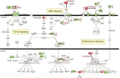
separation between growth media and osteogenic media, and further between growth media and osteogenic media plus 10% cyclic tensile strain. (b) Rotated (45 degrees) plot for 3D view. ... 26 Figure 2-3. Downregulation of TGF-β Signaling, Wnt/β-Catenin Signaling, and BMP
Signaling by hASC in 3D collagen culture in response to osteogenic induction media. Red indicates upregulated molecule (also indicated by ↑), green indicates downregulated
xvi indicates downregulated molecules. Color intensity indicates the level of expression. (see Supplementary Table 7 for full names and expression profile data). ... 32 Figure 2-6. Real-time RT-PCR results (a) Upregulation of corin during hASC osteogenesis. Corin expression in hASC was compared to its expression in bone tissue (expression set to 1). (b) Upregulation of PDLIM4 and VEGF A by hASC in response to 10% uniaxial cyclic tensile strain. Gene expression in osteogenic media set to 1, GAPDH normalization. ... 34 Figure 3-1. hMSC from three aged, postmenopausal female osteoporotic donors are able to osteogenically differentiate after chemical stimulation in osteogenic induction media for 14 days in 2D monolayer culture. Alizarin red staining for calcium accretion (A) in growth media or (B) osteogenic media. Endogenous alkaline phosphatase (EALP) activity of hMSC after 14 days (C) in growth media or (D) in osteogenic induction media. EALP activity indicated in bright green; nuclei shown in blue. ... 85 Figure 3-2. Quantitative analysis of cell-mediated calcium accretion normalized to cellular protein content after 14 days of hMSC culture in complete growth medium (CGM), or osteogenic medium (ODM). hMSC isolated from osteoporotic donors (A, B, C) exhibited significantly (*p<0.05, **p<0.01) higher calcium/protein ratio than hMSC isolated from young, non-osteoporotic donors (D, E)... 86 Figure 3-3. Comparative analysis of average proliferation rate (normalized to day 1) of hMSC isolated from osteopotic donors (OP) and hMSC isolated from young,
xvii significantly (*p<0.05) less than hMSC from non-osteoporotic donors at all time points in both growth and osteogenic differentiation media... 87 Figure 3-4. Function analysis indicates 10% uniaxial cyclic tensile strain induces expression of genes associated with cellular movement, growth, and proliferation in hMSC from osteoporotic donors as shown in the top ten molecular and cellular functions (A); and, expression of genes associated with system development and function for skeletal, muscular and cardiovascular systems as shown in the top ten categories expressed within physiological and system development and function (B). Left Y-axis is –log(p-value) shown as bar chart. Right Y-axis is number of genes associated with functions shown as dot(.) with number. ... 90 Figure 3-5. Potential key molecules affected in hMSC from osteoporotic donors in response to 10% uniaxial cyclic tensile strain are jun D proto-oncogene (JUND), plasminogen
activator, urokinase (PLAU), plasminogen activator, urokinase receptor (PLAUR), vascular endothelial growth factor A (VEGF A), and follistatin (FST) as centered in the first (A), and ubiquitin C (UBC) as centered in the second (B) ranked networks (highlighted with blue circles). The first ranked network was generated with 22 focus genes previously found to be associated to cellular movement, cell morphology, and skeletal and muscular system
development and function (A). The second ranked network was generated with 19 focus genes previously found to be associate with tissue morphology, cardiovascular disease, and molecular transport (B). Red indicates upregulated molecules; green indicates
downregulated molecules. Color intensity indicates the level of expression (see
xviii Figure 3-6. Canonical pathways affected in hMSC from osteoporotic donors by 10% uniaxial cyclic tensile strain. Top 10 canonical pathways showed that 5 genes (FZD8, TCF4,
PIK3CD, WNT5B, and BMP1) are associated and repeatedly mapped in multiple pathways (A). Mapped canonical pathway, role of osteoblasts, osteoclasts and chondrocytes in rheumatoid arthritis, showed the integrated signals of these genes in Wnt/BMP/PIKs
signaling (B)... 93 Figure 3-7. RT-PCR results. Upregulation of six genes identified with microarray analysis: vascular endothelial growth factor a (VEGF A), PDZ and LIM domain 4 (PDLIM 4), wingless-type MMTV integration site family, member 5B (WNT5b), phosphoinositide-3-kinase, catalytic, delta polypeptide (PIK3CD), jun D proto-oncogene (JUND), plasminogen activator, urokinase receptor (PLAUR) in hMSC from osteoporotic donors in response to 10% uniaxial cyclic tensile strain had expression confirmed with RT-PCR. Gene expression in growth media without the application of strain set to 1, GAPDH normalization. ... 95 Figure 3-8. Comparative analysis of average metabolic activity (normalized to day 7) of control knockdown (Control KD) or PDLIM4 knockdwon (PDLIM4 KD) hMSC in
complete growth medium (CGM) or osteogenic medium (ODM) on days 10 in 2D monolayer culture. Proliferation of hMSC did not alter by PDLIM4 knockdwon (CGM) but metabolic activity was significantly (p<0. increased during osteogenesis (ODM). ... 96 Figure 3-9. Upregulation of bone marker genes, Corin and PDLIM4 in hMSC during
xix Figure 3-10. Upregulation of PDLIM4 and Corin during osteogenesis of hMSC; expression knockdown resulted in an increase of bone markers Runx2 and Col1a2 at day 3 in both hMSC culture in normoxia (a) and in hypoxia (b). Gene expression normalized to GAPDH and fold change compared to mRNA level in normal human adult bone tissue (set to equal 1.0) (*p<0.05, **p<0.01) ... 99 Figure 3-11. Alkaline phosphatase activity of hMSC at day 14. PDLIM4 knockdown
1 Preface
Tissue engineering holds great potential for treating critical sized defects; particularly the creation and use of engineered tissue from autologous adult stem cells. Adult stem cells can be derived from various sources such as bone marrow, epidermal tissue, and adipose tissue 1-3. From these source tissues, bone marrow derived mesenchymal stem cells (MSC) have been the most extensively studied given their early identification as having multipotent lineage specification capability 4-7. More recently, adipose derived stem cells (ASC) have gained increasing interest as they have also been shown to undergo multilineage differentiation including, but not limited to, osteoblasts, chondrocytes, adipocytes, myoblasts, cardiomyocytes, neural and liver cells 8,9. Moreover, ASC can be more easily harvested with less invasive surgery (liposuction) from a relatively abundant tissue source relative to MSC, which are usually harvested via iliac crest needle biopsy 10-12. However, it is still challenging to mimic tissue in vivo and engineer functional tissue with both material and chemical properties consistent with native tissue using progenitor cells such as ASC or MSC in vitro. Understanding and elucidating the process of stem cell differentiation and lineage specification in response to specific inductive environments provided by both physical and chemical stimuli will help us to optimize the use of MSC and ASC for patient specific tissue engineering and regenerative medicine applications.
2 of hASC to chemical stimuli in the form of soluble inductive factors within specified culture medium or by seeding within a human, decellularized extracellular matrix; and, 2) molecular mechanisms underlying their response to 10% uniaxial cyclic tensile strain, a mechanical stimulus we have previously shown accelerates and increases osteogenic differentiation of these stem cells
3 1 Proliferation of Human Adipose Derived Stem Cells Stem Cells in a
Needle-Punched, Porous, Decellularized Human Allograft-Derived Meniscus and Synthetic Aligned PLA scaffold
1.1 Introduction
The meniscus plays a crucial role in knee joint function by providing joint stability, shock absorption, load transmission, and stress distribution within the knee joint. Meniscal tears are the most common of knee injuries with an annual reported incidence of 60-70 per 100,000 persons 13,14. With limited natural repair of meniscus, arthroscopy repair is the gold standard for meniscus tear. However, not all meniscus tear can be repaired, especially tears in the avascular inner-third and complex or degenerative tears in older patients with limited healing potential. The clinical evidence showing the detrimental effects of partial or total removal of the meniscus on the knee joint, as it increases the contact stresses on the articular surface of the tibia 15. Previous biomechanical studies have shown that at least 50% of the compressive load of the knee joint is transmitted through the knee joint in extension while up to 85% of the knee load is transmitted in 90 degrees of flexion 16. In the knee with a
menisectomy, the contact area between the tibia and the femur is reduced by 50 percent17. For clinicians, meniscal preservation or replacement has become more important as our understanding of the devastating long-term consequences of meniscal removal have become better understood.
Meniscal allograft transplantation is a treatment utilized when the native meniscus has been removed or ceases to function due to injury to protect the meniscally deficient knee
18
4 integrate with host knee, and degradation over time due to a lack of cellular incorporation after implantation 19,20. Previous human retrieval studies and animals studies have noted incomplete cellular incorporation, absence of cell proliferation, and a microscopic immune response 21-23. It has been postulated that the lack of cellular incorporation may result from the high density of the meniscus. Without successful ingrowth and cellular re-population, menisical allografts lack the capacity to remodel and perform necessary maintenance. In longer term studies, 10 year failure rates for meniscal allografts are as high as 25-50 percent
24-26
.
Removing donor cellular component and increasing porosity to allow loading of a transplanted meniscus with a biologically active substrate (growth factors, platelets or MSC) have been studied to better improve the success rate of allograft transplant using an animal models 27-31. To date, little research has focused on the possibility of modifying the human meniscus with human stem cells to evaluate the meniscus as a potential scaffold and also the biological responses of human stem cells to human meniscal allograft.
In this study, a human meniscus was decellularized to remove donor cells and
5 1.2 Materials and Methods
1.2.1 Decellularization and Porosity Enhancement of Human Meniscus
Gamma sterilized, frozen human menisci were provided by the Musculoskeletal Transplant Foundation. Menisci were defrosted at room temperature. Chemical
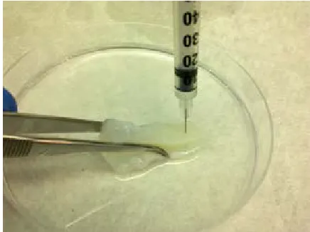
decellularization was performed to remove donor cells 27. In brief, whole meniscus was placed in deionized water at 37oC for 48 hours, followed by 24 hours enzymatic digestion in 0.05% trypsin EDTA, 24 hours trypsin neutralization in complete growth media (CGM) (α -MEM supplemented with 10% FBS, 2 mM L-glutamine, 1% Pen/Strep) followed by 48 hours in 2% Triton X-100/ 1.5% peracetic acid solution 27. The allograft was then subjected to multiple washes in deionized water. To further enhance porosity, a 28G microneedle was used to manually punch 360 µm pores through the meniscus in a superior to inferior pattern (Figure 1).
6 1.2.2 Cell seeding and cell culture
Human ASC were seeded throughout the meniscus by centrifugal seeding at 2,000 rpm, 5 times at 1 minute intervals with gentle resuspension of unattached cells after each cycle to increase cell-meniscus contact 3. Cell seeded menisci were cultured for up to 4 weeks in complete growth medium (CGM) at 37°C humidified 5% CO2 atmosphere. The
medium was changed every three days.
1.2.3 Nuclei staining
After four weeks, the cell-seeded menisci were dissected and fixed in 10% formalin for 24 hours, followed by two washes in phosphate buffered saline (PBS), ethanol
dehydration and paraffin embedding. Menisci were sectioned into 10-µm thick slices with a rotary microtome and sections were stained with 4,6-diamidino-2-phenylindole (DAPI) staining to identify residual nuclei debris.
1.2.4 Cell survival and histology
Live cells were stained with Calcein AM for visualization (Molecular Probes,
7 1.2.5 Scanning Electron Microscopy
Tissue specimens were fixed with 2.5% glutaraldehyde, followed by serial ethanol dehydration. They were then critical point dried in liquid CO2 and gold-platinum sputter
coated for scanning electron microscopy (SEM) analyses.
1.3 Results
1.3.1 Decellularized of Human Meniscus
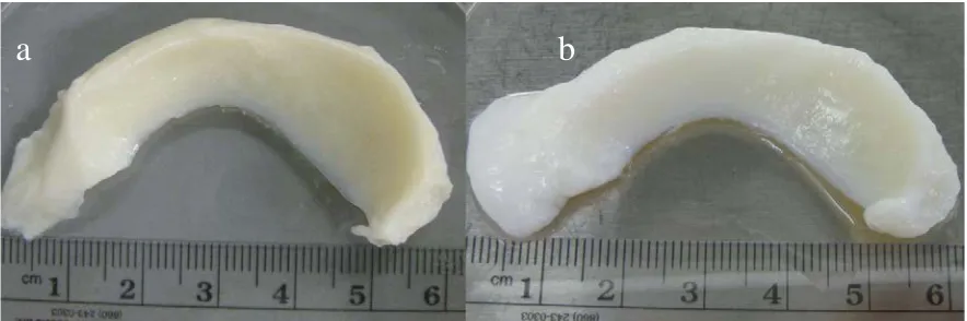
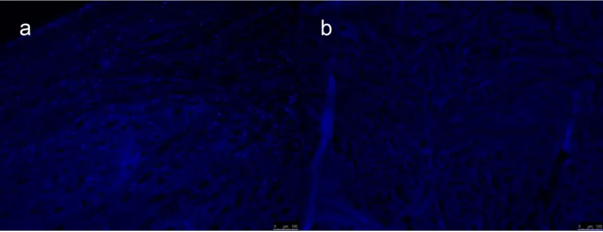
The general shape and architecture of meniscus was maintained after needle-punching and decellularizing process (Figure 2). Successfully remove of donor nucleic acid can be observed with a relative absence of nuclear staining with DAPI in the decellularized meniscus relative to the intact meniscus (Figure 3).
Figure 1-2. Gross inspection of human meniscus before decellularization (a) and after decellularization (b). Overall shapes were maintained for decellularized compare to intact meniscus
8 Figure 1-3. Cellular and nuclear content (blue dots) as indicated via DAPI staining of intact meniscus (a) and decellularized meniscus (b).
Scanning electron microscopy revealed that chemical decellularization removed the donor cell membrane, loosened but did not unpack the collagen bundle, and generated gaps and micropores within the collagen network (Figure 4).
Donor cell membrane
a
b
c
9 1.3.2 Proliferation of hASC on Decellularized Meniscus
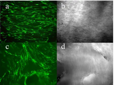
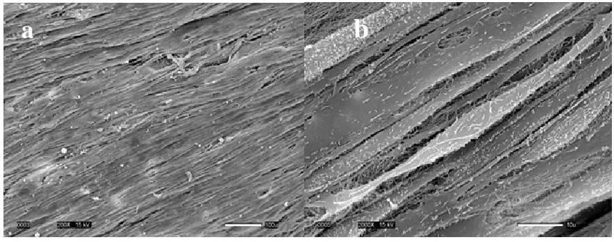
Live staining showed that hASC remained viable for both decellularized and needle-punched decellularized menisci (Figure 5). SEM revealed elongated hASC aligned parallel to the native collagen fibers of the decellularized meniscus (Figure 6).
10 Figure 1-6. hASC proliferation and alignment in hASC-seeded meniscus. hASC were well organized and aligned parallel to the collagen fiber extracellular matrix of decellularized menical allograft: scanning electron micrograph at 200x (a), and at 2000x (b).
1.3.3 Histology
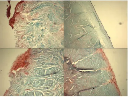
Histological analyses of the re-seeded meniscal allograft showed that cells were able to proliferate and migrate through the outer periphery region where vascular channels were present (Figure 7a). However, decellularization alone did not allow for cells to migrate further than the surface of the inner (non-vascularized) regions (Figure 7b). With needle punching, cells were able to migrate through the pores from the surface into the central regions of the meniscal tissue (Fig 7c,d).
12 1.4 Discussion
Meniscus allograft transplant has shown favorable results in various situations at short- and medium-term up, and even, in some cases, at long-term (>10 years) follow-up. With the first meniscal allograft transplantation 28 years ago in 1984, currently long-term studies in meniscal allograft transplant are in the length of 10 years. The survive rate for 10 years follow-up in cryopreserved and fresh-frozen meniscal allograft transplants is in between 70%-80% with defined failure as tearing, sub/total destruction requiring repair/partial meniscectomy or removal of the allograft 25,32-34. However, a magnetic resonance imaging study on the width and thickness of fresh frozen meniscal transplants showed that shrinkage in the width (89%) and increase in thickness (115%) can be observed within the first year of transplant 35.
Immunohistocomistry analyses of fresh, cryopreserved and frozen human menisci showed the presence of HLA, and ABH antigen that could complicate the meniscus allograft transplant 23,36. In this study we have successfully remove donor cells while maintain the overall structure of meniscal allograft. Reseeding the decellularized allograft with hASC showed promising result as cells are viable, proliferate and organized parallel to the structure of collagen fiber extracellular matrix within the meniscus. The ability of hASC to align parallel to the native collagen fibers in the meniscal allograft is promising for collagen production by hASC to overcome the degeneration of the meniscal allograft after long term implantation. It has been shown that alignment of a cell-produced collagen matrix is
13 Successful tissue engineering utilizing hASC would allow meniscal allograft
14 2 Microarray Analysis of hASC in 3D Collagen Culture: Osteogenesis Inhibits BMP
and Wnt Signaling Pathways and Cyclic Tensile Strain Causes Upregulation of Proinflammatory Cytokine Regulators and Angiogenic Factors
To better implement the use of stem and progenitor cells for cell-based therapy and tissue engineering, understanding how these cells are committed to or differentiate into a specific cell lineage is needed. While hASC have shown great promise for bone tissue engineering as discussed in Chapter 2, molecular mechanisms underlying their potential are as yet unknown.
In this chapter, osteogenecity of hASC was investigated at the gene expression level of over 47,000 transcripts by cDNA microarray analysis. A three-dimensional collagen type I matrix was used as the culture system to mimic the bone ECM, osteogenic differentiation of hASC was induced with osteogenic media containing the soluble osteogenic inductive factors; β-glycerol phosphate, ascorbic acid, and dexamethasone, and 10% uniaxial cyclic tensile strain was applied as the mechanical loading. The significantly modulated genes were analyzed and compared to the available canonical pathways along with the function and network analysis to filter for the key factors or signals.
15 2.1 Introduction
Autologous stem-cell-based bone tissue engineering holds great potential for treating bone trauma and pathologies in a patient specific manner. Adult stem cells can be derived from various source tissues such as bone marrow, epidermal tissue, and adipose tissue 1,44. Initially, bone-marrow-derived mesenchymal stem cells (MSC) received the most attention for bone tissue engineering applications given their known osteogenic capability. More recently however, adipose-derived stem cells (ASC) have received increasing interest for tissue engineering applications due to their relative ease of harvest, abundance, and multilineage differentiation potential 9,44,45.
When cultured in monolayer in the presence of osteogenic media containing ascorbic acid, β-glycerolphosphate, and dexamethasone, ASC have been shown to undergo osteogenic differentiation, deposit calcium phosphate, and express osteoblast-associated gene markers, osteocalcin, alkaline phosphatase, and osteopontin in vitro 9, 46. In order to better mimic the in vivo environment, researchers have also utilized three-dimensional (3D) culture conditions.
Culture of human ASC (hASC) in 3D collagen gels has been found to promote osteogenic differentiation of hASC by elevating bone marker mRNA expression 47. As with 3D culture, the mechanical environment also plays an important role in stem cell growth, differentiation, and function.
At the tissue level, predominant mechanical stimuli in bone include fluid shear stress and tensile strain48. In a mechanobiological investigation of mandibular distraction
16 tensile strains of these magnitudes have also been found to promote cell proliferation and upregulation of bone marker genes in MSC, osteoblasts, and periosteal cells 49-51. Previous studies in our lab with hMSC and hASC have shown that 10% uniaxial cyclic tensile strain enhances osteogenesis of these stem cells by increasing bone markers and calcium mineral deposition52-54. However, it is still an ongoing challenge to mimic natural bone and engineer functional, weight-bearing bone tissue from progenitor cells such as ASC. Understanding and elucidating the process of bone formation, along with the molecular biology of
osteoprogenitor cells and the osteoinductive environment provided by both physical and chemical stimuli, will help us to optimize use of ASC for functional bone tissue engineering.
In this study, we analyzed differences in gene expression profiles and calcium accretion of hASC in three-dimensional (3D) collagen culture during exposure to varying chemical and mechanical stimuli. Human ASC were cultured in 3D type I collagen gels and maintained in either growth or osteogenic differentiation medium (varied chemical stimulus) in either the presence or absence of 10% cyclic tensile strain (varied mechanical stimulus), a strain magnitude we have previously shown accelerates and increases osteogenic
differentiation and calcium accretion of hASC52. Gene expression profiles were determined and analyzed using advanced microarray analysis, including an evaluation of the canonical pathways affected, to investigate the mechanisms underlying the ability of hASC to undergo osteogenic differentiation and their response to cyclic tensile strain while undergoing
17 2.2 Materials and Methods
2.2.1 Cell isolation, culture, and characterization
Excess human adipose tissue from abdominoplasty procedures was obtained from three female donors (45-year-old African American, 31-year-old Caucasian, and 35-year-old Caucasian) in accordance with an approved IRB protocol at UNC-Chapel Hill (IRB
04:1622). Human ASC were isolated from the tissue using a method based on density and differential adhesion, as previously described 9, 55. In brief, adipose tissue was digested with 0.075% collagenase typeI (Worthington Biochemical Corp., Lakewood, NJ) for 30 min. A dense cell fraction was separated from the adipocytes by centrifugation and resuspended in 160 mM ammonium choloride to lyse the blood cells. Cells were then pelleted by
18 2.2.2 Fabrication of collagen gels
Human ASC were seeded into collagen gels consisting of 70% type I collagen (BD Biosciences, San Jose, CA) (pH adjusted to 7.0), 20% 5x MEM and 10% FBS at 60,000 cells/ 200 µl gel solution. The cell-seeded gel solutions were loaded into Tissue Train® collagen I-coated six-well culture plates (Flexcell International, Hillsborough, NC) to create linear three-dimensional collagen constructs and were allowed to polymerize for 2 hours prior to application of growth media (α-MEM supplemented with 10% FBS (lot selected; Atlanta Biologicals, Lawrenceville, GA), 2 mM L-glutamine, 100 units/mL penicillin, and 100 µg/mL streptomycin).
2.2.3 Osteogenic differentiation and application of 10% cyclic tensile strain
Beginning 24 hours after cell seeding, the constructs were cultured for an additional two weeks in either growth or osteogenic medium in the presence or absence of 10% uniaxial cyclic tensile strain. Osteogenic medium consisted of growth medium supplemented with 50 µM ascorbic acid, 0.1 µM dexamethasone, and 10 mM β-glycerolphosphate 55-57. Cell-seeded constructs were subjected to 14 days of 10% uniaxial cyclic tensile strain at 1 Hz for 4
hours/day using a computer-driven strain device (FX-4000T; Flexcell International). The cell-seeded collagen constructs are fully polymerized around non-woven anchors at each end, resulting in a 10% uniaxial global tensile strain to the 3D construct58. Constructs were
collected on days 1, 7, and 14 for further analyses.
19 for calcium analyses were dissolved in 0.5 N HCl overnight at 4°C and supernatant analyzed with a colorimetric Calcium LiquiColor® assay (Stanbio Laboratory Boerne, TX). Cellular DNA was extracted from constructs for DNA analyses with DNeasy blood and tissue kit (Qiagen, Valencia, CA) and evaluated using a Nanodrop (Thermo-Fisher Scientific, Wilmington, DE). Data were subjected to a one way ANOVA. n = 2-3.donors, each with single or duplicate samples per condition.
2.2.4 RNA isolation and real-time RT-PCR analysis
Constructs were washed twice in PBS, placed in lysis buffer containing β-Mercaptoethanol, and frozen at -80 °C until RNA could be isolated. A high cell-seeding density of 60,000 cells/construct was implemented to ensure adequate yield of total RNA. As expected, this resulted in higher contraction of the constructs over time and in some instances construct breakage prior to day 1460. Therefore, only 1 donor had day 14 samples under the strain alone condition (i.e., cultured in complete growth medium, not osteogenic
differentiation medium, in the presence of 10% cyclic tensile strain) due to contraction-associated breakage of the constructs by this time; therefore, these samples were only used for RT-PCR validation analysis and not for microarray analysis.
20 pool of cDNA was reverse transcribed from 3-100 ng of each RNA sample using
SuperScript™ III (Invitrogen, Carlsbad CA) with oligo dT primers.
Real-time RT-PCR was performed using an ABI Prism® 7000 Sequence Detection System (Applied Biosystems, Foster City, CA). TaqMan based PCR Assays-on-DemandTM (Applied Biosystems) were used for gene expression analysis of corin, PDZ and LIM domain 4 (PDLIM4), vascular endothelial growth factor A (VEGF A) and
glyceraldehyde-3-phosphate dehydrogenase (GAPDH), the endogenous control. Expression levels were determined using the DDCT method 61, and presented as fold change. Corin expression in
hASC was compared to mRNA level in normal human adult bone tissue (BioChain, Hayward CA), which was set to 1.0; n = 2 donors, each with duplicate or triplicate samples per
condition. PDLIM4 and VEGF A fold change expression was compared between osteogenic media (set equal to 1), and osteogenic media plus strain; n = 2-3 donors each with single or duplicate samples per condition. Data were subjected to a two-tailed student’s t-test to determine significant difference (p<0.05) from control (growth media). Data are presented as mean ± standard error.
2.2.5 Biotin labeling, streptavidin antibody staining, scanning and detection
21 oven using the Affymetrix Eukaryotic Target Hybridization Controls and protocol. Slides were stained and washed as indicated in the Antibody Amplification Stain for Eukaryotic Targets protocol using the Affymetrix Fluidics Station FS450. Arrays were then scanned with an Affymetrix Scanner 3000. Data were obtained using the Genechip® Operating Software (Version 1.2.0.037); n = 3 donors.
2.2.6 Microarray data analysis
Data preprocessing, normalization, and error modeling was performed with the Rosetta Resolver system (version 7.2.) (Rosetta Biosoftware, Kirkland, WA). Principal component analysis (PCA) was performed on all samples and all probes to characterize the variability present in the data. Intensity profiles were combined by weight-averaging into Intensity Experiments. When required, Intensity Experiments were built into ratios representing treated/control (osteogenic induction media compared to complete growth media, or osteogenic induction media plus 10% uniaxial cyclic tensile strain compared to osteogenic induction media), as described by Stoughton and Dai (2005) 62. An
error-weighted ANOVA with Bonferroni test was used to reduce the number of false positives with p<0.01.
23 2.3 Results
2.3.1 Osteogenesis of hASC after 14 days in 3D collagen culture in response to osteogenic media and 10% uniaxial cyclic tensile strain
hASC from three donors used in this study had been pre-selected for positive
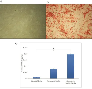
mesenchymal stem cell markers, and capability for adipogenic and osteogenic differentiation in 2D culture55. All three selected cell lines were also verified to deposit mineral over an area spanning at least 50% of the tissue culture well, determined via Alizarin red staining (Figure 1 a, b).
25 2.3.2 Validation of microarray data
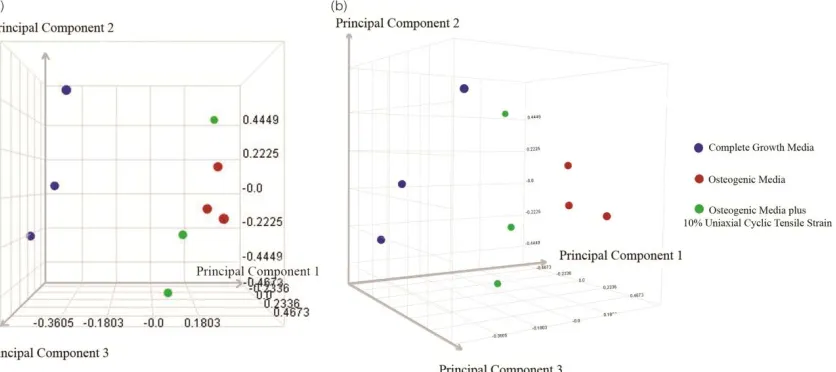
To characterize variability present in the microarray data, principal component analysis (PCA) was performed. PCA results showed discrimination between three
treatments: 1) hASC cultured in growth media; 2) hASC cultured in osteogenic media; and, 3) hASC cultured in osteogenic media in the presence of 10 % uniaxial cyclic tensile strain (Figure 2). As expected, variability was also found between human donors within each treatment. Osteogenic media alone and osteogenic media plus cyclic tensile strain exhibited gene expression profiles closer to each other than hASC cultured in growth media, since both of those populations were undergoing osteogenic differentiation (Figure 2).
Genes differentially expressed by hASC in response to 3D collagen culture in
osteogenic media or osteogenic media plus 10% uniaxial cyclic tensile strain were identified by error-weighted analysis of variance (ANOVA). 1288 gene identifiers were detected as osteogenic sensing and 184 gene identifiers were detected as cyclic tensile strain sensing. Of the previously reported top 20 upregulated genes during osteogenesis of hASC for 14 days in 2D culture 57, we found 43 relevant gene identifiers for 15 genes which were also upregulated during osteogenesis of hASC in 3D collagen I gel culture (Table 1). Four of our top five upregulated genes: alcohol dehydrogenase 1B (ADH1B), glycoprotein M6B (GPM6B), monoamine oxidase A (MAOA), and FK506 binding protein 5 (FKBP5) were also found by Liu et al. as top upregulated genes during osteogenic differentiation of hASC in 2D culture
57
26 results for corin by previous investigators during osteogenic differentiation of hMSC versus hASC when analyzing cell response in 2D culture63, and our current microarray finding that corin was highly upregulated by hASC in 3D culture, we confirmed corin expression using real time RT-PCR with analysis at a greater number of time points, as described later in the real-time RT-PCR analysis section.
Figure 2-2. Principal Component Analysis (PCA). PCA results indicated a distinct
27 2.3.3 Genes regulated during osteogenic differentiation of hASC in 3D collagen
culture for 14 days
Of the 1,288 gene identifiers that were significantly and differentially expressed by all three donors’ hASC in response to osteogenic differentiation medium, 1218 identifiers were mapped to 847 genes (Supplementary Table 1 and 2). 864 identifiers were eligible for network analysis with IPA; 832 identifiers were eligible for function and pathway analyses.
31 2.3.4 Genes regulated by hASC in 3D collagen culture in response to 10% uniaxial
cyclic tensile strain
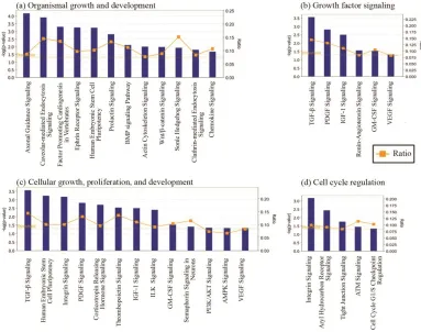
To identify the effect of 10% uniaxial cyclic tensile strain on hASC undergoing osteogenic differentiation, the gene expression profile of hASC cultured in osteogenic media plus 10% uniaxial cyclic tensile strain were compared to the gene expression profile of hASC cultured in osteogenic media alone (0% strain). 184 transcripts were modulated by 10% uniaxial cyclic tensile strain (Supplementary Tables 4 and 5). Of the 184 transcripts, 176 transcripts were mapped to 147 genes with IPA. 146 identifiers were eligible for network analysis and 135 identifiers were eligible for function and pathway analyses.
Cardiovascular system development and function was the top function when
categorizing the genes modulated by 10% uniaxial cyclic tensile strain (24 genes, p<10-7-10
-3
) with upregulation of 16 genes including fibroblast growth factor 2 (FGF2), IL1RN
(interleukin 1 receptor antagonist), matrix metalloproteinase 2 (MMP2), vascular endothelial growth factor A (VEGF A), and transforming growth factor beta receptor 1 (TGFBR1) (Table1).
32 scored at 41 (scores of 2 or higher have at least a 99% confidence of not being generated by random chance alone) with 22 focused molecules and 36 total molecules in the network (Figure 5 and Supplementary Table 7). Three focused molecules, IL1RN (interleukin 1 receptor antagonist), JUND (jun D proto-oncogene), and SOCS3 (suppressor of cytokine signaling 3) were located in the center of the network and linked with 5 or more other molecules.
33 indicates downregulated molecules. Color intensity indicates the level of expression. (see Supplementary Table 7 for full names and expression profile data).
2.3.5 Real-time RT-PCR analysis to validate results for corin, the typeII serine protease upregulated during osteogenesis of hASC, PDLIM4, an actin binding protein, and VEGF A, an angiogenic factor, upregulated in response to 10% uniaxial cyclic tensile strain in 3D culture.
34 Two genes upregulated in response to 10% cyclic tensile strain during osteogenesis of hASC were also validated with RT-PCR: 1) PDZ and LIM domain 4 (PDLIM4), one of the top five upregulated genes which has been shown to have polymorphisms found to associate with bone mineral density regulation 64,65; and, 2) VEGF A, a known angiogenic factor. Real time RT-PCR results confirmed that expression of both genes was significantly increased by hASC in response to 10% uniaxial cyclic tensile strain (Figure 6 b).
35 2.4 Discussion
Differentiation of mesenchymal progenitor cells into the osteoblastic lineage has been studied extensively in transgenic animal models66,67,67. While animal models provide an in vivo system to identify the pivotal role of specific factors, there are possibilities of
interspecies differences. In vitro studies on human cell lines are thus required to fully develop our knowledge for clinical application. Research on osteoblastic differentiation of human mesenchymal stem cells from multiple source tissues has been performed 68,69, but they have lacked incorporation of a three-dimensional, mechanically-loaded culture system that provides bone ECM-mimetic 3D culture and mechanical loading, and the global analysis to obtain insight into associated regulatory mechanisms of osteogenesis.
In the present study, collagen type I was used to mimic the organic ECM of bone and 10% uniaxial cyclic tensile strain was applied as an appropriate mechanical load to promote greater osteogenic differentiation and calcium accretion of hASC 70,52,71,54. Calcium
accretion data indicated that hASC differentiated down an osteogenic pathway by accreting calcium in 3D collagen culture when in the presence of soluble osteogenic inductive factors. 10% uniaxial cyclic tensile strain further increased calcium accretion of hASC (Figure 1c), as we have previously shown in 2D culture52. Global analysis of over 47,000 gene identifiers from microarray data revealed the canonical pathways involved with osteogenic
36 cells from cell cycle or self-renewal toward cell maturation with associated changes in cell growth, cell development, and cell morphology. Many canonical pathways affected by osteogenic media were also reported as signaling required for bone formation. After 14 days, common bone marker genes alkaline phosphatase (ALP), runt-related transcription factor-2 (RUNX2), and osteocalcin/ bone gamma-carboxyglutamate (gla) protein (BGLAP) were not significantly upregulated relative to culture in 3D type I collagen gels without soluble osteogenic inductive factors. The time point and culture system chosen likely caused this result, as we have found that endogenous ALP activity highly increases in hASC without chemical or mechanical stimuli after culture in linear 3D type I collagen gels for 10 days (Supplementary Figure 1). Further, it has been previously shown that when soluble osteogenic inductive factors are included in the culture media, three-dimensional collagen culture of hASC causes increased mRNA expression of bone markers RUNX2, ALP,
osteonectin, osteopontin, Collagen I, and JNK2/MAPK9 (mitogen-activated protein kinase 9) compared to hASC cultured in 2D monolayer on tissue culture plastic47.
37 direct induced modulation of platelet derived growth factor D (PDGF-D) and platelet-derived growth factor receptor, alpha polypeptide (PDGFRA). PDGF-D is a newly found member of the PDGF family 75. A recent study on PDGF-D function in transgenic mice showed that PDGF-D induces macrophage recruitment and blood vessel maturation during angiogenesis
75
.
Our data on IGF-1 mRNA expression indicated that neither the secreted factor (IGF-1) nor its receptor (IGF1R) was directly affected during osteogenic differentiation of hASC. However, IGF-1 signaling was affected with the activation of insulin receptor substrate 1 and 2 (IRS1 and IRS2) and its downstream cascades including PI3K. Secreted IGF-1 from osteoblasts has been reported to act as a chemotactic factor with its function potentially inhibited by phosphatidyl inositol 3 kinase (PI3K) inhibitor76. Both IRS-1 and IRS-2 are expressed in bone. IRS-1 is important for maintaining bone turnover (bone formation and bone resorption). IRS-1 expression is also required for skeletal growth and fracture healing
77,78, 79
. IRS-2 is not required for bone healing but plays a critical role in the coupling of bone resorption to bone formation 80,81. These findings suggest that IGF-1 signaling, even without the modulation of mRNA expression of IGF-1 itself, may play a critical role for osteogenic differentiation of hASC.
38 Interestingly, another pathway found to be downregulated was the antigen
presentation pathway with the suppressed expression of major histocompatibility complex – A,-B,-C,-G (HLA-A,-B,-C,-G), and calnexin (CANX). Donor-specific HLA antibodies have been associated with graft dysfunction and failure 84. A study on immunological properties of hASC and hMSC has shown low immunogenecity and suggested the potential application of these cell types for allogenic transplantation 85. Our data suggest that osteogenic induction also suppresses the immune characteristics of hASC further, indicating their potential use for allograft bone tissue engineering.
39 In addition to the pathway analysis, of interest was the gene corin, which was found to be upregulated by 100-fold but was not matched with any canonical pathways. Corin is a serine protease normally associated with the heart, where it has been found to help maintain blood pressure levels88. Corin has also been found to be expressed in developing bone near hypertrophic chondrocytes and in perichondrocytes 89. This latter report suggested that corin may be associated with chondrogenesis. Recently, corin was found to be relatively highly upregulated compared to other genes during osteogenic differentiation of hMSC, but not during osteogenic differentiation of hASC in 2D culture at days 3 and 14 57. Corin has not previously been studied in the context of its potential role in osteogenesis or chondrogenesis. In the present study, we have shown that corin may be involved with osteogenesis of hASC in 3D culture in response to soluble osteogenic inductive factors but not by 10% uniaxial cyclic tensile strain. Taken together with its presence in vivo, more studies on the role of corin in both chondrogenesis and osteogenesis might lead to a better understanding of its role in endochondral and potentially intramembranous bone formation.
Other genes found to be highly expressed during osteogenic differentiation of hASC in 3D collagen culture were glycoprotein M6B (GPM6B), monoamine oxidase A (MAOA), FK506 binding protein 5 (FKBP5), and zinc finger and BTB domain containing 16
(ZBTB16), which have also been found to be upregulated during osteogenic differentiation of hASC and hMSC after 14 days in 2D culture 57. Likewise, alcohol dehydrogenases may be involved in retinoic acid synthesis 90, which has been found to induce osteogenic
40 Finally, mechanical load, a critical factor in bone formation and resorption, was also found to significantly affect hASC during osteogenic differentiation. Application of 10% uniaxial cyclic tensile strain to hASC induced upregulation of chondroitin sulfate
biosynthesis and endoplasmic reticulum stress pathways. Depletion of heparan/ chondroitin sulfate in MSC has been reported to result in altered BMP and Wnt activity 92. Further, XBP1 and ATF4 from the ER stress pathway have been found to express and regulate the onset of osteoblast differentiation 93-95. This suggests that chondroitin sulfate biosynthesis and the ER stress pathway may play key roles in hASC response to 10% cyclic tensile strain.
Since many affected genes have not been fully studied, such as the mechanosensing genes of hASC in this study, our analysis had to be based on known function and canonical pathways which could conceivably bias the focus to some of the extensively studied genes. The network analysis was generated to identify key regulatory genes. The network analysis of genes differentially expressed by 10% uniaxial cyclic tensile strain showed that interleukin 1 receptor antagonist (IL1RN), and suppressor of cytokine signaling 3 (SOCS3) centered in the first rank network. Interestingly, both of these genes are inhibitors of inflammatory signaling. IL1RN is a competitive inhibitor of interleukin-1 (IL-1), and has been shown to reduce osteoclast formation, bone loss, and bone resorption in estrogen deficient
(ovariectomized) mice 96-98. SOCS3 negatively regulates IL-6, and mRNA expression of SOCS3 is stabilized by tumor necrosis factor α (TNF-α) 97,99,100
41 IL-1 and IL-6 genes. A previous study on the role of proinflammatory cytokines during bone fracture healing showed that the expression of IL-1 and IL-6 is temporal, with peaks at 24 hours and 7 days 102. As we also found one of the top classified biofunctions was
cardiovascular development with the upregulation of many genes that promoted angiogenesis such as fibroblast growth factor 2 (FGF2), matrix metalloproteinase 2 (MMP2), and vascular endothelial growth factor A (VEGF A), this suggests that 10% uniaxial cyclic tensile strain may induce angiogenesis in hASC, possibly through the activation of IL-1 and IL-6, thus requiring IL1RN and SOCS3 to negatively control the temporal expression of IL-1 and IL-6.
In summary, our data suggest that after 14 days of osteogenic differentiation in 3D collagen I gels in the absence of cyclic tensile strain, hASC downregulated TGF-β signaling, Wnt/β-Catenin signaling, and BMP-signaling through some of their inhibitor molecules. Further, osteogenic differentiation of hASC in 3D collagen culture elevated the expression of some genes, e.g. corin, in a distinctly different response from a previous analysis of hASC osteogenic differentiation in 2D culture 57. Pathway analysis with non-overlapping results by the addition of 10% cyclic tensile strain relative to the effects of osteogenic differentiation media alone suggested that increased calcium accretion by hASC in response to mechanical loading is associated with a different set of genes than those affected by soluble osteogenic inductive factors. Of particular interest was the upregulation of PDZ and LIM domain 4 (PDLIM4), one of the top five upregulated genes which has been shown to have
42 bone regeneration: 1) proinflammatory cytokine regulators IL1RN and SOCS3; and, 2) angiogenic inductors FGF2, MMP2, and VEGF A.
In conclusion, this is the first study to investigate the effects of both 3D collagen culture and 10% uniaxial cyclic tensile strain on hASC osteogenic differentiation. A complete microarray analysis investigating both the separate effect of soluble osteogenic inductive factors, and the combined effects of chemical and mechanical stimulation was performed on hASC undergoing osteogenic differentiation. We have identified specific genes and pathways associated with mechanical response and osteogenic potential of hASC, thus providing significant information toward improved understanding of our use of hASC for functional bone tissue engineering applications.
Acknowledgements:
43 2.5 Supplementary Data
Supplementary Table 2-1: Genes upregulated by hASC in 3D collagen culture in response to osteogenic induction media.
Symbol Entrez Gene Name Fold
Change p-value
ADH1B alcohol dehydrogenase 1B (class I), beta polypeptide 100 1.32E-09
CORIN corin, serine peptidase 100 3.46E-10
GPM6B glycoprotein M6B 100 6.07E-12
MAOA monoamine oxidase A 100 6.07E-12
FKBP5 FK506 binding protein 5 81.612 6.07E-12
ZBTB16 zinc finger and BTB domain containing 16 70.665 6.07E-12
GALNTL2 UDP-N-acetyl-alpha-D-galactosamine:polypeptide N-acetylgalactosaminyltransferase-like 2 52.185 6.07E-12
FAM107A family with sequence similarity 107, member A 48.251 6.07E-12
PRELP proline/arginine-rich end leucine-rich repeat protein 46.755 4.09E-06
RERG RAS-like, estrogen-regulated, growth inhibitor 41.131 6.07E-12
RTN1 reticulon 1 40.335 5.30E-04
NEBL nebulette 40.084 4.39E-09
ZNF385B zinc finger protein 385B 36.828 6.07E-12
CD36 CD36 molecule (thrombospondin receptor) 36.21 5.35E-03
LMO3 LIM domain only 3 (rhombotin-like 2) 34.837 6.07E-12
STMN2 stathmin-like 2 33.972 6.07E-12
PPL periplakin 31.538 6.07E-12
CNR1 cannabinoid receptor 1 (brain) 29.986 9.80E-04
GLUL glutamate-ammonia ligase (glutamine synthetase) 26.818 6.07E-12
EFNB2 ephrin-B2 26.296 9.58E-06
PDK4 pyruvate dehydrogenase kinase, isozyme 4 26.223 6.07E-12
INHBB inhibin, beta B 24.706 1.21E-11
PTK2B PTK2B protein tyrosine kinase 2 beta 24.227 6.07E-12
AOX1 aldehyde oxidase 1 23.088 6.07E-12
GABBR2 gamma-aminobutyric acid (GABA) B receptor, 2 22.935 6.00E-05
SORBS1 sorbin and SH3 domain containing 1 21.679 6.07E-12
CFD complement factor D (adipsin) 21.341 6.07E-12
GPX3 glutathione peroxidase 3 (plasma) 19.242 6.07E-12
PPP1R14A protein phosphatase 1, regulatory (inhibitor) subunit 14A 19.201 8.30E-04 NPR1 natriuretic peptide receptor A/guanylate cyclase A (atrionatriuretic peptide receptor A) 17.775 4.02E-06
SCARA5 scavenger receptor class A, member 5 (putative) 17.051 7.45E-08
STC1 stanniocalcin 1 16.305 6.07E-12
MCTP1 multiple C2 domains, transmembrane 1 15.027 6.07E-12
MT1M metallothionein 1M 14.57 4.13E-03
CCDC68 coiled-coil domain containing 68 14.453 6.85E-08
CIDEC cell death-inducing DFFA-like effector c 13.933 6.07E-12
C10ORF10 chromosome 10 open reading frame 10 13.078 6.07E-12
CEBPA CCAAT/enhancer binding protein (C/EBP), alpha 12.856 1.94E-03
CHRDL1 chordin-like 1 12.676 6.90E-08
44 Supplementary Table 2-1: continued
NOG noggin 11.83 5.50E-08
FOXO1 forkhead box O1 11.81 6.07E-12
PRUNE2 prune homolog 2 (Drosophila) 10.774 6.07E-12
GHR growth hormone receptor 10.486 6.07E-12
FGD4 FYVE, RhoGEF and PH domain containing 4 10.344 6.07E-12
SORT1 sortilin 1 10.338 6.07E-12
EDNRB endothelin receptor type B 10.271 4.79E-08
PRKAR2B protein kinase, cAMP-dependent, regulatory, type II, beta 9.813 2.06E-10 SLC7A2 solute carrier family 7 (cationic amino acid transporter, y+ system), member 2 9.599 1.70E-04
METTL7A methyltransferase like 7A 9.52 6.07E-12
MAMDC2 MAM domain containing 2 9.493 9.71E-11
C13ORF15 chromosome 13 open reading frame 15 9.441 6.07E-12
NRCAM neuronal cell adhesion molecule 9.404 8.30E-04
APCDD1 adenomatosis polyposis coli down-regulated 1 9.287 2.10E-04
NEDD9 neural precursor cell expressed, developmentally down-regulated 9 9.135 5.54E-07
MYO16 myosin XVI 9.133 3.00E-05
TIMP4 TIMP metallopeptidase inhibitor 4 9.086 4.80E-04
CCDC69 coiled-coil domain containing 69 8.89 6.07E-12
GGT5 gamma-glutamyltransferase 5 8.86 5.89E-10
LEPR leptin receptor 8.063 6.07E-12
MGP matrix Gla protein 7.716 5.00E-05
MOBKL2B MOB1, Mps One Binder kinase activator-like 2B (yeast) 7.693 6.07E-12
AFAP1L1 actin filament associated protein 1-like 1 7.666 6.07E-12
GPD1L glycerol-3-phosphate dehydrogenase 1-like 7.402 6.07E-12
NAV2 neuron navigator 2 7.13 6.07E-12
FMO2 flavin containing monooxygenase 2 (non-functional) 7.1 6.07E-12
SOX13 SRY (sex determining region Y)-box 13 7.085 2.63E-03
VIT vitrin 7.017 5.58E-07
ADH1C alcohol dehydrogenase 1C (class I), gamma polypeptide 6.987 8.89E-03
PELO pelota homolog (Drosophila) 6.958 4.25E-11
TJP2 tight junction protein 2 (zona occludens 2) 6.843 6.07E-12
MMP7 matrix metallopeptidase 7 (matrilysin, uterine) 6.74 2.56E-03
NETO2 neuropilin (NRP) and tolloid (TLL)-like 2 6.719 2.00E-05
HSPA2 heat shock 70kDa protein 2 6.626 1.03E-10
PER1 period homolog 1 (Drosophila) 6.347 2.63E-09
FAM65B family with sequence similarity 65, member B 6.315 5.85E-06
SYNE2 spectrin repeat containing, nuclear envelope 2 6.032 6.07E-12
B3GNT5 UDP-GlcNAc:betaGal beta-1,3-N-acetylglucosaminyltransferase 5 5.864 5.91E-06
S1PR3 sphingosine-1-phosphate receptor 3 5.813 6.41E-07
USP53 ubiquitin specific peptidase 53 5.812 1.00E-05
45 Supplementary Table 2-1: continued
DUSP10 dual specificity phosphatase 10 5.768 3.53E-06
CD1D CD1d molecule 5.765 6.30E-04
ADAMTS5 ADAM metallopeptidase with thrombospondin type 1 motif, 5 5.706 6.07E-12 PTGS1 prostaglandin-endoperoxide synthase 1 (prostaglandin G/H synthase and cyclooxygenase) 5.56 1.52E-10
MYPN myopalladin 5.531 2.24E-06
RAMP1 receptor (G protein-coupled) activity modifying protein 1 5.472 2.00E-05
TSC22D3 TSC22 domain family, member 3 5.4 6.07E-12
ANGPT1 angiopoietin 1 5.266 4.55E-09
TMEFF2 transmembrane protein with EGF-like and two follistatin-like domains 2 5.2 2.57E-06
RNF144B ring finger protein 144B 5.164 1.00E-05
RRM2 ribonucleotide reductase M2 5.133 6.07E-12
SLC40A1 solute carrier family 40 (iron-regulated transporter), member 1 5.074 8.00E-05
OPN3 opsin 3 4.992 6.07E-12
MITF microphthalmia-associated transcription factor 4.956 6.07E-12
NEGR1 neuronal growth regulator 1 4.952 2.43E-11
SPRY1 sprouty homolog 1, antagonist of FGF signaling (Drosophila) 4.911 1.50E-04
KIAA0146 KIAA0146 4.86 5.28E-06
SEPP1 selenoprotein P, plasma, 1 4.851 1.00E-05
OLFML2A olfactomedin-like 2A 4.787 7.30E-04
ARHGAP6 Rho GTPase activating protein 6 4.698 2.00E-05
MRVI1 murine retrovirus integration site 1 homolog 4.695 1.80E-04
SYTL4 synaptotagmin-like 4 4.683 2.90E-03
MMD monocyte to macrophage differentiation-associated 4.587 6.07E-12
LAMA2 laminin, alpha 2 4.583 6.07E-12
C1ORF21 chromosome 1 open reading frame 21 4.568 5.15E-08
AURKA aurora kinase A 4.511 2.40E-04
PPARG peroxisome proliferator-activated receptor gamma 4.488 3.19E-09
ITGA10 integrin, alpha 10 4.476 2.52E-07
CCBE1 collagen and calcium binding EGF domains 1 4.47 1.21E-11
REPS2 RALBP1 associated Eps domain containing 2 4.458 1.21E-11
MCAM melanoma cell adhesion molecule 4.439 9.10E-04
MTSS1 metastasis suppressor 1 4.397 1.31E-07
LMOD1 leiomodin 1 (smooth muscle) 4.392 1.27E-10
MLPH melanophilin 4.367 6.07E-12
HIPK2 homeodomain interacting protein kinase 2 4.35 1.00E-05
PDE7B phosphodiesterase 7B 4.299 1.21E-11
CDC2 cell division cycle 2, G1 to S and G2 to M 4.292 1.82E-11
MT1F metallothionein 1F 4.292 4.24E-06
ADRA2C adrenergic, alpha-2C-, receptor 4.284 5.09E-03
SETMAR SET domain and mariner transposase fusion gene 4.28 1.40E-10
46 Supplementary Table 2-1: continued
BIRC5 baculoviral IAP repeat-containing 5 4.267 6.60E-04
CUGBP2 CUG triplet repeat, RNA binding protein 2 4.25 5.80E-04
OSR2 odd-skipped related 2 (Drosophila) 4.23 1.07E-07
CAMK2N1 calcium/calmodulin-dependent protein kinase II inhibitor 1 4.154 6.20E-09
TGFBR3 transforming growth factor, beta receptor III 4.152 6.07E-12
ABAT 4-aminobutyrate aminotransferase 4.114 1.00E-05
NR2F1 nuclear receptor subfamily 2, group F, member 1 4.093 6.07E-12
TBC1D8 TBC1 domain family, member 8 (with GRAM domain) 4.029 6.07E-12
KCNT2 potassium channel, subfamily T, member 2 4.016 2.21E-03
CFH complement factor H 4.008 6.07E-12
ITPR1 inositol 1,4,5-triphosphate receptor, type 1 3.959 6.07E-12
DKK1 dickkopf homolog 1 (Xenopus laevis) 3.932 6.07E-12
CEBPD CCAAT/enhancer binding protein (C/EBP), delta 3.904 1.09E-07
ENDOD1 endonuclease domain containing 1 3.859 2.43E-11
ADM adrenomedullin 3.837 2.67E-08
ZNF367 zinc finger protein 367 3.824 5.00E-05
ZFP36 zinc finger protein 36, C3H type, homolog (mouse) 3.819 6.07E-12
SLC27A3 solute carrier family 27 (fatty acid transporter), member 3 3.807 1.21E-11
LASS6 LAG1 homolog, ceramide synthase 6 3.773 6.07E-12
ACACB acetyl-Coenzyme A carboxylase beta 3.755 1.72E-06
TFPI tissue factor pathway inhibitor (lipoprotein-associated coagulation inhibitor) 3.742 5.00E-05 RASSF4 Ras association (RalGDS/AF-6) domain family member 4 3.709 1.57E-03
CRYAB crystallin, alpha B 3.697 6.07E-12
OSBPL5 oxysterol binding protein-like 5 3.694 3.71E-08
MT1E metallothionein 1E 3.676 3.84E-06
AHCY adenosylhomocysteinase 3.65 6.07E-12
SLC44A1 solute carrier family 44, member 1 3.637 6.07E-12
ADAMTS1 ADAM metallopeptidase with thrombospondin type 1 motif, 1 3.632 6.07E-12
MT1H metallothionein 1H 3.618 3.98E-09
CD302 CD302 molecule 3.612 6.07E-12
ITGA9 integrin, alpha 9 3.594 2.80E-04
ANGPTL4 angiopoietin-like 4 3.587 6.73E-03
ADARB1 adenosine deaminase, RNA-specific, B1 (RED1 homolog rat) 3.546 2.67E-10
C5ORF30 chromosome 5 open reading frame 30 3.502 4.81E-03
PIK3R1 phosphoinositide-3-kinase, regulatory subunit 1 (alpha) 3.499 3.89E-07
ABCA6 ATP-binding cassette, sub-family A (ABC1), member 6 3.475 1.00E-05
SESTD1 SEC14 and spectrin domains 1 3.461 1.44E-07
TOP2A topoisomerase (DNA) II alpha 170kDa 3.449 8.34E-03
SVEP1 sushi, von Willebrand factor type A, EGF and pentraxin domain containing 1 3.434 4.88E-08
PDGFD platelet derived growth factor D 3.418 6.07E-12
47 Supplementary Table 2-1: continued
PCDH9 protocadherin 9 3.375 6.22E-07
MT1X metallothionein 1X 3.365 9.17E-10
SMPDL3A sphingomyelin phosphodiesterase, acid-like 3A 3.335 2.73E-10
KLF6 Kruppel-like factor 6 3.33 6.07E-12
GSN gelsolin (amyloidosis, Finnish type) 3.315 6.07E-12
ATP2B4 ATPase, Ca++ transporting, plasma membrane 4 3.311 5.98E-06
CNKSR3 CNKSR family member 3 3.279 6.07E-12
NFIA nuclear factor I/A 3.232 3.16E-10
PHF17 PHD finger protein 17 3.229 6.07E-12
DIAPH2 diaphanous homolog 2 (Drosophila) 3.228 6.07E-12
ALCAM activated leukocyte cell adhesion molecule 3.224 6.07E-12
AMPH amphiphysin 3.204 3.54E-08
PRC1 protein regulator of cytokinesis 1 3.194 3.19E-03
IFI44L interferon-induced protein 44-like 3.151 2.00E-03
WNT5B wingless-type MMTV integration site family, member 5B 3.151 8.08E-09
APOE apolipoprotein E 3.15 4.60E-04
FAM167A family with sequence similarity 167, member A 3.148 3.30E-04
EFHD1 EF-hand domain family, member D1 3.133 4.58E-08
HIP1R huntingtin interacting protein 1 related 3.131 1.89E-03
COL7A1 collagen, type VII, alpha 1 3.104 1.33E-06
ELL2 elongation factor, RNA polymerase II, 2 3.086 6.07E-12
TFPI2 tissue factor pathway inhibitor 2 3.081 2.51E-03
STK17B serine/threonine kinase 17b 3.08 6.07E-12
DYNC1I1 dynein, cytoplasmic 1, intermediate chain 1 3.06 7.00E-04
ANKRD28 ankyrin repeat domain 28 3.045 6.07E-12
PRR16 proline rich 16 3.035 6.07E-12
TRIM35 tripartite motif-containing 35 3.003 2.25E-10
PDLIM1 PDZ and LIM domain 1 2.999 3.00E-05
PTGER4 prostaglandin E receptor 4 (subtype EP4) 2.989 6.07E-12
FANCI Fanconi anemia, complementation group I 2.983 6.34E-06
PHF15 PHD finger protein 15 2.982 3.35E-09
MT2A metallothionein 2A 2.981 6.07E-12
TBC1D1 TBC1 (tre-2/USP6, BUB2, cdc16) domain family, member 1 2.95 7.22E-03
NEXN nexilin (F actin binding protein) 2.936 3.71E-07
IRS2 insulin receptor substrate 2 2.935 3.42E-03
CPPED1 calcineurin-like phosphoesterase domain containing 1 2.918 5.53E-06
TCEAL1 transcription elongation factor A (SII)-like 1 2.916 6.07E-12
ALDH3A2 aldehyde dehydrogenase 3 family, member A2 2.916 6.07E-12
PRKD1 protein kinase D1 2.891 6.07E-12
TXNIP thioredoxin interacting protein 2.885 3.20E-04
AR androgen receptor 2.883 8.00E-04
NUSAP1 nucleolar and spindle associated protein 1 2.877 3.00E-05