Visual control of posture:
the role of motion parallax and cognitive processes
by Michel Guerraz
MRC Human Movement and Balance Unit, Institute of Neurology,
National Hospital for Neurology and Neurosurgery, Queen Square, London.
Thesis submitted for the degree of PhD, in the Faculty of Science of the University of London.
ProQuest Number: U642916
All rights reserved
INFORMATION TO ALL USERS
The quality of this reproduction is dependent upon the quality of the copy submitted.
In the unlikely event that the author did not send a complete manuscript and there are missing pages, these will be noted. Also, if material had to be removed,
a note will indicate the deletion.
uest.
ProQuest U642916
Published by ProQuest LLC(2016). Copyright of the Dissertation is held by the Author.
All rights reserved.
This work is protected against unauthorized copying under Title 17, United States Code. Microform Edition © ProQuest LLC.
ProQuest LLC
789 East Eisenhower Parkway P.O. Box 1346
ABSTRACT
The thesis investigates visual control of posture in man, in particular the role of visual motion parallax. The moving room paradigm, which simulates self-motion, was used in most experiments. In the first series of experiments the effects of room motion in the presence or absence of a foreground fixation target were investigated. Directionally specific postural responses opposite to background motion were observed when subjects were fixating the foreground target. Neither induced motion, ocular convergence nor binocular vision were sufficient to evoke such responses. It was concluded that such postural response and more generally postural control could be influenced by motion parallax. In experiments carried out in a normal stationary room, it was found that the motion parallax a subject generates during spontaneous body sway is used by the visuo-motor system to control body oscillations. The parallax-based effect was, however, limited to low frequency components of body sway (<0.75 Hz).
The influence of cognitive processes in visuo-postural control was also investigated. It was found that the predictability of the forthcoming displacement of a moving visual scene helped observers distinguish visual flow due to self-motion from that due to object-motion and therefore inhibit postural readjustments induced by object motion.
ACKNOWLEDGMENTS:
Publications incorporated in this thesis
Guerraz M, Gianna C, Burchill P, Gresty MA, Bronstein AM. (in press). Effect of visual surrounding motion on body sway in a 3 dimensional environment.
Perception & Psychophysics.
Guerraz M, Thilo KV, Bronstein AM, Gresty MA. (in press) Influence of action and expectation on visual control of posture. Cognitive Brain Research.
Guerraz M, Shallo-Hoffmann J, Yarrow K, Thilo KV, Bronstein AM, Gresty MA. (2000) Visual control of postural orientation and equilibrium in congenital nystagmus. Investigative Ophthalmology & Visual Science, 41:3798-804.
Guerraz M, Sakellari V, Burchill P, Bronstein AM. (2000) Influence of motion parallax in the control of spontaneous body sway. Experimental Brain Research, 131:244-52.
Other relevant publications
Guerraz M, Yardley L, Bertholon P, Poliak L, Gresty MA, Bronstein AM. (submitted) Visual vertigo: Symptom-assessment, spatial orientation and postural control.
Bronstein A, Guerraz M. (1999) Visual-vestibular control of posture and gait: Physiological mechanisms and disorders. Current Opinion in Neurology, 12, 5-
Table of contents
Title page 1
Abstract 2
Acknowledgments 3
Publications incorporated in this thesis 4
Other relevant publications 4
Tables of contents 5
List of tables 10
Abbreviations 11
Chapter 1
General introduction
A. Multi-sensory control of posture 12
B. Role of visual information in postural control 15
1. Visual control of spontaneous body sway 16
2. Visually induced body sway: the moving room paradigm 17
C. Physio-geometrical aspects of visuo-postural control 20
1. Effect of head, trunk and gaze orientation 20
2. Static versus dynamic visual information. 20
3. Functional specialization of the peripheral retina ? 21
4. Structure of the visual flow 23
D. Motion parallax and spatial orientation 24
1. Definition and geometrical aspects of motion parallax 24
2. Motion parallax as a cue for depth perception. 27
3. Motion parallax as a cue for spatial orientation and postural control. 28
Chapter 2
Motion parallax and visually induced body sway
A. Abstract 31
B. Introduction 32
C. General method 34
1. Apparatus 34
2. Procedure 34
3. Data analysis 36
D. Experiment 1; Transient versus sustained response 37
1. Method 37
2. Results and discussion 38
E. Experiment 2; Induced movement 46
1. Method 46
2. Results and discussion 47
F. Experiment 3: Ocular convergence 51
1. Method 52
2. Results and discussion 52
G. Experiment 4: Monocular versus binocular fixation 56
1. Method 56
2. Results and discussion 57
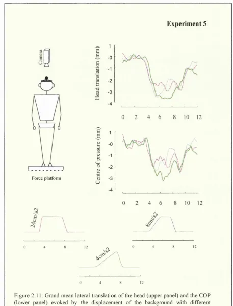
H. Experiment 5: Background acceleration 58
1. Method 59
2. Results and discussion 60
I. Discussion 63
1. Motion parallax as a relevant cue for postural control 63
2. Directionally specific response to an ecological stimulus. 64
3. A peripheral dominance of postural control ? 65
4. About the nature of the initial body response. 66
Chapter 3
Motion parallax and spontaneous body sway
A. Abstract 68
B. Introduction 69
C. General method 71
1. Apparatus 71
2. Procedure 71
3. Parameters of body sway 72
D. Experiment 1: Motion parallax and spontaneous body sway 73
1. Method 73
2. Results 74
E. Experiment 2: Monocular versus binocular vision 80
1. Method 80
2. Results 81
F. Discussion 84
1. Afferent and efferent visual control of body sway 85
2. Motion parallax affects only slow body re-orientation 88
Chapter 4
Influence of action and predictability on visual control of posture
A. Abstract 89
B. Introduction 90
C. General method 92
1. Subjects 92
2. Apparatus 92
3. Procedure 93
D. Experiment 1; Effect of action 95
1. Procedure and visual conditions 95
2. Results 96
E. Experiment 2: Expectation versus action 99
1. Procedure and visual conditions 99
2. Results 100
F. Experiment 3: Directionally specific information 103
1. Procedure and visual conditions 103
2. Results 103
G. Discussion 106
Chapter 5
Visual control of postural orientation and stabilization
in congenital nystagmus
A. Abstract 110
B. Introduction 111
C. General method 113
1. Subjects 113
2. Postural recordings 114
3. Eye movement recording 114
D. Experiment 1: Visual Control of Equilibrium 115
1. Method 115
2. Results 116
E. Experiment 2: Visually Induced Body S^vay 121
1. Method 121
2. Results 123
Chapter 6
General discussion
1. Motion parallax is a relevant cue for postural control 132
2. Discrete visual cues or visual flow ? 133
3. Postural orientation and postural stabilization 135
4. Do we use motion parallax in natural environments ? 136
References 138
List of tables
Table 2.1: Mean induced head / COP response (average and standard deviation) in mm in the different visual conditions in Experiment 1.
Table 2.2 Mean induced head/COP response (average and standard deviation) in mm in the different visual conditions in Experiment 2.
Table 2.3: Mean head/COP induced response (average and standard deviation) in mm in the different visual conditions in Experiment 3.
Table 2.4: Mean head/COP induced response (average and standard deviation) in mm of the head and the COP in the different visual conditions in Experiment 4.
Table 2.5: Mean head/COP response and slopes (average and standard deviations) in mm in the three conditions of acceleration manipulated in Experiment 5.
Table 4.1: Mean head/COP lateral induced response (average and standard deviation) in mm of the head and the COP in the different visual conditions in Experiment 1.
Table 4.2: Mean lateral head/COP induced response (average and standard deviation) in mm of the head and the COP in the different visual conditions in Experiment 2. Table 4.3: Mean lateral head/COP induced response (average and standard deviation) in mm of the head and the COP in the different visual conditions in Experiment 3. Table 5.1. Clinical details of the nine subjects with CN who took part in Experiment 1 (Subjects 1-6) and 2 (Subjects 1-9).
ABBREVIATION
COP: Centre of foot pressure LED: Light-emitting diode EGG: Electro-oculography CN: Congenital nystagmus SD: Standard deviation SwP: Sway path length
Chapter 1 : Introduction
Chapter 1
General introduction
Posture refers to a complex assemblage of the multiple body segments relative to each other controlled by a neuromuscular system. The orientation of the body and the maintenance of the human erect posture with respect to vertical in both the frontal and sagittal plane is a primary constraint in a world where the effect of gravity must be taken into account. Nashner and Cordo (1981) and later Amblard et al. (1985) and Roll et al. (1988) have suggested that the human postural control system was in charge of two important behavioral goals: postural orientation and equilibrium. Postural orientation was defined by Horak and Macpherson (1996) as the relative positioning of the body segments with respect to each other and to the environment and postural equilibrium as the state in which all the forces acting on the body are balanced so that the body tends to stay in the desired position and orientation (static equilibrium) or to move in a controlled way (dynamic equilibrium). Although human upright stance is considered as being a state of ‘static’ equilibrium (in contrast to locomotion), the body is constantly moving, and therefore requires a dynamic control. The main frequency of spontaneous body oscillations is below 0.5 Hz, but higher components (up to 10-15 Hz) of sway also exist, particularly important for the lower parts of the body (ankles for instance) as opposed to higher segments such as the trunk and the head (Benda et al. 1994; Amblard et al. 1985).
A. Multi-sensory control of posture
Chapter 1 : In trod uction
postural orientation an d eq u ilib riu m involves the integration o f these m ultiple
sen so ry signals that specify in form ation about the position o f bo d y se g m e n ts relative
to each oth er an d to the su rro u n d in g e n vironm e nt. T h e m ain re cep to rs in v o lv e d in
postural control are re p re se n te d in F igure 1.1.
Semi-circular canals
Visual receptors
M u scu lar receptors
M u sc u lo -a itic u la r receptors
Oto iths
V e stib u la r receptors
C u ta n e o u s re ceptors
Fig. 1.1: The main sensory receptors involved in the perception o f self-m otion and postural control: there are vestibular, visual and propriosomesthetic receptors. The later include receptors into the m uscles (G olgi receptors and muscular spindles), into the skin (cutaneous receptors, sensitive to pressure and shears) and in the articulations (adapted from Berthoz
1997).
S o m a to se n so ry afférents include c utane ous, m u sc u lo -a rtic u la r an d m u scu lar
receptors. Since these re cep to rs are distributed th ro u g h o u t the w h o le body, they are
critical for d eterm in in g b o d y co nfiguration, i.e. the relation o f each seg m e n t relative
to the adjacent ones. In contrast, vestibular an d visual receptors are located on the
head that can m o v e in d e p e n d e n tly o f the trunk. T herefore, in o rd e r to contrib u te to
body c o n figuration o r orientation, vestibular and visual signals require the
Chapter 1 : Introduction
signals, the integration of the position of the eye in the head (Wolsley et al. 1996). Vision must also be distinguished from the vestibular and the propriosomesthetic senses in that visual flow carries information relative both to object-motion and self- motion. Such an ambiguity can however be resolved by reference to information from other sensory systems specifying body movement, therefore indicating body motion instead of self-motion. As we will see in the fifth chapter of this thesis, this ambiguity can also be suppressed by the knowledge or predictability of the spatio- temporal aspect of any displacement of the moving environment. Although the contribution of auditory cues are limited, they can also provide spatial information allowing a reduction of body sway (Easton et al. 1998). Due to the limited implication of the auditory system to postural control in human, this fourth sensory system will not be further considered in the present thesis.
Chapter 1 : Introduction
Stabilization against external disturbances. However, in some circumstances, such as when the vestibular system does not specify any head acceleration (as during whole body rotation at a constant velocity), the displacement of the visual environment can be erroneously interpreted as a displacement of the self, giving rise to the illusion of self-motion, also called vection (Held et al. 1975; Dichgans & Brandt 1978; Thilo et al. 1999, 2000) in addition to postural readjustments (see below).
Although the respective influence of visual, vestibular and somatosensory information on postural control has been the subject of extensive research, the way these sensory channels are fused remains poorly understood. As reported recently by Jeka al. (2000), these sensory channels do not work in a simple additive way. The task of understanding sensory integration becomes particularly difficult when one considers the ability of the nervous system to learn to ignore (to some extent !) some inaccurate information and focus mainly on those which are relevant to the task. This ability can be observed in response to repetitive exposure to disturbances. For instance, the postural strategy adopted in response to an abrupt displacement of the support surface (Maki & Whitelaw 1993) or to a displacement of the visual environment (Bronstein 1986) can be profoundly influenced by prior experience of such a disturbance. Although prior experience prevents from falls or inappropriate postural reactions, it remains that, in normal subjects, false information can still induce postural re-adjustments, well within the limit of stability. A perturbation deployed to any of the sensory systems involved in self-motion perception (visual, vestibular and proprioceptive systems) can induce postural readjustments. These observations indicate that the sensory disturbance is centrally interpreted as due to self-displacement, and requires a compensatory response. In this thesis, we will focus mainly on the role of visual information in the control of postural orientation and equilibrium in standing subjects.
B. Role of visual information in postural control
Chapter 1 : Introduction
with obtaining information about the layout of the environment and objects. Proprioception, on the other hand, which is necessary for controlling any activity, has been generally considered the exclusive domain of the mechano-receptors within the body. As early as the works of Travis (1945), Edwards (1946) but also Wapner and Witkin (1950) it appeared however, that body stability was improved by vision. Accordingly, Gibson (1958, 1966, 1979, Gibson et al. 1955) suggested that vision could be integrated in the overall function of proprioception. The visual cues, or
'propriospecific’ visual information (in Gibson’s terminology) about observer’s own bodily movement, could be obtained through the changing optic array at the eye. Deformation of the retinal image due to ego-motion or transposition of objects in the environment (called optic flow) is then not considered just as a nuisance but actually as a rich source of information concerning the world and the relation of the subject into this world (Gibson et al. 1958).
1. Visual control o f spontaneous b o d y sw a y
A way to understand the nature of the visual basis of postural control consists of modulating the physical visual parameters available and then examine the resultant spontaneous body sway. The most popular protocol is the Romberg test (Dichgans et al. 1976; Black et al. 1982). It involves the comparison of an individual’s spontaneous sway under eyes closed and eyes open. Under these conditions, a reduction of body oscillations with eyes open can be observed in most people, across the different frequency components of body sway (Dichgans et al.
1976; Lestienne et al. 1977).
Chapter 1: Introduction
significant postural instability (or even falls) with eyes closed, then visual stabilization becomes particularly effective.
Modulations of the physical and physiological visual parameters have shown that visual stabilization depends critically upon the characteristics of the visual scene. For instance, both lateral and fore-aft spontaneous body sway and eye-target distance are linearly related such that sway decreases with decreasing eye-object distance (Lee & Lishman 1975; Bles et al. 1980; Paulus et al. 1984, 1989). The explanation of this phenomenon is based on the simple geometric rule that the retinal flow of a viewed scene is greater the nearer the objects are to the eyes. Accordingly, Bles et al. (1980) have shown that the maximum eye-target distance at which vision is still efficient is twice as long when subjects are made unstable by using compliant surfaces (visual stabilization up to 5m) than when subjects are standing still on a rigid support (visual stabilization up to 2.5 m). Other physical and physiological parameters affect spontaneous sway (See Bronstein and Guerraz 1999): we note the object size and localization (Paulus et al. 1984; Nougier et al. 1997), binocular disparity (Fox 1990), visual motion (Amblard et al. 1985) but also visual acuity (Paulus et al. 1984) and spatial frequency (Kunkel et al. 1998).
Further insight into the importance of visual flow on postural control has been gained by manipulating artificial optical flow. The most popular paradigm refers to the moving room paradigm, designed by Lee and collaborators.
2. V isually induced b o d y sway: the m o vin g room paradigm
C hapter 1: In trod uction
abse n ce o f visual m otion, the influence o f vision on postural sw a y can be detected.
T h e result o f such ex p e rim en ts s h o w ed directionally specific postural response: in
re sp o n se to linear fo rw ard an d b ac k w a rd m otion o f the visual en v iro n m e n t, subjects
sw ay in the direction o f m otion (Lee and L ish m a n 1975; S toffre gen 1985, 1986;
B ro n stein 1986, B ronstein et al. 1990). Small d isp la c e m e n t o f the ro o m such as 3
m m fo r a v iew in g distance o f 30 cm (Lee & L ish m an 1975), o r 2.5 c m for a view ing
distance o f 2 m (Stoffregen 1986) are sufficient to in d u ce postural sway. T h ese
am p litu d e s are so small that they are generally not co n sc io u sly p erceiv ed by
subjects. T h u s the sw ay induction threshold is lo w er than that o f explicit m otion
perception.
A d ra w in g o f the m o v in g room p aradigm is pre sen ted in F ig u re 1.2. In this
picture, from B ertenthal et al. (1997), a child is dep icte d falling b a c k w a rd as a
function o f the ro o m m o v in g tow ards him. This re sp o n se is usually c o n sid e re d to
arise from a c o n s e q u e n c e o f a m is-inteipretation o f the visual flow as due to self-
m otion instead o f object-m otion. U nlike in early o r p re -w alk e rs (B ertenthal et al.
1997; L ee & A ro n so n 1974; Stoffregen et al. 1987), in adults the d isp la c e m e n t o f the
room e v o k e s on ly small body oscillations in phase with the ro o m , instead o f falls
(Lee & L ish m an 1975).
Fig. 1.2: Schematic drawing o f the moving room paradigm. Depicted inside the room is a child falling backward as a function o f the room moving towards him (from Bertenthal et al.
Chapter 1 : Introduction
In addition to the real moving room, a wide variety of moving visual stimuli have been employed to investigate the phenomenon of visually induced body sway. These include, tilting rooms (Bles et al. 1980), projected displays simulating a moving visual wall, tunnel, floor or ceiling (Lestienne et al. 1977; Fluckiger & Baumberger 1988; van Asten et al. 1988a; Gielen & van Asten 1990; Dijkstra et al. 1994) and visual roll rotations (Dichgans et al. 1972; Clément et al. 1985; Dijkstra et al. 1992). Consistently, it has been reported that in rhythmic motion of the visual scene (sinusoidal motion) with either a linear or a rotational stimulus, the largest effect of vision is observed at fairly low frequencies (~ 0.2 Hz) at which the body is phase locked with the moving scene. At higher frequencies (> 0.3 Hz), this locking is lost and the overall effect of visual motion is reduced (van Asten et al. 1988a.b; Dijkstra et al. 1994; Giese et al. 1996). The amplitude of postural sway, at least in the antero-posterior direction has been shown to be particularly affected by visual properties such as the density of texture (Lestienne et al. 1977; van Asten et al. 1988a) and the velocity of motion (Lestienne et al. 1977; Masson et al. 1995). Lestienne et al. reported that fore-aft sway induced by linear visual motion is linearly related to the logarithm of both the mean spatial frequency in the visual scene and the velocity of motion. However, these results have not been systematically replicated for oscillating motion (see van Asten et al. 1988a).
The directionally specific postural response induced by the 'moving room' demonstrate clearly that the overall changes of the retinal image (retinal flow) due to self-motion or object-motion provide a rich source of information, not only about the environment but also about the orientation of the subject in it. On the basis of these findings, Lee and Lishman (1975) completed Gibson’s theory, and added to the
Chapter 1 : Introduction
C. Physio-geometrical aspects of visuo-postural control
1. E ffect o f head, tru n k and gaze orientation
An important aspect of the visually induced postural response is that its direction does not depend on the orientation of the head on the body nor on the orientation of the body relative to the stimulated motion (Stoffregen 1985; Gielen & van Asten 1990; Wolsley et al. 1996; Thurrell et al. 2000). Wolsley et al. (1996) showed that when subjects were fixating the centre of a rotating disk, the main direction of body sway was always re-orientated to be parallel to the disc, whether both the head and body were parallel to the disc or not. Similar results were previously reported by Gielen et al. (1990) using a visual flow simulating displacements in a tunnel. These observations demonstrate that the signals of eye-in- orbit and head-on-trunk are well integrated in order to redirect visuo-motor commands to the appropriate postural muscles. Also, Gielen et al. (1990) reported that when subjects were looking slightly sideways to the simulated self-motion through a tunnel, their postural responses were re-oriented to be perpendicular to the direction of gaze instead of being parallel to the simulated motion.
2. Static versus dyna m ic visua l inform ation.
Although visual flow appears to be a rich source of information for postural control, visually induced body re-orientation has also been reported with purely static displays such as a static tilted room (Bles 1979 reported by Isableu 1999), or a tilted frame (Isableu et al. 1997, 1998, 1999). Sway amplitude caused by static visual disturbances is however of a smaller amplitude ( < 1 ° for lateral sway) than sway amplitude caused by dynamic visual disturbances.
Chapter 1 : Introduction
performances close to those observed in the absence of visual cues (Amblard & Crémieux 1976; Kapteyn et al. 1979; Amblard & Carblanc 1980; Paulus et al. 1984; Amblard et al. 1985; Isableu et al. 1998). Power spectral analysis of spontaneous sway indicates that the high frequency components of sway (>2Hz), particularly important at the ankle level (and COP), are drastically affected by stroboscopic illumination. In contrast, sway frequency components below 2 Hz, dominants for higher segments of the body (the trunk and the head), are partially preserved (Amblard et al. 1985). Amblard and collaborators concluded that discrete visual information (‘static cues’) is sufficient to control the upper part of the body, which has predominantly low-frequency dynamics. Visual motion cues ('dynamic cues') control oscillations of the lower part of the body which extend through a higher frequency range. In agreement with the model proposed by Amblard et al. (1985), slow re-orientation of the body induced by either the displacement of a moving scene (Kapteyn et al. 1979) or by a static tilted frame (Isableu et al. 1997, 1998) are preserved under stroboscopic light.
Overall, the experiments with stroboscopic light indicate that an optimal stabilization requires a continuous recording of the visual afferents. In contrast, visual flow may not be necessary for the control of postural orientation; discrete visual sampling, providing only visual displacements instead of visual flow, seems sufficient. However, this statement does not imply that when available, ‘motion cues’ are not used to control body orientation.
3. Functional specialization o f the peripheral retina ?
Chapter 1 : Introduction
longitudinal body axis (in yaw) can be elicited with circular visual stimuli (drum) but only if the diameter of the stimuli sustained more than 30° in diameter. Similar results have been observed both with roll vection (Held et al. 1975) and linear vection (Berthoz et al. 1975). However, this dominance of peripheral vision on illusory self-motion has been challenged, and several studies have shown that vection could be elicited in central vision (Andersen & Braunstein 1985; Post 1988). Post (1988) showed that the magnitude of vection was greater with a larger stimulus area, but differences disappeared when an equal area stimulus was presented either centrally or in the periphery of the visual field.
C hapter 1 : Introduction
4. Structure o f the visual flo w
An aspect often neg lec ted in investigations base d on the d ic h o to m y betw een,
peripheral and central vision is that the visual flow g e n e ra te d d u rin g self-m otion is
not un ifo rm th ro u g h o u t the visual field. As sh o w n in F ig u re 1.3, w h e n a subject is
lo co m o tin g o r sim ply sw aying back and forth in a 3D en v iro n m e n t, looking straight
at its extrem ity, a radial flow having a focus o f ex p a n sio n in the direction o f m otion
is projec ted in the ce n te r o f the retina. At the peripheral e d g e s o f the field o f view,
the visual flow is nearly parallel to the line o f m otion. T h is flow structure at the
p erip h e ry has been term e d lam ellar flow. It m ust be n o te d that the relationship
b etw e en the structure o f the visual flow and the area o f the retina is sim ply reversed
for lateral disp lace m e n ts o r w hen the o b serv er is lo o king in the direction
p e ip e n d ic u la r to motion.
(a)
L a m e lla r flow
Radial flow
/
(b)
i
Radialflow...
I
L am ellar flow
f
, n
Fig. 1.3: Representation o f the optical flow field generated by forward displacement through a 3 dim ensional environment (a) with the focus o f expansion in the direction o f sway grading into lamellar flow in the orthogonal direction (b).
T h e differential effect o f radial versus lam ellar flow has been investigated in
Chapter 1 : Introduction
vision (15-20°), both lamellar and radial flow can induce directionally specific antero-posterior body sway (Stoffregen 1985; Andersen & Dyre 1989). In contrast, in the retinal periphery, only the lamellar flow (horizontal flow) can provoke body sway (Stoffregen 1985). The interaction between the retinal location and the structure of the visual flow was interpreted by Stoffregen as reflecting a differential sensibility of the peripheral and central retina to the different structures of the visual flow.
Irrespective of retinal area, regulation of antero-posterior body sway is commonly considered as being mainly controlled by radial flow (expansion/ contraction) when the observer in looking in the direction of self-motion and by lamellar flow (pure translation) when looking in the direction orthogonal to self- motion (Stoffregen 1985; van Asten et al. 1988a). Conversely, when looking in the perpendicular direction of motion, lateral body sway is largely regulated on the basis of lamellar flow. Often neglected, a natural 3D environment provides a pattern of differential horizontal movements between elements in the field related to the displacement of the observer. These relative movements are referred to as motion parallax. Since lateral sway is supposed to be mainly controlled by lamellar cues, motion parallax might be a relevant cue involved in its regulation.
D. Motion parallax and spatial orientation
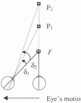
1. D efinition and geom etrical aspects o f m otion parallax
The term motion parallax or relative motion parallax is commonly used to denote the (horizontal) optical motion of a discontinuity produced by a near object against that produced by a far object due to a change in the observer’s position. (Fig. 1.4). It could also refer to change in the projective relations among elements in the visual field in condition of simulated motion while the observer is stationary and the scene moving. Motion parallax contains information about both the direction and the
C hapter 1: Introduction
to
Figure 1.4: Relative retinal displacements for an observer m oving from right to left, fixating an object F at an intermediate distance.
A s the o b se rv e r m o v es to the left while trac king an object F (fixation point),
the direction o f m otion o f the im ages o f other objects in the e n v iro n m e n t depends
on th e ir depth relative to the point o f fixation. It is im p o rtan t to note that the retinal
projection o f the fixation point {F) is not displaced on the retina d u rin g self-m otion;
it serves as an anchor. An object in the b ac k g ro u n d (b e h in d the fixation point)
appears to m o v e in the direction o f self-m otion; at tim e to, the retinal projection o f
the tree is on the right o f the line o f sight, at ry, it is to the left. R elative to the line o f
gaze, its im ag e has m o v e d to the left (Fig 1.4). An object in the fo reground, (in front
o f the fixation point), appears to m o v e in the o p p o site direction; at tim e to, the
im age o f the rose is on the left o f the line o f sight, at ry, it is to the right. Relative to
the line o f gaze, its im age has m o v e d to the right. T h e s e opp o site m otio n s o f the
b a c k g ro u n d -fo r e g ro u n d will be generated as long as the o b se rv e r d o es not look in
the direction o f his self-m otion but keeps looking at the fixation point. At som e
point b etw e en to and ? y , the point o f fixation F, the n ea r an d far o bjects are aligned
on the line o f sight, and therefore their retinal pro jec tio n s overlap. T h is point is
called the locus o f zero parallax.
T h u s, w hen the retinal im ages o f tw o objects m o v e opp o site to each other as a
result o f the o b s e r v e r ’s m otion, the o b serv er is fixating at so m e distan ce betw een the
tw o objects. T h e direction in w hich the retinal im ag e o f an ob ject m o v e s relative to
C hapter 1: Introduction
F u ith e r depth inform ation is gained from the m a g n itu d e o f d isp lace m e n t or
re lative an g u la r velocity o f the retinal im age o f the objects. O b jec ts o f w hich the
retinal im a g e s m o v e in the sam e direction as the line o f sight an d d isplay large retinal
disp la c e m e n ts (i.e. higher velocity) are farther from the fixation p o int than those
m o v in g in that direction but displaying sm aller d isp la c e m e n t (i.e. s lo w e r velocity); in
this case, both objects w o u ld be on the far side o f the fixation p o int (see Fig. 1.5).
O b jec ts o f w hich the retinal im ag e m oves in the o p posite direction to the o b s e r v e r’s
line o f sight an d display large retinal displacem ents, are c lo se r to the o b serv er than
those m o v in g in that direction but displaying sm a lle r disp la c e m e n ts; in this case,
both objects are on the n ear side o f the o b s e r v e r’s fixation point. T h u s, the extent o f
m otion parallax b etw een an object and a second object is proportional to the distance
in depth b etw een the two objects. In Figure 1.5 the angles 0i an d ôi represent the
relative disp lace m e n t o f objects P i and P2 re spectively on the re tina when an
ob se rv e r is fixating at point F w hile m o v in g from right to left. T h e d istance in depth
betw e en the o b se rv e r and the fixation point is also an im p o rtan t p a ra m e te r affecting
m otion parallax. Indeed, m otion parallax is proportional to the distan ce betw een the
different stimuli but inversely proportional to the d istance b etw e en the fixated object
and the observer.
E y e ’s m otion
Chapter 1 : Introduction
Fore-aft displacement of an observer in a 3D environment also generates motion parallax. It is maximum in the perpendicular direction and null in the direction of the displacement (heading). Far and close objects on the side of the heading will move in the same direction, opposite to the that of self-motion, with a velocity that is inversely proportional to the distance from the observer. Thus, the
magnitude of motion parallax during fore-aft displacement varies with the sway amplitude. However, as long as the observer is looking in the direction of self- motion, the direction of the retinal displacements of the objects is always in the opposite direction of motion. Thus, unlike lateral sway, fore-aft sway provides only variation in the magnitude of motion parallax, but no differential direction (as shown in Fig. 1.4 for lateral sway). This does not hold true when the subject is looking in the perpendicular direction of motion. For that reason, in this thesis, the role of motion parallax in the control of posture is investigated for lateral body sway instead of for ore-aft body sway.
Motion parallax must be distinguished from binocular disparity, also called binocular parallax. Binocular disparity and motion parallax are very different sorts of information about the 3 dimensional structure of objects and their layout. Lateral image disparity refers to the difference in the relative position of the visual images of objects on the two retina due to lateral separation of the eyes. Thus, binocular disparity is a binocular cue, essentially static whereas motion parallax is a dynamic monocular cue.
2. M o tion parallax as a cue fo r d ep th perception.
Chapter 1: Introduction
1995; Braunstein & Tittle 1988; Hayashibe 1993). Perception of the 3D structure of objects is, however, less ambiguous when the visual flow is generated actively, implying that extra-retinal infonnation also provides important cues (Wexler et al. 2001).
3. M otion parallax as a cue fo r spatial orientation and postu ra l control.
Gibson et al. (1955) pointed out the importance of optic flow patterns, including motion parallax, generated by observer movement as a source of information about the 3 dimensional layout of the environment but also as a rich source of information about the observer’s own movements. Accordingly, motion parallax has been shown to be an important cue in self motion perception and particularly to determine one’s direction of self-motion also called heading perception (Cutting et al. 1992; Frey & Owen 1999). During translation of an observer within a 3 dimensional environment, motion parallax occurs between elements at different distances: to the left of the heading, the images of nearer objects on the retina move to the left relative to those of more distant objects. To the right of the heading, the images of nearer objects move to the right relative to those of more distant objects. The images of objects that are collinear with the motion path remain visually aligned. This locus of zero parallax can therefore specify heading direction.
Chapter 1 : Introduction
flow comparable to what a standing subject would experience during spontaneous quiet lateral sway in a 3 dimensional environment. As shown in Figure 1.4, when an observer moves laterally while fixating a distant object, the images of nearer objects move in the opposite direction of the head. When the observer fixates a nearer object, the images of distant objects move in the same direction as the head. Therefore, postural adjustment in the opposite direction to background motion is consistent with the direction of image movement on the retina that a moving subject would experience in a stationary 3 dimensional environment. Motion parallax, i.e. the relative movement of images across the retina is useful in the determination of depth, perception of heading and during locomotion (Bardy, Warren & Kay 1996; Warren, Kay & Yilmaz 1996). It also appears to be a potential source of information for postural control.
E. Aim of the thesis
In the first experimental chapter (chapter 2), the contra-directional postural response observed by Bronstein and Buckwell (1997) in the foreground-background display was further investigated. Experiment 1 investigated whether this response was only a transient instability or could be a sustained repositioning of the body according to the visual reference. In the following experiments 2-4, alternative explanations to motion parallax were tested. Experiment 5 investigated whether this contra-directional response was sensitive to dynamic characteristics of the relative displacement of the background in relation to the foreground.
The third chapter (Experiment 1-2) reports an investigation on whether motion parallax generated by natural spontaneous body sway in a 3 dimensional environment could be used as a feedback by the postural control system to regulate spontaneous body sway.
Chapter 1: Introduction
giving the observer all the spatio-temporal characteristics of the forthcoming visual event (Experiment 1-3).
Chapter 2: Motion parallax and visually induced body sway
Chapter 2
Motion parallax and visually induced body sway
A. ABSTRACT
Chapter 2: Motion parallax and visually induced body sw ay
B. INTRODUCTION
The aim of Experiments 1-5 was to explore the contra-directional postural response observed by Bronstein and Buckwell (1997) in the foreground-background condition. In their experiment, these authors used a short duration stimulus of 2 sec with an overall displacement of the visual scene in the background of 1.4° of visual angle. Therefore the peak amplitude of the contra-directional re-adjustment observed in the foreground-background was as small as 1 mm at the level of the head and had a maximum duration of 1-2 sec. The aim of experiment 1 was to determine whether this contra-directional response in the foreground-background condition was only a transient phenomenon or whether it could be sustained for longer than l-2s. This would parallel findings on the co-directional antero-posterior sway observed with an uni-planar linearly moving visual environment (Lestienne et al. 1977) or a rotating disk (Dichgans et al. 1972).
Chapter 2: M otion parallax and visually induced body sway
contra-directional sway observed in the foreground-background display could be due to induced movement of the fixated foreground instead of motion parallax.
Experiment 3 investigated whether convergence of the eyes on the foreground target, in front of a moving background per se, independently of motion parallax could be responsible of the contra-directional response in the foreground- background display. To test this alternative explanation, we asked our subjects to fixate a close target coupled with head movement instead of earth fixed. As mentioned in introduction, the opposite motion of the foreground against the background is generated as long as the observer does not look in the direction of self-motion but keep looking at the fixation point. When the fixation point is coupled to head movement, subjects fixate in the direction of self-motion and so the opposite motion between foreground-background due to motion parallax does not occur.
Chapter 2: M otion parallax and visually induced body sway
C. GENERAL METHOD
1.
Apparatus
In Experiments 1-5, we recorded head and COP responses in the lateral direction for different visual conditions. Since the main goal of the study was to characterize the direction of visually evoked postural responses, discrete, unidirectional motion of a visual background was the chosen stimulus. The visual background consisted of a 2m x 3m flat board (67° x 90° of visual angle). Photoluminescent yellow-green stripes were used to create the picture of a house with one tree on each side (see Fig. 2.2). Background displacement was achieved by mounting the flat board on a chassis with four pneumatic wheels (bogie) running on a linear track. The bogie was driven by a pair of linear induction motors that generated thrust against a reaction plate situated along the middle of the track. The background was moved at 150 cm from the subject's eyes along an axis parallel to the inter-aural axis, in an otherwise dark room. In Experiments 1-4, the background was moved 58 cm (21°) leftwards or rightwards. A constant velocity of 6 cm/s was reached after approximately 1.25s of acceleration onset and sustained for 8.5s before the deceleration occurred. After each trial, the subjects were asked to close their eyes while the background was moved back to its starting position and re-illuminated with a lamp to keep a constant level of luminance. The experimental set-up can be seen in Figure 2.2. The velocity profile of background displacement was different in Experiment 5 and therefore will be defined in the method section of that experiment.
2.
Procedure
Chapter 2: M otion parallax and visually induced body sway
are measured by load cells, one located at each of the four corners of the platform. The recording of the movement of the body’s point of pressure to the foot support using a force measuring platform is a common method for studying body sway. However, the displacement of the COP is not only caused by a possible shift of the body’s point of gravity, but also by the acceleration and deceleration forces related to the movements of the inertial mass of the body. This dynamic component due to body inertia becomes particularly important with increasing body sway frequency (Gurfinkel 1973; Huffsmith et al. 1980). In addition, a shift of the COP can be caused by a movement of the standing subject as such, but also can be caused by a change in the configuration of the parts of the body in which case the measured shift is not representative for the subject sway. Thus, in addition to the COP, head sway was recorded throughout all the experiments presented in this thesis.
C hapter 2: M o tio n parallax and v isu a lly in d u ced b od y sw a y
3. D a ta a tta ly s is
O n each trial, c a lc u latio n s w ere m ade of: the m ean C O P /h e a d lateral p o sitio n
o v er the 4s b efo re stim u lu s o n set (m ean b ase lin e p o sitio n ) an d th e m ean lateral
p o sitio n o f C O P /h e a d o v er a 8.5s p eriod, from 1.25s to 9 .7 5 s afte r the o n set o f
stim u lu s m otion. T h e d iffe ren c e betw een these tw o m ean s is the ‘m ean am p litu d e o f
the C O P /h e a d re sp o n se o r ‘m ean resp o n se'; a m easu re o f how m u ch C O P /h e ad
d e v ia te s in re sp o n se to the stim u lu s (Fig. 2.1). T h e p e rio d o f 8.5s fro m 1.25s to 9.75s
c o rre sp o n d s to the p erio d o f co n stan t velo city o f stim u lu s m o tio n . S ince no
d iffe ren tial effect b etw een the tw o d irec tio n s o f m o tio n w as e x p e cted , re sp o n se s to
le ftw a rd and rig h w a rd stim uli w ere c o m b in ed by re v e rsin g re sp o n se s w ith leftw ard
stim u li. T h e in d u ced re sp o n se ca lc u lated fo r each trial w as then av e ra g e d fo r each
su b ject and visual con d itio n . O th er p aram eters o f bo d y sw ay , sp ec ific to a given
ex p e rim e n t, w ill be p re sen ted in the ap p ro p riate m eth o d s section.
a
a
c
20
§
CC
10
0
a
-10
OJ
Baseline
Mean position during stimulus motion
S tim u lu s m o tio n
Chapter 2: M otion parallax and visually induced body sway
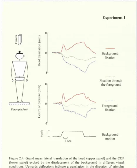
D. EXPERIMENT 1: Transient versus sustained response
1.
Method
1.1. Subjects
Twelve normal subjects (25-50 years old) with normal or corrected normal vision (normal visual acuity and stereopsis) gave their informed consent to participate in the experiment in accordance with the local ethics committee. All subjects were healthy without musculoskeletal or neurological disorders.
1.2. Visual Conditions
The three visual conditions to which each subject was exposed were:
1) Background fixation: The luminescent background was the only visible display in the otherwise completely dark room (see Fig. 2.2, left panel). The instruction given to subjects was: 'look straight ahead at the background but keep you eyes straight ahead (primary gaze position) and do not deliberately follow any part of the moving scene'\ This instruction was used throughout the experiments and for brevity is referred to as 'look straight ahead'.
2) Foreground fixation: Subjects had to fixate a cross ( 1 x 1 cm) placed at the centre of a purpose built earth fixed luminescent window frame (30 x 24 cm), 50 cm distant from the subject's eyes. This foreground was placed 100 cm in front of the background (see Fig. 2.2, right panel).
3) Fixation through the foreground: Subjects were asked to look straight ahead at the background through the 50 cm distant earth fixed window frame.
Each visual condition consisted of 24 trials, 8 with motion to the right, 8 to the left and 8 with stationary background as a control condition. The 24 trials were equally divided into two blocks of 12 pseudo-randomized stimuli. The first three blocks (one per visual condition), followed by the second three blocks, were presented in a
Chapter 2: M otion parallax and visually induced body sw ay
square design. A rest of 15 minutes was given to the subjects between the two test sessions.
An analysis of variance (ANOVA) was performed on the three experimental conditions and LSD post hoc test was used for pairwise comparisons. One sample t- tests were used to test whether the head/COP induced response in each visual condition is different from baseline level (zero) both for the group (based on the mean induced response) and for each individual subject (based on the induced response per trial). Finally, Spearman correlation coefficients were used to assess any relation between sway magnitudes for different conditions of visual fixation. A .05 significance level was adopted throughout.
2.
Results and discussion
Figure 2.2 shows a typical sample record of head and COP displacements during background and foreground fixation conditions. As can be seen in that figure, head and COP traces evolve in a relatively parallel manner although COP recordings contains higher frequency components of sway as compared to the head.
Background fixation
C hapter 2: M o tio n parallax and v isu a lly in d u ced b o d y sw a y
all in d iv id u a l averages w ere c o m b in ed (Fig. 2.4). T h e m ean slo p e o f the initial
p o stu ral re sp o n se (b ased on the reg ressio n line) c o m p u te d fo r each in d iv id u al subject
w as 6.4 m m /s fo r the C O P and 3.6 m m /s fo r the head. T h re e out o f the 12 subjects
sh o w e d no statistica lly sig n ifican t p ostural ad ju stm en t (p > .05). D a ta are p re sen ted in
T ab le 2.1.
Background fixation Foreground fixation
11 mm C
--H ead
6 cm/s
2 sec
V elocity ---^ <—>
p rofile 2 sec
7 mm
C O P
UA
Chapter 2: M otion parallax and visually induced body sway
Foreground fixation
During fixation of the foreground target, background motion induced a body sway in the opposite direction to motion as represented in the sample recording in Fig. 2.2 (right panel) and in the individual mean head translations in Fig. 2.3. The mean latency of the postural response was of circa 850 ms (Fig. 2.4). The initial postural displacement in the opposite direction to that of the visual stimulus was not followed by a sharp postural correction towards the baseline as in background fixation condition (Fig. 2.2). Instead, subjects continued to lean in the opposite direction to stimulus motion until it stopped. The average peak displacement in the opposite direction of motion was approximately 6 and 8 mm for the COP and the head respectively. 89% of responses in the foreground condition were in the opposite direction to stimulus motion. This postural effect was significant in all subjects except one. One sample t-test analysis confirmed that the displacement of both the COP and the head in the opposite direction to background motion were statistically significant (COP: r (ll) = 9.4 p<.01; head: r(ll) = 6.9 p<.01).
The slope of the initial postural response (regression line), measured for 600 ms Is after stimulus onset was less steep (COP: -3.01mm/s; head -2.6 mm/s) than in background fixation condition. Mean comparison based on the slope measured for each individual subject in both conditions indicated that the difference did reach significance for the COP (r(l 1) = 2.8 p<.05) but not for the head (r(l 1) = 1.5 p> .05).
Background fixation through the foreground
Chapter 2; M o tio n parallax and v isu a lly in d u ced b od y sw a y
15
10
£
£
c 0 1 5
c
I 0
s
X -5
2 sec
" : - i o
2 sec
X - 5
E xp erim en t 1
Background fixation
Foreground fixation
Fixation through the
foreground
2 sec
Chapter 2: M otion parallax and visually induced body sway
The ANOVA based on the mean induced response indicated that there was a significant effect of the visual condition both for the head (F 2,22= 16.4 p<.01) and
the COP (F 2,22= 24.2 p<.01). Post hoc mean comparison (LSD) indicated that the
three visual conditions differed from each other, both for the head and the COP (p<.05). In addition, the mean response in foreground and background fixation conditions were negatively correlated (Head: r =-0.60 p<.05; COP: r =-0.62 p<.05). Therefore, the more a subject deviated in the direction of motion during background fixation, the more this subject deviated in the opposite direction during foreground fixation. No correlation was observed between the condition of fixation through the foreground and the other two. No postural displacement was observed in the control condition, with stationary visual display (p>.05).
Two subjects reported spontaneously at the end of the experiment that in the foreground fixation condition they experienced the illusion they were moving with the earth-fixed in the opposite direction to background motion (vection). Therefore, both vection and self-displacement were in the same direction, both opposite to background motion. The implication of this anecdotal finding is discussed in the general discussion of the thesis.
Table 2.1: Mean induced head / COP response (average and standard deviation) in mm in the different visual conditions in Experiment 1.
Background Foreground Fixation through Stationary
fixation fixation the foreground background
M SD M SD M M SD
Head sway 3.41 4.01 -5.38 2.7 -2.42 3.07 0.27 0.61
COP 2.96 2.67 -4.01 1.63 -1.27 1.97 0.26 0.74
C hapter 2: M o tio n p arallax and v isu a lly in d u ced b o d y sw ay
E xp erim en t 1
Q
Force plalfonn
T3ce
<u X
£
¥
C/D £ o-'o£ s u
-8
-6
B a ck g ro u n d fix atio n
F ix a tio n through th e fo reg ro u n d
F o reg ro u n d fix atio n
1 [ _
/
\
2 sec
B a ck g ro u n d m o tio n
Chapter 2: Motion parallax and visually induced body sway
S u p p le m en tary analysis: body stab ility w ith statio n ary b ac k g ro u n d
B o d y stab ility in the lateral d irection w as m e a su re d w ith the sw a y p ath length
d u rin g the control trials fo r each o f the 3 co n d itio n s w ith statio n ary b a c k g ro u n d . T he
sw ay path (Sw P ) is the length o f the path d esc rib ed by the h ea d /C O P , an d is d efin e d
as the sum o f the d istan ces b etw een sequential p o in ts sam p led d u rin g the analy sis
p erio d (8.5s).
E q u atio n 1: SwP = ^ + 1 - j
D ata are p re sen ted in F ig u re 2.5. T h e A N O V A sh o w ed th at th ere w as
sig n ifican t d iffe ren c es betw een co n d itio n s (n= 3) both fo r the h ead ( ¥2,2 2= 7.6 p <.01)
and the C O P (¥2,2 2= 20.5 p <.01). M ean co m p ariso n s in d ic a te d th at th e sw ay path
length w as sh o rter w hen the fo reg ro u n d w as ad d ed in front o f th e b a c k g ro u n d
irre sp ectiv e o f the fixation point both for the head and the C O P (p < .0 5 ). T h e re w as
no d iffe ren c e betw een the fo reg ro u n d fixation and b ac k g ro u n d fix a tio n th ro u g h the
fo reg ro u n d . In terestin g ly , in the b ack g ro u n d fix atio n co n d itio n , the sw ay path length
w ith statio n ary b ack g ro u n d w as sig n ifican tly c o rre la te d (p < .0 1 ; S p earm an n
c o e ffic ie n t) w ith the slope o f the initial postural re sp o n se fo llo w in g the o n set o f
stim u lu s m otion (head: r= .72; C O P: r= .76). Such co rre la tio n w as n o t fo u n d fo r the
fo re g ro u n d con d itio n .
3
-9
a
I 5
Foreground Fixation Background fixation through the fixation
foreground
Foreground Fixation Background fixation through the fixation
foreground
Chapter 2: M otion parallax and visually induced body sway
In summary, Experiment 1 replicated the observation of Bronstein and Buckwell (1997) showing that by fixating a stationary foreground placed between the subject and a moving background, the postural response is reversed in direction. In addition, we showed that 1) this contra-directional postural response was not transient as it was sustained during the 11 s stimulus and 2) the amplitude of the postural responses induced by our visual display was bigger than that reported by Bronstein and Buckwell (1997). Indeed these authors reported a peak displacement of a magnitude of 1mm at the level of the head in both the condition of background and foreground fixation. In Experiment 1, we observed peak displacements in similar visual conditions of a magnitude of 6-8 mm. Two factors could explain this difference. First, the characteristics of the stimulus was rather different in the two studies. The displacement of the background used in Experiment 1 lasted longer, was faster and traveled a longer distance than that used previously by Bronstein and Buckwell (1997). Second, the stimulus in the present research consisted of the displacement of a real object while in the previous studies, the stimulus consisted of the displacement of a checkerboard projected onto a screen.
Chapter 2: M otion parallax and visually induced body sway
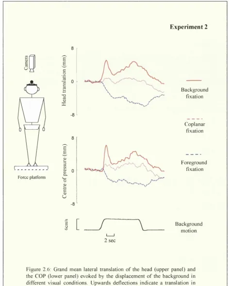
E. EXPERIMENT 2: Induced movement
The aim of experiment 2 was to investigate the respective role of motion parallax and induced movement in the contra-directional postural response observed in Experiment 1. While induced movement is most effectively elicited when induced and inducing stimulus are as close as possible in all three dimensions of space (adjacency principle: Gogel & koslow 1972; Gogel & MacCracken 1979; Reinhardt- Ruthland 1988), motion parallax increases with increasing distance between two objects. As a consequence, if the contra-directional sway observed in foreground fixation condition (Experiment 1) was elicited by induced movement of the foreground, it should be even greater and more consistent when the earth fixed foreground is coplanar with the moving background than when the foreground and background are at 100 cm from each other.
1.
Method
1.1 Subjects
Eight normal subjects (24-50 years of age) with normal or corrected normal vision gave their informed consent to participate in the experiment. Three out of these eight subjects took part in Experiment 1.
1.2. Visual Conditions
Parameters of background motion were the same as in Experiment 1. Subjects were exposed to three visual conditions:
1) Background fixation: Subject were asked to look straight ahead at the background as in the background fixation condition in Experiment 1.
Chapter 2: M otion parallax and visually induced body sway
cm from the background. In that visual environment, motion of the background or motion of the subject generate motion parallax.
3) Coplanar fixation: The subjects were asked to fixate an earth-fixed laser point projected directly onto the background. In that condition, motion of the background should induce an apparent movement of the earth fixed point in the opposite direction to background motion, but not motion parallax.
Each condition consisted of 21 trials, 7 with motion of the background to the right, 7 to the left and 7 with stationary background (control), divided in two blocks of 10 or 11 pseudo-randomized stimuli. The first three blocks (one per condition) followed by the second three blocks were presented in a Latin-square design. At the end of each block with the laser spot as a fixation point, subjects were asked to report whether they perceived this point as moving with respect to the background and in what direction. An analysis of variance (ANOVA) based on the mean induced response was performed on the three experimental conditions and LSD post hoc test was used for pairwise comparisons. A .05 significance level was adopted throughout.
2.
Results and discussion
Subjective reports
In the coplanar condition, 7 out of the 8 subjects tested reported to see either systematically or from time to time, the earth-fixed laser point moving in the opposite direction of background motion (induced movement). In the foreground fixation condition, only one subject reported to see the laser point as moving.
Postural re-adjustments
C hapter 2; M o tio n parallax and v isu a lly in d u ced b o d y sw a y
E xp erim en t 2
8
-Force plalfonn
£
B
c 0
1
0c ce
-8
-£ & e 3 c /5 C/5 £ 0
Cl
O s § U
-8
B ack g ro u n d fix atio n
C o p la n a r fix atio n
F o reg ro u n d fix atio n
I [
/
\
B ack g ro u n dm otio n
2 sec
F ig u re 2.6: G ran d m ean lateral tra n sla tio n o f th e h ea d (u p p e r p a n e l) and th e C O P (lo w e r pan el) e v o k e d by th e d isp lace m e n t o f th e b a c k g ro u n d in
C hapter 2: M o tio n parallax and v isu a lly in d u ced b od y sw a y
In the c o p la n a r co n d itio n , w hen the fix atio n p o in t w as p ro je c te d onto the
m o v in g scene, no co n sisten t sw ay w as ob serv ed ; 59 % o f the re a d ju stm e n ts w ere in
the d irec tio n o f m otion w h ile 41 % w ere in the o p p o site d irec tio n . W hen individual
d ata w ere co m b in ed , a sm all shift in the d irectio n o f m o tio n em erg es, (see Fig. 2-6)
b ut w as no t o b jectiv ated statistica lly (C O P; p = .1 2 ; H ead: t(l)= 0.8 p =.43).
M ean h ea d in d u ced re sp o n ses in both the co p la n a r an d th e fo re g ro u n d fixation are
p lo tte d in Fig. 2.7. In 7 out o f the 8 sub ject tested, the lateral d isp la c e m e n t o f the
head (an d C O P ) in the o p p o site d irectio n to m otion w as m o re im p o rta n t than in the
c o p la n a r co n d itio n . In terestin g ly , the su b ject fo r w hich th is w as no t true, had alread y
a te n d e n c y to lean in the o p p o site directio n to m otion in the c o p la n a r co n d itio n .
b - 3
-6
- 9
C o p l a n a r c o n d i t i o n F o r e g r o u n d f i x a t i o n
F ig u re 2.7: M ean in d u ced re sp o n se o f the head in both the co n d itio n o f co p lan a r fix atio n (in d u ced m otion) and fo reg ro u n d fix atio n (m o tio n p arallax ). P o sitiv e values in d ic a te a d isp lace m e n t in the d irection o f b a c k g ro u n d m o tio n w h ile n eg a tiv e values in d icate a d isp lace m e n t in the o p p o site d irection.
T h e A N O V A in d icated that there w as a sig n ifican t e ffe c t o f the visual
c o n d itio n both fo r the head (F 2,14= 17.2 p< .01) an d the C O P (F 2,14= 31.2 p< .01).
M ean c o m p a riso n (L S D ) re v ealed that the d iffe ren c e b etw e en the c o p la n a r co ndition
(in d u c e d m o v em en t) and the fo reg ro u n d fix atio n c o n d itio n (m o tio n p arallax ) w as