Title: Cytomegalovirus-associated Venous and Arterial Thrombotic Disease Running Head: CMV infection associated with VTE & ACS
Amar H. Kelkar, M.D.1 Brian L. Loc, D.O.2
Michael D. Tarantino, M.D.2,3,4 Anita Rajasekhar, M.D., M.S.1 Huaping Wang, Ph.D.2 John J. Farrell, M.D.2,5
1University of Florida College of Medicine, Department of Medicine, Division of Hematology & Oncology, Gainesville, FL, U.S.A.
2University of Illinois College of Medicine at Peoria, Department of Medicine, Peoria, IL, U.S.A. 3Bleeding & Clotting Disorders Institute, Peoria, IL, U.S.A.
4University of Illinois College of Medicine at Peoria, Department of Pediatrics, Peoria, IL, U.S.A. 5Microbiology & Immunology, OSF System Lab, Peoria, IL, U.S.A.
Keywords
Acquired Coagulation Disorders; Arterial thrombosis: prophylaxis, diagnosis, and treatment; Infections in immunocompromised hosts; Viral Infection; Venous thromboembolism: prophylaxis, diagnosis, and treatment
Corresponding Author: Amar H. Kelkar, M.D.
Department of Internal Medicine Division of Hematology & Oncology University of Florida College of Medicine
Fax: (352) 273-7849
Email: [email protected]
Alternate Corresponding Author: Brian L. Loc, D.O.
Department of Internal Medicine Division of Cardiology
University of Illinois College of Medicine at Peoria 1 Illini Dr, Peoria, IL 61605
Phone: (512) 497-6801 Fax: (309) 655-3297
Email: [email protected]
Article Type: Major Article Abstract: 250 words Text: 3039 words
Tables & Figures: 3 tables, 2 figures
Key Points
Abstract
Background: Cytomegalovirus (CMV) infection has been associated with venous thromboembolism (VTE) and acute coronary syndromes (ACS).
Methods: A retrospective study was conducted within the OSF HealthCare System in Peoria, Illinois. The objectives were to determine the incidence of acute VTE and ACS within one year of CMV testing. The “study group”
included patients with positive CMV IgM or positive CMV Polymerase Chain Reaction (PCR). The “seropositive control” group included patients with positive CMV IgG and negative IgM. The “seronegative control” group included patients with negative CMV IgG and IgM, or negative PCR.
Results: Within one year of CMV infection, 38 of 379 patients (10.0%) developed VTE in the study group compared to 41 of 1334 patients (3.1%) in the seropositive control and 37 of 1249 (3.0%) in the seronegative control.
Adjusting for age and gender, both control groups were less likely to have VTE than the study group within one year (Seropositive Control: OR = 0.3, 95% CI 0.2-0.5, p <0.0001; Seronegative Control: OR = 0.4, 95% CI 0.2-0.6, p <0.0001). ACS was more likely to occur in the study group, with incidence of 7.7% compared to 4.7% (p <0.0001) in the seropositive control and 1.9% (p <0.0001) in the seronegative control. Adjusting for age and gender, the seronegative control was less likely to develop ACS than the study group within one year (OR = 0.4, 95% CI 0.2-0.7, p = 0.003).
Introduction
Cytomegalovirus (CMV) is a heterogenous DNA virus in the herpesviridae family capable of infecting a broad range of tissue types within a host.1 CMV is most often associated with heterophile-negative mononucleosis and invasive infections in primary immunocompromised hosts, patients on immunosuppressants, and infants.1 CMV was first described in association with venous thromboembolism (VTE) in 1984.2 Since that time, dozens of cases of CMV-associated VTE in both immunocompromised and immunocompetent hosts have been described.
Numerous risk factors have been linked to VTE. Irreversible risk factors include inherited thrombophilic disorders, advanced age, prior VTE, and underlying malignancy. Reversible risk factors include recent surgery, trauma, prolonged immobility, obesity, pregnancy, estrogen use, indwelling catheters, acute infection, and current or recent hospitalization.3,4 However, few studies have explored CMV as a risk factor for VTE.
CMV has also been associated with acute coronary syndrome (ACS) and other arterial vascular diseases. CMV antigens were first identified in cultured human arterial smooth muscle cells from atherosclerotic carotid and aortic plaques in 1983.5 This relationship has been further explored in vitro; however, results in vivo have been limited.6,7 The relationship between CMV and thrombotic complications is particularly interesting given the unique biology of the severe acute respiratory syndrome coronavirus 2 (SARS-CoV-2) and vascular disease being described during the COVID-19 pandemic.{Jean, 2020, COVID-19 and its implications for thrombosis and anticoagulation}
Herein, we present retrospective data collected from the OSF HealthCare System to determine the incidence of acute VTE and ACS within one year of CMV testing. The purpose was to determine whether biological models of CMV thrombogenicity translate to increased incidence of thrombotic events.
Methods
Reaction (PCR) testing, or an ICD-9 (078.5) or ICD-10 (B25.9) diagnosis code for CMV disease during the study period were included. CMV immunoglobulin testing was performed in the OSF System Laboratory on a BioPlex® 2200 automated analyzer (Bio-Rad Laboratories, Hercules, CA). CMV deoxyribonucleic acid (DNA) polymerase chain reaction (PCR) is a plasma-based assay performed in the OSF System Laboratory with the Cobas® AmpliPrep/Cobas® TaqMan® CMV quantitative PCR (Roche Diagnostics, Indianapolis, IN). A data collection form was developed for systematic retrieval of anonymized epidemiologic, historic, diagnostic, and treatment data. Patients of all ages including children and pregnant women were included, but incarcerated patients were excluded. The study was approved by the OSF HealthCare System Research Administration Department and the University of Illinois College of Medicine at Peoria Institutional Review Board.
The primary outcome was incidence of acute VTE within one year of CMV testing. These data were collected by database query of the sample population within one year of the CMV detection date, initially using ICD-9 (415.1, 452, 453.4, 453.8) and ICD-10 (I26.90, I26.99, I81, I82, I82.40, I82.90) codes for VTE and all subtypes to identify diagnosis and date. Manual review of the charts was performed to confirm diagnoses using available imaging or provider documentation. VTE was defined as deep vein thrombosis (DVT) of the upper or lower extremities, pulmonary embolism (PE), or splanchnic vein thromboses. Catheter-related DVTs were included. PE was confirmed by results of computed tomography with pulmonary angiography (CTPA). DVT or splanchnic vein thrombosis were confirmed by venous doppler ultrasound, abdominal ultrasound, or computed tomography with angiography. The majority events were confirmed by in-network studies; however, scanned reports in the electronic health record from outside facilities were also reviewed and incorporated. Demographic data were also collected.
Data was de-identified after extraction and review. Univariate and multivariate analysis included logistic regression models, chi square test, Kruskal-Wallis test, and Wilcoxon two-sample test, followed by the post hoc test once statistical significance was determined, all performed using SAS® 9.4 software (Cary, NC). Age- and gender-adjusted analysis was also performed. Patient race was collected with initial demographic data but was not consistently recorded and was thus excluded from further analysis.
Results
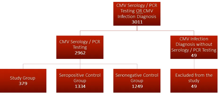
The initial sample consisted of 3011 patients, and the medical records were manually reviewed for quality-control. Forty-nine patients were excluded due to lack of CMV IgG, IgM, or DNA PCR testing to confirm date of CMV diagnosis. The remaining population of 2962 patients was then divided into three groups. The “study group” included all patients with acute or recent CMV infection, defined as positive CMV IgM or detection of CMV DNA by PCR in plasma. The remaining patients were divided into two control groups. The “seropositive control” group was defined as patients with positive CMV IgG and negative IgM or PCR. The “seronegative control” group was defined as patients with negative CMV IgG, IgM, and/or PCR. Three patients included in the study group had positive CMV PCR, but negative IgM testing. The study design is depicted in Figure 1.
The study population of patients with acute CMV infection consisted of 379 patients. The seropositive control group included 1334 patients. The seronegative control group included 1249 patients.
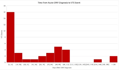
For primary outcome data, 38 of 379 patients (10.0%) in the study group were diagnosed with VTE within one year of acute CMV diagnosis. The vast majority of these events occurred within the first six months, as depicted in Figure 2. This is compared to 41 of 1334 patients (3.1%) in the seropositive control group and 37 of 1249 patients
(3.0%) in the seronegative control group (Table 2). The seropositive control group had lower incidence of VTE than the study group (3.1% versus 10.0%, p <0.0001). The seronegative control group also had lower incidence of VTE than the study group (3.0% versus 10.0%, p <0.0001). There was no statistically significant difference between the incidence of VTE in the seropositive control group and seronegative control group (3.1% versus 3.0%, p = 0.9), suggesting that the effect of acute CMV infection on VTE incidence may be transient.
Multivariate analysis was performed after adjustment for age and gender. VTE in the study group was higher than either control group (Table 2). The seropositive control group was less likely to have VTE than the study group within one year (OR = 0.3, 95% CI 0.2-0.5, p <0.0001). The seronegative control group was also less likely to have VTE than the study group within one year (OR = 0.4, 95% CI 0.2-0.6, p <0.0001).
Secondary outcome data demonstrated that ACS was more likely to occur in the study group, with incidence at one year of 7.7%, compared to 4.7% (p <0.0001) in the seropositive control group and 1.9% (p <0.0001) in the seronegative control group (Table 2). Multivariate analysis of incidence adjusted for age and gender demonstrated that the seronegative control group was less likely to develop ACS than the study group within one year (OR = 0.4, 95% CI 0.2-0.7, p = 0.003). However, the seropositive control group failed to demonstrate significant differences compared the study group (OR = 0.7, 95% CI 0.4-1.2, p = 0.2). The difference between the two control groups in the secondary outcomes data was significant, with the seropositive control group more likely to develop ACS within one year than seronegative control group (OR = 1.8, 95% CI 1.1-2.9, p = 0.03).
Discussion
suggest that CMV may have contributed to a prothrombotic state in patients with active infection or seropositive state.
These data align with several studies in the past decade supporting the association between CMV and VTE. The MAISTHRO retrospective registry case-control study reported 4.3% with CMV IgM positivity amongst patients with acute VTE, compared to 0.6% with CMV IgM positivity in matched control patients without acute VTE (OR 7.3).10 One large single-center prospective case-control study observed patients with acute CMV infection and matched CMV IgM negative controls for 6 months and found 3.06 cases of acute VTE per 1000 in those with positive IgM compared to 1.36 cases of acute VTE per 1000 in those with negative IgM (OR 2.25, 95% CI 1.38– 3.66, p = 0.003).11 A retrospective case-control study of 140 patients with acute CMV infection showed an incidence of concurrent VTE of 2.9%, which was significantly greater than 0% VTE in 140 matched controls without acute CMV infection.12 A meta-analysis concluded that between 1.9% and 9.1% of patients hospitalized with acute venous thromboembolism had concurrent acute CMV infection.13
The secondary outcome data collected in this study similarly showed a measurable difference between the
seronegative control group and the study group, with regard to incidence of ACS. However, incidence of ACS was not significantly different between the seropositive control group and the study group. While there are no prior studies of similar design to directly compare these data, the discrepancies might be explained by the different mechanisms involved in arterial thrombosis. Studies examining the role of CMV in arterial disease fall into two groups. CMV IgG seropositivity and chronic CMV endothelitis have been linked with the pathogenesis of
atherosclerosis, while acute CMV infection has been associated with ACS.6,7,16-18 Both primary ACS and restenosis were counted amongst the ACS incidence data in our study, possibly explaining the similar incidence rate of ACS in both the seropositive control group and the study group.
Despite these limitations, these data support the existence of CMV-associated VTE. There are also several known and proposed mechanisms for a causative relationship have been described in vitro. The majority of models are based on host monocyte and endothelial cell infection. Direct infection of vascular endothelial cells has been demonstrated to increase release of von Willebrand factor and increase cell-surface expression of tissue factor.7,19 This has been shown to promote inhibition and depletion of natural anticoagulants as well as inhibition of
antithrombin III and fibrinolysis.7,20 CMV infection also increases leukocyte adhesion to CMV-infected endothelial cells, which has been shown to promote a procoagulant microenvironment through similar mechanisms, possibly acting as cofactors.21,22 Direct infection of monocytes also resulted in increased secretion of tissue factor, thus promoting thrombosis.7 Another model described an immunologic response to CMV envelope phospholipids, including phosphatidylserine, which has procoagulant prothrombinase properties, reducing the factor Xa clotting time by bypassing most of the coagulation cascade and directly activating thrombin.20
One proposed model describes transient elevations of antiphospholipid antibodies in the context of acute CMV and other viral infections including Epstein-Barr Virus, Hepatitis C Virus, Parvovirus, Varicella Zoster Virus, and Adenovirus.23 A study in mice demonstrated that immunization with a CMV capsule peptide known as TIFI (an analog of human beta-2-glycoprotein I [β2GPI]), induced lupus anticoagulant activity and resulted in thrombotic complications.24,25 Subsequent study demonstrated that co-injection of TIFI with exogenous antiphospholipid antibodies decreased incidence of vascular thrombosis in the setting of mechanical injury to the mice. These mice did not form endogenous antiphospholipid antibodies, suggesting a potentially protective effect of the exogenous antibodies.26 Case reports in humans have described transient increases of serum antiphospholipid antibodies in acute CMV, specifically anticardiolipin IgM.27,28 This would support the applicability of the findings in mice to humans. However, at this time there are no published studies connecting TIFI to formation of anti-β2GPI antibodies in human patients.29 If this link can be established, the homology between CMV TIFI and the 5th domain of human β2GPI could explain why CMV has been more commonly associated with VTE than other viral infections.
preceding urinary tract and respiratory tract infections in the preceding six months. They found that the incidence risk ratios of DVT (2.10, 95% CI 1.56–2.82) and PE (2.11, 95% CI 1.38–3.23) were significantly higher after urinary tract infections, particularly in the first two weeks after infection. Respiratory tract infections also had increased risk of VTE, but diagnostic data were more confounded.30 One key difference in our study design was following a single infection across a broad cohort of patients, rather than looking for incidence of preceding infection in a cohort of patients with VTE. Additionally, past studies did not consider disease-specific causes of VTE. They cite studies discussing transient alterations to endothelial function, increased inflammation measured by surrogate markers such as C-reactive protein, the role of neutrophil activation and neutrophil extracellular traps, tissue factor activation of thrombotic pathways, inhibition of endogenous anticoagulant pathways, and inhibition of fibrinolysis.4,30-32 While there are other known associations between acute infection and thrombosis, they neither address nor negate the evidence presented for a potentially unique causal relationship between CMV and
thrombosis. This is further evidenced by the presence of increased odds of VTE in the CMV seropositive group of patients suggesting a more prolonged prothrombotic state than has been presented in other infection models of thrombosis.
Conclusions
Our findings support the hypothesis of a link between acute CMV infection and thrombotic events such as acute VTE and ACS. In particular, the adjusted odds ratios for acute VTE were convincing of a difference between the study and control populations related to CMV status. The secondary outcome data contribute to the limited existing evidence for a role of acute CMV infection in ACS. Additional in vivo studies are needed to further clarify the relationship between acute CMV infection and thrombosis and account for other known risk factors for VTE, especially in high-risk populations. These studies should include data on incidence of anti-β2GPI antibody
formation based on in vitro findings.28 No prior studies have evaluated VTE prophylaxis in patients with acute CMV infection. These concepts should be considered in future clinical trials if an association between CMV infection and VTE is confirmed.
All authors had access to the data and had a role in writing the manuscript. Amar H. Kelkar M.D. and Brian L. Loc D.O. wrote the preliminary draft manuscript. The study was designed by Amar H. Kelkar M.D., Brian L. Loc D.O., and John J. Farrell M.D. Data collection and review were performed by Amar H. Kelkar M.D. and Brian L. Loc D.O. Data analysis was performed by Amar H. Kelkar M.D. and Huaping Wang, Ph.D. All authors made critical revisions related to the content of the manuscript and approved the final version of this article for submission.
Funding Sources
The authors received no specific funding for this work from any funding agency in the public, commercial, or not-for-profit sectors.
Conflict of Interest
None of the authors have any relevant conflicts of interest or disclosures. However, it should be noted that Anita Rajasekhar has served on an advisory board for Alexion, Baxter, Bayer, Kedrion Biopharma Octapharma Plasma, and her institution has received research support on her behalf from Alnylam (Sanofi Genzyme), Baxalta (Shire), Biomarin, Dimensions Therapeutics, Genetech, Janssen Pharmaceuticals, and Roche.
Acknowledgements
Special thanks to Mona Kelkar M.S. M.B.A. for technical support for data visualization and Janet Dean M.L.S. as the lead clinical laboratory scientist in the OSF HealthCare System serology laboratory.
References
1. Ho M. The history of cytomegalovirus and its diseases. Med Microbiol Immunol. Jun 2008;197(2):65-73. 2. Boers M, Haak A. Cytomegalovirus infection with perfusion defects on the lung scan. Infection. 1984
Jul-Aug 1984;12(4):265-267.
4. Cohoon KP, Ashrani AA, Crusan DJ, Petterson TM, Bailey KR, Heit JA. Is Infection an Independent Risk Factor for Venous Thromboembolism? A Population-Based, Case-Control Study. Am J Med. Mar
2018;131(3):307-316.e302.
5. Melnick JL, Petrie BL, Dreesman GR, Burek J, McCollum CH, DeBakey ME. Cytomegalovirus antigen within human arterial smooth muscle cells. Lancet. Sep 1983;2(8351):644-647.
6. Nikitskaya E, Lebedeva A, Ivanova O, et al. Cytomegalovirus-Productive Infection Is Associated With Acute Coronary Syndrome. J Am Heart Assoc. 08 2016;5(8).
7. Squizzato A, Gerdes VE, Büller HR. Effects of human cytomegalovirus infection on the coagulation system. Thromb Haemost. Mar 2005;93(3):403-410.
8. Amsterdam EA, Wenger NK, Brindis RG, et al. 2014 AHA/ACC guideline for the management of patients with non-ST-elevation acute coronary syndromes: a report of the American College of
Cardiology/American Heart Association Task Force on Practice Guidelines. Circulation. Dec 2014;130(25):e344-426.
9. Nishimura RA, Otto CM, Bonow RO, et al. 2014 AHA/ACC Guideline for the Management of Patients With Valvular Heart Disease: executive summary: a report of the American College of
Cardiology/American Heart Association Task Force on Practice Guidelines. Circulation. Jun 2014;129(23):2440-2492.
10. Schimanski S, Linnemann B, Luxembourg B, et al. Cytomegalovirus infection is associated with venous thromboembolism of immunocompetent adults--a case-control study. Ann Hematol. Apr 2012;91(4):597-604.
11. Paran Y, Shalev V, Steinvil A, et al. Thrombosis following acute cytomegalovirus infection: a community prospective study. Ann Hematol. Jul 2013;92(7):969-974.
12. Atzmony L, Halutz O, Avidor B, et al. Incidence of cytomegalovirus-associated thrombosis and its risk factors: a case-control study. Thromb Res. Dec 2010;126(6):e439-443.
13. Justo D, Finn T, Atzmony L, Guy N, Steinvil A. Thrombosis associated with acute cytomegalovirus infection: a meta-analysis. Eur J Intern Med. Apr 2011;22(2):195-199.
15. Kelkar AH, Jacob KS, Yousif EB, Farrell JJ. Venous thromboembolism related to cytomegalovirus infection: A case report and literature review. Medicine (Baltimore). Dec 2017;96(51):e9336.
16. Gabrylewicz B, Mazurek U, Ochała A, et al. Cytomegalovirus infection in acute myocardial infarction. Is there a causative relationship? Kardiol Pol. Oct 2003;59(10):283-292.
17. Guetta E, Scarpati EM, DiCorleto PE. Effect of cytomegalovirus immediate early gene products on endothelial cell gene activity. Cardiovasc Res. Jun 2001;50(3):538-546.
18. Levi M. CMV endothelitis as a factor in the pathogenesis of atherosclerosis. Cardiovasc Res. Jun 2001;50(3):432-433.
19. Goeijenbier M, van Wissen M, van de Weg C, et al. Review: Viral infections and mechanisms of thrombosis and bleeding. J Med Virol. Oct 2012;84(10):1680-1696.
20. Pryzdial EL, Wright JF. Prothrombinase assembly on an enveloped virus: evidence that the cytomegalovirus surface contains procoagulant phospholipid. Blood. Dec 1994;84(11):3749-3757. 21. Toyoda M, Galfayan K, Galera OA, Petrosian A, Czer LS, Jordan SC. Cytomegalovirus infection induces
anti-endothelial cell antibodies in cardiac and renal allograft recipients. Transpl Immunol. Jun 1997;5(2):104-111.
22. Visseren FL, Bouwman JJ, Bouter KP, Diepersloot RJ, de Groot PH, Erkelens DW. Procoagulant activity of endothelial cells after infection with respiratory viruses. Thromb Haemost. Aug 2000;84(2):319-324. 23. Uthman IW, Gharavi AE. Viral infections and antiphospholipid antibodies. Semin Arthritis Rheum. Feb
2002;31(4):256-263.
24. Gharavi AE, Pierangeli SS, Espinola RG, Liu X, Colden-Stanfield M, Harris EN. Antiphospholipid antibodies induced in mice by immunization with a cytomegalovirus-derived peptide cause thrombosis and activation of endothelial cells in vivo. Arthritis Rheum. Feb 2002;46(2):545-552.
25. de Groot PG, Urbanus RT. The significance of autoantibodies against β2-glycoprotein I. Blood. Jul 2012;120(2):266-274.
26. Ostertag MV, Liu X, Henderson V, Pierangeli SS. A peptide that mimics the Vth region of beta-2-glycoprotein I reverses antiphospholipid-mediated thrombosis in mice. Lupus. 2006;15(6):358-365. 27. Delbos V, Abgueguen P, Chennebault JM, Fanello S, Pichard E. Acute cytomegalovirus infection and
28. Nakayama T, Akahoshi M, Irino K, et al. Transient antiphospholipid syndrome associated with primary cytomegalovirus infection: a case report and literature review. Case Rep Rheumatol. 2014;2014:271548. 29. Chamley LW, McKay EJ, Pattison NS. Cofactor dependent and cofactor independent anticardiolipin
antibodies. Thromb Res. Feb 1991;61(3):291-299.
30. Smeeth L, Cook C, Thomas S, Hall AJ, Hubbard R, Vallance P. Risk of deep vein thrombosis and
pulmonary embolism after acute infection in a community setting. Lancet. Apr 2006;367(9516):1075-1079. 31. Fuchs TA, Brill A, Duerschmied D, et al. Extracellular DNA traps promote thrombosis. Proc Natl Acad Sci
U S A. Sep 2010;107(36):15880-15885.
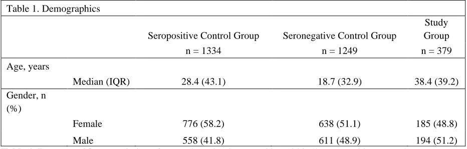
Table 1. Demographics
Seropositive Control Group Seronegative Control Group
Study Group
n = 1334 n = 1249 n = 379
Age, years
Median (IQR) 28.4 (43.1) 18.7 (32.9) 38.4 (39.2)
Gender, n
(%)
Female 776 (58.2) 638 (51.1) 185 (48.8)
Male 558 (41.8) 611 (48.9) 194 (51.2)
Table 1. Demographics Description of age and gender demographics within the seropositive control group, seronegative control group, and study group.
Table 2. Incidence of VTE and ACS in Control Groups versus Study Group Seropositive Control Group Seronegative Control Group Study
Group Unadjusted Results
Age- and Gender- Adjusted Results
n = 1334 n = 1249 n = 379 p value
OR (95%
CI) p value
OR (95% CI) VTE,
n (%)
41 (3.1) 37 (3.0) 38 (10.0)
Seropositive Control Group vs.
Study Group
<0.0001 0.3
(0.2-0.5) <0.0001
0.3 (0.2-0.5)
Seronegative Control Group vs.
Study Group
<0.0001 0.3
(0.2-0.4) <0.0001
0.4 (0.2-0.6)
Seropositive Control Group vs.
Seronegative Control Group
0.9 1.0
(0.7-1.6) 0.5
0.9 (0.5-1.4)
ACS, n (%)
62 (4.7) 24 (1.9) 29 (7.7)
Seropositive Control Group vs.
Study Group
0.02 0.6
(0.4-0.9) 0.2
0.7 (0.4-1.2)
Seronegative Control Group vs.
Study Group
<0.0001 0.2
(0.1-0.4) 0.003
0.4 (0.2-0.7)
Seropositive Control Group vs.
Seronegative Control Group
0.0002 2.5
(1.5-4.0) 0.03
1.8 (1.1-2.9)
Table 2. Incidence of VTE and ACS in Control Groups versus Study Group Incidence of VTE and ACS in the seropositive control group, seronegative control group, and study group with calculated odds ratios (OR), 95% confidence intervals (CI), and p values between each of the control groups and the study group. Age- and gender-adjusted results were also calculated.
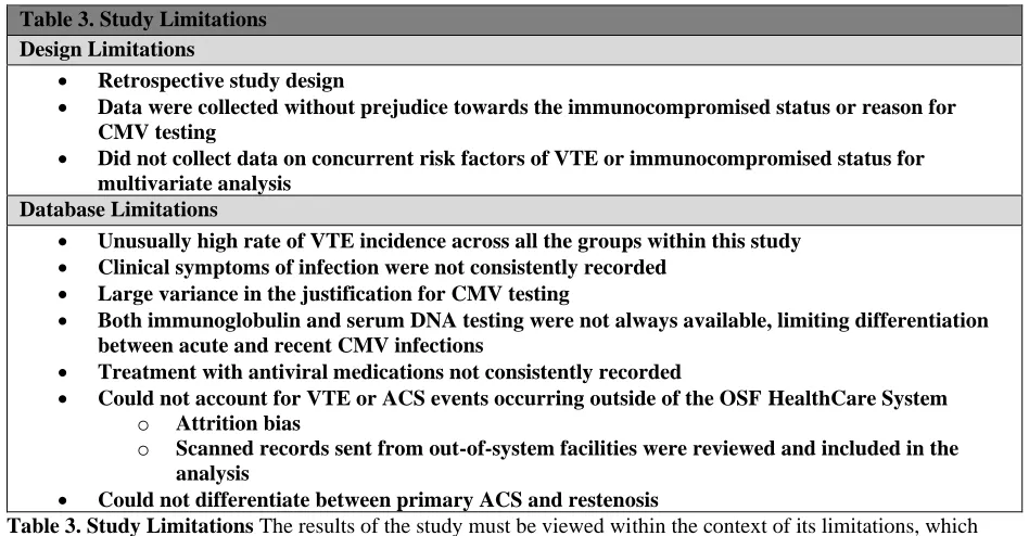
Table 3. Study Limitations Design Limitations
• Retrospective study design
• Data were collected without prejudice towards the immunocompromised status or reason for CMV testing
• Did not collect data on concurrent risk factors of VTE or immunocompromised status for multivariate analysis
Database Limitations
• Unusually high rate of VTE incidence across all the groups within this study • Clinical symptoms of infection were not consistently recorded
• Large variance in the justification for CMV testing
• Both immunoglobulin and serum DNA testing were not always available, limiting differentiation between acute and recent CMV infections
• Treatment with antiviral medications not consistently recorded
• Could not account for VTE or ACS events occurring outside of the OSF HealthCare System o Attrition bias
o Scanned records sent from out-of-system facilities were reviewed and included in the analysis
• Could not differentiate between primary ACS and restenosis
Table 3. Study Limitations The results of the study must be viewed within the context of its limitations, which include its retrospective study design, large volume of patients with missing or untested data points, and database search limitations.
Figure 1. Study Design The patient population was divided into three groups after excluding patients with no serology or PCR testing: “study group” (IgM+/PCR+), “seropositive control” group (IgG+/IgM-/PCR-), “seronegative control” group (IgG-/IgM-/PCR-).
Figure 2. Time from Acute CMV Diagnosis to VTE Event Within the study group, the majority of VTE events occurred within six months of the initial acute CMV diagnosis, supporting a temporal relationship.