THE ROLE OF SEROTONIN AND OTHER
NEUROTRANSMITTER SUBSTANCES IN THE
DEVELOPMENT OF CORTICAL CELL TYPES:
AN
IN VITRO
STUDY
By
AUDREY ELAINE DOOLEY
Submitted to the University o f London f o r the Degree o f
Doctor of Philosophy
Department o f Anatomy and Developmental Biology
University College London
Gower Street
WCIE 6BT
ProQuest Number: 10106663
All rights reserved
INFORMATION TO ALL USERS
The quality of this reproduction is dependent upon the quality of the copy submitted.
In the unlikely event that the author did not send a complete manuscript and there are missing pages, these will be noted. Also, if material had to be removed,
a note will indicate the deletion.
uest.
ProQuest 10106663
Published by ProQuest LLC(2016). Copyright of the Dissertation is held by the Author.
All rights reserved.
This work is protected against unauthorized copying under Title 17, United States Code. Microform Edition © ProQuest LLC.
ProQuest LLC
789 East Eisenhower Parkway P.O. Box 1346
ACKNOWLEDGEMENTS
My appreciation and thanks go foremost to John Pamavelas for his guidance and supervision throughout this thesis and in whose laboratory these investigations were conducted. I would next like to thank Dr. loannis Pappas for his additional valuable guidance throughout many aspects of this work and Dr. Marina Mione for her helpful comments and advice.
My thanks also go to the following for the teehnical assistance and advice gratefully received: Dr. Mimika Thomaidou for her help in labelling dying cells, Brett Harris for developing and printing techniques, John Cavanagh for photography techniques, Peter Boardman for his help in maintenance of eulture equipment. Dr. Bagi Nadarajah and Dr. Alex Lavdas for their helpful advice and also Dr. Stephen Davies for the use of equipment in his laboratory.
ABSTRACT
The different neuronal and glial cell types that form the mammalian cerebral cortex are generated from a population of epithelial cells lining the embryonic telencephalic ventricles. Cells within this germinal layer proliferate and migrate to form the characteristic six-layered structure of the neocortex. The factors that control the proliferation of cortical progenitor cells and their differentiation into different cell types are still mainly unknown, but recent studies point to the importance of signals from the cells’ microenvironment in their commitment to a particular phenotype and laminar fate. Such signals may be provided by growth factors, hormones, neuropeptides, extracellular matrix molecules, or neurotransmitters produced and regulated locally by differentiated cortical neurons or afferent axonal systems.
A host of neurotransmitters have been implicated in regulating the proliferation and differentiation of cortical cell types. Chief among them are the catecholamines, noradrenaline and dopamine, and the indoleamine, 5-hydroxytryptamine (serotonin; 5- HT). The early development of the monoaminergic systems has prompted speculation that they may play a role in a number of developmental processes in the cortex.
the survival o f cortical cells during development and point to a preferential survival of postmitotic cortical neurons.
PUBLICATIONS ARISING FROM THESIS
TABLE OF CONTENTS
A cknowledgements... 2
A bstract... 3
Publications Arising Front Thesis... 4
Table o f Contents... 5
Abbreviations...8
CHAPTER 1: Introduction
1.1 Preview and Background Information... 91.2 Overview of Cortical Development... 11
1.3 Cell Proliferation and Death in the Cerebral Cortex... 15
1.3.1 Cell proliferation and the ventricular zone... 15
1.3.2 Cell death in the cerebral cortex... 19
1.4 Early Theories on Cortical Cell Fate (Historical Foundations)... 21
1.5 Cortical Cell Fate and Migration... 22
1.5.1 Commitment to cell phenotype in the cerebral cortex... 22
1.5.2 Commitment of cells to laminar and regional fates in the cerebral cortex 25 2. Overview of Environmental Factors... 27
2.1 Growth Factors in the Developing C N S...29
2.1.1 Growth factors and cell survival... 29
2.1.2 Growth factors and cell proliferation... 30
2.1.3 Growth factors and cell differentiation...32
2.2 N euro transmitters in the Cerebral Cortex: Appearance and Distribution... 35
2.3 Serotonin in Development...42
2.3.1 5-HT and early embryogenesis...42
2.3.2 5-HT and cell proliferation... 43
2.3.3 5-HT and the regulation of cell differentiation and growth...43
2.3.4 5-HT, cortical plasticity and maintenance... 45
3. Serotonin Receptors and Intracellular Pathways... 47
3.1 5-HT receptors in the developing nervous system ...47
3.2 Intracellular pathways...50
3.3 Regulation of cell proliferation and differentiation...51
4. Other Neurotransmitters in Development... 53
4.1 Noradrenergic actions, receptors and intracellular pathways...53
5. Summary and Aims of Study...58
CHAPTER 2: Methods and Materials
6.1 Preparation of Cortical Cultures...606.2 MTT Survival Assay... 61
6.3 Immunohistochemistry... 62
6.3.1 BrdU staining... 63
6.4 Tunel Labelling... 63
6.4.1 Double immunolabelling... 64
6.5 Cell Counts and Statistical Analysis... 64
6.6 Morphological Analysis of MAP-2 Labelled C ells... 65
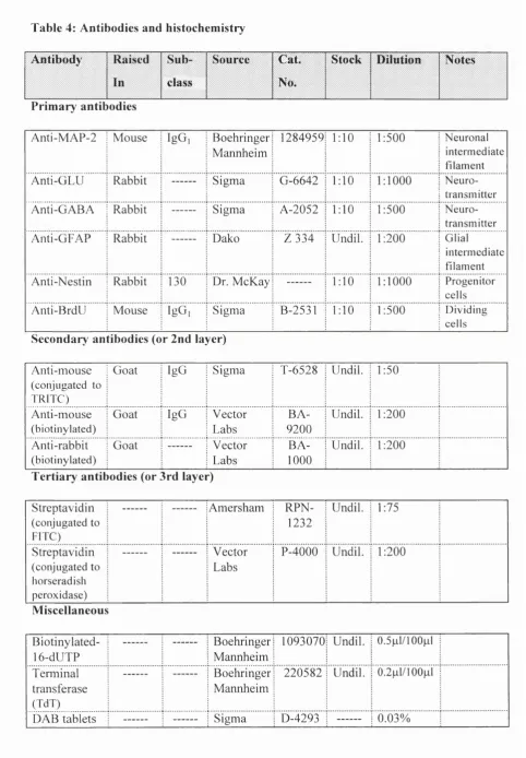
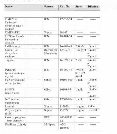
6.7 Antibodies, Chemical Reagents, and Culture M edia... 65
CHAPTER THREE: Serotonin and the Survival of Embryonic
Cortical Cells
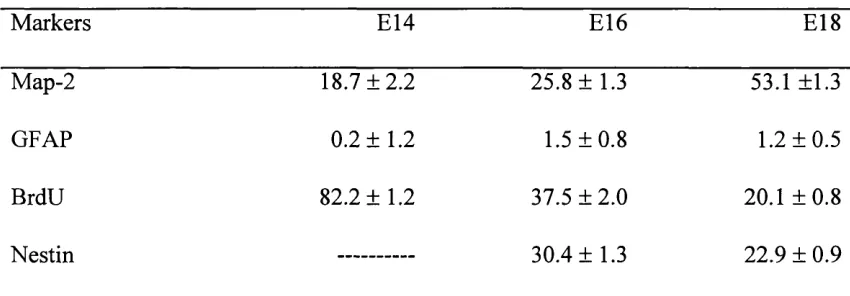
7.1 Introduction... 707.2 Cell Types in Cultures Prepared from E14, E l 6 and E l 8 Cortices...70
7.3 Standardizing Culture Conditions... 76
7.3.1 Fetal calf serum ... 76
7.3.2 Vehicles used and culture density...78
7.4 Serotonin Promotes the Survival of Developing Cortical C ells... 82
7.4.1 5-HT promotes cortical survival in an age dependent manner...82
7.4.2 5-HT promotes cortical survival in a concentration dependent manner.... 84
7.4.3 Time period o f 5-HT action... 84
7.4.4 5-HT decreases cortical cell death in E l 6 cultures...84
7.5 Serotonin Does Not Affect Cortical Cell Proliferation... 87
7.6 Discussion... 90
7.6.1 Standardizing culture conditions...90
7.6.2 5-HT promotes the survival of cortical cells in vitro... 90
CHAPTER FOUR: An Analysis of the Survival of Cortical Cells by
Serotonin
8.1 Introduction...94
8.2 The Effect of Serotonin on Cell Phenotype... 95
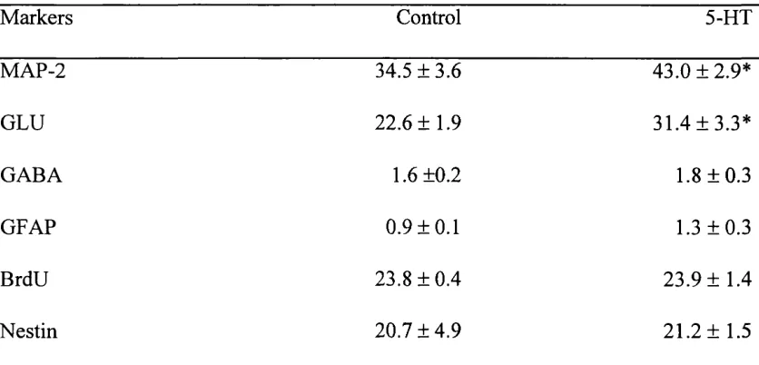
8.2.1 5-HT promotes the survival of glutamatergic neurons...95
8.2.2 5-HT increased cell body area...96
8.3 Mechanism of Serotonin Action on Cortical Cultures...102
8.3.1 5-HT2a receptors mediate the effects of 5-H T...102
8.4 Discussion...106
8.4.1 The effect of 5-HT on cell phenotype... 106
8.4.2 5-HT increases cell body area...108
8.4.3 5-HT2a receptors mediate the survival effects of 5-H T ... 108
CHAPTER FIVE: Serotonin and Comparison with Other
Neurotransmitters
9.1 Introduction... 1109.2 Noradrenaline and Dopamine Promote E l 6 Cortical Survival... I l l 9.2.1 DA mediated survival is concentration dependent... 114
9.3 Noradrenergie and Dopaminergic Increased Survival is Mediated by Spécifié Reeeptors... 117
9.4 Intracellular Mechanisms Governing the Increased Survival of Cortieal Cells by Serotonin, Noradrenaline, and Dopamine... 121
9.5 Discussion... 126
9.5.1 NA and DA promote the survival of cortical cells by specific receptors 126 9.5.2 Intracellular meehanisms involved in the survival of embryonie cortical cells... 127
CHAPTER SIX: Conclusions
10.1 Neuroactive Substances Regulate Cortical Development...12910.2 The Role of Serotonin and Other Neurotransmitters in Cortical Development... 130
ABBREVIATIONS
ACh Acetylcholine
bFGF Basic fibroblast growth factor
BDNF Brain derived neurotrophic growth factor
BrdU Bromodeoxyuridine
Calcium
cAMP Cyclic adenosine monophosphate
CNS Central nervous system
DA Dopamine
DAB Diaminobenzidene
DIV Days in vitro
DMEM Dulbecco’s modified eagles medium
DNA Deoxyribonucleic acid
E Embryonic day (e.g. E l 6)
FCS Fetal calf serum
GABA Gamma amino butyric acid
GFAP Glial fibrillary acidic protein
GLU Glutamate
HBSS Hanks balanced salt solution
5-HT Serotonin
MAP-2 Microtubule associated protein-2
NA Noradrenaline
NGF Nerve growth factor
NT Neurotrophin
6-OHDA 6-hydroxydopamine
PBS Phosphate buffered saline
pCPA p-cholorophenylalanine
PLC Phospholipase C
PNS Peripheral nervous system
PYE Pseudostratified ventricular epithelium SPP Secondary proliferative population
SYZ Subventricular zone
TdT Terminal transferase
TH Tyrosine hydroxylase
CHAPTER 1
INTRODUCTION
1.1 Preview and Background Information
The mature central nervous system (CNS) of mammals presents us with a precisely organized and structured ensemble of areas and connections which serve to orchestrate our response to various sensory stimuli. As the tools available to scientists became further advanced, so we have been able to follow and describe in detail the events which lead to the presentation of this adult structure.
There are two main classes of cells encountered in the CNS: neurons and glia. Each type of cell itself presents us with many variations dependent upon the area of the CNS to which it belongs. Cell types of the cerebral cortex for example, the largest structure of the CNS, display an array of morphologically, chemically and physiologically distinct neuronal cell types (Szentagothai, 1973; McCormick et al., 1985; Pamavelas et al., 1989; DeFelipe, 1993).
Cortical neurons are subdivided into two principal types: pyramidal and nonpyramidal. Pyramidal cells are the projection neurons known to exert excitatory action in the cortex and in subcortical target areas by releasing L-glutamate (GLU) or L- aspartate (Fagg and Foster, 1983; Dori et al., 1992). The nonpyramidal cells, the cortical intemeurons, utilize the inhibitory neurotransmitter y-aminobutyric acid (GABA) (Houser et al., 1984; Peters and Jones, 1984). Separate developmental patterns have been established for these neuronal cell types (Mione et al., 1997).
markers, has enabled us to determine at least narrow time periods during which cell potentials are restricted (Price and Thurlow, 1988; Luskin et ah, 1993; Mione et ah,
1994). While such lineage experiments have provided important data as to when cell fate is specified, exactly what factors and signals are involved in cortical cell generation and differentiation is not entirely known. In the peripheral nervous system (PNS), instructional substances such as the neurotrophins, have been found to regulate the survival and differentiation of particular neurons (Snider, 1994). Within the CNS, such factors have also proved instructional (Rohrer, 1990; McConnell, 1991; Pappas and Pamavelas, 1997). There is yet ample evidence as to the actions of growth factors in the regulation of cell proliferation, survival and differentiation in various areas of the CNS including the cerebral cortex. A further source of environmental signals has been found to be provided by neurotransmitter substances (Mattson, 1988; Pamavelas et ah, 1988; Lauder, 1993). In particular, evidence suggests that the neurotransmitter serotonin (5- HT) plays a significant role in cortical development (Lauder, 1990).
1.2 Overview of Cortical Development
FIG. 1. DEVELOPMENT OF CORTICAL LAYERS
Pial Surface
E 1 2
E 1 3
E 1 4 -
E l 5
E l 6
E l 7
E l 8
E 2 0
A d u l t
Thus, the generation of neurons and final formation of layers in the cortex results in groups of cells which share similar patterns of connectivity and age in relation to their position. In addition to this well-documented scheme of cortical development, further studies indicated an overall frontal to caudal (Miller 1988; Bayer and Altman, 1991) and lateral to medial (Raedler et al., 1980; Bayer and Altman, 1991; Ignacio et al., 1995) gradient of neurogenesis, such that layer specific neurons generated in frontal and lateral regions arrived earlier to their destinations than their caudal and medial counterparts.
The period of production of neurons in the rat occurs between embryonic day 12 (E l2) and the time of birth around E21 (Uylings et al, 1990; Bayer and Altman, 1991). Neuronal generation proceeds from the proliferation of progenitor cells lining the neuroepithelium. Eventually, these immature neurons leave their site of origin and migrate to their destinations along processes of radial glial cells (Rakic, 1972; Hatten, 1990) where they establish and maintain synaptic connections. Less is known about glial production and their patterns of colonization in the cortex, suffice to say that the generation of macroglia occurs slightly later than the production of neurons taking place predominantly in postnatal life (Frederiksen and Mckay, 1988; Skoff and Knapp, 1991).
1.3 Cell Proliferation and Death in the Cerebral Cortex
We start this section by focusing on the events of cell proliferation and cell death, the first involving a mechanism designed to produce more cells, and the latter involved in the elimination of cells from the developing cerebral cortex. These two events at first glance seem mutually opposed in their purpose, yet both processes exhibit certain morphological similarities, such as the retraction and loss of neurites, and the rounding up and bipolar appearance of the cell body (Heintz, 1993; Rubin et ah, 1993). It is these characteristics as well as recent biochemical data implicating cell cycle proteins in the process of cell death (Freeman et ah, 1994) that has strengthened the hypothesis that the two events may be related. It is now postulated that an inappropriate activation of proliferation could cause a cell to abort and instead follow a pathway leading to death. We shall, therefore, consider both processes in the developing cerebral cortex.
1.3.1 Cell proliferation and the ventricular zone in the cerebral cortex
The mechanics of cell division in the cortex, by which an accurate number and ratio o f the major cell types is produced in the correct locations, has been investigated extensively in the mouse cortex (Takahashi et al., 1994). The cell cycle, which results in division of a cell, is divided into 4 distinct phases- G l, S, 0 2 and M. These involve periods of active synthesis and replication of chromosomes (S phase), nuclear and cellular division (M phase) and intervals of preparation in between (G1/G2 phase). Each phase lasts a defined period of time according to the stage of cortical development and each cell also leaves the cell cycle after a defined number of divisions to become postmitotic. Alternatively, the cell can re-enter the cell cycle to continue the process of division. Thus, the regulation of stem cell proliferation was seen to involve the control o f at least four distinct decisions- that of activation and maintenance o f the proliferative state coupled with a later stop signal and maintenance of cell cycle repression.
Studies in the mouse found that the developing cerebral wall contains two proliferative populations categorized on the basis o f the cell classes which it produces and their proliferative behaviour. The first and largest of the two is the pseudostratified ventricular epithelium (PVE), which has long been known to be the source o f most, if not all, neocortical neurons as well as some cells o f the glial lineage (Sauer, 1935). The second population of proliferative cells is known as the secondary proliferative population (SPP).
concluded that the increase in the cell cycle length observed during development is essentially due to a four-fold increase in the Gl phase, the only phase that varies systematically. Thus, any extrinsic signals that may be involved in the regulation of neuronal number is most likely to affect and modulate the G l phase of the cell cycle. It has also been postulated that differences in the size of the cortex across species could be regulated by factors which prolong the early period of proliferation, the time of expansion of the progenitor pool. Such factors could achieve this result by shortening the cell cycle or by providing a continuous mitogenic signal (Caviness, 1995).
The significantly smaller population of proliferative cells, the SPP, is principally a source of glial cells arising from the PVE, and extends from the interface of the ventricular and subventricular zone towards the base of the cortical plate (Altman and Bayer, 1990; Takahashi et al., 1993). Studies that focused on the SPP found a high rate of proliferation between El 4-El 6. The population increased nearly six-fold antecedent to the production of glial cells throughout the neocortex. Quantitatively, it was estimated that 87% of SPP cells would re-enter S-phase during this interval, while only 13% would exit the cell cycle (Takahashi et al., 1995b).
Recent studies have begun to investigate the molecular mechanisms which might regulate cell proliferation in vertebrates. Of note are the investigations in mutant mice expressing a deficiency for the gene BF-1 (Xuan et al., 1995) in the telencephalic neuroepithelium. These mice showed a dramatic reduction in the size of their cerebral hemispheres. It was postulated that this cell intrinsic transcription factor may regulate the machinery required for the progression of the cell cycle or enhance the effects of mitogens on neuroepithelial cells. However, the importance of a number o f genes in cell proliferation has become evident in invertebrate studies which have used the host
The mode of cell production in the developing cerebral cortex also influences the
number of cells generated during proliferation. Symmetrical division, produces two daughter cells of which both re-enter the cell cycle. In this process the plane of cleavage of cell division is orientated vertically (or perpendicular) to the ventricular surface. Asymmetrical division, however, results in one of the two daughter cells continuing to proliferate while the other cell becomes postmitotic. In this situation, the plane of cleavage is horizontal to the VZ. Observations made by Takahashi et al. (1995a), showed that many proliferative cortical cells are produced within a short time. They thus hypothesized that the progenitor pool is expanded by divisions of a symmetrical nature. Direct evidence for symmetrical and asymmetrical divisions within the proliferative zone came from imaging labelled progenitor cells with confocal microscopy (Chenn and McConnell, 1995). How the cell divisions observed produce distinct cell types that display a different proliferative nature has been studied at a molecular level using
Drosophila. It was observed that products of the genes Numb (a membrane associated protein) and Prospero (a nuclear protein), become differentially segregated during asymmetrical neural precursor cell division (Hirata et al., 1995; Knoblich et al., 1995; Spana et al., 1995; Spana and Doe, 1995). Similar studies in the cortex of vertebrates have shown that the protein Notch 1 is localized close to the upper pial surface of mitotic cells (Chenn and McCormell, 1995). Asymmetrical divisions result in the uneven distribution of this protein and as such provide a mechanism by which distinct cell types can be generated in the mammalian species.
the adult (Morshed et al., 1994; Gritti et al., 1996). Such studies have introduced the idea of using adult stem cells in transplantation to replace dying neurons during pathological diseases (Gage et al., 1995).
1.3.2 Cell death in the cerebral cortex
Cell death in many systems is mainly classified into two major types based on morphological and biochemical criteria. Programmed cell death (apoptosis) is prevalent during development, and cells undergoing apoptosis exhibit features such as plasma and nuclear membrane blebbing, cell shrinkage, chromatin condensation and DNA fragmentation. Necrosis, which is the type of cell death apparent in neurodegenerative diseases, involves cell swelling and eventual eruption. The distinction between both these forms of cell death is now less clear than traditionally believed due to similar changes occurring in both pathways i.e: elevated levels of calcium (Ca^^) (Choi, 1992; Ricter, 1993).
In the developing nervous system apoptosis occurs in a variety of circumstances. Major periods featuring apoptosis include:
1) Death of cells after the terminal division of the cell cycle, but before the formation of synaptic contacts. An example of such dying cells are found in the cortical subplate; these cells die after acting as targets of early thalamic afferents (McConnell et al., 1994). 2) Cell death resulting from competition between neurons for target derived factors. Such types of cell death has been demonstrated in the chick spinal cord using limiting amounts of target derived factors (Oppenheim, 1991 ; Raff et al., 1993).
Additionally, an earlier scene of cell death is observed when limited amounts of a survival factor are made available to proliferating or early postmitotic cells. This is demonstrated in cells of the oligodendrocyte lineage in vitro which show survival responses to various combinations of growth factors (Barres et al., 1992).
Of interest in the field of cell death in the cerebral cortex has been the extent to which this process occurs. Earlier studies in the cortex (Ferrer et al., 1992), as well as retina and spinal cord (Young, 1984; Homma et al., 1994) reported insignificant levels of cell death. More recent investigations using an enhanced sensitive assay, reported large amounts (up to 70%) of cell death in the proliferative areas and upper postmitotic layers of the cortex (Blaschke et al., 1996), although these high figures may be due to the sensitive nature of the technique used (Voyvodic, 1996). Recent work in our laboratory has shown a high correlation between dying cells and those that were in Gl phase of the cell cycle (Thomaidou et al., 1997). These results indicate that apoptosis is
prominent amongst the proliferative population of the developing cortex and that it is
1.4 Early Theories on Cortical Cell Fate (Historical Foundations)
The generation of neuronal and glial phenotypes in the mammalian cortex (i.e: how the specification of neurons occurs from an apparently homogeneous population of stem cells in the VZ and at what time does this happen?) is among the earliest questions asked and investigated in this field. Studies in this area concentrated on the production o f the major constituent cell types of the cortex- neurons and glia. By visualizing the ultrastructure of cells contained within the VZ (using electron microscopy and autoradiographic criteria), an hypothesis was formed which suggested that the uniform population of stem cells gave rise first to neuronal elements and later switched to glial production (Fujita, 1963, 1964). Subsequent discoveries, however, revealing the apparent co-existence of neuronal and glial elements in the developing VZ of the cortex (Levitt et al., 1981), led to two early theories gaining prominence. The first postulated by His in 1889, proposed separate cell lineages for neuronal and glial cell types in the proliferative zone, whereas Schaper in 1897 suggested single multipotential cells which gave rise to the two cell types (reviewed by McConnell, 1988).
The advent of techniques which permanently trace the lineage o f cells during their development has revealed new data on cortical cell fate. The use o f recombinant retroviruses in cell lineage experiments, for example, in which the gag, p a l and env
1.5 Cortical Cell Fate and Migration
1.5.1 Commitment to cell phenotype in the cerebral cortex
Information regarding the determination of the neurochemical, morphological and physiological fate of a cell has rapidly increased by the use of retroviral mediated gene transfer to study cell lineage. Experiments that utilized this method labelled cortical cells in vivo in the period of neurogenesis (E14-E21), and later analyzed the composition and distribution of cell clones at adult stages. Clones of cells produced which were homogeneous in composition indicated the presence of progenitor cells which at the point of infection were restricted in potential. However, the existence of heterogeneous clones suggested the presence of multipotential stem cells in the VZ. These studies reported that clones of cells analyzed are almost always exclusively composed of one cell type i.e: either neurons, astrocytes or oligodendrocytes (Price and Thurlow, 1988; Grove et al., 1993; Luskin et al., 1993). Thus, it appeared that progenitor cells from an early stage of cortical development (E l6) were restricted in their developmental potential. Further evidence for the existence of separate precursor cells came from experiments which showed that the neuronal subtypes- pyramidal and non-pyramidal cells also had defined lineages as early as E l 5 (Pamavelas et al., 1991; Mione et al., 1994).
local environmental factors, may produce the homogenous clones observed in the adult. Further complications were evident by the definitions of the spatial dimensions that constituted a clone of cells. Clones were usually defined as a spatial clustering o f labelled cells in a 500 pm cortical strip, separated from another clone of cells by at least 500 pm. Such clones were considered to be separate and unrelated (Lavdas et al., 1996). The basis for these definitions came from earlier observations of the migratory behaviour of cortical progenitor cells. As such, studies focusing on the migratory behaviour of progenitor cells revealed that there was potential for movement over large distances (average of 100 pm over 8 hr) and in the lateral direction (Rakic, 1972; Fishell et al., 1993; O’Rourke et al., 1995). This mode of movement by progenitor cells provided a mechanism by which widespread related clones could be produced. The existence of these dispersed clones was conclusively identified by use of a modified retroviral technique (Walsh and Cepko, 1992, 1993).
produces a different but related homogeneous cluster of another cell type (Reid et al., 1995).
In vitro studies also provides evidence for multipotential cells in the cerebral cortex (Davis and Temple, 1994; Reynolds and Weiss, 1996) it should be noted, however, that while in vivo retroviral analysis documents the ultimate fate o f clonally related cells, tissue culture manipulations reflect the range of cell potential. In other regions of the developing CNS the early presence of multipotential cells is well- established and as such has emphasized the importance of environmental factors in specification of cell fate (Wetts and Fraser, 1988; Galileo et al., 1990; Turner et al., 1990; Halliday and Cepko, 1992).
Studies aimed at identifying genes which specify commitment to the neuronal or glial cell lineages have focused on Drosophila and to a lesser extent the mammalian species. These investigations revealed the existence of prorvQmdX genes in Drosophila
such as the Achaete-Scute complex (AS-C) and atonal which endow progenitor cells with the ability to adopt a neuronal fate (Jan and Jan, 1995). Vertebrate homologues of these genes (termed Mash 1, Math 1, Math 2 and neuroD) are also reported (Johnson et al., 1990; Akazawa et al., 1995; Lee et al., 1995). As well as proneural genes, neurogenic genes are also evident which confer neuronal differentiation (Jan and Jan,
cell fate, reported that loss o f gem resulted in failure o f glial cell differentiation. Gain of
gem however, promoted presumptive neuroblasts to a glial fate.
1.5.2 Commitment o f eells to laminar and regional fates in the eerebral eortex
Neurons with similar morphologies, functional properties, and patterns of connectivity divide the cortex into distinctive layers. Thymidine autoradiography studies established that neurons in a given layer are bom at about the same time (reviewed by McConnell, 1988). Recent investigations (McConnell and Kaznowski, 1991) has shown that the laminar identity o f a cell is determined during the S-phase of a cells final mitotic division in the VZ. Such studies, using cell transplantation, have tested the importance of the environment and cell lineage in this aspect o f specification. They observed that environmental signals, which may be cell-cell interactions, were able to change the final position of a cell, however, only during the S-phase of the cell
cycle.
plasticity in axonal connections and cytoachitechture, yet there remain regional differences across the VZ from an early period during development.
2. Overview of Environmental Factors
Environmental factors play an integral role in the process of cell survival, proliferation, differentiation, growth, and plasticity during the development of the CNS. The nature of these signals can be broadly divided into four categories of 1) growth factors, 2) neurotransmitters and neuropeptides, 3) extracellular matrix molecules and 4) cell-cell interactions (reviewed by Loughlin and Fallon, 1993). This study will focus on the above two categories of epigenetic stimuli, namely, growth factors and neurotransmitters.
Growth factors are small, soluble, polypeptides synthesized and secreted by cell types. These factors include the major groups of neurotrophins, fibroblast growth factors, epidermal growth factors, and insulin-like growth factors, as well as other molecules such as platelet-derived growth factor (PDGF) (Loughlin and Fallon, 1993). Within the neurotrophin family, 4 main members have been identified: nerve growth factor (NGF: Thoenen et al., 1987), brain-derived neurotrophic factor (BDNF: Leibrock et al., 1989), neurotrophin (NT)-3 (Emfors et al., 1990), and NT-4/5 (often termed NT- 4: Berkmeier et al., 1991). All of these neurotrophins bind to the low-affmity receptor p75, as well as a specific receptor tyrosine kinase of the Trk family (Trk A for NGF, Trk B for BDNF and NT-4, and Trk C for NT-3). The involvement of neurotrophins in a spectrum of biological processes is widespread (reviewed by Bonhoeffer, 1996; Henderson, 1996). The FGF family is composed of at least seven heparin-binding proteins that share a 40%-60% sequence homology. Their main members include basic FGF (bFGF or FGF2), and acidic FGF (aFGF or FGFl) (Morrison et al., 1986; Walicke,
Neurotransmitters, although conventionally assigned roles as intercellular mediators of communication within the adult nervous system, are now regarded to play a wider neurotrophic role in development (Lauder, 1988; Mattson, 1988a; Lipton and Kater, 1989; Schwartz, 1992). Such ideas have followed the early localization and transient nature of expression shown by neurotransmitters substances and their receptors during neurogenesis, as well as the significant pharmacological plasticity which can be induced by their reduction or depletion. Within the CNS, such neurotransmitters include the amino acids glutamate (GLU) acting through ionotropic and metabotropic receptors (Watkins et al., 1990) and y-aminobutyric acid (GABA), the physiological actions of which are mediated through GABA^ and GABAg receptors (Barnard, 1988a,b). Additionally, there are the monoamines, serotonin (5-HT), noradrenaline (NA), and
dopamine (DA) (reviewed further in latter sections), and finally acetylcholine (ACh) which interacts with muscarinic (Bonner et al., 1987) and nicotinic receptors (Deneris et al., 1991). Furthermore, a proportion of GABA-containing nonpyramidal neurons have been shown to contain biologically active peptides (Papadopoulos et al., 1987; Pamavelas er al., 1989). Peptides demonstrated to be localized in cortical neurons include: somatostatin, neuropeptide Y (NPY), vasoactive intestinal polypeptide (VIP), cholecystokinin (CCK), and corticotrophin-releasing factor (CRF) (Pamavelas et al.,
1989).
2.1 Growth Factors in the Developing CNS
2.1.1 Growth factors and cell survival
The role of growth factors in the regulation of cell survival was first demonstrated for NGF in the PNS (Levi-Montalcini, 1987). These studies showed that treatment of neonatal mice or rats with anti-NGF antiserum resulted in virtual degeneration of sympathetic neurons. Further studies revealed that the effects of NGF on sympathetic neuronal survival were closely related to connectivity with its target i.e: whereas axotomy of these neurons resulted in their degeneration, treatment with NGF prevented axotomy-induced cell death (Hendry, 1975a, b; Levi-Montalcini, 1987). Thus, the final cohort of a developing population of cells may be regulated as they compete for limited amounts of target-derived or afferent provided trophic factor (Oppenheim,
1991). Within the CNS, for example, this has been shown by developing retinal ganglion neurons which project to the optic tectum. These neurons die upon transection o f the optic nerve (Perry and Cowey, 1979; Linden and Perry, 1983), but BDNF, which is released by tectal cells was found to promote the survival of retinal ganglion neurons (Barde, 1989; Rodriguez-Teber et al., 1989).
In vitro experiments have provided appropriate conditions to expose the survival effects of growth factors on different populations of neurons. Particularly, bFGF is noted to affect neurons of the cerebral cortex and hippocampus (Morrison et al., 1986; Walicke 1986), while BDNF is involved in activity-dependent survival of developing cortical neurons (Ghosh et al., 1994). O f additional interest is the action of NTs in promoting the survival of progenitor cells. Several groups have distinguished the survival action of NT-3 on sympathetic and sensory PNS cells to affect proliferating
et al., 1993, ElShamy and Emfors, 1996; ElShamy et al., 1996). Also studies with oligodendrocytes and their precursors indicated that long-term survival may be regulated by multiple factors (Barres et al., 1992, 1993). Similar investigations have shown that distinct populations of cells respond to more than one growth factor i.e: BDNF and bFGF can both promote the survival of cultured dentate neurons (Lowenstein and Arsenault, 1996), while BDNF, NT-3 and NT-4/5 together increase striatal survival (Ventimilia et al., 1995).
The prominence of NTs in regulating the survival of the developing nervous system is also reflected in the adult where they prevent degeneration and facilitate the recovery of injured neurons (reviewed by Hefti et al., 1989; Gage et al., 1990). These factors have been proposed as potential therapeutic agents for treatment of the structural deterioration that occurs in neurodegenerative diseases, aging and trauma. Attention in the CNS has focused on the survival of cholinergic neurons of the forebrain (Knusel et al., 1991), as well as dopaminergic mesencephalic neurons (Hyman et al., 1994).
2.1.2 Growth factors and cell proliferation
bFGF can interact with other factors in the stimulation of neuronal synthesis. This is shown by the addition of NGF to murine embryonic striatal neuroectoderm cells which significantly augmented bFGF induced proliferation (Cattaneo and McKay,
1990), while in the neuroepithelium bFGF acted in conjunction with insulin-like growth factors to stimulate DNA synthesis (Drago et al., 1991). It is interesting to note, however, that early experiments using PNS sympathetic neuronal precursors assigned a differentiative action to bFGF and NGF (Greene and Tischler, 1976; Anderson and Axel, 1986) and as such underlines the diverse effects of these factors upon different cell types in various systems.
Many studies in this area have asked the question as to which type of progenitor cell responds to the mitogenic actions of bFGF? Multipotential cells from the adult SVZ have been implicated (Gritti et al., 1996) as well as neuronal precursor cells from the embryonic striatum (Vescovi et al., 1993), olfactory epithelium (DeHamer et al., 1994) and the cerebral cortex (Vaccarino et al., 1995). In the cortex it is shown that bFGF promotes the proliferation of a restricted neuronal progenitor cell dedicated to glutamatergic production (Vaccarino et al., 1995). Further information has also been obtained as to how the termination of progenitor cell division is maintained. Oligodendrocyte precursor cells undergo a fixed number of cell divisions in vitro
2.1.3 Growth factors and cell differentiation
FIG. 2. SUMMARY OF ACTIONS OF NEUROTROPHINS IN THE
DEVELOPMENT OF CORTICAL CELL TYPES IN VITRO
Proliferation bFGF
MULTIPOTENTIAL STEM CELL
Differentiation NT3/BDNF
Proliferation bFGF
RESTRICTED NEURONAL PROGENITOR CELL
RESTRICTED GLIAL PROGENITOR CELL
Differentiation NT3/BDNF
Survival and Differentiation bFGF/BDNF/NT3
2.2 Neurotransmitters in the Cerebral Cortex: Appearance and
Distribution
FIG. 3 SCHEMATIC DIAGRAM ILLUSTRATING THE MAIN
SEROTONERGIC PATHWAYS INNERVATING THE ADULT RAT
CORTEX
Cerebral cortex
Cerebellum
o
External Capsule
Nucleus caudate
Fornix
Rostral raphe nuclei
V Caudal rapj -I’VS. nuclei Stria term.
Stria med. MFB
In addition, a transient expression of serotonergic immunoreactivity is reported during the postnatal development of somatosensory areas (D’Amato et al., 1987; Rhoades et al., 1990). By P I2 an adult-like distribution is achieved. Other groups of 5-HT neurons of the medulla oblongata, namely the caudal raphe neurons (or Bl-3 group), are shown to send descending projections to the spinal cord. However their generation and distribution of projections is not further reviewed here.
FIG. 4. SCHEMATIC DIAGRAM ILLUSTRATING THE MAIN
NORADRENERGIC PATHWAYS INNERVATING THE ADULT RAT
CORTEX
External
capsule Cerebral cortex
Olfactory bulb
Cerebellum
Locus coeruleus
Fornix
Spinal cord Dorsal
catecholamine bundle
Central tegmental
The dopaminergic afferents to the cortex show a select and restricted pattern o f innervation both during development and in the adult in contrast to 5-HT and NA. In the rat, discrete dopaminergic projections arise from cell groups in the ventral tegmental area and the substantia nigra (Lindvall and Bjorklund, 1984) (See Fig. 5). Cells o f these nuclei differentiate between E ll- E l 5 and their projections reach the anterior frontal cortex by E l6 (Voom et al., 1988). Upon reaching the cortex they are restricted to the intermediate zone until birth where they extend into the cortical plate o f the frontal cortex and project caudally to the limbic cingular cortex. By the termination o f postnatal weeks 2 and 3, a distribution resembling the adult is apparent in both the motor and limbic areas (Berger and Vemey, 1984; reviewed by Pamavelas et al., 1988).
FIG. 5. SCHEMATIC DIAGRAM ILLUSTRATING THE MAIN
DOPAMINERGIC PATHWAYS INNERVATING THE ADULT RAT CORTEX
Anterior cingulatc cortex
Hippocampus Corpus callosum
Prefrontal cortex
Habenula
AlO Perirhinal
cortex A8 Locus coeruleus
Striatum
A9 Central
nucleus
amygdala Pyriform
cortex
Lateral parabrachial nucleus
The major excitatory neurotransmitter GLU is distributed widely throughout the CNS (Fonnum, 1984). Widespread effects of this neurotransmitter are mediated through activation of ionotropic and metabotropic receptors, and in the adult CNS these receptors are implicated in neurotoxicity (Choi, 1988). The developmental profile of glutamatergic projection neurons during the development of the mammalian cortex, however, has remained limited in comparison to that of the monoaminergic substances, yet evidence for a trophic role of GLU in development has continued to increase (Cohen-Cory et al., 1991; Lindholm et al., 1994; Liu et al., 1995; Meyer-Franke et al.,
1995).
2.3 Serotonin in Development
2.3.1 5-HT and early embryogenesis
Studies focusing on the early control of cell division in fertilized eggs of sea urchins and chicks reported regulatory actions for 5-HT in development (reviewed by Buznikov et ah, 1996). These investigations showed that various neurotransmitter substances are synthesized and released by yolk granules following egg fertilization. Levels of 5-HT, in particular, consistently showed variation at the start of cell division, while 5-HT receptor antagonists specifically inhibited cleavage in these cells. Such effects by 5-HT on cell proliferation were associated with the regulation of intracellular levels o f cyclic adenosine monophosphate (cAMP) and Ca^^. Thus, it is inferred that the regulation of these secondary messengers may be an important aspect in the control of cell proliferation (reviewed by Buznikov et al., 1996). Interestingly, 5-HT is associated with the regulation of proliferation in other studies, where 5-HT was found to stimulate meiosis in bivalve molluscs (Deguchi and Osanai, 1995). The important mediators in this investigation were observed to be Ca^^ and pH.
these studies provided evidence for the role of 5-HT as a dose-dependent regulatory signal at these sites.
2 3 .2 5-HT and cell proliferation
Studies into the effect of 5-HT on cell proliferation are primarily on non neuronal cells. In vitro, bovine smooth muscle cells increase in proliferation during exposure to concentrations of 5-HT between InM-lOpM. Mitogenesis of these cells labelled by ^H-thymidine incorporation required a prolonged exposure time to 5-HT (20-24 hr) for stimulation of DNA synthesis to continue (Nemecek et al., 1986). 5-HT is also shown to have a synergistic action, potentiating the mitogenic effects of PDGF. Further studies on the mitogenic effect of 5-HT have been conducted using fibroblast cells (Seuwen et al., 1988). These investigations reported an interaction between 5-HT and other growth factors (in this case bFGF) on mitogenicity, although 5-HT was observed to have no significant effect on its own. Regulation of proliferation by 5-HT is proposed to be mediated by specific surface receptors and secondary transduction pathways. Interestingly, NIH 3T3 fibroblasts transfected with cDNA clones encoding the 5-HTic receptor gene, could transform fibroblasts into actively proliferating tumor cells (Julius et al., 1989).
2.3.3 5-HT and the regulation o f differentiation and growth
5-HT is required as a differentiation signal. Experiments on the role of 5-5-HT in the cerebral cortex and hippocampus, however, have focused on the postnatal period of development. In studies that utilized organotypic cultures of the rat neocortex, 5-HT was observed to promote morphofunctional development such as stimulation of glia proliferation, axon myelination, neuronal differentiation and synaptogenesis (Chubakov et al., 1986). A similar stimulatory effect by 5-HT was observed in postnatal hippocampal cultures (Gromova et al., 1983). A recent report defining a differentiative action of 5-HT on postnatal GABAergic intemeurons of the cerebral cortex correlated this action with stimulation of specific serotonergic receptors (Kostner and Homung,
1995).
Influences of 5-HT on cellular fate have also been reported by studies that used serotonergic neurons of the midbrain raphe nuclei in the snail Helisoma. Data from in vivo and in vitro experiments point to a role of 5-HT in the autoregulation of raphe neuronal development. This is dependent on the concentration of 5-HT applied (Whitaker-Azmitia and Azmitia, 1986; Shemer et al., 1991). Low concentrations of 5- HT inhibit neuronal differentiation and neurite outgrowth, whereas high concentrations activate serotonergic glial receptors. In turn, the glia growth factor S-100 is released which stimulates growth of the serotonergic neurons (Whitaker-Azmitia et al., 1990). Thus, the effects of 5-HT appeared dependent on the cell type present and class of receptor expressed. Further studies reported an additional survival action as well as regulation of somal size by 5-HT on serotonergic neurons (Liu and Lauder, 1991). The effect of 5-HT on growth cones and neuritic elongation was demonstrated, however, in
class o f neurons was observed to withdraw filopodia in response to low concentrations o f 5-HT, while other neurotransmitter substances such as DA induced inhibition of mobility of a different subset of neurons. Further results showed that 5-HT induced withdrawal of neuronal axons could be reversed by ACh. These results indicate the specificity of action of neurotransmitter substances and the interactions that may occur in regulating growth and differentiation of neurons. In the adult CNS, there are many situations showing the interactions between monoaminergic systems in growth and maintenance. Perhaps the most interesting of these studies is the interaction of monoamine systems in the pathophysiology of affective disorders and schizophrenia (Hsiao et al. 1993; Kahn and Davidson, 1993).
2.3.4 5-HT, cortical plasticity and maintenance
These studies mainly expose monoaminergic nuclei to pharmacological lesions during postnatal or adult stages of life. The effects o f these lesions are assessed in the target regions o f the depleted monoaminergic terminals. The role o f the transient hyperinnervation of layer IV in the rat somatosensory cortex by 5-HT axons during the first postnatal week has been investigated using this technique (Fujimiya et al., 1986; D’Amato et al., 1987). Such studies reported that the disruption o f 5-HT afférents resulted in decrease in monoaminergic and non-monoaminergic synapses within the
\
Recently, new data indicate that the levels of 5-HT present during this period are essential to the formation o f these barrels (Cases et al., 1996).
3. Serotonin Receptors and Intracellular Pathways
5-HT has been shown to have profound and diverse actions also in the adult CNS; effects on mood, sleep, arousal, vomiting, cardiovascular regulation, thermoregulation, pain, anxiety and depression have so far been reported (Wilkinson and Dourish, 1991; Leonard, 1996). How can a single neurotransmitter exert multiple actions is indicated by the presence of an extreme diversity of pharmacologically and physiologically identified receptor subtypes. Originally, three main groups of 5-HT receptors were classified, each recognising different synthetic ligands (Bradley et al., 1986). However, cloning of 5-HT receptors has revealed a vast heterogeneity of receptor subtypes. At present there are 13 different mammalian receptor subtypes which are divided into seven distinct families (5-HT1-5-HT7) based on the sequence of their amino acids and coupling to secondary messengers. These receptors have been found in both the adult and developing CNS to display regional heterogeneity, however, an additional indication as to the importance of these receptors in ontogeny is the appearance of transient patterns and over-expression. The adult distribution of 5-HT receptors and their coupling to secondary messengers is briefly reviewed in Table 1. The appearance of 5-HT receptors during development and in particular those associated with the cerebral cortex (namely 5-HTi, 5-HT2 and 5-HTg), are further considered in the following two sections.
3.1 5-HT receptors in the developing nervous system
synchronized overproduction of neurotransmitter receptors has been documented (Lidow et al., 1991). These studies indicate that dividing or newly generated cortical neurons are capable of receiving specific signals from neurotransmitters located within their environment. Interestingly, studies that have investigated the synaptic properties of serotonergic growth cones in the embryo show that at El 5 and E20, before synapses are formed, the growth cones display features of mature serotonergic synapses (Ivgy-May et al., 1994). Other studies have observed the release of neurotransmitters prior to the development of synaptic contacts (Young and Poo, 1983; Sun and Poo, 1987).
Table 1. Characteristics and distribution of adult 5-HT receptors
Receptor Subtype Effector Mechanism mRNA Distribution
5-HTi (G- la inhibits adenylate Hippocampus, raphe nueei.
protein cyclase amygdala, septum
coupled) lb/1 dp Id a le I f Striatum, hippocampus, ganglion cells, subthalamic nuclei, entorbinal and cingulate cortex, cerebellum, spinal cord, raphe nuclei
Striatum, nucleus accumbens, dorsal raphe nuclei,
hippocampus
Caudate putamen, parietal cortex, frontoparietal motor cortex, olfactory tubercle Hippocampus, spinal cord, uterus, mesentery
5-HT2 (G- 2a activates PLC Cerebral cortex, hippocampus.
protein striatum, spinal cord, olfactory
coupled)
2b 2c
bulb
Stomach fundus, intestine, heart, kidney, lung, brain Choroid plexus, medulla, pons, striatum, hippocampus,
hypothalamus, spinal cord
5-HT3 Cerebral cortex, hippocampus.
(ligand-gated amygdala, spinal cord, olfactory
ion channel) bulb, dorsal root ganglia
5-HT4 (G- activates adenylate Striatum, brain stem, thalamus.
protein cyclase hippocampus, olfactory bulb.
coupled) heart, gastrointestinal tract
5-htg 5a ? Hippocampus, cerebral cortex.
(unknown cerebellum, olfactory bulb.
linkage)
5b
babenula, spinal cord Hippocampus, dorsal raphe nuclei, babenula
5-HT6 (G- activates adenylate Caudate putamen, olfaetory
protein cyclase tubercle, nucleus accumbens.
coupled) cerebral cortex, hippocampus
5-HT7 (G- activates adenylate Hippocampus, hypothalamus.
protein cyclase superior colliculus, raphe
coupled) nuclei, intestine, heart
The 5-HT2C and 5-HT2a receptors both display an early pattern of onset within the rat CNS with expression beginning at E l4. The highest signal for 5-HT2c receptors is found to be localized in the choroid plexus with this signal increasing throughout brain stem areas at later ages. There are no or undetectable signals in the cortical plate. 5-HT2a receptor transcripts, conversely, were undetectable in the choroid plexus with expression shown in the cortical plate, subplate, and other areas by E l 8 (Hellendall et al., 1993). Other investigations report a relative over-expression of this receptor, reaching a peak at the end of the second postnatal week followed by a gradual decline to adult levels (Morilak and Ciranello, 1993). These studies implicate a role for 5-HT in the cortex in the late embryonic and early postnatal period. One may speculate that this role is in regulating an aspect of differentiation since much of the period of proliferation is over. Interestingly, however, in the neuroepithelium of the cerebral cortex, hippocampus, amygdala, strionucleus, hypothalamus and stria terminalis, 5-HT3 receptor mRNA transcripts are detected from E l4 to E l 8 (the last stage analyzed) (Johnson and Heineman, 1995).
3.2 Intracellular pathways
Interactions o f secondary messengers in developmental decisions, for example, is shown by the effect o f GLU on the neuroarchitecture of hippocampal pyramidal neurons (Mattson et al., 1988a, b). High levels o f GLU cause cell death in certain mammalian central neurons (Rothman, 1985; Choi, 1992) and are thus implicated in
neurodegenerative diseases. This group reported the regression of hippocampal neuronal dendrites in response to low levels of GLU (lO-lOOpM), while high concentrations (ImM) caused cell death. They noted that the effects o f GLU on neurite outgrowth and cell survival were mediated by parallel intracellular pathways, i.e: neuritic outgrowth was inhibited by low levels of Ca^^ or protein kinase C while high levels o f activators for these substances caused cell death. In addition stimulation of cAMP by forskolin could increase dendritic outgrowth (Mattson et al., 1988a, b). A similar study (Meyer- Franke et al., 1995) noted the effects of neurotransmitters and growth factors in the activation of cAMP and proposed that they together with electrical activity acted to regulate cell survival. Other studies which implicate, in particular, intracellular Ca^^
levels in the spectrum of modulatory growth effects by neurotransmitters (in this study 5-HT, DA and ACh), are documented using identified neurons of Helisoma (Lipton and Rater, 1989; Goldberg et al., 1991).
3.S Regulation o f proliferation and differentiation
FIG. 6. INTRACELLULAR PATHWAYS ACTIVATED BY
NEUROTRANSMITTERS
Depolarization N eurotransm itter
Hormones Transmitters Sensory stimulation
Ion Channel Receptor
Adenylate cyclase PLA2, PLC, PLD
cAMP DAG, lipid mediator
Cam kinase Protein Kinase A Protein Kinase C
4. Other Neurotransmitters in Development
4.1 Noradrenergic actions, receptors and intracellular pathways
Noradrenergic actions in the rat CNS have been documented to include the regulation of survival, proliferation, growth and differentiation of neurons and glia, and plasticity o f regional structures during development. Noradrenergic receptors at present are subdivided into 4 main categories (a^, «2, Pi, P2) the adult distribution of which is summarized in Table 2 (Nicholas et al., 1996).
Table 2. Distribution of Adult Noradrenergic Receptors
Receptor Subtype Effector Mechanism mRNA Distribution
a (G-protein coupled)
lb
Id
activates PLC Cerebral cortex, thalamus, dorsal raphe, pineal gland, medulla, spinal cord, olfactory bulb, hypothalamus,
intermediolateral cell column Olfactory bulb, hippocampus, inferior olivary nucleus, cerebral cortex, spinal cord, cerebellum
2a inhibits adenylate cyclase
Locus coeruleus,
intermediolateral cell column, cerebral cortex, medulla,
nucleus tractus solitarius, spinal cord
2b Thalamus
2c Olfactory bulb, hippocampus,
striatum, sympathetic ganglia, cerebral cortex, cerebellum, thalamus, hypothalamus, pineal gland, spinal cord
(3 (G-protein coupled) 1 2 activates adenylate cyclase
Pineal gland, cerebral cortex, thalamus, sympathetic ganglia Olfactory bulb, cerebral cortex, thalamus, hypothalamus, pineal gland, hippocampus, spinal cord
Many actions of NA within the cerebral cortex have been reported in postnatal animals. These studies have shown that the catecholamine neurotoxin 6- hydroxydopamine (6-OHDA), which ablates NA axons, produces extensive and severe morphological alterations to pyramidal neurons (Felten et al., 1982). Pyramidal neurons were found to exhibit decreased length and branching of basolateral dendrites, loss of dendritic spines and an increased number of cells with rounded somatic contours. However, other studies using 6-OHDA neurotoxicity or ablation o f the locus coeruleus by electrolytic lesions, have reported a less severe influence (Maeda et al., 1974; Wendlant et al., 1977) or no effect on pyramidal dendrites (Ebersole et al., 1981; Lidov and Molliver, 1982c). Further investigations have documented effects o f 6-OHDA toxicity on serotonergic sprouting and the morphological organization of the somatosensory cortex (Blue and Molliver, 1987; Loeb et al., 1987).
4.2 Dopaminergic actions, receptors and intracellular pathways
neurons is influenced by local environmental factors. Such phenotypic plasticity is well characterized in neurons of the PNS (Patterson et al., 1978; Teitelman et al., 1985; Landis et al., 1988).
Table 3. Distribution of adult dopaminergic receptors
Receptor Subtype Effector Mechanism mRNA Distribution
Di-like D, can activate adenylate cyclase and PLC
Neostriatum, nucleus
accumbens. Islands o f Calleja
Ds activates adenylate cyclase
Hippocampus, parafascicular nucleus
D2-like D2 inhibits adenylate cyclase and activates PLC
Neostriatum, core o f nucleus accumbens, substantia nigra and ventral tegmental area
Da inhibits adenylate cyclase
Nucleus accumbens. Islands of Calleja, cerebellum, substantia nigra and ventral tegmental area, hippocampus
Ü4 inhibits adenylate cyclase
Frontal cortex, hippocampus, cerebellum
5. Summary and Aims of Study
This thesis aims to investigate the actions of 5-HT during the embryonic period of cortical development. Specifically, utilizing dissociated cultures of E14, E16 and E l 8 cortices we established the effects of 5-HT on cortical cell survival and proliferation. 5- HT is established as an instructive environmental factor in the development of the PNS and CNS (see sections 2.3 and 3). Studies have so far examined the effects of 5-HT predominantly in target areas in the CNS such as the hippocampus, cerebral cortex, and craniofacial regions, while there are few regional embryonic studies. The origin of serotonergic innervation the brainstem raphe nuclei, has also been a focus of many investigations.
Progress in the definition of specific actions for growth factors within the developing cerebral cortex is apparent. Our in vitro studies (Cavanagh et al., 1997; Pappas and Pamavelas, 1997) have respectively highlighted the roles of bFGF and BDNF in regulating the division of embryonic cortical progenitor cells and inducing the expression of calbindin positive intemeurons. Such studies have also complemented lineage analyses of cell fate specification, cell death and proliferation in the developing cortex in vivo (Mione et al., 1994, 1997; Lavdas at al., 1996; Thomaidou et al., 1997).
al., 1997). A previous study has indicated the involvement of 5-HT in the establishment of differentiated cortical embryonic neurons in vivo (Lauder and Krebs, 1978, Lauder, 1990). Our studies, using dissociated and organotypic cell cultures, have sought to further characterize the actions of 5-HT within the developing cortex (Dooley et al., 1997; Lavdas et al., 1997). Part of the results of this investigation appear in Dooley et al., 1997.
The following chapters cover the dissociated cortical culture investigations involving 5-HT and focuses on three main areas as follows:
• Chapter 3- Serotonin and the survival of embryonic cortical cells.
We characterized the effect of 5-HT on embryonic cortical cells using defined environmental conditions. We reasoned that due to the appearance of 5-HT at a time well into the period of neurogenesis, it would be unlikely for it to influence neuronal proliferation, but rather, affect neuronal survival and differentiation.
• Chapter 4- An analysis of the survival of cortical cells by serotonin.
We identified the receptors through which 5-HT mediates its actions on developing cortical cells. In situ hybridization, receptor binding and immunohistochemical studies have jointly identified the presence of three different serotonergic (5-HTi, 5-HT2 and 5- HT3) receptors during these early stages of cortical development (Whitaker-Azmitia et al., 1987; Hellendal et al., 1993; Morilak and Ciranello, 1993; Johnson and Heinemann, 1995). As such, either of these receptors were ideally placed to regulate the observed effects.
• Chapter 5- Serotonin and comparison with other neurotransmitters.
CHAPTER 2
METHODS AND MATERIALS
6.1 Preparation of Cortical Cultures
replaced every 2-3 days and are listed in Tables 5 and 6 (see section 6.7). (Modified after Bottenstein and Sato, 1979).
Experiments which involved the switching of medium (i.e: from control to 5- HT-treated medium, or 5-HT-treated to control) were conducted as follows: cortical cultures (E l6) were prepared and maintained for 24 hr in 10% PCS according to the above protocol. Cells were cultured in control or 5-HT-treated medium. At specified time points cells were washed x 3 in warm DMEM for 10 min and culture medium was switched. All cultures were grown up to 7 days.
6.2 MTT Survival Assay
6.3 Immunohistochemistry
6.3.1 BrdU staining
BrdU (Bromodeoxyuridine; 10|j,M) was added to cultures for 2 hr during days 1- 5 and 7, or 16 hr at 7 DIV to determine the BrdU labelling index among progenitor cells. Briefly, after the incubation period, cultures were fixed in 4% paraformaldehyde and then treated for 1 hr with 2N hydrochloric acid, washed in PBS, neutralized with 0.2 M borate buffer (pH 8.5; 15 min) and incubated in 5% normal goat serum in PBS (20 min). A monoclonal anti-BrdU antibody (Sigma; 1:500) in PBS/0.5% Triton X-100 was used overnight at 4°C and cells visualized by using the peroxidase coupled avidin-biotin staining kit (Vectastain) with DAB as a substrate. Coverslips were mounted in PBS/glycerol (1:1).
6.4 Tunel Labelling
min. Coverslips were mounted in Citifluor and examined using a Leica D-20 microscope equipped with the appropriate fluorescein filter.
6.4.1 Double immunolabelling
After 1, 4, or 7 DIV cultures were fixed using 4% paraformaldehyde in PBS for
1 0 min and processed for double immunolabelling using standard fluorescence methods. Briefly, cultures were first incubated with a monoclonal anti-MAP-2 (1:500) primary antibody at 4°C overnight. Following incubation in a second layer of goat anti-mouse antibody conjugated to TRITC (rhodamine; 1:50) for 2 hr at room temperature, cultures were processed using the TUNEL protocol as described above in the dark. A similar double-labelling procedure using a primary polyclonal anti-BrdU (1:500) antibody following TUNEL staining was also applied. BrdU staining was accomplished using a second layer consisting of goat anti-mouse conjugated to TRITC (rhodamine; 1:50) for 2 hr at room temperature. All coverslips were mounted in Citifluor and analyzed using the Leica D-20 microscope equipped with the appropriate filters.
6.5 Cell Counts and Statistical Analysis
6.6 Morphological Analysis of MAP-2 Labelled Cells
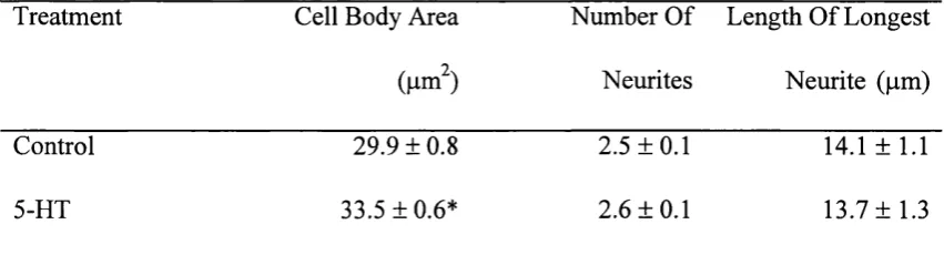
Cultures were fixed in 4% paraformaldehyde at 7 DIV and labelled for MAP-2. Only cells with clearly delineated processes were analyzed and drawn using a 40 X objective lens with the aid of a camera lucida attachment to a Leica D-20 bright field microscope. Neuronal images were traced onto a computer screen with a digitalizing mouse and then measured utilizing the TABLYT computer program (Dr. J. Cook, UCL, London) which automatically calculates cell body area, and length of neurites. 50 cells per coverslip were drawn, and 3 coverslips per treatment group assessed. Experiments were repeated twice.