International Journal of Nanomedicine 2017:12 7589–7602
International Journal of Nanomedicine
Dove
press
submit your manuscript | www.dovepress.com 7589
O r I g I N a l r e s e a r c h
open access to scientific and medical research
Open access Full Text article
Identification of lncRNAs involved in biological
regulation in early age-related macular
degeneration
Wei Zhu1,* Yi-Fang Meng1,* Qian Xing1 Jian-Jun Tao1 Jiong Lu1 Yan Wu2
1Department of Ophthalmology, Changshu No. 2 People’s Hospital, Changshu, China; 2Department of Ophthalmology, First Hospital Affiliated to Soochow University, suzhou, china
*These authors contributed equally to this work
Background: Age-related macular degeneration (AMD) is one of the most common causes of adult blindness in developed countries. However, the role of long noncoding RNAs (lncRNAs) in the development and progression of early AMD is unclear.
Methods: We established the lncRNA profile of early AMD by reannotation of microarrays from the gene expression omnibus database. Quantitative real-time polymerase chain reaction was used to determine the expression of selected lncRNAs.
Results: The expression profiles of 9 cases of AMD and 7 controls were studied. A total of 266 differentially expressed genes (DEGs) were detected (94 upregulated and 172 downregulated). Among all the DEGs, 64 were lncRNAs. Advanced bioinformatics analyses demonstrated that differentially expressed lncRNAs could play significant roles in visual perception, sensory perception of light stimulus, and cognition. The pathway analyses showed that the two most significantly influenced Kyoto Encyclopedia of Genes and Genomes pathways were those of phototransduction and purine metabolism. By the analyses of the key lncRNAs, it was found that RP11-234O6.2 was downregulated in the aging retinal pigment epithelium (RPE) cellular model. Exogenous RP11-234O6.2 treatment led to increased cell viability and improved apop-tosis but it did not affect the cell migration ability of aging RPE cells.
Conclusion: This study indicated that lncRNAs are differentially expressed in early AMD and may produce important regulative effects. An lncRNA, RP11-234O6.2, might be involved in the biological regulation of early AMD and have therapeutic potential.
Keywords: age-related macular degeneration, lncRNAs, microarray, bioinformatics analyses
Introduction
Age-related macular degeneration (AMD) is an age-related multifactorial disease that is regarded as the most common cause of adult blindness in many developed countries.1 It is the primary cause of loss of central vision and significantly decreases
the individual’s quality of life. In general, AMD is divided into early and late stages. Late AMD is regarded as neovascular AMD characterized by neovascularization from the choriocapillaris.2 At present, anti-vascular endothelial growth factor is used to treat
neovascular AMD and has been shown to benefit patients with AMD. Considering the increasing prevalence of the elderly in the population, the incidence rate of AMD is predicated to be higher in the future.3 Despite the fact that significant efforts have been
made to identify risk factors of AMD, currently, there are no recognized methods to prevent it.4 For these reasons, there is an urgent need to develop effective interventions
to prevent the progression of early AMD to late AMD. Except for smoking cessation and avoidance of cigarette smoke exposure, effective treatment options for early AMD Correspondence: Jiong Lu
Department of Ophthalmology, Changshu No. 2 People’s Hospital, No 99, Xiannan Street, Changshu, Jiangsu, 215000 China email cslujiong@163.com
Yan Wu
Department of Ophthalmology, First Hospital Affiliated to Soochow University, No 188, Shizi Street, Suzhou, Jiangsu, 215000 China email txwuyan@suda.edu.cn
Journal name: International Journal of Nanomedicine Article Designation: Original Research
Year: 2017 Volume: 12
Running head verso: Zhu et al
Running head recto: Differentially expressed lncRNAs in early AMD DOI: http://dx.doi.org/10.2147/IJN.S140275
International Journal of Nanomedicine downloaded from https://www.dovepress.com/ by 118.70.13.36 on 23-Aug-2020
For personal use only.
Number of times this article has been viewed
This article was published in the following Dove Press journal: International Journal of Nanomedicine
Dovepress
Zhu et al
cases are absent.5 Thus, more knowledge is required on the
pathogenesis of AMD; especially key would be insights into the pathophysiology of early AMD.
Given the aging population and the multifactorial nature of AMD, currently, no exact and single etiology of AMD has been identified. However, there are several fairly well-accepted molecular mechanisms that have been reported to be involved in the development and progression of AMD, including inflammation, oxidative damage, and abnormal angiogenesis. Interventions for these potential key factors for its pathogenesis may produce rational treatments for AMD. For instance, polyunsaturated fatty acid, which is both an antioxidant nutritional supplement and an anti-inflammatory agent, has been reported to provide significant benefits in AMD management.6,7 More advanced clinical and
labora-tory studies are now needed to characterize the detailed pathogenesis and to develop effective therapeutic agents for AMD targeting on the key pathways.
Long noncoding RNAs (lncRNAs), which are a class of noncoding RNAs with more than 200 nucleotides, play critical roles in the development of different kinds of human diseases.8 As reported in the previous studies, lncRNAs can
significantly affect epigenetic modification, cell differentia-tion, transcriptional reguladifferentia-tion, immune responses, and other biological processes.9 In a previous study on the murine
model of ocular neovascularization by microarray analyses, it was found that lncRNAs were significantly dysregulated.10
The results from advanced bioinformatics analyses suggested that lncRNAs play important roles in ocular neovascular-ization. However, no previous studies have focused on the role of lncRNAs in early AMD. Considering the extensive effect of lncRNAs on physiological function,11 it is
reason-able to think that lncRNAs can significantly influence the development and progression of early AMD. In the present study, we identified a set of lncRNAs that were differentially expressed in early AMD, confirmed the expression patterns of selected lncRNAs in an in vitro model, and performed a preliminary investigation into the biological effects of dys-regulated lncRNA on AMD development.
Materials and methods
Data sets
Expression profiles of retinal pigment epithelium (RPE)/ choroid from AMD cases and controls were downloaded from the gene expression omnibus (GEO) database (https:// www.ncbi.nlm.nih.gov/geo).12 All of the profiles were from
participants from the Department of Ophthalmology and Visual Sciences, the University of Iowa. A total of 9 AMD
cases and 7 controls were included in the data analyses. Early AMD in this study was defined as RPE changes (depigmen-tation or hypo pigmen(depigmen-tation) and/or macular drusen without geographic atrophy or choroidal neovascularization. A total of 5 females and 4 males were included in the AMD group, while there were 3 females and 4 males in the control group (P=0.500). The age distribution in the AMD (84.22±6.553) and control (83.00±5.657) groups was matched in this study (P=0.873). As complement factor H (CFH) was regarded as one of the most important genetic risk factors, the single nucleotide polymorphism rs1061170 of CFH was assessed. It was found that the distribution of high-risk genotypes (YH/ HH) and low-risk genotypes (YY) was matched between the case and control groups (P=0.596). An exon-based array (the Affymetrix Human Exon ST 1.0 array) was obtained for the microarray analysis.
Data processing and probe reannotation
Microarray re-annotations were divided into two processes: detection of differentially expressed genes (DEGs) and differ-entially expressed lncRNAs. For the detection of DEGs, the downloaded cell data of GSE50195 based on GPL17629 were added to the information of protein classes (protein_coding, lncRNA, and miRNA). The linear models for microarray data (LIMMA) package was obtained for bioinformatics analyses using R and Bioconductor.13 All the DEGs were presented
as both heatmap and volcano plots. In order to extract the lncRNAs from the DEGs, the lncRNA label in protein classes was chosen, and all the differentially expressed lncRNAs were presented in an independent heatmap and were used for advanced functional analyses.
construction of lncrNa–mrNa
coexpression network
To date, the exact biological roles of most lncRNAs are still unclear, and it was reasonable to conduct the bioinformatics functional analyses of the coexpressed mRNAs of related lncRNAs. Functionally related genes generally demonstrated similar expression profiles, and this suggests that these genes may have regulatory relationships. In this study, weighted correlation network analysis14 was used to identify all the
DEGs (adjacency threshold =0.6). Among all the coex-pression nodes, we selected the nodes that demonstrated a coexpression relationship between lncRNA and mRNA and thus constructed an lncRNA–mRNA coexpression network. In order to verify the lncRNA–mRNA coexpression results, advanced validation analyses by sequencing results were con-ducted by using RIsearch analysis software;15 this software
International Journal of Nanomedicine downloaded from https://www.dovepress.com/ by 118.70.13.36 on 23-Aug-2020
Dovepress Differentially expressed lncRNAs in early AMD
could detect the interaction between the selected lncRNAs and all coding genes (free energy #20). The lncRNA and mRNA pairs, which demonstrated both a coexpression rela-tionship and interactions, were then recorded as an extreme lncRNA–mRNA coexpression network, and a graphical output was obtained by using Cytoscape.16
Bioinformatics analyses
The lncRNA-coexpressed mRNAs were used in the func-tional enrichment analysis. In order to investigate the potential biological role of related mRNAs, the analyses were conducted by using the Database for Annotation, Visualiza-tion and Integrated Discovery (DAVID) for the annotaVisualiza-tion and functional analysis.17 Kyoto Encyclopedia of Genes and
Genomes (KEGG) pathway analyses were also conducted to show gene set enrichment analysis. In the current study, the clusterProfiler R package was used to conduct the pathway analyses of the DEGs,18 and the Pathview was used to
visualize the results of the enrichment pathway analyses.19
Cell culture and the in vitro aging model
Cultured retinal pigmented epithelium (ARPE-19) cells were purchased from American Type Culture Collection (Rockville, MD, USA). The ARPE-19 cells were grown in Dulbecco’s Modified Eagle’s Medium nutrient mixture F-12 (DMEM/F12; Invitrogen Corporation, Carlsbad, CA, USA) supplemented with 10% fetal bovine serum (FBS) and 1% penicillin/streptomycin. The ARPE-19 cells were used at 80% confluence for each experiment and were pas-saged every 3 days after being transferred into the culture medium. All the cells were incubated at 37°C in a humidified atmosphere of 5% CO2.
As reported in our previous study, a pulsed H2O2 exposure aging cell model was obtained in this study.20 In general,
the cultured ARPE-19 cells were incubated with complete medium containing 800 µM H2O2 for 2 hours every day, and the aging ARPE-19 cells on the 8th day could be used as aging groups.
RNA extraction and quantitative
polymerase chain reaction (PCR)
Total RNA was isolated from the ARPE-19 cells by using TRIzol® reagent according to the manufacturer’s instructions
(Invitrogen). The concentration and quality of the extracted RNA were measured by using a NanoDrop™ 2000 spectro-photometer (Thermo Fisher Scientific, Waltham, MA, USA). Quantitative PCRs were conducted in a 20 µL reaction vol-ume using an SYBR Green mix. Glyceraldehyde-3-phosphate
dehydrogenase was used as an internal control. The fold changes of all RNA expression levels were calculated by using the 2−ΔΔCt method.
Vector construction and transfection
The full-length sequence of RP11-234O6.2 was synthesized and cloned into the pcDNA3.1 (Invitrogen, Shanghai, China) vector according to the manufacturer’s instructions. The transfection of pcDNA3.1–RP11-234O6.2 into ARPE-19 cells was done by using Lipofectamine 2000 (Invitrogen) at a final concentration of 50 nM. The relative expression levels of RP11-234O6.2 were detected by quantitative PCR.
Cell viability assay
Cell viability was monitored by using the Cell Counting Kit 8 (CCK-8; Dojindo, Shanghai, China). The ARPE-19 cells in each group were seeded onto 96-well plates at an initial density of 5,000 cells/well. Then, 10 µL of CCK-8 solution was added to each well, and they were incubated for 2 hours. Cell viability was measured at a wavelength of 450 nm (SpectraMax® Plus 384 Microplate Reader;
Molecular Devices, Sunnyvale, CA, USA). The data were calculated using the following formula: relative viability = A450(target)− A450(blank).
Cell migration detection
The impact of lncRNA on aging ARPE-19 cell migration was examined by the cell scratch test. ARPE-19 cells in each group were placed on poly-D-lysine-coated 12-well plates (Corning Inc., Corning, NY, USA) with a cell density of 2.5×10⁵ cells/well in DMEM/F2 1:1 culture media without
FBS. A 200-µL pipette tip was used to perform scratches, and all the processed cells were divided into control and lncRNA-treated groups. After 36 hours, distances from the initial scratches were measured. Each detection test was repeated with three independent experiments.
apoptosis detection
In this study, the apoptosis level was detected by using a flow cytometry method with an annexin V-fluorescein isothiocya-nate apoptosis detection kit. The cultured cells in each group were harvested and washed with ice-cold PBS for 2 hours. All the cells were then treated with ice-cold 70% ethanol and then resuspended in 1 mL PBS. Annexin-V fluorescein iso-thiocyanate and propidium iodide (PI) solutions were added and incubated in the dark for 20 minutes. The results were expressed as the percentage of both early (annexin V+/PI−)
and late apoptosis (annexin V+/PI+) cells.
International Journal of Nanomedicine downloaded from https://www.dovepress.com/ by 118.70.13.36 on 23-Aug-2020
Dovepress
Zhu et al
Statistical analysis
The results were expressed as means ± SD of the three repeated experiments. The difference between the means in each index was calculated by using the Student’s t-test. A p-value of ,0.05 was considered statistically significant. All the sta-tistical analyses were conducted by using Prism GraphPad 5 software (GraphPad Software Inc., La Jolla, CA, USA).
Results
DEGs and dysregulated lncRNAs
From the GEO database, the cell files of GSE50195 were downloaded for advanced probe reannotation. Using LIMMA with a P-value ,0.01 and |logFC| .0.5 as the threshold, a total of 266 DEGs were identified (94 upregulated and 172 downregulated). As shown in Figure 1A, the DEGs were
8
6
4
2
0
–4 –2 0
logFC
–log10
(P
-value
)
2 4
Color key and histogram
Control group AMD group
800
600
Count 400
200
0 Value 2 6 10
Control group AMD group
140 100
Count
Color key and histogram
Value 80 60 40 20 0
2 4 6 8
A
B
C
Figure 1 Differentially expressed genes and lncRNAs of early AMD cases (9 cases versus 8 controls).
Notes: (A) Volcano plot of differentially expressed genes for early AMD. The red points are marked as genes of significance. (B) Clustering heatmap of 266 differentially expressed genes. (C) Clustering heatmap of 64 differentially expressed lncRNAs. AMD group (GSM1215520–GSM1215528) and control group (GSM1215529–GSM1215535) are marked. Red is for high expression, and green is for low expression. Each column represents one sample, and each row represents one gene/lncRNA.
Abbreviations: AMD, age-related macular degeneration; lncRNA, long noncoding RNA.
International Journal of Nanomedicine downloaded from https://www.dovepress.com/ by 118.70.13.36 on 23-Aug-2020
Dovepress Differentially expressed lncRNAs in early AMD
marked red in the volcano plot. Cluster analysis for all the DEGs was conducted, and the expression levels were marked from red to blue (Figure 1B). In advanced analyses, it was found that a total of 64 lncRNAs were differentially expressed when comparing early AMD cases with normal controls. Among the 64 detected lncRNAs, 36 lncRNAs were downregulated and 28 lncRNAs were upregulated. Figure 1C shows the heatmap plot of cluster analysis for the 64 differentially expressed lncRNAs. Table 1 lists the top 20 differentially expressed lncRNAs. The gene ID and symbol as well as P-values were also recorded.
lncrNa–mrNa coexpression network
analyses
As the detailed functions of lncRNAs were not completely known, the analyses of the coexpressed mRNA would help in the advanced function annotations. In general, it was reasonable to consider that a close expression pattern predicated related gene function. In this study, the lncRNA– mRNA coexpression analysis detected a total of 6 lncRNAs: RP11-234O6.2, RP11-11K13.1, AF131216.6, AC000124.1, LINC00948, and RP11-22M7.2. Figure 2 presents the graphical lncRNA–mRNA coexpression relationship, and it was found that RP11-234O6.2 demonstrated the largest number of coexpression relationship. Considering that RP11-234O6.2 was coexpressed with one lncRNA (LINC00948) and 20 mRNAs, it could be conjectured that it might play an important role in the early AMD development.
The validation of lncRNA–mRNA relationship was also done using RIsearch by the sequence alignment. Through the interaction relationship analysis, we selected the lncRNA– mRNA pairs, which demonstrated both coexpression and interaction relationships, for advanced analyses. As reported in Table 2, a total of three lncRNAs (ie, RP11-11K13.1, AF131216.6, and RP11-234O6.2) and their related mRNAs were described.
Functional enrichment analyses
Functional enrichment analyses, which have been widely used in high-throughput data analysis to indicate the function of large-scale genes, would help to understand the role of dys-regulated lncRNAs in the development of AMD. Among all the functional enrichment methods, the DAVID is one of the most commonly used integrated data-mining environments, which was used in this study. In this study, a total of 19 gene ontology (GO) terms were detected, and Figure 3A presents these GO terms as well as their related DEGs. Among all the GO terms of significance, visual perception (GO: 0007601, false discovery rate [FDR] =2.38e−18) and sensory perception
of light stimuli (GO: 0050953, FDR =2.38e−18) demonstrated
the most important effects. Figure 3 presents all the detected GO terms and the significance indexes.
Meanwhile, KEGG pathway analysis was a useful tool in the visualization of the enriched pathways. In this study, two independent pathways, hsa04744 (phototransduction) and hsa00230 (purine metabolism), were associated with the
Table 1 Top 20 differentially expressed long noncoding RNAs in early AMD
ID logFC t P-value Symbol
ENSG00000225649 −0.775314345 −6.260045806 7.65e–06 AC064875.2
ENSG00000248677 0.561298878 5.77399922 2.03E–05 CTD-2044J15.1
ENSG00000233705 0.514802877 5.531509843 3.33E–05 SLC26A4-AS1
ENSG00000205452 0.849909105 5.441902994 4.01E–05 RP11-812E19.3
ENSG00000258683 −0.671719098 −5.007665761 9.99e–05 RP11-11K13.1
eNsg00000257120 −1.012223924 −4.705291702 0.000190741 CTD-2503I6.1
ENSG00000229245 −0.707584769 −4.673941743 0.000204068 RP11-542K23.7
ENSG00000249797 −0.954373352 −4.651608226 0.000214138 CTD-3179P9.1
ENSG00000253898 0.572160865 4.584293323 0.000247655 RP11-51M18.1
ENSG00000255311 −0.827712084 −4.369916079 0.000394412 RP11-160H12.2
ENSG00000255480 0.810803659 4.330604017 0.000429689 RP4-710M3.2
eNsg00000229267 0.748573134 4.262839387 0.000498169 ac072062.1
ENSG00000249568 −0.704593601 −3.051932202 0.005047204 RP11-234O6.2
eNsg00000259285 0.569040604 4.256555244 0.000505054 CTD-2330J20.2
ENSG00000240758 −0.948553638 −4.207281836 0.000562481 RP11-155G14.6
ENSG00000236837 −0.600514564 −3.955002408 0.00097778 AC139712.4
ENSG00000242339 0.709975451 3.944008193 0.00100167 RP11-735B13.2
ENSG00000246863 −0.591744536 −3.921314683 0.001052852 RP11-325N19.3
ENSG00000254610 −1.025245637 −3.841380386 0.001254953 RP5-903G2.2
ENSG00000233620 −1.415375474 −3.789946295 0.00140511 RP11-22M7.2
Abbreviation: AMD, age-related macular degeneration.
International Journal of Nanomedicine downloaded from https://www.dovepress.com/ by 118.70.13.36 on 23-Aug-2020
Dovepress
Zhu et al
development of early AMD. Figure 4 presents the two KEGG pathways, and the downregulated genes are marked green in the pathways. When the DEGs were considered, there were 9 and 6 gene counts in hsa04744 and hsa00230, respectively.
Downregulation of RP11-234O6.2 in
early AMD
By the functional enrichment analysis, it was found that RP11-234O6.2 played a key role in the AMD development by both coexpression and interaction analyses. Advanced independent analyses showed that coexpressed mRNAs of RP11-234O6.2 were mainly listed in phototransduction, which was one of the most significant KEGG pathways.
These advanced analyses on the expression of RP11-234O6.2 demonstrated an important role. RP11-RP11-234O6.2, whose transcript is ENST00000508189.1, was located in Chromosome 4.
From the microarray analyses of RPE/choroid samples from early AMD cases, it was found that the relative expression of RP11-234O6.2 was significantly decreased compared with controls. In order to detect the expression pattern of RP11-234O6.2 in the aging RPE in vitro model, a real-time PCR method was used to detect the content of RP11-234O6.2. Compared with the normally cultured ARPE-19, it was found that RP11-234O6.2 was significantly downregulated in the aging RPE model. Figure 5A and B show the detailed expression data of RP11-234O6.2 in both tissue samples and in vitro models.
RP11-234O6.2 regulates biological
function of aging RPE cells
In order to detect the biological effects of RP11-234O6.2 on the RPE in vitro aging model, we conducted a series of exper-iments, including cell viability, migration, and apoptosis. In this study, it was found that RP11-234O6.2 treatment would upregulate the expression level of RP11-234O6.2 in the aging model (Figure 5C). When considering the general function, it was found that cell viability was significantly decreased in
Figure 2 Coexpression of differentially expressed lncRNA and related mRNAs.
Notes: The blue circle represents lncRNAs, the green circle represents protein-coding genes, and the relationship between the two circles represents the coexpression relationship between them.
Abbreviation: lncRNA, long noncoding RNA.
Table 2 list of lncrNa–mrNa coexpression network after interaction analysis
lncRNA Protein coding Adjacency cor Free energy
RP11-11K13.1 MAP2 0.618646 −22.87
RP11-11K13.1 rDh8 0.604374 −24.17
RP11-11K13.1 MPP4 0.60982 −33.46
AF131216.6 SNAP25 0.609362 −20.47
LINC00948 OPN1MW 0.673562 −25.71
RP11-234O6.2 ROM1 0.605834 −24.38
LINC00948 GPR98 0.664874 −26.55
AF131216.6 TMEM244 0.617153 −22.13
Abbreviations: cor, correlation; lncRNA, long noncoding RNA.
International Journal of Nanomedicine downloaded from https://www.dovepress.com/ by 118.70.13.36 on 23-Aug-2020
Dovepress Differentially expressed lncRNAs in early AMD
GO:0009314 response to radiation
GO:0009881 photoreceptor activity
GO:0050908 detection of light stimulus
GO:0009584 detection of visible light
GO:0007602 phototransduction
GO:0051606 detection of stimulus
The detected GO terms
The number of related genes GO:0050877 neurological system process
GO:0050890 cognition
GO:0007601 visual perception
0 5 10 15 20 25
GO:0009416 response to light stimulus
A
B
Figure 3 Functional enrichment of the differentially expressed lncRNA-related mRNAs.
Notes: (A) Graphical presentation of the detected GO terms. Diamonds represent GO term and circles represent genes. The red circles show the upregulated gene and the green circles the downregulated genes. (B) The GO terms of significance are listed with their values of significance.
Abbreviations: GO, gene ontology; lncRNA, long noncoding RNA.
the aging model. After exogenous RP11-234O6.2 treatment, cell viability was significantly improved compared with the aging group (Figure 6; P,0.01). By the cell scratch tests, it was found that the migration rate of the aging RPE cell group
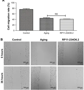
was significantly decreased compared with the normal group, whereas it was found that RP11-234O6.2 treatment did not influence the migration effects of aging RPE cells (Figure 7). Through flow cytometry detection, it was found that both the
International Journal of Nanomedicine downloaded from https://www.dovepress.com/ by 118.70.13.36 on 23-Aug-2020
Dovepress
Zhu et al
International Journal of Nanomedicine downloaded from https://www.dovepress.com/ by 118.70.13.36 on 23-Aug-2020
Dovepress Differentially expressed lncRNAs in early AMD
Figure 4
The detected KEGG pathway of significance.
Notes:
(
A
)
Phototran
sduction
pathway.
(
B
)
Purine
metabolism
pathway.
Each
panel
represents
one
gene.
Red
is
for
high
expression,
and
green
is
for
low
expression.
Reprodu
ced
with
permission
from
Kanehisa
M,
Furumichi
M,
Tanabe
M,
Sato
Y,
Mor
ishima
K.
KEGG:
new
perspectives
on
genomes,
pathways,
diseases
and
drugs.
Nucleic
Acids
Res
. 2017;45(D1):D353–D361.
©
The
Author(s)
2016.
Published
by
Oxford
University
Press
on
behalf
of
Nucleic
Acids
r
esearch.
33
Abbreviations:
KEGG, Kyoto Encyclopedia of Genes and Genomes;
+
p, phosphorylation
; hv, rovided energy under light conditions; h, Planck constant; v, freque
ncy.
International Journal of Nanomedicine downloaded from https://www.dovepress.com/ by 118.70.13.36 on 23-Aug-2020
Dovepress
Zhu et al
early and late apoptosis rates were significantly increased in the aging group. After RP11-234O6.2 treatment, early apoptosis (P=0.0008), but not late apoptosis (P=0.0848), was significantly improved (Figure 8).
Discussion
LncRNAs demonstrate important effects and participate in different biological processes. AMD is a multifactorial disease and might also be associated with the dysregula-tion of lncRNAs. The primary purpose of this study was to detect their global expression patterns in the early AMD. A total of 64 differentially expressed lncRNAs were identi-fied in this study. The advanced bioinformatics analyses of the coexpressed mRNAs showed that a total of 19 GO terms were detected. KEGG pathway analyses showed that two independent pathways, phototransduction and purine metabolism, were associated with the development of early AMD. Based on the functional analyses, one of the differ-entially expressed lncRNAs, RP11-234O6.2, demonstrated important potential effects. Advanced in vitro studies based on the aging RPE model showed that RP11-234O6.2 could improve cell viability and early apoptosis.
Early AMD has been reported to be influenced by oxida-tive stress, cell apoptosis, abnormal lipid metabolism, and dysfunction of the immune system,21 whereas lncRNAs
&RQWURO $JLQJ 532
&HOOYLDELOLW\
Figure 6 Cell viability of aging RPE cells influenced in RP11-234O6.2 in vitro.
Notes: Cell viability was significantly decreased in the aging group, and RP11-234O6.2 could improve the cell viability significantly. ***P,0.001.
Abbreviation: RPE, retinal pigment epithelium.
&RQWURO
$0'FDVHV
0LFURDUUD\RIFOLQLFDO VDPSOHV
5HODWLYHH[SUHVVLRQ RI53
2
&RQWURO 'D\ 'D\ 'D\
573&5RILQYLWUR VDPSOHV
5HODWLYHH[SUHVVLRQ RI53
2
$
%
5HODWLYHH[SUHVVLRQ RI53
2
$JLQJ 532
WUHDWHG
&
Figure 5 RP11-234O6.2 was differentially expressed in early AMD.
Notes: (A) RP11-234O6.2 was downexpressed in early AMD cases by microarray analyses; (B) RP11-234O6.2 was downexpressed during aging RPE cell model by RT-PCR; (C) RP11-234O6.2 vector treatment could significantly increase the expression of RP11-234O6.2 in aging ARPE-19 cells. *P,0.05, **P,0.01, ***P,0.0001.
Abbreviations: AMD, age-related macular degeneration; RPE, retinal pigment epithelium; RT-PCR, real-time polymerase chain reaction.
International Journal of Nanomedicine downloaded from https://www.dovepress.com/ by 118.70.13.36 on 23-Aug-2020
Dovepress Differentially expressed lncRNAs in early AMD
P P P
P P P
&RQWURO
KRXUV
KRXUV
$JLQJ 532
%
16
&RQWURO $JLQJ 532
&HOOPLJUDWLRQUDWH
$
Figure 7 Cell migration ability of aging RPE cells influenced in RP11-234O6.2 in vitro.
Notes: (A) The graphs of each scratch test for 0 and 36 hours. (B) Results for cell migration test. It was found that RP11-234O6.2 treatment did not influence the migration effects of aging RPE cells.
Abbreviations: NS, not significant; RPE, retinal pigment epithelium.
might also be associated with these effects in different disorders. In a previous study on RNA-Seq data sets, dys-regulated lncRNAs were detected which were associated with oxidative stress in cholangiocarcinoma.22 It was reported
that H19 and HULC were upregulated by oxidative stress and regulated cholangiocarcinoma cell migration and inva-sion by targeting interlukin-6 and CXCR4. In a study on diabetic retinopathy (DR), the results indicated that lncRNA MIAT regulated apoptosis in DR.23 Lipid metabolism was
also regarded as a key process in AMD incidence, and a previous study demonstrated that 13 potential lncRNA– mRNA gene pairs were associated with the dysregulation of lipid metabolism in thyroid gland.24 In a study on
myo-cardial infarction and lncRNA expression profiles, the functional analysis demonstrated that a total of 9 lncRNAs were involved in myocardial innate immune responses, and the dysregulation of these lncRNAs led to abnormalities of the immune system.25
Knowledge on the regulatory effects of lncRNAs on AMD progression, and the detailed functions of lncRNAs on the incidence of early AMD, is quite limited. In a previous study, the detection of dysregulated lncRNAs in pathological neovascularization provided evidences of late AMD develop-ment.10 However, it focused on late AMD, and most attention
was paid to neovascularization, which was quite different during early AMD. Several previous studies reported that lncRNAs were associated with epithelial–mesenchymal tran-sition of RPE cells,26 maintenance of retinal architecture,27
and regulation of microvascular dysfunction in DR.28 In this
study, we conducted an overall review of the expression patterns of lncRNAs in early AMD. It was found that a total of 64 lncRNAs were dysregulated in early AMD. Through advanced functional analyses, it was found that lncRNAs may play a key role in the early AMD development. The mapping of lncRNA-related dysregulated mRNAs showed that most DEGs were located in phototransduction and purine
International Journal of Nanomedicine downloaded from https://www.dovepress.com/ by 118.70.13.36 on 23-Aug-2020
Dovepress
Zhu et al
4 4
4 4
),7&$ =/-&21
$
%
&
'
3,$
4 4
4 4
),7&$ =/-,0
3,$
4 4
4 4
),7&$
=/-6%,0
3,$
&RQWURO $JLQJ 532
$SRSWRVLVUDWH
(DUO\DSRSWRVLV /DWHDSRSWRVLV
Figure 8 The apoptosis changes in each group by flow cytometry detection.
Notes: (A) The normal group; (B) the aging group; (C) aging RPE cells after RP11-234O6.2 treatment; (D) results for apoptosis test; early apoptosis (P=0.0008), but not late apoptosis (P=0.0848), was significantly improved by RP11-234O6.2.
Abbreviations: FITC, fluorescein isothiocyanate; PI, propidium iodide; RPE, retinal pigment epithelium.
metabolism KEGG pathways. As reported in a previous study, the downregulation of phototransduction was a key process in blue light damage of retinas.29 Considering that
light damage is regarded as a risk factor for AMD and the light damage model was used in the early AMD research, it was quite reasonable to regard phototransduction as the most significantly influenced pathway. The other pathway addressed in the analyses of microarray data was purine metabolism. Purinergic signaling has been reported to be
involved in the degeneration of the injured retina.30 Based
on previous research, purine metabolism was associated with the proliferation of retinal glial cells and demonstrated protective effects on retinal degeneration. The finding of this study provided improved understanding of the role of lncRNAs on AMD development by both phototransduction and purine metabolism. Further studies on lncRNA regulation of these two processes may help elucidate the development of AMD.
International Journal of Nanomedicine downloaded from https://www.dovepress.com/ by 118.70.13.36 on 23-Aug-2020
Dovepress Differentially expressed lncRNAs in early AMD
In this research, we evaluated the expression level of RP11-234O6.2 in vitro and conducted a series of laboratory studies addressing the biological effects of RP11-234O6.2 on the aging model. As far as we know, there are no previous studies on the biological effects of RP11-234O6.2 in other disorders. In this study, the initial data showed that RP11-234O6.2 was downregulated in early AMD. Advanced exogenous RP11-234O6.2 applications showed protective effects by improving cell viability and decreasing early apoptosis rate. The data in this study expand the repertoire of lncRNAs and mRNAs and contribute to our understanding of the molecular mechanisms involved in the development of early AMD. However, one limitation of this study should be recognized. It was difficult to conduct a functional bioinfor-matics analysis on lncRNAs at this time. The strategy used for analysis, obtained from a previous work,32 was to examine
the functions of coexpressed mRNAs. The basic idea was that the relationship between lncRNAs and the coexpressed mRNAs would reflect a potential regulatory relationship. However, the effects of lncRNAs were highly variable and were usually demonstrated directly on DNA and proteins.31
The bioinformatics analyses in this study mainly focused on lncRNA–mRNA regulation, and this certainly could lead to misperceptions of the role of lncRNA on AMD develop-ment. Thus, more regulated methods will be required, and the conclusions based on the results of these bioinformatics analyses should be considered with caution.
Conclusion
This study was designed to detect expressed lncRNAs in early AMD by integrating public microarray data. Based on the reannotations of microarray data and functional analyses, it was found that lncRNA–mRNA coexpression appears to play an important role in early AMD development. Among the 64 detected lncRNAs, RP11-234O6.2 was one of the key lncRNAs and is predicated to exert significant regulatory effects. The pattern of expression of RP11-234O6.2 was vali-dated in an in vitro model, and RP11-234O6.2 demonstrated protective effects on the aging RPE model. Considering that preliminary data were obtained in this study, further experi-ments will be needed to verify the expression and detailed mechanisms of dysregulated lncRNAs in early AMD.
Acknowledgments
This work was supported in whole or in part by the Founda-tion for Young Scholars of Suzhou, China (grant number kjxw2015044) and the Foundation for Young Medical Talents
of Jiangsu Province, 2016 (grant number QNRC2016211). The sponsor and funding organizations had no role in the design or conduct of this research.
Disclosure
The authors report no conflicts of interest in this work.
References
1. Deemer AD, Massof RW, Rovner BW, Casten RJ, Piersol CV. Functional outcomes of the low vision depression prevention trial in age-related macular degeneration. Invest Ophthalmol Vis Sci. 2017; 58(3):1514–1520.
2. Balaiya S, Abu-Amero KK, Kondkar AA, Chalam KV. Sirtuins expression and their role in retinal diseases. Oxid Med Cell Longev. 2017;2017:3187594.
3. McGuinness MB, Karahalios A, Finger RP, Guymer RH, Simpson JA. Age-related macular degeneration and mortality: a systematic review and meta-analysis. Ophthalmic Epidemiol. 2017;24:141–152. 4. Pennington KL, DeAngelis MM. Epidemiology of age-related macular
degeneration (AMD): associations with cardiovascular disease pheno-types and lipid factors. Eye Vis (Lond). 2016;3:34.
5. Garg A, Yang J, Lee W, Tsang SH. Stem cell therapies in retinal dis-orders. Cells. 2017;6(1):pii: E4.
6. Koskela A, Reinisalo M, Petrovski G, et al. Nutraceutical with res-veratrol and omega-3 fatty acids induces autophagy in ARPE-19 cells.
Nutrients. 2016;8(5):pii: E284.
7. Zhu W, Wu Y, Meng YF, Xing Q, Tao JJ, Lu J. Fish consumption and age-related macular degeneration incidence: a meta-analysis and systematic review of prospective cohort studies. Nutrients. 2016; 8(11):pii: E743.
8. Cao MX, Jiang YP, Tang YL, Liang XH. The crosstalk between lncRNA and microRNA in cancer metastasis: orchestrating the epithelial-mesenchymal plasticity. Oncotarget. 2016;8(7):12472–12483. 9. Wan P, Su W, Zhuo Y. Precise long non-coding RNA modulation in visual
maintenance and impairment. J Med Genet. 2016;54(7):450–459. 10. Xu XD, Li KR, Li XM, Yao J, Qin J, Yan B. Long non-coding RNAs:
new players in ocular neovascularization. Mol Biol Rep. 2014;41(7): 4493–4505.
11. Degirmenci U, Lei S. Role of lncRNAs in cellular aging. Front
Endo-crinol (Lausanne). 2016;7:151.
12. Whitmore SS, Braun TA, Skeie JM, et al. Altered gene expression in dry age-related macular degeneration suggests early loss of choroidal endothelial cells. Mol Vis. 2013;19:2274–2297.
13. Diboun I, Wernisch L, Orengo CA, Koltzenburg M. Microarray analysis after RNA amplification can detect pronounced differences in gene expression using limma. BMC Genomics. 2006;7:252.
14. Langfelder P, Horvath S. WGCNA: an R package for weighted cor-relation network analysis. BMC Bioinformatics. 2008;9:559. 15. Wenzel A, Akbasli E, Gorodkin J. RIsearch: fast RNA-RNA interaction
search using a simplified nearest-neighbor energy model.
Bioinformat-ics. 2012;28(21):2738–2746.
16. Shannon P, Markiel A, Ozier O, et al. Cytoscape: a software environ-ment for integrated models of biomolecular interaction networks.
Genome Res. 2003;13(11):2498–2504.
17. Dennis G Jr, Sherman BT, Hosack DA, et al. DAVID: database for annotation, visualization, and integrated discovery. Genome Biol. 2003; 4(5):P3.
18. Yu G, Wang LG, Han Y, He QY. clusterProfiler: an R package for comparing biological themes among gene clusters. OMICS. 2012;16(5): 284–287.
19. Luo W, Brouwer C. Pathview: an R/Bioconductor package for path-way-based data integration and visualization. Bioinformatics. 2013; 29(14):1830–1831.
International Journal of Nanomedicine downloaded from https://www.dovepress.com/ by 118.70.13.36 on 23-Aug-2020
International Journal of Nanomedicine
Publish your work in this journal
Submit your manuscript here: http://www.dovepress.com/international-journal-of-nanomedicine-journal
The International Journal of Nanomedicine is an international, peer-reviewed journal focusing on the application of nanotechnology in diagnostics, therapeutics, and drug delivery systems throughout the biomedical field. This journal is indexed on PubMed Central, MedLine, CAS, SciSearch®, Current Contents®/Clinical Medicine,
Journal Citation Reports/Science Edition, EMBase, Scopus and the Elsevier Bibliographic databases. The manuscript management system is completely online and includes a very quick and fair peer-review system, which is all easy to use. Visit http://www.dovepress.com/ testimonials.php to read real quotes from published authors.
Dovepress
Dove
press
Zhu et al
20. Zhu W, Wu Y, Meng YF, et al. Effect of curcumin on aging retinal pigment epithelial cells. Drug Des Devel Ther. 2015;9:5337–5344. 21. Handa JT, Cano M, Wang L, Datta S, Liu T. Lipids, oxidized lipids,
oxidation-specific epitopes, and age-related macular degeneration.
Biochim Biophys Acta. 2017;1862(4):430–440.
22. Wang WT, Ye H, Wei PP, et al. LncRNAs H19 and HULC, activated by oxidative stress, promote cell migration and invasion in cholangiocar-cinoma through a ceRNA manner. J Hematol Oncol. 2016;9(1):117. 23. Zhang J, Chen M, Chen J, et al. Long noncoding RNA MIAT acts
as a biomarker in diabetic retinopathy by absorbing of miR-29b and regulating cell apoptosis. Biosci Rep. 2017;37(2):pii: BSR20170036. 24. Shen Y, Mao H, Huang M, et al. Long noncoding RNA and mRNA
expression profiles in the thyroid gland of two phenotypically extreme pig breeds using Ribo-Zero RNA sequencing. Genes (Basel). 2016;7(7):pii: E34.
25. Sun C, Jiang H, Sun Z, Gui Y, Xia H. Identification of long non-coding RNAs biomarkers for early diagnosis of myocardial infarction from the dysregulated coding-non-coding co-expression network. Oncotarget. 2016;7(45):73541–73551.
26. Yang S, Yao H, Li M, Li H, Wang F. Long non-coding RNA MALAT1 mediates transforming growth factor beta1-induced epithelial-mesen-chymal transition of retinal pigment epithelial cells. PLoS One. 2016; 11(3):e0152687.
27. Krol J, Krol I, Alvarez CP, et al. A network comprising short and long noncoding RNAs and RNA helicase controls mouse retina architecture.
Nat Commun. 2015;6:7305.
28. Yan B, Yao J, Liu JY, et al. lncRNA-MIAT regulates microvascular dysfunction by functioning as a competing endogenous RNA. Circ Res. 2015;116(7):1143–1156.
29. Calzia D, Panfoli I, Heinig N, et al. Impairment of extramitochondrial oxidative phosphorylation in mouse rod outer segments by blue light irradiation. Biochimie. 2016;125:171–178.
30. Reichenbach A, Bringmann A. Purinergic signaling in retinal degeneration and regeneration. Neuropharmacology. 2016;104: 194–211.
31. Engreitz JM, Haines JE, Perez EM, et al. Local regulation of gene expression by lncRNA promoters, transcription and splicing. Nature. 2016;539(7629):452–455.
32. Zhang Q, Ge H, Jiang Y, Cheng B, Zhou D, Xu N. Microarray profil-ing analysis of long non-codprofil-ing RNAs expression in tendinopathy: identification for potential biomarkers and mechanisms. Int J Experi
Pathol. 2015;96(6):387–394.
33. Kanehisa M, Furumichi M, Tanabe M, Sato Y, Morishima K. KEGG: new perspectives on genomes, pathways, diseases and drugs. Nucleic
Acids Res. 2017;45(D1):D353–D361.
International Journal of Nanomedicine downloaded from https://www.dovepress.com/ by 118.70.13.36 on 23-Aug-2020