THE CLINICAL USE OF
NUCLEAR MAGNETIC
RESONANCE
SPECTROSCOPY FOR
STUDYING HUMAN
MUSCLE METABOLISM
E. B. Cady
Department of Medicine, Rayne Institute
University College London School of Medicine
ft D. Griffiths
Department of Medicine, Rayne Institute
University College London School of Medicine
ft H. T. Edwards
Department of Medicine, Rayne Institute
University College London School of Medicine
INTRODUCTION
Nuclear magnetic resonance (NMR) imaging has recently become an accepted technique in the medical practitioner's armory (38). NMR spectroscopy (44) is a subtly different application of the same physical principles underlying NMR im-aging, but the clinical potential for this modality is currently still under evaluation. The most important application of clinical NMR spectroscopy is for the
nonin-The research at University College London School of Medicine (UCLSM) is supported by the Well-come Trust, Action Research, the Special Trustees of University College Hospital, and the Muscular Dystrophy Group of Great Britain. The studies have been carried out with the assistance of Dr. P. S. Tofts, Mr. D. Delpy and Mr. A. Chu of the Department of Medical Physics (UCLSM), and Mr. N.Taylor of Kings College London. The advice and assistance of Prof. D. R. Wilkie, FRS, is gratefully acknowledged.
632 CADY ET AL.
vasive monitoring of changes in metabolite levels and intracellular pH of intact tissues during physiological stress or in response to pharmacological agents or disease. The 31phosphorus (3IP) nucleus has been the most commonly investigated in muscle disease (39) but the applications of proton ('H), (4,5,8) and l3carbon (I3C), (2,7) are currently being explored.
The main difference between NMR imaging and NMR spectroscopy is that in the former, protons (mainly in water) are positionally labeled by the application of magnetic field gradients so that the resonant frequency depends on the location of the 'H nucleus in the tissue. By the use of suitable combinations of field gra-dients, a three-dimensional proton distribution can be derived which can give an image in any selected plane. In NMR spectroscopy, a highly homogeneous, static magnetic field is applied to the sample. This enables the nuclei in different chem-ical species to resonate at resolvable frequencies and so produce a spectrum. The separation of signals from various chemical species is made possible by the fact that each nucleus experiences a total magnetic field consisting of the main field plus a local field depending on chemical structure, and hence the resonances are chemicaly shifted and separated in the frequency spectrum.
The information acquired from NMR spectra consists of determinations of relative metabolite concentrations from measurements of resonance line areas and estimates of the intracellular pH from the chemical shifts of inorganic phos-phate (Pi), (36) or the C4-H, C2-H, and N-H protons of carnosine (5).
The NMR method has some advantages over needle or open biopsy proce-dures. Being noninvasive, harmless (42), and painless, NMR spectra can be col-lected as frequently as the signal-to-noise ratio will allow, and the procedure is acceptable to young children. The metabolites are assayed in situ, and it is believed that realistic estimates are obtained (47), whereas using the needle biopsy pro-cedure, even the most skilful operator takes a few seconds to obtain and freeze the sample, allowing time for metabolite levels to change. Using NMR, typically 10 to 50 cm3 of tissue are examined, and the metabolite and pH results are more representative of overall muscle pathology, whereas the fact that needle biopsy gives only a small sample can be a problem in inhomogeneous tissue. Histological information, however, is not obtained, and hence NMR spectroscopy augments but does not replace the biochemical and histological uses of biopsy and other techniques.
METHODS
In order to pick up the NMR signal, it has been found most practical to use a surface coil antenna consisting of one or more loops of wire placed on the skin adjacent to the tissue of interest (1). The surface coil is used for pulse transmission to perturb the nuclei as well as for reception of the subsequent free induction decay (FID). Insulation between the patient and the coil is usually provided by a thin (approximately 1 mm) sheet of polytetrafluoroethylene (PTFE). Coils are tuned to the Rf for the particular nucleus and matched to the appropriate imped-ance while the limb is in position. The addition of a simple circuit allows the same coil to be "double tuned" for 'H, and this allows the homogeneity of the main field to be optimized (shimmed) using the strong H2O signal. Both these tasks can
be accomplished usually within 5 minutes. Parameters such as coil size and tuning (15), pulse length (related to flip angle), and pulse interval also need to be opti-mized to get maximum signal per unit time (23,31). For short pulse intervals, correction factors have to be determined for each metabolite to allow for insuf-ficient relaxation of the spin system between pulses. These factors can be esti-mated usually by collecting data with the normal pulse interval and then with a pulse interval several times the longest Tl (the spin-lattice relaxation time con-stant) so that the spin system is very nearly relaxed. Direct comparison of the resonance line areas then gives correction factors which can be applied to all subsequent data.
The signal has to be localized so that data are acquired only from the required tissue, and this can be achieved in several ways. The spatial sensitivity of circular surface coils as a function of flip angle and pulse repetition rate has been examined in detail (23,31). A degree of localization can be obtained by judicious positioning of the coil adjacent to the muscle and by appropriate selection of the transmitted pulse length, interval, or sequence (9,10,11). For 31P-NMR investigations, great benefit is obtained from the fact that most surface tissues do not contain detectable amounts of phosphorus metabolites, and simple muscle studies can be made by the use of a suitable size surface coil only. An alternative approach is to super-impose high-order profiled magnetic field gradients onto the main field (25), which are arranged so that only a defined volume of muscle experiences a homogeneous field. Nuclei in the field gradients resonate over a range of frequencies, and this broad signal can be removed by mathematical processing (26) to leave only the narrow resonance lines from the homogeneous region. In studies of muscle, signal localization has so far been achieved by the use of combinations of all these methods.
At the moment, techniques involving the application of field gradients are being developed to give the user more flexible localization and ability to define the shape, size, and position of the volume from which data is collected (6,12,43). These methods are somewhat handicapped by the low concentrations and sen-sitivities of most potentially interesting nuclei. For muscles near to the skin sur-face, the user may still be better off in many applications using the simple surface coil technique.
struc-a).
15 -10 -15
b).
RCOOR'
Glycerol C-1,3
C-2
- C H ,
PPM 200 150 50 -50
Figure 1. (a) 'H spectrum obtained using a 2.5 cm diameter coil placed adjacent to the extensor digitorum muscle. 8 x 75 (is pulses with a pulse interval of 1.256 s. The main peak is due to H2O protons; the other smaller resonances are due mainly to subcutaneous fat. (b) A 13C spectrum collected with a 4 cm surface coil
ture of organic molecules, I3C spectra usually consist of resonance line multiplets which complicate spectral analysis. These multiplets can be condensed to single resonances by simultaneously applying decoupling Rf at the 'H frequency. How-ever, as this can cause tissue heating, the use of decoupling has to be approached very cautiously in living systems. 'H studies of tissue are handicapped by the presence of a very large H2O resonance which completely dominates the
spec-trum. Various pulse sequence techniques exist for the reduction of this signal (33,40) although their usefulness with surface coils remains to be shown. The application of 'H-NMR to muscle shows great promise because of the greater sensitivity of the proton and the large number of potentially detectable metabolites including lactate, phosphocreatine (PCr), and creatine (4,5).
CLINICAL APPLICATIONS OF NMR SPECTROSCOPY TO MUSCLE DISEASE
Normal Muscle
In order to use NMR spectroscopy to monitor physiological or pathological changes in muscle tissue, it is necessary to know the normal ranges for resting metabolite levels and intracellular pH. Figures 1 and 2 show 'H, 13C, and 31P spectra obtained from normal, resting muscles. The 'H spectrum (Figure la) con-sists mainly of a strong H2O resonance and a small peak due to -CH2 protons in
triglycerides. Other resonances from metabolites of millimolar concentration such as creatine, phosphocreatine, and lactic acid are swamped by the strong H2O and
-CH2 signals. If no precautions are taken to reduce surface tissue signals, the
size of this -CH2 proton peak will depend largely on the subcutaneous fat
thick-ness. In the coupled I3C spectrum (Figure lb), the predominant feature is the triplet, due to -CH2 carbon nuclei in triglyceride chains. Other prominent
reso-nances are detected from -CH3, Glycerol, - O C - , and RCOOR' carbons. So far,
t h e m o s t illuminating results from m u s c l e tissue h a v e been obtained using 3 IP
NMR. The spectrum from normal, resting extensor digitorum (Figure 2) shows several prominent resonances from ATP (the 7-, a-, and (3-phosphorus nuclei), phosphocreatine (PCr), and inorganic phosphate (Pi). Signals from monoesters (mainly sugar-phosphates (SP) and phosphodiesters (PD) are also detected along with broad signals from immobile phosphorus in bone and phospholipids which in this spectrum have been removed mathematically to give a flat baseline. The PCr resonance usually has a very good signal-to-noise ratio and is commonly used as an internal reference for the determination of 3IP-NMR chemical shifts. The PCr line profile is narrow and its chemical shift is not pH dependent, although there is some variation with the Mg2+ concentration. In most muscle studies the PCr resonance makes a good reference. However, in instances of PCr depletion,
636 CADY ET AL.
PCr
15 10 -IB -15 -20 - 2 5 PPM
Figure 2. A 3IP spectrum obtained from the extensor digitorum muscle using the same coil as in Figure la. 256 x 13 (xs pulses with an interval of 2.256 s. Because of the very low concentrations of mobile 3IP metabolites in other adjacent tissues, metabolite levels determined from this spectrum are very close to those pertaining to the muscle tissue. Spectrum obtained with the assistance of N. Taylor.
it may be necessary to use other approaches, such as 'H H2O referencing (17).
Mean values for the metabolite levels (relative to a total mobile and NMR-visible phosphate pool of 1) and pH of normal, resting, male calf muscle tissue are given in Table 1 (28).
Changes in normal metabolite levels and pH can be induced by ischemia (19) or exercise (46), and time resolution better than a minute can usually be obtained using the NMR technique. Figure 3 shows the response of muscle tissue to oc-clusion of arterial blood flow to the arm by a sphygmomanometer cuff for 35 minutes. Gradual depletion of PCr and increase of Pi occurs until the cuff is released. PCr + Pi, ATP, and pH appear to stay constant throughout the study.
Table 1. Metabolite Ratios (Relative to Total Mobile Phosphate) for Normal Calf, Forearm, and Duchenne Dystrophy Calf.
P-ATP PCr Pi P-diester PCr/ATP PCr/Pi PCr + Pi
PH Normal n = (Mean) 0.11 0.47 0.08 0.06 4.23 6.02 0.55 7.05 Calf 5 (SD) 0.01 0.02 0.01 0.02 0.12 0.49 0.03 0.03 Normal n = (Mean) 0.11 0.49 0.08 0.03 4.44 6.71 0.57 7.04 Forearm = 24 (SD) 0.01 0.03 0.01 0.02 0.49 1.54 0.03 0.05 Duchenne Dystrophy Calf n = (Mean) 0.11 0.39a 0.14a 0.06 3.7Oa 2.92a 0.53 7.08 53 (SD) 0.02 0.04 0.03 0.02 0.75 0.81 0.03 0.10
" Significant difference (p < .001) between normal and Duchenne calf.
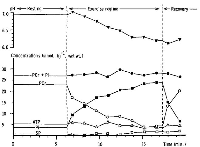
the ability to run complete studies of this type with minimal operator intervention. Figure 4 shows the response of normal muscle tissue to a typical exercise test. PCr is rapidly consumed with accompanying accumulation of Pi and fall in pH due to lactate accumulation from glycolysis. Recovery of metabolites and pH to resting values is complete within about 5 minutes. Studies of normal exercizing muscle tissue in young and old populations have shown no significant differences, indicating that muscle changes in the elderly are not due to alterations in energy production (45).
Duchenne Muscular Dystrophy
638 C A D Y E T A L .
P-METABOLITE AND pH CHANGES AT REST WITH ISCHAEMIA
•g 30 .
"3
e
e
Q-20 .
10 .
Ischaemia
....-,, s-**-*~~
PCr
y
p H
..°
••Pi
Tcr
Pi
* 7.2
PH units
7.0
J6.8
10 20 30 40 50
time min
Figure 3. Changes in 3IP metabolites and pH in normal resting forearm muscle
induced by occlusion of the arterial blood supply. Spectra were collected using a 6 cm surface coil placed over the forearm flexor muscle compartment. The pulse length was optimized for maximum signal: 32 pulses, pulse delay 2.256 s.
Other Myopathies
Abnormal resting muscle spectra have been seen in only a few cases of myopathy apart from muscular dystrophy. The abnormalities reveal themselves as decreased PCr/Pi ratios (24,39), a reported increase in phosphodiester signal in hypothyroid myopathy (34) or alkaline pH (39). Exercise studies produce more interesting results.
Several cases of phosphofructokinase (PFK) deficiency have been examined (18,22,30). Although resting metabolite levels and pH are normal, exercise leads to the detection by 31P-NMR of rapid accumulation of fructose-6-phosphate, a metabolite in the glycolytic pathway (see Figure 5). No significant change in pH has been reported in any study confirming the inability to produce lactic acid. On cessation of exercise, there is a slow recovery of PCr to the resting level. This could be due to the trapping of phosphate by fructose-6-phosphate generated by the enzyme block.
pH •«—Resting 7.0 -J
6 . 5
-Recovery
15 Time (min.)
Figure 4. Response of normal muscle tissue to anaerobic contractions. During the exercise regime the arterial circulation was occluded by the application of a sphygmomanometer cuff controlled by compressed air. Exercise consisted of 32 x 1 sec. 20% maximum isometric voluntary contractions interleaved with data acquisition every 2.256 sec. Subject performance was monitored by the use of a strain gauge applied to the back of the hand. On alternate data collections the muscle was relaxed. At the end of the complete exercise sequence the cuff was let down and the metabolite levels allowed to recover. Conditions of data col-lection as for Figure 3. Metabolite concentrations were estimated using a total mobile phosphate concentration of 49.5 mmol. kg"1 wet wt. Source: Reference
20.
anaerobic glycolysis because of a deficiency of the enzyme phosphorylase. This has been studied in four patients using 31P-NMR (39,41). The resting metabolite levels were normal, and average resting pH was 7.05. During exercise there was a marked decrease in PCr and an increase in Pi—but no intracellular acidosis— in the patients with McArdle's syndrome, while in normal subjects pH decreased significantly due to lactic acid production. In fact, during exercise in the my-ophosphorylase-deficient muscle, pHs were consistently alkaline and could be estimated very well because of the high Pi and PCr signal-to-noise ratios.
ab-640 CADY ET AL
15 10 -10 -15 -20 - 2 5 PPM
Figure 5. 31P NMR spectrum of phosphofructokinase- (PFK) deficient forearm muscle during aerobic exercise. Note the greatly increased sugar phosphate peak at about 7 ppm due mainly to the accumulation of unmetabolized fructose-6-phosphate. Data acquisition conditions as for Figure 3, but 64 pulses. The peak assignments are as for Figure 2.
normality may be due to a change in the relative contributions of glycolytic and oxidative processes. A low general mitochondrial activity in subjects with muscle pain has been described (35), and this therefore is not too surprising.
Hypothyroid myopathy has been investigated in two subjects. In one case, a raised phosphodiester level was reported (34), but in the other case (30), no sig-nificant abnormality at rest or during exercise and recovery was shown both before and 6 weeks after commencement of thyroxine, despite the subjects' regaining normal muscle function.
In a large number of patients with undiagnosed muscular pain and fatigue during or after exercise 31P-NMR has so far not shown any abnormality in spite of the possibility of energy supply defects. One group reports that out of 68 such cases, only 20% have shown a deviation from normal NMR results (39).
Monitoring Chemotherapy
a). Before.
12 3 4 5 6 7
15 10 5 0 -5 -10 -15 -20 -25
b). After.
PPM 15 10 - 1 0 - 1 5 - 2 0 - 2 5 PPM
642 CADY ET AL.
The most striking features were the presence of conspicuous peaks in the phos-phomonoester and diester regions of the spectra and reduced PCr/Pi and PCr/ ATP ratios when compared with the normal subject's. The only differences be-tween the tumor spectra before and after treatment were reductions in PCr/ATP to 75% and PCr/Pi to 70% of their initial values. In accord with the overall clinical situation and the absence of tumor resorption, this was taken to indicate deteri-oration. The ability to follow such changes indicates the potential for monitoring the effect of different cytotoxic chemotherapeutic regimes.
FUTURE DEVELOPMENT OF CLINICAL NMR SPECTROSCOPY FOR MUSCLE STUDIES
The more general application of NMR spectroscopy to muscle disease must be considered in the light of some of the other major clinical applications. 31P-NMR spectroscopy has already made a significant contribution to the study of neonatal cerebral metabolism (14,32,48). Further advances in the investigation of the brain and other organs in adults and children will require the use of large-bore whole-body spectrometers which are recently available. Systems are also being devel-oped to combine NMR imaging and spectroscopy (13). The considerable invest-ment of financial and personnel resources in the manageinvest-ment of a clinical NMR system will depend on its application to a broad medical horizon including both imaging and spectroscopy of many different organs. However, advantages arise from experience gained in studying other tissues, particularly in terms of the optimization of signal localization and sensitivity.
At present, the use of NMR in the study of muscle and other tissues is hand-icapped to some extent by the reliance on surface coil techniques. The devel-opment of methods by which the operator can know fairly precisely the location and shape of the tissue volume from which the NMR signal originates will be very beneficial. Some sensitivity problems are encountered when investigating deeper tissues. These may be avoided in the future if the potential of 'H NMR is realized, especially if studies of the safety of NMR allow the introduction of higher magnet field strengths and hence better resolution and signal-to-noise ratios. The variety of metabolites that are available for study by 'H-NMR may make it unnecessary to use other nuclei in general, although there will always be special applications.
REFERENCES
1. Ackerman, J. J. H., Grove, T. H., Wong, G. G., Gadian, D. G., & Radda, G. K. Mapping of metabolites in whole animals by 31P NMR using surface coils. Nature, 1980, 283, 167-70.
2. Alger, J. R., & Shulman, R. G. Metabolic applications of high resolution 13C nuclear magnetic resonance spectroscopy. British Medical Bulletin, 1984, 40, 160-64. 3. Arnold, D. L., Bore, P. J., Radda, G. K., Styles, P., & Taylor, D. J. Excessive
intracellular acidosis of skeletal muscle on exercise in a patient with post-viral ex-haustion/fatigue syndrome. The Lancet, 1984, 1, 1367-69.
4. Arus, C , Barany, M., Westler, W. M., & Markley, J. L. 'H NMR of intact tissues at 11. IT. Journal of Magnetic Resonance, 1984, 57, 519-25.
5. Arus, C , Barany, M., Westler, W. M., & Markley, J. L. 'H NMR of intact muscle at 1 IT. FEBS Letter, 1984, 165, 231.
6. Aue, W. P., Muller, S., Cross, T. A. & Seelig, J. Volume selective excitation: A novel approach to topical NMR. Journal of Magnetic Resonance, 1984, 56, 350-54. 7. Barany , M., Doyle, D. D., Graff, G., Westler, W. M., & Markley, J. L. Natural
abundance I3C NMR spectra of human muscle, normal and diseased. Magnetic Res-onance in Medicine, 1984, 1, 30-43.
8. Behar, K. L., den Hollander, J. A., Stromski, M. E., Ogino, T., Shulman, R. G., Petroff, O. A. C , & Pritchard, J. W. High-resolution 'H nuclear magnetic resonance study of cerebral hypoxia in vivo. Proceedings of the National Academy of Sciences, 1983, 80, 4945-48.
9. Bendall, M. R., & Gordon, R. E. Depth and refocussing pulses designed for multipulse NMR with surface coils. Journal of Magnetic Resonance, 1983, 53, 365-85.
10. Bendall, M. R., & Aue, W. P. Experimental verification of depth pulses applied with surface coils. Journal of Magnetic Resonance, 1983, 54, 149-52.
11. Bendall, M. R., & Pegg, D. T. DEPT at depth: Polarization transfer and sample lo-calization combined using surface coils. Journal of Magnetic Resonance, 1984, 57, 337-43.
12. Bottomley, P. A. Localized NMR spectroscopy by the sensitive point method. Journal of Magnetic Resonance, 1982, 50, 335-38.
13. Bottomley, P. A., Hart, H. R. Jr., Edelstein, W. A., Schenk, J. F., Smith, L. S., Leue, W. M., Mueller, O. M., & Redington, R. W. Anatomy and metabolism of the normal human brain studied by magnetic resonance at 1.5 Tesla. Radiology, 1984, 150, 441-46.
14. Cady, E. B., Dawson, M. J., Hope, P. L., Tofts, P. S., Costello, A. M. de L., Delpy, D. T., Reynolds, E. O. R., & Wilkie, D. R. Non-invasive investigation of cerebral metabolism in newborn infants by phosphorus nuclear magnetic resonance spectros-copy. The Lancet, 1983, 1, 1059-62.
15. Cady, E. B., Delpy, D. T., & Tofts, P. S. Clinical 31P NMR spectroscopy. In R. A. Lerski, (ed.), Physical principles and clinical applications of NMR. Bristol: Adam Hilger, 1984.
16. Cady, E. B., Edwards, R. H. T., Griffiths, R. D., & Wilkie, D. R. 31P nuclear magnetic resonance studies of leg muscle metabolites in Duchenne muscular dystrophy. Proc. Phys. Soc. April, 1984, 57.
17. Cady, E. B., & Wilkie, D. R. Estimation of cerebral intracellular pH by 31P NMR spectroscopy. In P. Rolfe, (ed.), Fetal and neonatal physiological measurements. Lon-don: Butterworths, in press.
644 CADY ET AL.
19. Cresshull, I. D., Dawson, M. J., Edwards, R. H. T., Gadian, D. C , Gordon, R. E., Radda, G. K., Shaw, D., & Wilkie, D. R. Human muscle analyzed by 31P nuclear magnetic resonance in intact subjects. Journal of Physiology, 1981, 317, 18.
20. Dawson, M. J. Quantitative analysis of metabolite levels in normal human subjects by 3IP topical magnetic resonance. Bioscience Reports, 1982, 2, 727-33.
21. Edwards, R. H. T. Energy metabolism in normal and dystrophic human muscle. In L. P. Rowland, (ed.), Pathogenesis of human muscular dystrophies. Amsterdam: Ex-cerpta Medica, 1977, 416-28.
22. Edwards, R. H. T., Dawson, M. J., Wilkie, D. R., Gordon, R. E., & Shaw, D. Clinical use of nuclear magnetic resonance in the investigation of myopathy. The Lancet, 1982, 1, 725-31.
23. Evelhoch, J. L., Crowley, M. G., & Ackerman, J. J. H. Signal-to-noise optimization and observed volume localization with circular surface coils. Journal of Magnetic Resonance, 1984, 56, 110-24.
24. Gadian, D. G., Radda, G. K., Ross, B., Hockaday, J., Bore, P., Taylor, D. J., & Styles, P. Examination of a myopathy by phosphorus nuclear magnetic resonance. The Lancet, 1981, 2, 774-75.
25. Gordon, R. E., Hanley, P. E., Shaw, D., Gadian, D. G., Radda, G. K., Styles, P., Bore, P. J., & Chan, L. Localization of metabolites in animals using 3IP topical mag-netic resonance. Nature, 1980, 287, 736-38.
26. Gordon, R. E., Hanley, P. E., & Shaw, D. Topical magnetic resonance. Progress in NMR Spectroscopy, 1982, 15, 1-47.
27. Griffiths, J. R., Cady, E. B., Edwards, R. H. T., McCready, V. R., Wilkie, D. R., & Wiltshaw, E. 3IP NMR studies of a human tumor in situ. The Lancet, 1983, 1, 1435-36.
28. Griffiths, R. D., Cady, E. B., Edwards, R. H. T., & Wilkie, D. R. Muscle energy metabolism in Duchenne dystrophy studied by 31P NMR: Controlled trials show no effect of allopurinol or ribose. Muscle and Nerve. Submitted for publication. 29. Griffiths, R. D., Cady, E. B., Edwards, R. H. T., & Wilkie, D. R. 3lphosphorus nuclear
magnetic resonance used in a double blind trial of allopurinol in Duchenne muscular dystrophy. Clinical Science, 1984, 66, 16.
30. Griffiths, R. D., Edwards, R. H. T., & Cady, E. B. 31-P NMR studies of human myopathy. Proceedings of INCONSIM (30th May, 1984) Lisbon, Portugal. Portuguese Society of Radiology and Nuclear Medicine.
31. Haase, A., Hanicke, W., & Frahm, J. The influence of experimental parameters in surface coil NMR. Journal of Magnetic Resonance, 1984, 56, 401-12.
32. Hope, P. L., Costello, A. M. de L., Cady, E. B., Delpy, D. T., Tofts, P. S., Chu, A., Hamilton, P. A., Reynolds, E. O. R., & Wilkie, D. R. Cerebral energy metabolism studied wth phosphorus NMR spectroscopy in normal and birth-asphyxiated infants. The Lancet, 1984, 2, 366-70.
33. Hore, P. J. Solvent suppression in Fourier transform nuclear magnetic resonance. Journal of Magnetic Resonance, 1983, 55, 283-300.
34. lies, R. A., Stevens, A. N., & Griffiths, J. R. NMR studies of metabolites in living tissue. Progress in NMR Spectroscopy, 1982, 15, 49-200.
35. Mills, K. R., & Edwards, R. H. T. Investigative strategies for muscle pain. Journal of Neurological Sciences, 1983, 58, 73-88.
36. Moon, R. M., & Richards, J. H. pH by 3IP magnetic resonance. Journal of Biological Chemistry, 1973, 248, 7276-78.
37. Newman, R. J., Bore, P. J., Chan, L., Gadian, D. G., Styles, P., Taylor, D. J., & Radda, G. K. Nuclear magnetic resonance studies of forearm muscle in Duchenne dystrophy. British Medical Journal, 1982, 284, 1072-74.
39. Radda, G. K., Bore, P. J., & Rajagopalan, B. Clinical aspects of 31P NMR spectros-copy. British Medical Bulletin, 1984, 40, 155-59.
40. Redfield, A. G., Kunz, S. D., & Ralph, E. K. Dynamic range in Fourier transform proton magnetic resonance. Journal of Magnetic Resonance, 1975, 19, 114-17. 41. Ross, B. D., Radda, G. K., Gadian, D. G., Rocker, G., Esiri, M., & Falconer-Smith,
J. Examination of a case of suspected McArdle's syndrome by 31P NMR. New England Journal of Medicine, 1981, 304, 1338-42.
42. Saunders, R. D., & Smith, H. Safety aspects of NMR clinical imaging. British Medical Bulletin, 1984, 40, 148-54.
43. Scott, K. N., Brooker, H. R., Fitzsimmons, J. R., Bennett, H. F., & Micks, R. C. Spatial localization of 3IP nuclear magnetic resonance signal by the sensitive point method. Journal of Magnetic Resonance, 1982, 50, 339-44.
44. Slichter, C. P. Principles of magnetic resonance. 2nd Edition. Springer Verlag, 1978. 45. Taylor, D. J., Crowe, M., Bore, P. J., Styles, P., Arnold, D. L., & Radda, G. K.
Examination of the energetics of aging skeletal muscle using nuclear magnetic reso-nance. Gerontology, 1984, 30, 2-7.
46. Taylor, D. J., Bore, P. J., Styles, P., Gadian, D. G., & Radda, G. K. Bioenergetics of intact human muscle. A 31P nuclear magnetic resonance study. Mol. Biol. Med.
1983, 1,77-94.
47. Wilkie, D. R., Dawson, M. J., Edwards, R. H. T., Gadian, D. G., & Shaw, D. 3IP NMR studies of resting muscle in normal human subjects. In G. H. Pollack & H. Sugi (eds.), Contractile mechanisms in muscle. Vol. 2: Mechanics, energetics and molec-ular models. New York: Plenum Press, 1984, 333-47.