Electronic Thesis and Dissertation Repository
August 2012
The Effects of Exercise Training on Indices of
Cardiovascular Autonomic Neuropathy in
STZ-Induced Type 1 Diabetic Rats Treated with Insulin
Kenneth N. GriseThe University of Western Ontario
Supervisor
Dr. C.W. James Melling
The University of Western Ontario
Graduate Program in Kinesiology
A thesis submitted in partial fulfillment of the requirements for the degree in Master of Science
© Kenneth N. Grise 2012
Follow this and additional works at:https://ir.lib.uwo.ca/etd Part of theLife Sciences Commons
This Dissertation/Thesis is brought to you for free and open access by Scholarship@Western. It has been accepted for inclusion in Electronic Thesis and Dissertation Repository by an authorized administrator of Scholarship@Western. For more information, please contacttadam@uwo.ca,
wlswadmin@uwo.ca.
Recommended Citation
Grise, Kenneth N., "The Effects of Exercise Training on Indices of Cardiovascular Autonomic Neuropathy in STZ-Induced Type 1 Diabetic Rats Treated with Insulin" (2012).Electronic Thesis and Dissertation Repository. 822.
THE EFFECTS OF EXERCISE TRAINING ON INDICES OF
CARDIOVASCULAR AUTONOMIC NEUROPATHY IN
STZ-INDUCED TYPE 1 DIABETIC RATS TREATED WITH INSULIN
Spine Title: Exercise and diabetic cardiovascular autonomic neuropathy
Thesis Format: Integrated Article
By
Kenneth Neil Grisé
Graduate Program in Kinesiology
A thesis submitted in partial fulfillment of the requirements for the degree of Master of Science
The School of Graduate and Postdoctoral Studies The University of Western Ontario, London, Ontario, Canada
ii
THE UNIVERSITY OF WESTERN ONTARIO SCHOOL OF GRADUATE AND POSTDOCTORAL STUDIES
CERTIFICATE OF EXAMINATION
Chief Advisor Examining Board
Dr. Jamie Melling Dr. Earl Noble
Dr. Shauna Burke Advisory Committee
Dr. Earl Noble Dr. Dwayne Jackson
The thesis by:
Kenneth Neil Grisé
Entitled:
The Effects of Exercise Training on Indices of Cardiovascular
Autonomic Neuropathy in STZ-induced Type 1 Diabetic Rats Treated
with Insulin
is accepted in partial fulfillment of the requirements for the degree of Master of Science
___________________________ __________________________
iii
ABSTRACT
This study investigated whether regular aerobic exercise training could prevent the
dysregulation of autonomic cardiovascular (CV) control in a streptozotocin
(STZ)-diabetes model designed to represent clinical type 1 (STZ)-diabetes mellitus (T1DM). Rats were
divided into control (C), control exercise (CX), diabetic (D) and diabetic exercise (DX)
groups. Baroreflex sensitivity (BRS), heart rate variability (HRV) and vascular
sympathetic tone (VST) were measured following 10 weeks of exercise.
Parasympathetic-mediated bradycardia BRS was reduced in D compared to C and DX
(p<0.05). The HF (parasympathetic) HRV was reduced in D compared to CX and DX
(p<0.05) and the LF/HF ratio (sympathetic HRV) was elevated in D compared to all other
groups (p<0.05). The VST was increased in D compared to all other groups (p<0.05).
Diabetes caused a CV autonomic imbalance, which was prevented by exercise training.
Thus, this model paralleled clinical T1DM and demonstrated that exercise can prevent
autonomic dysfunction.
Keywords: type 1 diabetes, aerobic exercise, diabetic autonomic neuropathy,
cardiovascular autonomic neuropathy, sympathetic overactivity, heart rate variability,
iv
CO-AUTHORSHIP
Dr. Jamie Melling was involved in the design of the experiment, interpretation of the
v
DEDICATION
To my parents and grandparents, who set before me a path of opportunity paved with
unconditional support and who have been role models for living with integrity and
vi
ACKNOWLEDGEMENTS
“Those who learned to collaborate most effectively have prevailed” – Charles Darwin
This project could not have prevailed without the collaboration of many.
Foremost, I would like to thank my supervisor, Dr. Jamie Melling for his years of support
and guidance throughout internship, summer studentship, the work-study program and
master’s studies. It has been a pleasure and a privilege to have a mentor who cultivates
the scientific potential of his students through trust, patience and steadfast
encouragement. I would also like to thank Dr. Earl Noble for his support and counsel
throughout my master’s studies, your enthusiasm toward and aptitude for science have
been an inspiration.
I would like to thank Matt McDonald for being a stalwart soldier in the battle for
knowledge. Without you in the trenches each day, operation master’s thesis would not
have been a success. I would also like to thank T. Dylan Olver, Adwitia Dey and Hana
Kowalchuk for their tireless efforts throughout this study. I look forward to seeing all of
your names up in lights (or rather, in pixels, after a pubmed search).
I would also like to thank the rest of the former and current members of the
exercise biochemistry Lab: Dr. Mao Jiang, Dr. Juan Murias, Robbie Li, Kate Hall,
Mehrbod Estaki, Oscar Campos, Jordan Silver and Tom Dzialoszynski. You were all an
integral part of my time as kinesiology graduate student.
Lastly and especially, I would like to thank my family for all of their support:
my parents, Joan Winchester, Frank Grisé and Deb Grisé; my sisters, Heather and
Melanie Gowland; and my brothers, Gordon Grisé, Simon Gowland, Brett Nash, Kyle
vii
TABLE OF CONTENTS
TITLE PAGE i
CERTIFICATE OF EXAMINATION ii
ABSTRACT iii
COAUTHORSHIP iv
DEDICATION v
ACKNOWLEDGEMENTS vi
TABLE OF CONTENTS vii
LIST OF TABLES ix
LIST OF FIGURES x
LIST OF ABBREVIATIONS xi
CHAPTER 1 1
1.1 Introduction 1
1.2 Etiology of Type 1 Diabetes Mellitus 2
1.3 Pathogenesis of Neuropathy in T1DM 4
1.4 The Autonomic Nervous System and Mechanisms of Autonomic
Cardiovascular Regulation
7
1.5 Autonomic Dysfunction and Cardiovascular Dysregulation in T1DM 11
1.6 The Effects of Exercise on Neuropathy, Autonomic Regulation and
Cardiovascular Dysregulation in T1DM
17
1.7 Summary 22
1.8 References 26
CHAPTER 2 40
2.1 Introduction 40
2.2 Materials and Methods 45
2.3 Results 53
viii
2.5 Conclusion 76
2.6 References 78
APPENDIX A 87
A1. Streptozotocin Protocol 87
A2. Insulin Pellet Injection Protocol 89
A3. Intravenous Glucose Tolerance Test Protocol 91
APPENDIX B 93
B.1 Ethics Approval 93
ix
LIST OF TABLES
x
LIST OF FIGURES
Figure 1.1 Protein glycation as the common cause of neuropathy 24
Figure 1.2 The baroreflex circuit 25
Figure 2.1 Typical regression line used to generate BRS slope 51
Figure 2.2 Weekly body weights 57
Figure 2.3 Weekly blood glucose concentrations 58
Figure 2.4 IVGTT KG values at week 1 and week 10 59
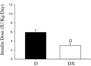
Figure 2.5 Insulin dosages 60
Figure 2.6 NIBP values at week 1 and week 10 61
Figure 2.7 Heart Rate 62
Figure 2.8 Total HRV (SDNN) 63
Figure 2.9 High Frequency HRV 64
Figure 2.10 Low frequency to high frequency HRV ratio 65
Figure 2.11 Vascular sympathetic tone 66
Figure 2.12 Tachycardia BRS 67
xi
LIST OF ABBREVIATIONS
ACE Angiotensin-converting-enzyme
ACTH Adrenocorticotropic hormone
ADH Antidiuretic hormone
AGE Advanced glycation end-product
ANG II Angiotensin II
ANOVA Analysis of variance
ANS Autonomic nervous system
AT1 ANG II type 1 receptor
BG Blood glucose
BP Blood pressure
BRS Baroreflex sensitivity
C Non diabetic sedentary control group
CAN Cardiovascular autonomic neuropathy
ChAT Choline acetyltransferase
CNS Central nervous system
CRH Corticotropin releasing hormone
CV Cardiovascular
CVLM Caudal ventrolateral medulla
CX Non diabetic exercise control group
D Diabetic sedentary group
xii
DNA Deoxyribonucleic acid
DX Diabetic exercise group
FFT Fast Fourier transform
GABA Gama-aminobutyric acid
GADA Glutamic acid decarboxylase antibody
GLUT1 Glucose Transporter 1
GLUT4 Glucose Transporter 4
GLUT3 Glucose Transporter 3
HF High frequency
HLA Human leukocyte antigen
HPA Hypothalamo-pituitary-adrenocortical
HR Heart rate
HRV Heart rate variability
IAA Insulin autoantibody
IA-2A protein tyrosine phosphatase-2 antibody
ICA Islet cell antibody
IML Intermediolateral
IP Intraperitoneal
IVGTT Intravenous glucose tolerance test
KG Glucose clearance rate
LF Low frequency
MAP Mean arterial pressure
xiii
NIBP Non-invasive blood pressure
nNOS Neuronal NOS
NO Nitric oxide
NOS Nitric oxide synthase
NTS Nucleus of the solitary tract
PE Phenylephrine
PKC Protein Kinase C
PSNS Parasympathetic nervous system
PVN Paraventricular nucleus
RAAS Renin-angiotensin-aldosterone system
RAS Renin-angiotensin system
ROS Reactive oxygen species
RVLM Rostral ventrolateral medulla
SBP Systolic blood pressure
SDNN Standard deviation between normal pulse peaks
SNP Sodium nitroprusside
SNS Sympathetic nervous system
SOD Superoxide dismutase
STZ Streptozotocin
TPR Total peripheral resistance
T1DM Type 1 Diabetes Mellitus
T2DM Type 2 Diabetes Mellitus
xiv
CHAPTER 1
1.1 Introduction
Type 1 diabetes mellitus (T1DM) is an idiopathic autoimmune disease that
results in the destruction of pancreatic β cells and subsequently, the inability to
produce endogenous insulin (4, 7, 144). This results in chronic hyperglycemia and
imbalances of fat, protein, and carbohydrate metabolism that lead to progressive
complications, such as: retinopathy, nephropathy, neuropathy and increased risk of
microvascular and macrovascular disease (4, 7, 24). These complications can be so
severe that, even with insulin therapy, a child diagnosed with diabetes at 10 years of
age will lose an average of 19 years of life due to the disease (97). Epidemiological
studies have shown that T1DM incidence is continuously rising in both high and low
incidence populations; particularly, in the 15 years of age and younger demographic
(63, 99, 163). For example, Onkamo et al. (99) reported that the global incidence rate
of the under 15 demographic from 1960 to 1996 increased at 3% per year. The
DiaMond Project Group (26), a multinational effort to investigate and monitor the
incidence patterns of childhood type 1 diabetes, reported a yearly global increase in
incidence of 3.4% for the years 1995 to 1999. In Europe it has been predicted that by
2020 the number of children under the age of 5 afflicted with T1DM will double and
the number of cases for children under the age of 15 will increase by 70% (104).
Clearly, this demonstrates that T1DM is a problem that is continuously growing in
prevalence and importance. Also of importance are the financial costs of T1DM on
patients and society at large (130). In fact, it was found that patient costs for
individuals with T1DM are disproportionately higher than costs for those with type 2
diabetes mellitus (T2DM) (142). Furthermore, the societal costs of T1DM in the USA
142). In light of such profound health and economic impacts, understanding the
predictors, onset and treatment of T1DM has been a focus of extensive investigation;
yet, a substantial amount about the disease and its complications remains to be
elucidated. Although it has been long understood that exercise can generate beneficial
health outcomes, more recently and increasingly, exercise is being regarded as a
therapy and is being prescribed by physicians as medicine (123, 128). The benefits of
exercise in the prevention and treatment of T1DM-related complications has been
well documented (36, 105, 112). In particular, diabetic autonomic neuropathy (DAN)
is known to be one of the earliest complications of diabetes which not only precludes
a variety of related complications but is also directly linked to increased mortality
(30, 40, 100, 149). Thus, the ability of exercise therapy to reduce DAN is a research
focus of great importance (9, 18, 105, 150). This chapter will provide an overview of
T1DM onset and its mediation of neuropathy, the function of the autonomic nervous
system and its regulation of the cardiovascular system, how neuropathy leads to
autonomic and cardiovascular dysfunction and the efficacy of exercise in mitigating
this dysfunction.
1.2 Etiology of Type 1 Diabetes Mellitus
Susceptibility to T1DM is determined by both genetic and environmental
factors (7, 31, 58, 144). The genetic basis of the disease is strongly exhibited by
studies of monozygotic and dizygotic twins. It has been found that monozygotic twins
can demonstrate over a 50% concordance rate of T1DM, which is far greater than the
concordance between dizygotic twins (50). Due to the fact that monozygotic twins
share the same DNA and developmental environment, whereas dizygotic twins have
concluded that the difference in concordance between the two types of twins is
resultant of a genetic influence (31). Through such twin studies, as well as familial
linkage studies, it has been established that the human leukocyte antigen (HLA)
encoding genes, which code for many different components of the immune system,
are the major factors conferring genetic susceptibility to T1DM (24, 31, 48, 50).
Although a high percentage of T1DM susceptibility can be attributed to a genetic
component, even at 50% concordance, there remain at least equally substantial
non-genetic factors that are involved in the manifestation of T1DM.
Environmental factors, including viral infections, diet, growth, toxins and
stress, have been implicated in the pathogenesis of T1DM (3). D’Angeli et al. (28)
provide evidence for the “hygiene hypothesis” by demonstrating that a clean
environment during childhood decreases antigenic stimulation and results in a greater
vulnerability to developing T1DM. Further evidence of an environmental
determinant of T1DM is the apparent role of geography and climate in its prevalence.
In both Europe and North America there is a north to south, high to low, gradient of
incidence (58, 144). In accordance with a climatic influence, a lower incidence of
T1DM during warm seasons than cold seasons has been observed within populations
(58, 68). Although these genetic, regional and climatic insights provide evidence of
risk factors for developing T1DM, they are not necessarily predictors of great utility.
The environmental factors may be correlated with T1DM but most cannot realistically
be avoided or managed, whereas the genetic factors alone do not yield disease onset;
as evidenced by the finding that, at most, only 10% of individuals with the highest
risk susceptibility genes actually become afflicted with T1DM (146).
The most effective predictors of T1DM are a complement of autoantibodies
detected during infancy (12, 61). The autoantibodies that predict T1DM are: islet cell
antibodies (ICA), insulin autoantibodies (IAA), glutamic acid decarboxylase
antibodies (GADA) and protein tyrosine phosphatase-2 antibodies (IA-2A) (71, 80).
These autoantibodies direct the infiltration of immune cells, mostly T cells, to the
islets and facilitate their inflammation and the destruction of β cells (19, 25, 71). It
has been observed that the detection of two or more of these autoantibodies in
relatives of T1DM patients has a 90% predictive value for their eventual development
of T1DM (147). In the general population, marginally elevated levels of 3 to 4
autoantibodies indicates a risk of roughly 60% for the development of T1DM (12).
Despite the predictive value of autoantibody detection, no autoantibody based
treatments exist to avert the onset of T1DM (25). Therefore, for now, the only way to
delay or prevent T1DM associated morbidity and mortality is to understand and
ameliorate the progression of the disease-related complications (4, 25, 80). However,
it has been suggested that exercise could ameliorate the severity of autoantibody
mediated inflammation and subsequently, reduce T1DM initiation and progression
(66).
1.3 Pathogenesis of Neuropathy in T1DM
The neurological complications of T1DM are understood to arise from either a
metabolic etiology, a vascular etiology or a combination of both (30, 41, 137, 156).
However, the pathology of metabolism and vasculature in T1DM, as well as the wider
breadth of complications that develop, arises primarily due to chronic hyperglycemia
(24, 41, 149). Indeed, the duration and severity of hyperglycemia have both been
indicated as predictors of the degree of diabetic neuropathy (30, 41). Fittingly then,
leads to irreversible binding of glucose molecules to intracellular and extracellular
proteins, have been implicated as a causative link between vascular and metabolic
dysfunction in T1DM (41).
The metabolic factors, whose dysfunction primarily contributes to diabetic
neuropathy, are many. For example, T1DM leads to increased polyol pathway activity
and decreased myo-inositol, which are associated with decreased nerve conduction
velocity (30, 41, 137). It also causes decreased nerve Na/K ATPase activity, the
function of which is necessary to maintain the transmembrane ionic gradient required
for electrical impulse conduction (41, 137). Further, T1DM is associated with
decreased synthesis and transport of axonal proteins, which are supplied from the cell
body to maintain structural and functional integrity of the axon (41, 87, 137) and
diminished incorporation of glycolipids and amino acids into myelin, thereby
reducing axon insulation and conduction velocity (30, 41, 139). T1DM also leads to
decreased nerve protein kinase C (PKC) activity, which can further reduce Na/K
ATPase activity (137). Dyslipidemia, consisting of increased triglycerides, increased
low-density lipoprotein and decreased high-density lipoprotein, is a metabolic factor
that contributes to neuropathy via the atherosclerotic impairment of the vasculature
(30, 41, 107).
Vascular factors that contribute to diabetic neuropathy mostly involve
pathology of the endoneurial capillaries (92, 152). These vascular impairments
include: basement membrane thickening and endothelial cell swelling and
proliferation, resulting in reduced luminal diameter, decreased nerve blood flow and
endoneurial hypoxia (41, 83, 143); degeneration of pericytes, which are integral for
maintaining endothelial cell function at the blood-nerve barrier (41, 129); and
nutrient exchange between axons and blood vessels across the endoneurium (30, 41,
129, 143). There is also evidence that hyperglycemia can have tissue-specific,
bidirectional effects on PKC activity (138). While hyperglycemia decreases PKC
activity in the nerve as mentioned above, it also becomes overactive in vascular
endothelium and leads to increased endothelial permeability, the dysregulation of
endothelial-derived vasodilator nitric oxide (NO) and thus, diminished blood flow
(127, 138).
Despite these metabolic and vascular factors often being considered discrete
etiologies of diabetic neuropathy, it is more likely that they arise concomitantly and
share protein glycation as their common cause (see Figure 1.1) (30, 41). For example,
glycation of antioxidant enzymes hinders their ability to remove reactive oxygen
species (ROS). This results in the accumulation of ROS that damage membranes,
cellular proteins and DNA, all of which contribute to neuropathic progression (41).
Glycation of the nitric oxide synthase (NOS) enzyme is likely a mechanism by which
NO production is decreased, driving the vascular dysfunction described above (41,
138). Also, glycated elastin and collagen in the endothelium can restrict access of NO
to the smooth muscle, further inhibiting NO-mediated vasodilation, reducing nerve
blood flow and nerve oxygenation (41). The manifestation of AGEs in the basement
membrane, elastic lamina and endothelial cells of the blood vessels causes
macrophage infiltration, which release macrophage-derived growth factors , leading
to smooth muscle proliferation and atherogenesis (41). Due to evidence that
inflammatory cytokines also cause vascular damage by inducing excessive ROS, it is
plausible that AGE-mediated inflammation is the primary cause of neurovascular
dysfunction in diabetic neuropathy (36, 134). AGE levels in the endoneurium and
atrophy in nerve biopsies; with AGE immunoreactivity detected in 90% of peripheral
nerves of diabetics compared to a complete absence of AGEs in controls (91).
Whether or not AGEs can be said to underlay both the metabolic and vascular causes
of neuropathy, they certainly do represent a link between them. Ultimately,
hyperglycemia-mediated AGEs, metabolic dysregulation and vascular dysfunction are
all fundamental to nerve dysfunction and degeneration in T1DM.
1.4 The Autonomic Nervous System and Mechanisms of Autonomic Cardiovascular
Regulation
The autonomic nervous system (ANS) is a network of efferent and afferent
nerves linked to structures of the central nervous system (CNS) that, together, govern
the motor control of visceral organs (40, 76). Since targets of the ANS include
multiple endocrine organs, the cardiovascular (CV) system and regions of CNS, the
ANS is a powerful regulator of homeostasis throughout the entire body (76, 124). The
ANS differentially regulates these systems by using two antagonistic branches, the
sympathetic nervous system (SNS) and the parasympathetic nervous system (PSNS),
which mutually inhibit one another (116). The main neurochemical difference
between the SNS and PSNS is the neurotransmitters involved in signaling to the target
organs. Preganglionic neurons of both branches utilize acetylcholine as their primary
neurotransmitter, whereas the postganglionic neurons of the PSNS emit primarily
acetylcholine and the postganglionic neurons of the SNS release primarily
norepinephrine (76, 116). The use of these different neurotransmitters underlies the
antagonistic physiological actions of the two branches of the ANS (76, 116). To
understand the discrete roles of the SNS and PSNS in controlling bodily functions, the
The SNS directs oxygen and energy toward organ systems that are recruited in
stressful or active situations; for example, it acts to increase heart rate (HR) and
decrease enteric motility. The PSNS, on the other hand, promotes conservation and
restoration of energy and so, stimulates functions that are more useful at rest while
inhibiting others; for example, it decreases blood pressure (BP) and facilitates the
excretion of waste (76, 116). When functioning properly, these two branches maintain
chronic homeostasis of visceral organ function (116).
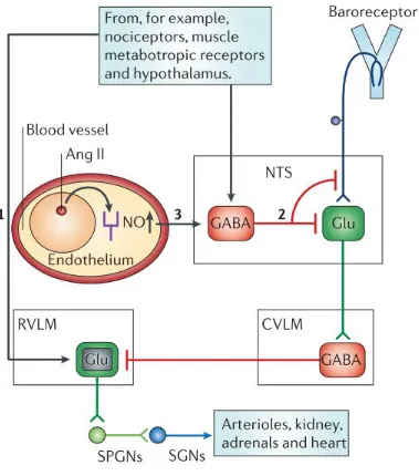
One such autonomically-mediated mechanism of homeostasis is the regulation
of CV function by the baroreflex (see Figure 1.2). Afferent arterial baroreceptors are
stretch-sensitive mechanoreceptors located in the aortic arch, carotid sinus and
elsewhere in the vasculature that send frequency coded signals to the brainstem in
order to signal increases and decreases in BP (40). Once these signals arrive at the
brainstem they are processed by several structures that govern the efferent
sympathetic and parasympathetic output to the heart, the vasculature, kidneys and
other visceral organs (40, 116). The four major brain regions involved in processing
these signals include the hypothalamus and three regions of the brainstem: the rostral
ventrolateral medulla (RVLM), the caudal ventrolateral medulla (CVLM) and the
nucleus of the solitary tract (NTS) (40, 76, 116). When a rise in BP above the set
physiological range occurs, the baroreceptors fire rapidly and stimulate the NTS. The
NTS in turn stimulates the CVLM, which provides inhibitory input to the RVLM.
Since the RVLM has excitatory input to sympathetic preganglionic neurons in the
intermediolateral (IML) column of the thoraco-lumbar spinal cord, when
baroreceptors fire the RVLM, and by extension sympathetic outflow, is inhibited and
the autonomic balance shifts towards greater parasympathetic activity; the outcome of
Alternatively, when arterial pressure declines below the set physiological range,
baroreceptor firing decreases, removing the stimulation of the NTS and CVLM and
disinhibiting the RVLM, resulting in increased sympathetic outflow, HR and again,
the restoration of BP to the physiological range (40, 116).
The baroreflex also has an important influence on total peripheral resistance
(TPR); which is the resistance of all peripheral vasculature in the systemic circulation
(145). Peripheral resistance and the variables that influence it can be described by
Poiseuille’s law, 𝑅 = 𝛥𝑃
𝑄 ; where R is resistance, ΔP is the change in pressure between
two points and Q is the blood flow, which is equivalent to cardiac output (90).
Resistance can also be represented as𝑅 = 8ɳ𝐿
𝜋r4; where ɳ is the viscosity of the fluid, L
is the length of the vessel, and r is the radius (90). Blood pressure and blood flow
(cardiac output) are acutely regulated by the baroreflex (77). However, beat-to-beat
changes in TPR are difficult to measure and thus, most studies examine feedback
control of HR as opposed to TPR when assessing the baroreflex (10).
The paraventricular nucleus (PVN) of the hypothalamus also modulates
sympathetic outflow (11, 21, 40). The PVN receives inputs from circulating hormones
and molecules (such as norepinephrine and glucose), visceral receptors that detect
osmolarity and blood volume and other regions of the brain (11). The PVN has many
complex effector subnuclei but its effects can be summed through its three major
connections. The magnocellular neurons of the PVN are large cells that interact with
the posterior pituitary to release oxytocin and vasopressin (also known as antidiuretic
hormone, ADH; a vasoconstrictor and mediator of water absorption in the kidneys)
into the blood (113). The medial parvocellular neurons synthesize corticotropin
releasing hormone (CRH), which stimulates the release of adrenocorticotropic
stimulate aldosterone (a mediator of water retention in the kidneys)and cortisol
production (which stimulates gluconeogenesis in the liver); this constitutes what is
known as the hypothalamo-pituitary-adrenocortical (HPA) axis (11, 125). Another
subset of parvocellular PVN neurons have direct connections to the RVLM in the
brainstem and the IML in the spinal cord (8). Thus, these PVN neurons can directly
and indirectly influence sympathetic nerve activity via inputs to preganglionic
sympathetic neurons and by modulating RVLM activity (8). Indeed, it has been
demonstrated that sympathoexcitation is elicited through the PVN in response to a
drop in blood volume, without a drop in pressure, via the activation of both RVLM
and ILM connected neurons (8). Conversely, the autonomic neurons of the PVN have
been reported to selectively decrease renal sympathetic activity and increase cardiac
sympathetic activity in response to blood volume expansion (21). This demonstrates
that the PVN’s influence on BP and the CV system is mediated through hormones,
sympathetic neurons and interactions with the kidney. The stimulation of sympathetic
activity by PVN neurons in response to increased circulating angiotensin II (ANG II),
generated by the renin-angiotensin-aldosterone system (RAAS), is another example of
its interaction with the kidneys (11, 54).
Renin release from juxtaglomerular cells of the kidney primarily occurs in
response to hypovolemia and increased osmolarity but can also be produced by
sympathetic stimulation (11, 20, 38). Once released, the renin enzyme acts locally and
systemically in the blood to cleave angiotensinogen, which is constitutively secreted
by the liver into circulation, to form angiotensin I (38). Angiotensin I is then cleaved
by angiotensin-converting-enzyme (ACE), which is found in blood plasma, vascular
endothelial cells, the kidneys, the brain, the lungs and many other tissues (38, 40).
of effects on numerous organs and tissues. For instance, ANG II is a potent
vasoconstrictor, it acts on presynaptic receptors of sympathetic nerve terminals to
potentiate norepinephrine release, it decreases glomerular filtration rate and increases
reabsorption of sodium and water into the blood and it stimulates aldosterone release
from the adrenal cortex, thus completing the RAAS (38). It also acts on the
subfornical organ (a circumventricular organ of the brain) to stimulate PVN mediated
sympathoexcitation to the heart and kidneys (20, 40, 54). However, it is very
important to note that this classic, endocrine view of the RAAS has undergone
profound conceptual change due to the discovery of local, tissue-intrinsic
renin-angiotensin systems (RAS – note the distinct acronyms denoting local and circulatory
renin-angiotensin systems) and its importance in autocrine and paracrine signalling
which act independently of the circulating RAAS factors (32, 115).
Thus, both neural and humoral mechanisms interact with the ANS to modulate
autonomic activity and CV function. For example, the PVN and the RAAS influence
blood volume, vascular tone and BP; baroreceptors regulate BP and maintain it within
a set range by modulating cardiac function; ANG II produced by the RAAS can act
directly on peripheral sympathetic synapses and can modulate PVN activity; and the
PVN can directly modulate sympathetic outflow and influence the brainstem circuitry
that mediates the baroreflex (40). All of these systems are integrated through the ANS
and cooperate to maintain CV homeostasis.
1.5 Autonomic Dysfunction and Cardiovascular Dysregulation in T1DM
Dysfunction of the ANS is a common and serious complication of T1DM that
can result in dysregulation of all of the organ systems under its control (30). Known
reported to vary widely, ranging from roughly 10-90% in diabetics depending on the
assessment criteria used (30, 149). The clinical manifestations of DAN are also
variable due to the systemic influence of the ANS and can include orthostatic
hypotension, tachycardia, hypertension, silent myocardial ischemia, gastroparesis,
xerostomia, decreased heart rate variability (HRV), anhidrosis and many others
symptoms (30, 33, 69, 95, 149). However, it should be noted that experimental forms
of diabetes do not always faithfully replicate the full spectrum of clinical
complications; in some cases, opposing outcomes have been observed, such as resting
bradycardia and hypotension (6, 47, 81). However, unlike the clinical population,
most of these studies do not use insulin therapy to manage hyperglycemia, which has
been shown to reverse those effects (47). DAN can also be a considerably deadly
complication of T1DM as it underlies diabetes-mediated CV dysfunction (85, 100).
Indeed, based on an analysis of the Pittsburgh Epidemiology of Diabetes
Complications Study, Orchard et al. (100) found that mortality increased fourfold in
patients with DAN. Similarly, a meta-analysis by Maser et al. (85) found that
cardiovascular autonomic neuropathy (CAN), a subset of DAN, significantly
increased the relative risk of mortality by a factor of 3.45. Another form of mortality
in T1DM is the occurrence of sudden nocturnal death (100, 154). This so called “dead
in bed” syndrome is thought to be exacerbated by increased susceptibility to nocturnal
hypoglycemia resulting from abnormal pancreatic α-cell function and impaired
glucagon release (140, 154). However, even this impaired glucagon response is
thought to be mediated by autonomic dysfunction and furthermore, sudden death in
T1DM is more greatly attributed to cardiac arrhythmia than hypoglycemia; thus,
“dead in bed” syndrome is likely resultant of DAN (140, 154). DAN, therefore, has a
T1DM; the most deadly consequences of which are mediated by loss of CV control
via CAN (30, 101, 149, 151).
As described previously, the etiology of general diabetic neuropathy and thus,
DAN, in T1DM is linked to chronic hyperglycemia (127, 155). Evidence of this was
reported by the Diabetes Control and Complications Trial/Epidemiology of Diabetes
Interventions and Complications (DCCT/EDIC) Study Research Group (98) who
observed that maintenance of near normoglycemia via intensive insulin therapy
reduced neurologic complications of T1DM and resulted in a 42% decrease in the risk
of any CV disease event, such as myocardial infarction, stroke or death. The
neuropathic effects of hyperglycemia that mediate DAN usually affect longer nerves
first and since the single longest ANS nerve, the vagus nerve, facilitates 75% of all
parasympathetic activity, a sympathovagal imbalance leading to sympathetic
overactivity is purported to be an early cause of CAN (30, 149, 151). Furthermore,
hyperglycemia can lead directly and indirectly to increased sympathetic outflow,
indicating peripheral and central mechanisms are behind ANS and CV dysfunction in
T1DM (108, 148). It is pertinent then to understand how the regulation of autonomic
activity and CV function are affected by T1DM.
Although there has been some evidence for increased RAAS activity in T1DM
and hyperglycemia, most of the literature has demonstrated that systemic components
of the RAAS, such as plasma renin, aldosterone and ANG II, are suppressed (13, 22,
73, 89). When considering this in the context of the physiological action of circulating
ANG II (vasoconstriction, volume expansion and sympathoexcitation), it should
follow that it is not involved in the increased BP and sympathetic tone observed in
T1DM; however, this is not the case (115, 148). More recent evidence implicates the
complications (109, 115). Hyperglycemia has been shown to increase local RAS
activity in numerous tissues and this process is being connected to the onset of many
downstream complications of T1DM; including, nephropathy, retinopathy and cardiac
myopathy (27, 34, 67, 110). This widespread deleterious effect of local RAS
overactivity in diabetes is likely interconnected to oxidative damage mediated by
locally produced ANG II (34, 115). Thus, RAS activation in diabetes is believed to
contribute to microvascular complications and has a probable role in the vascular
etiology of neuropathy (86, 109). Due to the presence of RAS in the brain and the
known ability of ANG II in the subfornical organ to elicit PVN-mediated increases in
sympathetic activity, there exists a plausible mechanism by which local RAS may be
driving increased sympathetic outflow in T1DM (11, 148).
The PVN of the hypothalamus is known to influence the basal level of
sympathetic outflow (60). In T1DM there are increases in both basal PVN activity and
sympathetic activity (35, 56). It has been demonstrated that the excitation of the PVN
by ANG II is enhanced in rats with streptozotocin (STZ)-induced diabetes; an
experimental form of T1DM produced by STZ-mediated destruction of β-cells (103).
This suggests that diabetes induced elevations of sympathetic outflow could be due to
increased levels of ANG II in the brain produced by local RAS system activation or
increased sensitivity to ANG II in the PVN (103). Another previously described
diabetes-related deficit that is also a mechanism contributing to central sympathetic
overactivity in T1DM is the loss of NOS activity and NO (138, 161). NO is a known
inhibitor of PVN activity that acts by increasing GABAergic input to the PVN (160).
Zheng et al. (161) found that STZ-diabetic rats had fewer neuronal NOS (nNOS)
positive cells and lower NO release in the PVN compared to controls. Thus, there is a
and thereby, contributes to sympathetic overactivity (114). Furthermore,
overstimulation of the PVN would also cause excitation of magnocellular neurons,
resulting in the release of vasopressin, which would contribute to increased BP via
direct vasoconstriction and volume expansion (70). However, the aberration of
sympathetic tone in diabetes is more directly influenced by the parvocellular neurons
of the PVN due to their association with the RVLM and IML; hence, PVN
dysfunction can dysregulate sympathetic outflow and CV parameters by modulating
neural control of HR, BP and the baroreflex (20, 60, 103).
The baroreflex itself is an important regulator of sympathetic and
parasympathetic outflow, BP, HR and TPR (84). Baroreflex dysfunction, in both
sensitivity and set point, is an established consequence of T1DM, with reductions in
sensitivity observed to occur as early as 5 days after STZ-diabetes induction (15, 23,
81). Thus, studies have investigated how each aspect of the baroreflex arc are
uniquely affected in T1DM; this arc consists of baroreceptors, baroreceptor afferents,
central regulators and sympathetic and parasympathetic efferents (74). Salgado et al.
(122) found that aortic baroreceptor neurons have reduced activity in STZ-diabetic
rats. Further, they determined that the aortic depressor nerves, the afferent nerves
between baroreceptor neurons and the brainstem, showed morphological signs of
axonal atrophy by measuring the G ratio; the ratio between axon diameter and whole
fiber diameter (122). Li et al. (72) observed that the excitability of aortic
baroreceptors, which mediates the set point of baroreceptor firing, was blunted in
STZ-diabetic rats and that later, this reduced excitability was due to up-regulated,
ANG II-NADPH oxidase derived, superoxide within the baroreceptor ganglion (73).
They also found that microinjection of an ANG II receptor antagonist into the nodose
local RAS activity being responsible for dysfunction of the afferent portion of the
baroreflex arc (73). Evidence for the dysfunction of central regulation of the
baroreflex was presented by Gu et al. (39) using OVE26 T1DM mice; a transgenic
model of T1DM that acquires β-cell damage through an overexpressed calmodulin
regulated by an insulin promoter. They found that these mice, compared to controls,
had a less prominent bradycardia response to aortic depressor nerve stimulation but
that the bradycardia response to vagal efferent stimulation was increased; suggesting a
central mechanism was responsible for the blunted response to baroreceptor
stimulation (39). Also, Chen et al. (16) documented a reduced firing rate in the NTS,
both basally and after phenylephrine injection, in STZ-induced diabetic rats compared
to controls. This decreased activity was implicated in their observation of decreased
baroreflex sensitivity (BRS) and could possibly indicate chronic disinhibition of the
RVLM; and therefore, increased sympathetic outflow (16). Of the two efferent arms
of the ANS, the PSNS appears to be the one primarily impaired in T1DM. Maeda et
al. (81) reported significant decreases in both basal PSNS tone and responsiveness to
hypertensive drugs in STZ-induced diabetic rats; but non-significant changes to
similar parameters in the SNS. Lund et al. (79) found that choline acetyltransferase
(ChAT, the enzyme that produces the chief neurotransmitter of the PSNS,
acetylcholine), had reduced activity in the hearts of diabetic rats as early as 4 weeks
post-STZ induction. After 8 weeks of diabetes, they measured a reduction in cardiac
ganglion size and number, prompting the conclusion that these PSNS deficits may
contribute to the impaired parasympathetic control of the heart, sympathetic
overactivity and baroreflex blunting that is common in T1DM (79).
A non-invasive functional measure capable of detecting such impaired
measured by time domain analyses, measuring the changes in the standard deviation
between R-R intervals or by frequency domain analyses, using Fourier analysis to
disintegrate HR variance into underlying high and low frequencies (136). Regardless
of which method is chosen, HRV has been described as one of the earliest indicators
of autonomic dysfunction in T1DM (126, 162). A variable HR has been known to be
a characteristic of a healthy heart for roughly a century (37). In frequency domain
analysis, high frequency (HF) variation (0.15-.4Hz) is considered to be mediated by
parasympathetic activity, whereas low frequency (LF) variation (0.04-0.15Hz) is
considered to be entrained by the oscillation of baroreceptor firing; and so, it is
modulated by both parasympathetic and sympathetic activity (37, 136). As described
above, T1DM results in baroreflex dysfunction that is largely attributed to damage of
the PSNS (29). Since PSNS dysfunction occurs first, a reduction in HF variability is
usually the first sign of autonomic dysfunction (52). Thus, in general, a reduction in
HRV in T1DM is associated with the onset of autonomic dysfunction and more
specifically, the loss of parasympathetic tone and baroreflex dysfunction (37, 126,
157). Now used as a standard indicator in the diagnosis of cardiac autonomic
neuropathy, reduced HRV is associated with cardiac arrhythmias that can have mortal
consequences for diabetics and is therefore an important marker of autonomic
function in T1DM (151).
1.6 The Effects of Exercise on Neuropathy, Autonomic Regulation and
Cardiovascular Dysregulation in T1DM
Exercise training is a well-established and widely prescribed
non-pharmacological therapy capable of ameliorating a wide breadth of diseases and their
improve heart disease, pulmonary disease, muscle disease, bone disease, joint disease,
cancer, depression, asthma, obesity, hypertension, dyslipidemia, insulin resistance,
type 2 and type 1 diabetes (18, 105, 123, 128). Beyond just treating disease, exercise
is also associated with preventing disease risk factors and manifestation. It has been
shown to improve cognitive performance, increase longevity and has a strong
negative correlation with all-cause mortality (1, 64, 78). One of the underlying
principles likely facilitating the benefits of exercise is the concept of “hormesis”;
wherein low level stress or damage causes a disruption of homeostasis and results in
an adaptive overcompensation and increased fitness of a physiological system (14,
36). In the case of exercise, the acute induction of low-level oxidative stress and
inflammation results in an increase of endogenous antioxidant enzymes and
anti-inflammatory mediators (59, 132). Continuous exercise training results in the chronic
upregulation of antioxidants and anti-inflammatory agents which confers constitutive
resistance to other sources of pathogenic insult (132, 133). Another outcome of
exercise, which is especially important in diabetes, is increased insulin sensitivity and
glucose tolerance (43). Exercise training increases the amount and function of
proteins in the insulin signalling cascade, which facilitates the translocation of glucose
transporter 4 (GLUT4) into the skeletal muscle membrane (53). Furthermore, muscle
contraction can upregulate GLUT4 translocation to the plasma membrane via an
insulin-independent mechanism (118). In T1DM, hyperglycemia mediated ROS act to
decrease insulin sensitivity by impairing the insulin cascade and GLUT4 translocation
(44). Therefore, the antioxidant, anti-inflammatory and glucoregulatory effects of
exercise make it a potent therapy for T1DM and many of its complications (9, 36).
The influence of exercise on insulin sensitivity and glucose tolerance both
endoneurium express GLUT4 or utilize it to transport glucose; instead, they use
GLUT1 and GLUT3 which do not require insulin for translocation and are
constitutively active (82, 131). Thus, exercise-mediated sensitization to insulin does
not occur within neural tissue, yet through improving glucose tolerance in other
tissues, exercise reduces exogenous insulin requirements and neural hyperglycemic
insult in T1DM (105, 112, 158). This moderated hyperglycemia reduces the level of
AGEs, which are known to be major contributors to neuropathy through ROS
formation and inflammation in the neurovasculature (36, 41, 117). Furthermore,
exercise-induced insulin sensitivity and AGE reduction also means increased NO
production and bioavailability, resulting in enhanced function of endoneurial
capillaries and oxygen delivery to peripheral nerves (117, 135, 138). Exercise training
also increases endothelial Cu/Zn-superoxide dismutase (SOD) and mitochondrial
Mn-SOD antioxidant enzymes independently of hyperglycemic status, while
simultaneously decreasing inflammatory cytokines and increasing anti-inflammatory
mediators (51, 59, 93). Thus, exercise training is capable of ameliorating neuropathy
through the alleviation of both its metabolic and vascular etiologies by improving
glucose metabolism and upregulating antioxidant and anti-inflammatory mediators
(36, 112, 158). Indeed, exercise training has been shown to both, prevent diabetic
neuropathy from occurring and improve nerve function in individuals previously
diagnosed with diabetic neuropathy (9, 62).
Another mediator of T1DM complications that is influenced by exercise is the
local RAS (115, 121). RAS overactivity has been shown to be mediated by
hyperglycemia and is implicated in causing insulin resistance (45, 148). As previously
described, exercise can reduce hyperglycemia and so can decrease
components of the RAS system, such as ACE, ANG II, ANG II type 1 receptor
(AT1), and NADPH oxidase (43, 57, 106, 121). Indeed, exercise has been shown to
decrease plasma ANGII and local RAS activity in cardiac muscle, blood vessels and
the brain (2, 121, 159). For example, Kar et al. (57) found that 3 weeks of treadmill
running decreased ACE mRNA in the NTS, RVLM, PVN and decreased sympathetic
outflow in rabbits with induced heart failure. Similarly, Pan et al. (102) found that
non-T1DM Sprague-Dawley rats had decreased AT1 and NADPH oxidase protein
expression and decreased superoxide production in the PVN following 3 to 4 weeks
of exercise training. As aforementioned, the PVN is activated in T1DM through
increased ANGII and decreased NO and this leads to sympathetic overactivity (103,
161). Thus, inhibiting RAS activity and ANGII production, while increasing NO
bioavailability, are likely mechanisms by which exercise reduces PVN mediated
sympathetic outflow (102, 117). This influence of exercise on RAS activity in the
PVN, NTS and RVLM makes it likely that exercise will also influence baroreflex
function in T1DM (55, 57, 102).
Since exercise can influence both vascular and neural function, both must be
taken into account when assessing the influence of exercise on BRS and baroreceptor
set point (65, 120). Indeed, both the neural structures involved in the baroreflex arc
and the carotid artery compliance can affect BRS and baroreceptor firing in diabetes
and exercise (65, 94, 120). Monahan et al. (94) found that both age-related and
exercise induced changes in the BRS corresponded to differences in carotid artery
compliance in healthy men. However, Komine et al. (65) determined that endurance
training increased BRS through alterations to the neural circuitry. In this vein, Miki et
al. (88) reported that exercise has been demonstrated to cause a shift in baroreflex
reported that diabetic neuropathy is a more significant determinant of BRS than
carotid artery elasticity in T2DM. Whether mediated by carotid artery compliance,
neural circuitry or a combination of both, exercise has been shown to increase BRS
after even a single bout of aerobic exercise in STZ-diabetic rats (55). Furthermore, the
benefits of aerobic exercise on baroreflex function have been shown to persist after 3
weeks of detraining subsequent to 10 weeks of moderate intensity training (50-70%
maximal running speed) in STZ-diabetic rats (96). Thus, exercise can be an effective
means to rescue or prevent baroreflex dysfunction and mitigate aberrations of CV
function in T1DM (42, 65, 102).
Since baroreflex function is directly linked to the neural control of HR,
fittingly, HRV is also influenced by exercise (119). In T1DM, there is generally a
reduction in HRV; an indication of autonomic dysfunction (17). However, in
individuals with T1DM there are inconsistent changes in HRV in response to
exercise. Chen et al. (17) reported that children with T1DM with moderate to high
physical activity did not differ in HRV compared to controls, whereas children with
low physical activity had reduced HRV, suggesting only higher intensity exercise
prevented a decline in HRV. On the other hand, Zoppini et al. (164) observed no
change in total HRV following twice-weekly exercise over 6 months but did find that
it increased HF power and lowered the LF/HF ratio, meaning there was a shift toward
more parasympathetic output in the sympathovagal balance. Howorka et al. (49)
tested HRV responses to 12 weeks of bicycle training in diabetic subjects who had
differing levels of cardiovascular autonomic neuropathy. They found that patients
without CAN had the greatest increases in HRV, patients with early CAN had lesser
increases in HRV and patients with severe CAN did not improve following training;
Therefore, the intensity of exercise and stage of disease both factor in to the ability of
exercise to modulate HRV in T1DM.
Some inconsistency in the effectiveness of exercise modulation of HRV is
reflected by the somewhat mixed outcomes for the influence of exercise on autonomic
function in T1DM. For example, De Angelis et al. (5) found that exercise training in
STZ-diabetic rats did not modify sympathetic tonus nor parasympathetic function. In
the same model, Wegner et al. (153) reported that training did not induce changes in
markers of parasympathetic innervation of the heart. However, vagal tonus and
function have been observed to improve after acute and prolonged training in the
STZ-diabetic model (96, 111). Indeed, the majority of literature supports the role of
exercise as an effective means to improve and maintain autonomic function in
diabetes (17, 150).
1.7 Summary
The pathogenesis of neuropathy in T1DM is a product of hyperglycemia-mediated
AGEs, oxidative stress, metabolic dysfunction, inflammation and microangiopathy
(41). Diabetic neuropathy affects peripheral and central aspects of the autonomic
nervous system, alters the homeostatic maintenance of the sympathovagal balance and
leads to a variety of systemic complications (30, 40). One of the most common and
serious complications of DAN is cardiovascular dysregulation (85). The local RAS,
PVN of the hypothalamus and baroreflex circuitry all interact to regulate autonomic
cardiovascular control (40, 41). In T1DM, hyperglycemia causes an increase in local
RAS, which in turn generates local ANGII mediated oxidative stress that can decrease
insulin sensitivity, blunt the excitability of baroreceptor neurons and increase PVN
mediators of the baroreflex and directly with preganglionic neurons to influence
sympathetic and parasympathetic outflow (8, 56). Thus, the baroreceptors, afferent
nerves, central neural structures and efferent nerves of the baroreflex arc are all
affected by T1DM (74). Together, dysfunction of these circuits underlie many of the
serious functional CV complications associated with DAN (40, 100). Exercise is not
only known to improve CV function directly but is also capable of improving ANS
regulation of CV control in diabetics (17, 105, 150). Through improving insulin
sensitivity and glucose metabolism, as well as the hormetic induction of antioxidant
and anti-inflammatory factors, exercise is capable of decreasing local RAS, reducing
PVN activity and improving the baroreflex and overall autonomic function in T1DM
via both molecular and physiological mechanisms (36, 43, 65, 102, 159). Therefore,
despite some inconsistencies between clinical and animal trials examining the
effectiveness of exercise in the improvement of autonomic control of CV function,
exercise remains an established, promising intervention for improving the prognosis
Figure 1.1. Protein glycation as the common cause of neuropathy; from Harati
1.8 References
1. Aberg MAI, Pedersen NL, Torén K, Svartengren M, Bäckstrand B,
Johnsson T, Cooper-Kuhn CM, Aberg ND, Nilsson M, Kuhn HG.
Cardiovascular fitness is associated with cognition in young adulthood. Proceedings of the National Academy of Sciences of the United States of America 106: 20906–11, 2009.
2. Agarwal D, Welsch MA, Keller JN, Francis J. Chronic exercise modulates
RAS components and improves balance between pro- and anti-inflammatory cytokines in the brain of SHR. Basic Research in Cardiology 106: 1069–85, 2011.
3. Akerblom HK, Vaarala O, Hyöty H, Ilonen J, Knip M. Environmental
factors in the etiology of type 1 diabetes. American Journal of Medical Genetics 115: 18–29, 2002.
4. Alberti KG, Zimmet PZ. Definition, diagnosis and classification of diabetes
mellitus and its complications. Part 1: diagnosis and classification of diabetes mellitus provisional report of a WHO consultation. Diabetic Medicine : A Journal of the British Diabetic Association 15: 539–53, 1998.
5. De Angelis KL, Oliveira AR, Dall’Ago P, Peixoto LR, Gadonski G,
Lacchini S, Fernandes TG, Irigoyen MC. Effects of exercise training on
autonomic and myocardial dysfunction in streptozotocin-diabetic rats. Brazilian Journal of Medical and Biological Research 33: 635–41, 2000.
6. De Angelis KL, Schaan BD, Maeda CY, Dall’Ago P, Wichi RB, Irigoyen
MC. Cardiovascular control in experimental diabetes. Brazilian Journal of Medical and Biological Research 35: 1091–100, 2002.
7. Atkinson MA, Eisenbarth GS. Type 1 diabetes: new perspectives on disease
pathogenesis and treatment. The Lancet 358: 221–9, 2001.
8. Badoer E. Hypothalamic paraventricular nucleus and cardiovascular
regulation. Proceedings of the Australian Physiological and Pharmacological Society Symposium : The Hypothalamus : 95–99, 2001.
9. Balducci S, Iacobellis G, Parisi L, Di Biase N, Calandriello E, Leonetti F,
Fallucca F. Exercise training can modify the natural history of diabetic peripheral neuropathy. Journal of Diabetes and its Complications 20: 216–23, 2006.
10. Batzel J, Baselli G, Mukkamala R, Chon KH. Modelling and disentangling
11. Benarroch EE. Paraventricular nucleus, stress response, and cardiovascular disease. Clinical Autonomic Research : Official Journal of the Clinical Autonomic Research Society 15: 254–63, 2005.
12. Bingley PJ, Bonifacio E, Williams AJ, Genovese S, Bottazzo GF, Gale EA.
Prediction of IDDM in the general population: strategies based on combinations of autoantibody markers. Diabetes 46: 1701–10, 1997.
13. Bojestig M, Nystrom FH, Arnqvist HJ, Ludvigsson J, Karlberg BE. The
renin-angiotensin-aldosterone system is suppressed in adults with Type 1 diabetes. Journal of the Renin-Angiotensin-Aldosterone System : JRAAS 1: 353–6, 2000.
14. Calabrese EJ, Baldwin LA. Hormesis: a generalizable and unifying hypothesis. Critical Reviews in Toxicology 31: 353–424, 2001.
15. Chapleau MW, Li Z, Meyrelles SS, Ma X, Abboud FM. Mechanisms
determining sensitivity of baroreceptor afferents in health and disease. Annals of the New York Academy of Sciences 940: 1–19, 2001.
16. Chen H-Y, Wu J-S, Chen J-JJ, Cheng J-T. Impaired regulation function in
cardiovascular neurons of nucleus tractus solitarii in streptozotocin-induced diabetic rats. Neuroscience Letters 431: 161–6, 2008.
17. Chen S-R, Lee Y-J, Chiu H-W, Jeng C. Impact of physical activity on heart rate variability in children with type 1 diabetes. Child’s nervous system : ChNS : official Journal of the International Society for Pediatric Neurosurgery 24: 741–7, 2008.
18. Chipkinn S, Klugh S, L C-T. Exercise and diabetes. Cardiology Clinics 19: 2001, 2001.
19. Conrad B. Evidence for superantigen involvement in insulin-dependent diabetes mellitus aetiology.pdf. Nature 371: 351–354, 1994.
20. Coote JH, Yang Z, Pyner S, Deering J. Control of sympathetic outflows by the hypothalamic paraventricular nucleus. Clinical and Experimental
Pharmacology and Physiology 25: 461–463, 1998.
21. Coote JH. A role for the paraventricular nucleus of the hypothalamus in the autonomic control of heart and kidney. Experimental Physiology 90: 169–73, 2005.
22. Cronin CC, Barry D, Crowley B, Ferriss JB. Reduced plasma aldosterone
concentrations in randomly selected patients with insulin-dependent diabetes mellitus. Diabetic Medicine : A Journal of the British Diabetic Association 12: 809–15, 1995.
23. Dall’Ago P, Silva VOK, De Angelis KLD, Irigoyen MC, Fazan R, Salgado
streptozotocin diabetic rats. Brazilian Journal of Medical and Biological Research 35: 843–9, 2002.
24. Daneman D. Type 1 diabetes. Lancet 367: 847–858, 2006.
25. Devendra D, Liu E, Eisenbarth GS, Davis B. Clinical review Type 1 diabetes : recent developments. British Medical Journal 328: 750–754, 2004.
26. Diamond PG. Incidence and trends of childhood Type 1 diabetes worldwide 1990-1999. Diabetic medicine : a journal of the British Diabetic Association 23: 857–66, 2006.
27. Durvasula RV, Shankland SJ. Activation of a local renin angiotensin system in podocytes by glucose. American Journal of Physiology. Renal Physiology 294: F830–9, 2008.
28. D’Angeli MA, Merzon E, Valbuena LF, Tirschwell D, Paris CA, Mueller
BA. Environmental Factors Associated With Childhood-Onset Type 1 Diabetes Mellitus. Archives of Pediatrics & Adolescent Medicine 164: 732–738, 2010.
29. Fazan R, Ballejo G, Salgado MC, Moraes MF, Salgado HC. Heart rate
variability and baroreceptor function in chronic diabetic rats. Hypertension 30: 632–5, 1997.
30. Fazan SVP, De Vasconcelos CCA, Valencia MM, Nessler R, Moore KC.
Diabetic Peripheral Neuropathies : A Morphometric Overview. International Journal of Morphology 28: 51–64, 2010.
31. Field LL. Genetic linkage and association studies of Type I diabetes: challenges and rewards. Diabetologia 45: 21–35, 2002.
32. Fleming I, Kohlstedt K, Busse R. The tissue renin-angiotensin system and intracellular signalling. Current Opinion in Nephrology and Hypertension 15: 8–13, 2006.
33. Fraser RJ, Horowitz M, Maddox AF, Harding PE, Chatterton BE, Dent J.
Hyperglycaemia slows gastric emptying in Type 1 (insulin-dependent) diabetes mellitus. Diabetologia 33: 675–680, 1990.
34. Frustaci A, Kajstura J, Chimenti C, Jakoniuk I, Leri A, Maseri A,
Nadal-Ginard B, Anversa P. Myocardial Cell Death in Human Diabetes. Circulation
Research 87: 1123–1132, 2000.
35. Ganguly PK. Neuropeptide Y level in paraventricular nucleus of experimental diabetic rats: correlation with sympathetic activity and body weight.
International Journal of General Medicine 3: 321–5, 2010.
37. Goldstein DS, Bentho O, Park M-Y, Sharabi Y. Low-frequency power of heart rate variability is not a measure of cardiac sympathetic tone but may be a measure of modulation of cardiac autonomic outflows by baroreflexes.
Experimental Physiology 96: 1255–61, 2011.
38. Greenstein B, Wood D. The endocrine system at a glance. Wiley-Blackwell 3rd Editio, 2011.
39. Gu H, Epstein PN, Li L, Wurster RD, Cheng ZJ. Functional changes in
baroreceptor afferent, central and efferent components of the baroreflex circuitry in type 1 diabetic mice (OVE26). Neuroscience 152: 741–52, 2008.
40. Guyenet PG. The sympathetic control of blood pressure. Nature Reviews. Neuroscience 7: 335–46, 2006.
41. Harati Y. Diabetic neuropathies: unanswered questions. Neurologic Clinics 25: 303–17, 2007.
42. Harthmann AD, De Angelis K, Costa LP, Senador D, Schaan BD, Krieger
EM, Irigoyen M-C. Exercise training improves arterial baro- and chemoreflex
in control and diabetic rats. Autonomic Neuroscience : Basic & Clinical 133: 115–20, 2007.
43. Henriksen EJ, Saengsirisuwan V. Exercise training and antioxidants: relief from oxidative stress and insulin resistance. Exercise and Sport Sciences Reviews 31: 79–84, 2003.
44. Henriksen EJ. Oxidative stress and antioxidant treatment: effects on muscle glucose transport in animal models of type 1 and type 2 diabetes. In:
Antioxidants in Diabetes Management. New York: Marcel Dekker, Inc., 2000.
45. Henriksen EJ. Improvement of insulin sensitivity by antagonism of the renin-angiotensin system. American Journal of Physiology. Regulatory, Integrative and Comparative Physiology 293: R974–80, 2007.
46. Hex N, Bartlett C, Wright D, Taylor M, Varley D. Estimating the current and future costs of Type 1 and Type 2 diabetes in the UK, including direct health costs and indirect societal and productivity costs. Diabetic Medicine (April 2012). doi: 10.1111/j.1464-5491.2012.03698.x.
47. Hicks KK, Seifen E, Stimers JR, Kennedy RH. Effects of streptozotocin-induced diabetes on heart rate, blood pressure and cardiac autonomic nervous control. Journal of the Autonomic Nervous System 69: 21–30, 1998.
48. Hitman GA. The immunogenetics of insulin-dependent diabetes. Eye 7: 209–
213, 1993.
49. Howorka K, Pumprla J, Haber P, Koller-Strametz J, Mondrzyk J,
patients with various degrees of cardiovascular autonomic neuropathy. Cardiovascular Research 34: 206–14, 1997.
50. Ikegami H, Ogihara T. Genetics of Insulin-Dependent Diabetes. Endocrine Journal 43: 605–613, 1996.
51. Inoue N, Ramasamy S, Fukai T, Nerem RM, Harrison DG. Shear stress
modulates expression of Cu/Zn superoxide dismutase in human aortic endothelial cells. Circulation Research 79: 32–7, 1996.
52. Javorka M, Javorkova J, Tonhajzerova I, Javorka K. Parasympathetic
versus sympathetic control of the cardiovascular system in young patients with type 1 diabetes mellitus. Clinical Physiology and Functional Imaging 25: 270– 4, 2005.
53. Jewell JL, Oh E, Thurmond DC. Exocytosis mechanisms underlying insulin
release and glucose uptake: conserved roles for Munc18c and syntaxin 4. American Journal of Physiology. Regulatory, Integrative and Comparative Physiology 298: R517–31, 2010.
54. Johns EJ. Angiotensin II in the brain and the autonomic control of the kidney. Experimental Physiology 90: 163–8, 2005.
55. Jorge L, da Pureza DY, da Silva Dias D, Conti FF, Irigoyen M-C, De
Angelis K. Dynamic aerobic exercise induces baroreflex improvement in
diabetic rats. Experimental Diabetes Research 2012: 108680, 2012.
56. Kang Y, Qin D, Elks C, Francis J. Pro-inflammatory cytokines increase neuronal activity in the hypothalamic paraventricular nucleus and contribute to the pathogenesis of streptozotocin-induced diabetes. FASEB Journal, 2009.
57. Kar S, Gao L, Zucker IH. Exercise training normalizes ACE and ACE2 in the
brain of rabbits with pacing-induced heart failure. Journal of Applied Physiology (Bethesda, Md. : 1985) 108: 923–32, 2010.
58. Karvonen M, Tuomilehto J, Libman I, LaPorte R. A review of the recent
epidemiological data on the worldwide incidence of Type 1 (insulin-dependent) diabetes mellitus. Diabetologia 36: 883–892, 1993.
59. Kasapis C, Thompson PD. The effects of physical activity on serum
C-reactive protein and inflammatory markers: a systematic review. Journal of the American College of Cardiology 45: 1563–9, 2005.
60. Kenney MJ, Weiss ML, Haywood JR. The paraventricular nucleus: an
important component of the central neurocircuitry regulating sympathetic nerve outflow. Acta Physiologica Scandinavica 177: 7–15, 2003.
61. Kimpimaki T, Kupila A, Ha A. The First Signs of Beta-Cell Autoimmunity
Population : The Finnish Type 1 Diabetes. The Journal of Clinical Endocrinology and Metabolism 86: 4782–4788, 2001.
62. Kluding PM, Pasnoor M, Singh R, Jernigan S, Farmer K, Rucker J,
Sharma NK, Wright DE. The effect of exercise on neuropathic symptoms,
nerve function, and cutaneous innervation in people with diabetic peripheral neuropathy. Journal of Diabetes and its Complications (June 2012). doi: 10.1016/j.jdiacomp.2012.05.007.
63. Knip M. Descriptive epidemiology of type 1 diabetes-is it still in? Diabetologia (March 9, 2012). doi: 10.1007/s00125-012-2522-4.
64. Kodama S, Saito K, Tanaka S, Maki M, Yachi Y, Asumi M, Sugawara A,
Totsuka K, Shimano H, Ohashi Y, Yamada N, Sone H. Cardiorespiratory
Fitness as a Quantitative Predictor of All-Cause Mortality and Cardiovascular Events. JAMA : the Journal of the American Medical Association 301: 2024– 2035, 2009.
65. Komine H, Sugawara J, Hayashi K, Yoshizawa M, Yokoi T. Regular
endurance exercise in young men increases arterial baroreflex sensitivity through neural alteration of baroreflex arc. Journal of Applied Physiology (Bethesda, Md. : 1985) 106: 1499–505, 2009.
66. Krause S, Ivo P, Bittencourt HD. Type 1 diabetes : can exercise impair the
autoimmune event ? The L -arginine / glutamine coupling hypothesis. Cell Biochemistry and Function 26: 406–433, 2008.
67. Kurihara T, Ozawa Y, Ishida S, Okano H, Tsubota K. Renin-Angiotensin
system hyperactivation can induce inflammation and retinal neural dysfunction. International Journal of Inflammation 2012: 581695, 2012.
68. LaPorte R, Matsushima M, Chang Y-F. Prevalence and incidence of
insulin-dependent diabetes. Diabetes in America. .
69. Lafferty AR, Werther GA, Clarke CF. Ambulatory blood pressure,
microalbuminuria, and autonomic neuropathy in adolescents with type 1 diabetes. Diabetes Care 23: 533–8, 2000.
70. Latchford KJ, Ferguson AV. ANG II-induced excitation of paraventricular nucleus magnocellular neurons: a role for glutamate interneurons. American journal of physiology. Regulatory, Integrative and Comparative Physiology 286: R894–902, 2004.
71. Leslie RD, Atkinson MA, Notkins AL. Autoantigens IA-2 and GAD in Type
I (insulin-dependent) diabetes. Diabetologia 42: 3–14, 1999.