The Brandy Creek fossil flora
Rachael Louise Keefe
Submitted in total fulfilment of the requirements of the
degree of Doctor of Philosophy
November 2012
“I, Rachael Louise Keefe declare that the PhD thesis entitled The Brandy Creek fossil flora is no more than 100,000 words in length including quotes and exclusive of tables, figures, appendices, bibliography, references and footnotes. This thesis contains no material that has been submitted previously, in whole or in part, for the award of any other academic degree or diploma. Except where otherwise indicated, this thesis is my own work”.
Signature Date
A detailed quantitative study of the fossil flora, palaeoclimate and
palaeoecology of the Eocene Brandy Creek fossil site, Bogong High Plains, Victoria, Australia was undertaken. Taxonomic assessment of Leaf macrofossils reveals 18 morphotypes that have affinity with nearest living relatives including Lauraceae genera Cryptocarya, Endiandra and Litsea and the families Cunoniaceae and Elaeocarpaceae. The pollen and spore record at Brandy Creek reveals 36 palynomorphs, with many of them having affinities with fossil and modern Dicksoniaceae, Araurcariaceae and Proteaceae and Nothofagus.
The Palaeoclimate of the Brandy Creek flora was reconstructed using Leaf Margin Analysis and Bioclimatic Analysis giving a Mean Annual Temperature (MAT) of 19.7 °C and 18.7 °C respectively. The climate profile of Brandy Creek is indicative of mesothermal rainforests of north eastern Queensland today.
Using both taxonomic information from leaf macrofossil and palynomorphs, combined with palaeoclimate data the Brandy Creek palaeoecology is reconstructed. The result show that the Brandy Creek flora is moderately diverse. The flora at Brandy Creek is representative of the flora that was present in south eastern
Australia during the Eocene. The diversity of the Brandy Creek flora is comparable to the modern day forest of north eastern Queensland and is characteristic of simple notophyll – microphyll vine forest with an MAT between MAT of 15.7– 21.7°C and a MAP of 107 – 320cm/yr which is indicative of mesothermal conditions.
I am extremely grateful to my supervisors Dr David Greenwood and Dr Randall Robinson, for their commitment, advice, support, patience, and understanding during the course of my research.
This project was financially supported by the Australian Research Council in the form of a research grant to Drs David Greenwood and John Webb: Stratigraphy and palaeoenvironments of Victorian Highland Tertiary macrofloras. I acknowledge the support of the Commonwealth Department of Education, Employment and Youth Affairs in the form of an Australian Postgraduate Award.
I’d also like to thank Dr Anthony Vadala, Dr Mark Scarr, Dr David Steart and Dr Stephen McLouglin for their assistance with field work. I’d also like to thank Dr Stephen McLouglin and Dr John Webb for assisting in the development of a
stratigraphic log of the Brandy Creek locality. I’d also like to thank Dr Alan Partridge and Dr Barbara Wagstaff for their assistance with the Brandy Creek palynology.
I am grateful to the University of Melbourne Botany Department for providing to me access to the scanning electron microscope which assisted greatly in the
identification of leaf cuticle characters, and to the Museum Victoria for housing the Brandy Creek fossil flora collection.
I’d also like to thank my work colleagues at Victoria University, especially Gary Carter, Laurie Farrugia, Allan Davidson and Jillian Bambach for their continued support, patience, and understanding over the protracted period.
I am especially grateful to my parents, Denise and Eric and my sister Lisa as well as my extended family who have sustained their support over the years. I like to give a special thank you to my closest friend Megan, who has supported me both
emotionally and practically over the years.
My biggest thankyou is to my partner Scott and my two beautiful children Sarah and William whose support, encouragement and understanding and particularly patience gave me the strength to finish this project.
Declaration of authenticity Abstract
Acknowledgements List of Tables List of Figures
ii iii iv v v
1. Introduction 1
2. Leaf macrofossils of the Brandy Creek Eocene Locality, Bogong High Plains, Victoria
2.1.Introduction
2.2.The use of leaf and cuticular morphology in the identification of leaf macrofossils
2.2.1. Leaf architecture 2.2.2. Leaf cuticle 2.2.3. Lauraceae Jussieu
2.2.4. Cunoniaceae R.Br and Elaeocarpaceae Juss 2.3.Materials and methods
2.3.1. Locality description 2.3.2. Site sampling 2.3.3. Fossil preparation 2.3.4. Taxonomic analysis 2.4.Results
2.4.1. Cluster analysis of Brandy Creek morphotypes
2.4.2. Taxonomic descriptions of the Brandy Creek macroflora 2.5.Discussion
2.5.1. Lauraceae
2.5.2. Cunoniacae/Elaeocarpaceae
2.5.3. Modern counterparts of the Brady Creek flora 2.6.Conclusions 2.7.References 9 9 11 11 11 13 16 17 17 18 18 20 21 21 24 33 33 35 37 39 39
3. The Brandy Creek Microflora (Spores and Pollen). 3.1.Introduction
3.1.1. The value of pollen and spores (palynology). 3.1.2. Limitations with the fossil pollen and spore record. 3.2.Methods and Materials
3.2.1. Fossil sampling on site
3.2.2. Extraction of pollen and spores
3.2.3. Taxonomic analysis of the palynological samples 3.2.4. Floristic composition
3.3.Results 3.4.Discussion
3.4.1. The Brandy Creek Pteridophytes 3.4.2. Brandy Creek Gymnosperms
3.4.5. Modern counterparts of the Brandy Creek pollen and spore flora 3.5.Conclusions 3.6.References 98 100 100
4. Palaeoclimate reconstruction of the Brandy Creek Eocene locality, Bogong High Plains, Victoria
4.1.Introduction
4.1.1. Palaeoclimate during the Eocene
4.1.2. Palaeoclimate reconstruction through palaeobotanical proxies 4.2.Materials and Methods
4.3.Results
4.3.1. Leaf margin analysis 4.3.2. Bioclimatic analysis 4.3.3. Epiphyllous Fungi 4.4.Discussion 4.5.Conclusions 4.6.References 113 113 114 117 124 125 125 126 127 127 130 131
5. Palaeoecological reconstruction of the Brandy Creek Eocene locality, Bogong High Plains, Victoria.
5.1.Introduction
5.1.1. Australia during the Eocene
5.1.2. Australian Eocene Climates and Vegetation 5.1.3. Reconstructing Ancient Plant Communities 5.2.Materials and Methods
5.3.Results
5.3.1. Overview of the Brandy Creek Fossil Flora
5.3.2. Trends in Diversity and Plant Community Composition 5.4.Discussion
5.4.1. Brandy Creek during the Eocene 5.4.2. Eocene Regional Diversity 5.4.3. Brandy Creek vs. modern flora 5.5.Conclusion 5.6.References 148 148 148 150 152 154 159 159 161 166 166 168 169 170 171 6. Conclusions
6.1.Major findings of the study 6.2.Further work
6.3.References
201 201 202 203
Appendix 1 Publication and manuscript Appendix 2 Leaf morphotypes scores Appendix 3 Leaf margin scores Appendix 4 Climate profiles
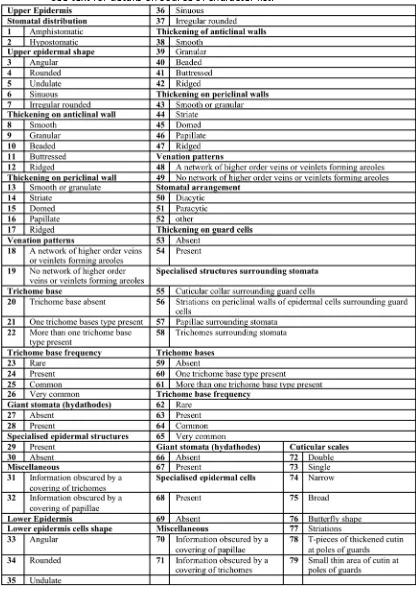
v Table 2.1. List of the 79 cuticular characters used to group cuticle
specimens in this study
49
Table 2.2. Main characters used to distinguish between morphotypes at Brandy Creek
50
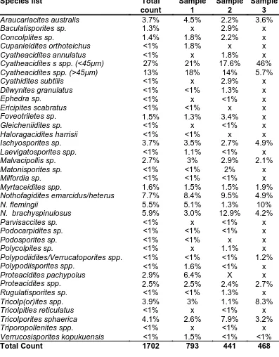
Table 3.1. Fossil and spore species (palynomorphs) list for Brandy Creek 107
Table 3.2. Comparison of pollen and spore count for Brandy Creek and other early to late Eocene localities in south eastern Australia
108
Table 4.1. Leaf margin equations for Australia and Global calibrations. 141
Table 4.2. Nearest living relatives of Brandy Creek and other Eocene flora. M= leaf macrofossils and P =pollen/spores.
142
Table 4.3. Key for identification of Fungal Germlings 143
Table 4.4. Leaf Margin Analysis and Bioclimatic climate estimates for south eastern Australian Eocene Floras.
144
Table 4.5. Comparison between Brandy Creek and other Eocene LMA estimates.
144
Table 4.6. Palaeoclimate estimates based on bioclimatic analysis of south eastern Australian Eocene Floras.
145
Table 4.7. Percentage of each grade of epiphyllous fungi found at Brandy Creek.
146
Table 5.1. List of Brandy Creek leaf morphotypes 182
Table 5.2. Percentage count of pollen and spores at Brandy Creek 183
Table 5.3. Comparison of pollen and spore counts for Brandy Creek and other Eocene localities in south eastern Australia
184
Table 5.4. Fossil palynomorphs at Brandy Creek individual samples 185
.
List of Figures
Fig. 2.1 Map of Australia showing the location of macroflora’s and microflora’s discussed in the text
51
Fig. 2.2. Stratigraphic log of the Brandy Creek outcrop 52
Fig. 2.3. Brandy Creek outcrop showing laminated siltstone and sandstone containing fossil plant material
53
Fig. 2.4. Dendogram showing the relationship between morphotypes at Brandy Creek
54
Fig. 2.5. Floristic composition of Brandy Creek and other Eocene localities in south eastern Australia
55
vi Fig. 2.16 – 2.17. Brandy Creek 003 Lauraceae aff. Cryptocarya 58
Fig. 2.18 – 2.19. Brandy Creek 003 Lauraceae aff. Cyptocarya 60
Fig. 2.20 - 2.23. Brandy Creek 009 Lauraceae aff. Cyptocarya 60
Fig. 2.24. Brandy Creek 009 Lauraceae aff. Cyptocarya 63
Fig. 2.25 – 2.29. Brandy Creek 015 Lauraceae aff. Cyptocarya 63
Fig. 2.30 – 2.34. Brandy Creek 004 Lauraceae aff. Endiandra 65
Fig. 2.35. Brandy Creek 005 Lauraceae aff. Endiandra 65
Fig. 2.36 – 2.38. Brandy Creek 005 Lauraceae aff. Endiandra 67
Fig. 2.39 – 2.41. Brandy Creek 006 Lauraceae aff. Endiandra 67
Fig. 2.42. Brandy Creek 006 Lauraceae aff. Endiandra 69
Fig. 2.43 – 2.45 Brandy Creek 007 Lauraceae aff. Endiandra 69
Fig. 2.46 – 2.47 Brandy Creek 008 Lauraceae aff. Endiandra 69
Fig. 2.48. Brandy Creek 008 Lauraceae aff. Endiandra 71
Fig. 2.49 – 2.52 Brandy Creek 010 Lauraceae aff. Endiandra 71
Fig. 2.53. Brandy Creek 010 Lauraceae aff. Endiandra 73
Fig. 2.54 – 2.57 Brandy Creek 013 Lauraceae aff. Endiandra 73
Fig. 2.58. Brandy Creek 014 Lauraceae aff. Endiandra 73
Fig. 2.59 – 2.62 Brandy Creek 014 Lauraceae aff. Endiandra 75
Fig. 2.63 – 2.64 Brandy Creek 011 Lauraceae aff. Litsea bennetti group 75
Fig. 2.65 – 2.67 Brandy Creek 011 Lauraceae aff. Litsea bennettii group 77
Fig. 2.68 – 2.70 Brandy Creek 012 Lauraceae aff. Litsea bennetti group 77
Fig. 2.71 – 2.75 Brandy Creek 016 aff. Cunoniaceae/Elaeocarpaceae 79
Fig. 2.76 – 2.80 Brandy Creek 017 aff. Cunoniaceae/Elaeocarpaceae 81
Fig. 2.81 – 2.83 Brandy Creek 018 aff. Cunoniaceae/Elaeocarpaceae 83
Fig. 3.1. Pollen and spore zones 109
Fig. 3.2. Stratigraphic log of the Brandy Creek outcrop 110
Fig. 3.3. Abundance histogram of the Brandy Creek palynomorphs
111
Fig. 3.4. Floristic composition of Brandy Creek and other Eocene floras based on palynomorph counts.
112
Fig. 4.1. Comparison of Eocene localities, taphonomic localities and observed MAT for modern rainforest in eastern Australia.
147
Fig 4.2. Comparison of estimates of Mean Annual Temperature (MAT) with associated errors of the estimate, for the Brandy Creek Eocene flora based on 3 different climate proxies.
147
Fig. 5.1. Early Paleogene palynostratigraphic schema for south eastern Australia
vii
Fig. 5.2. Stratigraphic log of the Brandy Creek outcrop 187
Fig. 5.3. Brandy Creek leaf morphotype rank abundance plot 188
Fig. 5.4. Brandy Creek palynomorph rank abundance plot 189
Fig. 5.5. Floristic composition of Eocene localities based on macrofloral record
190
Fig.5.6. Floristic composition of Eocene localities based on palynomorphs
191
Fig. 5.7. Dendogram showing relationship between Brandy Creek and other Eocene localities
192
Fig. 5.8 a -b SHE diagram for Brandy Creek macrofossil and pollen/spores
193
Fig.5.9. Rarefaction curves for summed plant leaf morphotypes and Palynomorphs
194
Fig. 5.10. Rarefaction curves for leaf morphotypes for Brandy Creek and other Australian Eocene localities
195
Fig. 5.11. Rarefaction curves comparing Eocene to modern tropical leaf assemblages
196
Fig. 5.12 a- b Dendogram showing the relationship between Brandy Creek samples for morphotypes and palynomorphs
197
Fig. 5.13. Spindle diagram of the Brandy Creek macrofossils 198
Fig. 5.14. Spore – pollen abundance diagram 199
Fig. 5.15 a - b Rarefaction curves for macrofossils and palynomorph samples at Brandy Creek
Chapter 1
Introduction
The late Eocene (37.2 - 33.9mya) marks the end of an interval during which the world’s biota evolved into forms recognisable today and the Northern Hemisphere continents adopted their current position, while Australia commenced its northward movement from high southern latitudes (Wing and Greenwood 1993; Quilty 1994; Zachos, Stott and Lohmann 1994; Greenwood and Wing 1995). Analysis of sea surface temperatures shows that the once warm oceans of the early Eocene and for a brief period during the Middle Eocene Climatic optimum (MECO) were between ~ 24 - 28°C and had begun to cool during the late Eocene to ~ 20°C (Huber and Caballero 2011). Carbon dioxide levels that were estimated to be 4400 ppm during the early Eocene and 2000 to 3000ppm again during the MECO had fallen by a further 1000 ppm by the late Eocene (Liu et al. 2009; Bajl et al. 2010; Pearson and Palmer 2000; Huber and Caballero 2011).
The Eocene has been used as a benchmark for understanding climates warmer than those of today, and provides the means to understand better trends in current global warming climate and how these can affect ecosystems (Gajewski 1993;
Greenwood and Basinger 1993; Shellito and Sloan 2006; Zachos et al. 2008; Huber and Caballero 2011; Pross et al. 2012; Smith et al. 2012).
The steady increase in the number of documented Eocene floras across Australia and the globe and the development of regional and global calibrations for both palaeoclimate and palaeoecological reconstruction has improved our
understanding of how plants adapt to changes in climate as a result of elevated
carbon dioxide levels and temperature (Peppe 2010; Greenwood et al. 2003 and 2004; Carpenter et al. 2007 and 2012; Smith et al.2010; Bannister et al. 2012; Lee et al. 2012).
environment, particularly those resulting in melting of ice at the poles, and increase in sea surface and terrestrial temperatures. Understanding how individual plants and plant communities adapt to shifts in environmental conditions is important to determine conservation and management programs particularly for rare and
endangered species. Insight into past environments can assist us to understand how best to manage future climate changes; the plant fossil record allows us to do this.
Paleogene plant fossils have been reported from localities on the Bogong High Plains and other sites from various altitudes in the Eastern Highlands of Victoria (Paterson 1935; Douglas 1978; Christophel 1980; Keefe 2000; Greenwood, Vadala and Douglas 2000; Greenwood et al. 2003; Carpenter et al. 2004). Of these, only three localities are at high altitudes; the late Middle Eocene to Late Eocene localities at Hotham Heights and Brandy Creek and the Oligocene Bundara River locality (Keefe 2000; Greenwood, Vadala and Banks 2000; Greenwood et al. 2003; Carpenter et al. 2004).
Previous research at Brandy Creek provided preliminary data on taxonomic diversity and abundance (Keefe 2000; Greenwood et al. 2003), whereas Carpenter et al. (2004) provided a preliminary taxonomic analysis of the Hotham Heights macro- and microfloras, but provided no quantitative assessment of either the flora
(palaeoecology) or estimates of the palaeoclimate.
The research presented in this thesis builds on the preliminary work by Keefe (2000); and reported in Greenwood et al. (2003) by undertaking a quantitative analysis using partial and entire leaf macrofossil. Leaf samples and concomitantly collected palynological samples are used to reconstruct the palaeoecology and
palaeoclimate of the Brandy Creek Eocene fossil locality. There were four objectives to the research:
1. Undertake a taxonomic analysis of the Brandy Creek leaf macrofossils; 2. Document the Brandy Creek pollen and spore microflora;
4. Identify trends in plant community dynamics over the time represented by the sediments exposed at Brandy Creek mine.
The following chapters were prepared as papers intended for publication; therefore, each chapter can be treated separately with each having its own introduction and literature review, methods, results, discussion and conclusions.
Chapter 2 “Leaf macrofossils of the Brandy Creek Eocene locality, Bogong High Plains, Victoria”, builds on the preliminary taxonomic assessment of the Brandy Creek flora by Keefe (2000). For the first time the flora is quantitatively sampled providing the foundations for palaeoclimatic (Chapter 4) and palaeoecological (Chapter 5) reconstructions. Cuticular and gross morphological characters are recorded and hierarchical clustering is used to determine the level of dissimilarity among morphotypes and where possible morphotypes are assigned to nearest living relatives.
Chapter 3 “The Brandy Creek Microflora (Spores and Pollen)”, provides additional information on the floristic character of the vegetation that is absent from the Brandy Creek macrofossil record; microfloral and macrofloral data are complimentary owing to differences in transport and preservation potential between spores-pollen and leaves, and between taxa for the same organ (Greenwood 1991). Using previous identification of pollen and spores across southern Australia, the pollen and spores at Brandy Creek are identified to stratigraphic spore-pollen taxa, and where possible assigned to a nearest living relatives. The data from this chapter contributes to a bioclimatic analysis of the Brandy Creek flora (Chapter 4) and an assessment of plant community structure in Chapter 5.
both regional and global calibrations, mean annual temperature for the Brandy Creek flora is calculated based on the proportion of toothed versus non-toothed dicot angiosperm species. A second method, Bioclimatic Analysis, uses nearest living relatives of the leaf, pollen and spores documented in Chapters 2 and 3, and applies the climate tolerances of modern floras to the fossil flora at Brandy Creek. Nearest living relative analogy assumes that the climate tolerances of fossil flora taxa are the same as those of their modern counterparts. The Mean Annual Temperature (MAT) for regional floras is updated with new climate profiles added to the original data set developed by Greenwood et al. (2003). These additional data improve the accuracy of the MAT for Brandy Creek and the other Eocene localities.
Additional indicators of Eocene climate at Brandy Creek are applied, including the presence and grade of epiphyllous fungi in a fossil flora (i.e., fungi found attached to leaf surfaces), as well as additional leaf morphological characters such as the presence of drip tips which can be indicators of wet climates.
Chapter 5 “ Palaeoecological reconstruction of the Brandy Creek Eocene locality, Bogong High Plains, Victoria, Australia”, the penultimate chapter, brings together the taxonomic analysis of the macrofossil record determined in Chapter 2 and the pollen and spores fossil record determined in Chapter 3, with the climate analysis in Chapter 4. Collectively, the data and taxonomic determinations from these chapters are used to reconstruct the palaeoecology of the Brandy Creek Eocene locality. Key elements of the chapter include: 1) a review of the floristic composition of the flora, using both the leaf macrofossil and pollen and spore record; and 2) analysis of trends in
community dynamics and diversity by comparing the 17 units that have been sampled vertically at the Brandy Creek locality.
Additional data afforded by the Brandy Creek leaf macrofossil and microfossil records contribute to the understanding of the regional (i.e. south eastern Australia)
Brandy Creek in the global context, looking at the common elements of floristic composition, diversity and climate as well as how the Brandy Creek flora compares to regional and global landscapes during this time.
Chapter 6 provides an overview of the key findings of the research and presents suggestions for further work leading on from this study.
Included in Appendix 1 are 2 papers that in part were generated from the research presented in this thesis, and for which I am co-author. The paper by
Greenwood et al. (2003) includes initial data from the Brandy Creek Eocene flora. The second work is an ‘in preparation’ manuscript prepared by Greenwood, Webb and Keefe for the peer-reviewed science journal ‘Geology’. In each case I contributed to the writing and interpretations presented.
References
Bijl, PK, Houben, AJP, Schouten, S, Bohaty, SM, Sluijs, A, Reichart, GJ, Sinninghe Damsté, JS, and Brinkhuis, H 2010, ‘Transient Middle Eocene Atmospheric C02
and Temperature Variations’, Science, vol. 30, p819, DOI: 10.1126/science.1193654
Bannister, JM, Conran, JG, and Lee, DE 2012, ‘Lauraceae from rainforest surrounding an early Miocene maar lake, Otago, southern New Zealand’, Review of
Palaeobotany and Palynology, vol. 178, pp. 13 – 34.
Carpenter, RJ, Hill, RS, Greenwood, DR, Partridge, AD, and Banks, MA 2004, ‘No snow in the mountains: an early Eocene flora from Hotham Heights, Victoria, Australia’, Australian Journal of Botany, vol. 52, part6, pp. 685 - 718. Carpenter, RJ and Jordan, GJ and Hill, RS 2007, ‘A toothed Lauraceae leaf from the
early Eocene of Tasmania, Australia’,International Journal of Plant Sciences, vol. 168, part 8, pp. 1191 - 1198.
Carpenter, RJ, Jordan, GJ, Macphail, MK and Hill, RS 2012, ‘Near-tropical Early Eocene terrestrial temperatures at the Australo-Antarctic margin, western Tasmania’, Geology, vol.40, no.3, pp.267 - 270.
south-eastern Australia’ Australian Journal of Botany, vol. 28, pp. 249-59.
Douglas, JG 1978, ‘Victoria’s oldest flowers’ Victorian Naturalist, vol. 95, pp. 137 -140. Gajewski, K 1993, ‘The role of palaeoecology in the study of global climate change’,
Review of Palaeobotany and Palynology, vol. 79, pp. 141-151.
Greenwood, DR, and JF Basinger 1993, ‘Stratigraphy and floristics of Eocene swamp forest from Axel Heiberg Island, Canadian Arctic Archipelago’, Canadian Journal of Earth Science, vol. 30, pp. 1914 - 1923.
Greenwood, DR 1991, ‘The Taphonomy of Plant Macrofossils’, In, Donovan, SK (Ed.) The Processes of Fossilization. Belhaven Press, London, 303p Ch. 7, pp. 141-169.
Greenwood, DR, Vadala, AJ, and Douglas, JG 2000, ‘Victoria Paleogene and Neogene macrofloras: a conspectus’, Proceedings of the Royal Society of Victoria, vol. 122, part 1, pp. 65 - 92.
Greenwood, DR, Vadala, AJ and Banks, M 2000, ‘Climate change and vegetation responses during the Paleocene and Eocene in southeastern Australia. pp. 65-66, In,Thematic issue on Early Paleogene Warm Climates and Biosphere Dynamics, Edited by Schmitz, B., B. Sundquist & F.P. Andreasson. GFF (Geologiska Föreningens i Stockholm Förhandlingar), 122(1): 1 – 192. Greenwood, DR, Moss, PT, Rowett AI, Vadala, AJ and Keefe, RL 2003, ‘Plant
communities and climate change in south-eastern Australia during the early Paleogene’. In SL Wing, PD Gingerich, B Schmitz, and E Thomas (eds), Causes and consequences of Globally Warm Climates in the Early Paleogene,
Geological Society of America Special Paper 369, Denver, Colorado. Greenwood, DR, Wilf, P, Wing, SL, and Christophel, DC 2004, ‘Paleotemperature
estimates using leaf margin analysis: Is Australia different?’, PALAIOS, vol. 19,
no. 2, pp. 129 – 142.
Greenwood, D and Wing SL 1995, `Eocene continental climates and latitudinal temperature gradients’, Geology, vol. 23, no.11, pp.1044 - 1048.
Huber M and R Caballero, 2011. The early Eocene equable climate problem revisited. Climate of the Past 7: 603 – 633.
Keefe, RL 2000, Windows on an ancient forest: The Palaeoecology of the Early Eocene flora of Brandy Creek Mine, Eastern Highlands, Victoria. Unpubl. Honours thesis. Victoria University of Technology, Melbourne.
Review, vol. 78, pp. 235 - 260.
Liu, Z, Pagani, M, Zinniker, D, Deconto, R, Huber, M, Brinkhuis, H, Shah, SR, Leckie, MR, and Pearson, A 2009, ‘Global Cooling During the Eocene-Oligocene Climate Transition’, Science, vol.323, pp. 1187 – 1190.
Paterson, HT 1935, ‘Notes on plant remains from Narracan and Darlimurla, South Gippsland’, Proceedings of the Royal Society of Victoria (new series), vol. 31, pp.362 - 363.
Pearson, PN, and Palmer, MR 2000, ‘Atmospheric carbon dioxide concentrations over the past 60 million years’, Nature, vol. 406, pp. 695 – 699.
Peppe, DJ, 2010, ‘Megafloral change in the early and middle Paleocene in the Williston Basin. North Dakota, USA’, Palaeogeography, Palaeoclimatology and
Palaeoecology, vol. 298, pp. 224 -234.
Pross J, Contreras, L, Bijl, PK, Greenwood, DR, Bohaty, SM, Schouten, S, Bendle, JA Röhl, U, Tauxe, L, Raine, JI, Huck CE, van de Flierdt, T, Jamieson, SSR, Stickley, CE, van de Schootbrugge, B, Escutia, C, Brinkhuis, H, and IODP Expedition 318 Scientists, 2012. ‘Persistent near-tropical warmth on the Antarctic continent during the early Eocene epoch’, Nature vol. 488 pp.73–77.
Quilty, PG 1994, ‘The background: 144 million years of Australian palaeoclimates and palaeogeography. pp. 14-43, In Hill, RS (Ed.) History of Australian vegetation: Cretaceous to Recent. Cambridge University Press, Cambridge.
Shellito, CJ and Sloan, LC 2006, ‘Reconstructing a lost Eocene paradise: Part I.
Simulating the change in global floral distribution at the initial Eocene thermal maximum’, Global and Planetary Change, vol. 50, no.1, pp. 1 -17.
Smith RY, Greenwood, DR, and Basinger, JF, 2010, ‘Coupling of globally warm temperatures and high levels of pCO2 during the Early Eocene Climatic Optimum: evidence from the Falkland flora of the Okanagan Highlands, Canada’ Palaeogeography, Palaeoclimatology, Palaeoecology vol. 293 pp.120– 131.
Smith, RY, Basinger, JF, and Greenwood, DR, 2012, ‘Early Eocene plant diversity and dynamics in the Falkland flora, Okanagan Highlands, British Columbia, Canada’, Palaeobiodiversity and Palaeoenvironments, Vol. 92, no 3 pp. 309-328. DOI: 10.1007/s12549-011-0061-5
Zachos, JC, Stott, LD and Lohmann, KC 1994, ‘Evolution of early Cenozoic marine temperatures’, Paleoceanography vol.9, pp. 353-387.
Chapter 2 Leaf macrofossils of the Brandy Creek Eocene locality,
Bogong High Plains, Victoria.
2.1 Introduction.
The use of leaf macrofossils has become a commonplace method for the
quantitative reconstruction of past floras and by extension past climates (e.g., Mosbrugger and Utescher 1997; Greenwood et al. 2003 and 2010; Uhl et al. 2007; Herman and Spicer 2010). Leaves of terrestrial species, unlike pollen of these very same species, are
particularly useful for the construction of local floras due to leaves having a limited
dispersal range. Leaves have a tendency to fall in close proximity to the parent plant while pollen, for the most part, is highly mobile and may travel many kilometers from the source of origin (Greenwood 1991; Cronin 1999). Because of the limited dispersal range of
leaves, very specific and locality sensitive data can be compiled from the examination of these leaf macrofossils and their use in comparison to extant species (Wilf et al. 1998).
Greenwood et al. 2003; Carpenter et al. 2004).
Records of plant macrofossils at Brandy Creek date back to the 1930’s when plant fossils were discovered during gold mining operations. Paterson (1935) recorded the presence of the gymnosperm Ginkgo L., and dicot taxa originally described by the authors as Laurus L., Eucalyptus L’Her. , and Ficus L., and the fern Lastraea. However, most of these initial identifications now have been shown to be incorrect after re-examination and comparison to additional, more recently collected and identified specimens (Greenwood et al. 2000). Subsequent investigations at Brandy Creek have indicated an infructescence of Gymnostoma Casuarinaceae R.Br). Initially described by Douglas (1978), the
identification of the infructescence was verified by Christophel (1980) as Casuarina subgenus Gymnostomae, and confirmed most recently as a species of Gymnostoma L.A.S Johnson by Scriven and Hill (1995).
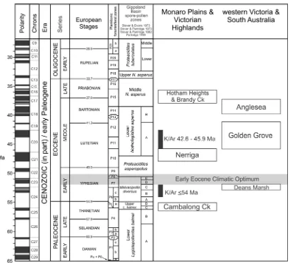
Palynofloras from Brandy Creek sediments were previously determined to belong to the upper Malvacipollis diversus zone of the Gippsland Basin palynostratigraphic scheme (Stover and Partridge 1973; Macphail et al. 1994), thus placing these sediments as Early Eocene (55.8 – 48.6 million years before present) (Scriven and Hill 1995; Partridge 1998; Greenwood et al. 2000; Carpenter et al. 2004). However, recent analysis by Holgate et al. (2008) correlated the Brandy Creek palynoflora to the Middle Nothofagidites asperus Zone, indicating a Late Eocene age (37.2 – 33.9my before present).
Several preliminary examinations of the Eocene flora at Hotham Heights and Brandy Creek by the author and others suggest that conditions were significantly warmer than at present (Keefe 2000; Vadala and Greenwood 2001; Greenwood et al. 2003; Carpenter et al. 2004). These warmer climates supported plant taxa typical of
now restricted to and considered endemic to the Humid Wet Tropical zone of north eastern Queensland (Keefe 2000; Vadala and Greenwood 2001; Greenwood et al. 2003; Carpenter et al. 2004).
This study builds on the preliminary taxonomic assessment of the Brandy Creek flora by Keefe (2000). Taxonomic assignment is based largely on cuticular character, with gross morphological characters recorded when possible. Hierarchical clustering is used to determine the level of dissimilarity among morphotypes and where possible morphotypes are assigned to nearest living relatives.
2.2 The use of leaf and cuticular morphology in the identification
of leaf macrofossils.
2.2.1 Leaf architecture.
The use of leaf architecture and morphology as a means of identifying plants was first attempted by von Ettingshausen in 1861 (Hickey 1973). More recent authors have developed sophisticated leaf architecture classification systems and attempted to
standardize leaf architecture terminology and classification (Dilcher 1974; Ellis et al. 2009). The use of leaf architecture has become refined to such a degree as to allow for the
delineation of some taxa into taxonomic orders. A prime example of this is a complex taxonomy of the Laurales based on particular characters including features such as; leaves simple, margin entire, venation pinnate, secondary veins brochidodromous and some cases of Lauraceae acrodromous, inter secondary veins common or tertiary veins reticulate to transverse (Hickey and Wolfe 1975). The limitations with these methods is that in many cases individual or a combination of characters, like the ones described above, are not restricted to one family or order, and cannot be used in isolation to identify fossil morphotypes.
As the use of leaf architecture has become more sophisticated, the shortcomings of the method have been highlighted. Of particular interest is the question: what
morphological features can be used for identification in the absence of clear leaf architectural features? Leaf cuticle characteristics have been identified to be of
considerable benefit in the diagnosis of macrofossil families even in the absence of leaf architecture, which can often be the case with fossil macroflora and as is the case at Brandy Creek which has only a small number of complete leaves resulting in cuticle characters being the main form of identification of the Brandy Creek leaf macroflora (Hyland 1989; Pole 2007). The cuticular membrane is highly resistant to oxidation,
therefore making it an ideal candidate for preservation in depositional environments and in providing key taxonomic information of past floras (Barclay et al. 2007). Identification of cuticular morphological characteristics to assign fossil morphotypes to an extant family and in some cases a genus or genera, is increasingly being utilised. Taxonomically useful characters include details of the stomatal complex, trichomes bases, and the form and arrangement of epidermal cells. Cuticular morphological identification has been applied to several families typically used to determine fossil floras in Australia, including:
Araucariaceae Jussieu (Stockey and Ko, 1986); Myrtaceae Jussieu (Christophel and Lys, 1986); Lauraceae (Hill 1986; Christophel 1986; Bannister et al. 2012); Cunoniaceae, and Elaeocarpaceae (Carpenter et al 2004). Although determination and assignment of particular cuticular morphological types to a given genus or family is possible, there are limitations. These limitations are primarily due to the differing levels of cuticle
preservation which can obscure some characteristics. Additionally, the long time period, coupled with evolutionary processes, between taxa of fossil flora and their nearest living relatives can obscure critical details. For the above reasons, it is prudent to assign
macrofossils as having only an affinity with a particular genus or family rather than taking the absolutist position.
Lack of clarity in the definitions of morphological and architectural characters used to score morphotypes can be problematic. Certain morphological and architectural
example of such a problem relates to epidermal anticlinal walls, which can be straight and easily identifiable; however, the difference between sinuous and curved anticlinal walls is often determined by individual assessment rather than a particular formula. Using a combination of characters to score morphotypes provides a much better and more robust method of identification. It is becoming increasingly common to include multiple
characteristics such as epidermal cells’ anticlinal and periclinal walls, guard cells, stomata and trichome bases. Using multiple characteristics has made identification and close affinity assignment to extant genera possible (Barclay et al. 2007). Additionally, these multiple character set identifications have allowed authors such as Christophel et al. (1987); Hill (1986); Barrett and Christophel (1990) and Carpenter et al. (2004) and (2007) to assign with some confidence, fossil morphotypes to family and genera based on cuticular morphology. Interestingly, for some families such as Lauraceae these multiple character set identifications are congruous with traditional morphological taxonomic identification tools such as fruits and flowers (Christophel and Rowett 1996).
2.2.3 Lauraceae Jussieu
The use of a combined analysis of leaf architecture and cuticular morphological characters as a method of identification of Lauraceae morphotypes and subsequent comparison of these types with extant genera has been almost universally adopted by authors in Australia and globally, Bandulska (1926), Hill (1986); Carpenter et al. (2004) Bannister et al. (2012) (New Zealand); Kovach and Dilcher, (1984) (North America), Lott et al. (2011) Costa Rica; Worobiec (2007) (Poland); Iglesias et al. (2008) (Patagonia). When considering the characters of particular families, not all cuticular features are
diagnostically useful. For example, there are a number of features that apply to all genera in the Lauraceae; all are hypostomatic and the stomatal arrangement is paracyctic.
combination with other more diagnosic features (Upchurch and Dilcher 1990; Christophel and Rowett 1996).
When assigning a fossil morphotype in the Lauraceae family, stomatal characters can be a significant diagnostic tool, with the stomata of particular genera having different configurations and characteristics; e.g., butterfly-shaped cuticular scales (Cryptocarya R.Br. and Beilschmeidia Nees) and double cuticular scales (Endiandra R.Br.). Individually, these distinctive stomatal characteristics do not assist in the delineation of genera. However, stomatal characteristics used in combination with other cuticular features, including epidermal cells and trichome bases, allow determination of affinities between modern and fossil genera of Lauraceae (Hill 1986; Christophel and Rowett 1996; Carpenter et al. 2007; Nishida and van der Werff 2007). Early identifications of Lauraceae using much less robust multiple character data sets ascribed many ‘Lauraceae’ to Laurophyllum (a form genus - a genus based on non-sexual morphological characters), indicating only general affinity with Lauraceae (Hill 1986; Carpenter and Pole 1995). More recently, work completed by Christophel, Hyland and Whiffin (1993), and Christophel and Rowett (1996), combining leaf venation, leaf shape and cuticle characters of extant Australian Lauraceae taxa, has been used to assign fossil taxa to extant Lauraceae genera.
Lauraceae fossil record
Fossil evidence of Lauraceae is found across the globe and includes flowers, fruits, inflorescences, leaves and wood dating from the mid Cretaceous to the late Paleogene (Drinnan et al. 1990; Herendeen 1991; Eklund and Kvaček 1998; Eklund 1999; Qiu, et al. 1999; Frumin et al. 2004; Renner 2004; Bannister et al. 2012). Lauraceae fossils have been found in the Maastrichtian in the Northern Hemisphere, and the upper Cretaceous in New Zealand (Pole 1992). Paleocene floras containing Lauraceae have been recorded in North America, Princeton chert, British Columbia, Canada (Little, Stockey and Penner, 2009) and central and eastern Europe (Crane 1987; Eklund and Kvaček 1998). The London clay floras include Lauraceae taxa such as Beilschmeidia, Endiandra and Litsea (Chandler 1964; Collinson 1983).
The use of cuticle structures to identify fossil Lauraceae has shown that there is an abundance of Lauraceae fossils in Paleogene floras across Australia. The earliest Australian record of Lauraceae is from the Late Paleocene at Cambalong Creek in New South Wales. The Cambalong Creek fossils contain specimens that have been identified as having an affinity with the Lauraceae genera Beischmiedia, Cyrptocarya, Endiandra and Litsea (Vadala and Greenwood 2001). Lauraceae, or species with an affinity to Lauraceae have been described from other Paleogene localities in Australia including Anglesea, Hotham Heights and Brandy Creek in Victoria, Golden Grove in South Australia, Nerriga in New South Wales and the Lefroy Paleodrainange (Pidinga formation) in Western Australia (Christophel, Harris & Syber 1987; Hill 1982, 1986; Carpenter and Pole 1995; Carpenter et al. 2004; Keefe 2000; Greenwood 2001).
Eklund 1999; Vadala and Greenwood 2001; Hably 2007; Pole 2007; Lee et al. 2012; Bannister et al. 2012).
2.2.4 Cunoniaceae R.Br and Elaeocarpaceae Juss
The assignment of fossil morphotypes to either the Cunoniaceae or
Elaeocarpaceae cannot be definitively carried out based on leaf and cuticle morphology alone due to the similarities of the two families (Pole 1996). Barnes and Hill (1999)
completed a detailed study of Cunoniaceae leaf morphology including cuticles, however a comparative study has not been done for Elaeocarpaceae. Cuticle characters typical of Cunoniaceae/Elaeocarpaceae include a dark staining ring on the inner stomatal ledge, thickened rim of the outer stomatal ledge and T pieces of thickened cuticle at poles of guard cells, although these characters do not occur together in all genera (Pole 1996; 2008).
Fruits and flowers resembling Cunoniaceae have been recorded from Australian localities including Fruits from Middle Eocene Maslin Bay (Barnes and Hill 1999), flowers from Early Oligocene Cethana (Barnes et al. 2001). The earliest record of a Cunoniaceae macrofossil is Eucryphia falcata R.S. Hill from the Late Paleocene (Barnes et al. 2001). Leaf macrofossils with an affinity to Cunoniaceae have been found at sites including Cethana and Regatta Point in Tasmania, Anglesea, Hotham Heights and the La Trobe Valley in Victoria, Maslin Bay and Golden Grove in South Australia, and West Dale in Western Australia (Barnes et al. 2001; Carpenter et al. 2004).
Fossil fruit, leaves, pollen and flowers of Elaeocarpaceae have been documented from Paleogene sediments in southeastern Australia, including Lake Eyre Basin (Early Eocene), Anglesea (Middle Eocene), and Golden Grove (Middle Eocene) (Rozefelds and Christophel 1996). The earliest record of Elaeocarpaceae in Australia was recorded by Duigan (1951) who documented 8 leaf macrofossils from various localities in southeastern Australia. Subsequent work by Hill (1988) dismissed Duigan’s (1951) taxonomic
Elaeocarpacae. Vadala (2001) described Elaeocarpaceae aff. Elaeocarpus L.from Cambalong Creek in Victoria.
Globally, Cunoniaceae/Elaeocarpaceae fossils have been found from the Miocene of New Zealand (Pole 2008; Lee et al. 2012), Eocene and Oligocene fossil wood aff. Cunoniaceae from Europe (Friis et al. 2011), Elaeocapraceae leaf cuticle from the
Oligocene of Italy (Hably 2007), fruits from the Paleogene of North America (Manchester 1999 and Manchester and Kvaček, 2009), and wood aff. Elaeocarpaceae from Antarctica (Francis et al. 2009).
2.3 Materials and Methods
2.3.1 Locality Description
Fossil leaves that form the basis of this research were collected from Paleogene age sediments that crop out in the abandoned Brandy Creek gold mine. The mine site is located 7.8 km east-southeast of Hotham Heights, Bogong High Plains, northeast of Melbourne, Victoria, Australia, (37° 01’ S, 147° 13’ E; Map reference 8323 Dargo 55HEV 030188), at an altitude of 1500 m asl (above sea level) (Figure. 2.1). Middle Eocene to Late Oligocene basalts that cover parts of the Bogong High Plains crop out at Brandy Creek. Sediments from the locality previously have been dated as Early to Middle Eocene (Scriven and Hill 1995; Partridge 1998). However recent analysis by Holdgate et al. (2008) dated the locality as Late Eocene biostratigraphically through correlation of the microflora with the Nothofagdities asperus zone of the Gippsland Basin palynostratigraphic scheme (Stover and Partridge 1973; Macphail et al. 1994; Partridge 1999).
which sediments are differentiated into discrete layers, with obvious leavespreserved as compression fossils (Figure 2.2 and 2.3)(Greenwood et al. 2000; Keefe 2000).
2.3.2 Site Sampling
Sampling from Brandy Creek outcrop provided more than 500 partial or entire leaves across the site for taxonomic analysis. Leaf samples from each unit (Figure 2.2) were selected if an entire leaf,margin, base or apex was evident. Leaf macrofossils were sampled quantitatively to obtain ecological information, such as species dominance and diversity. Field census data from laterally continuous fossiliferous sediments, and studies based on present day leaf litter, have shown that large numbers of specimens (greater than 350) can be used to closely approximate patterns of dominance and overall floristic richness of the original floral community (Burnham et al. 1989, 1993; Burnham et al. 1992; Wing et al. 1995; Wilf et al. 1998). Collections were made vertically to determine
temporal variability in taxonomic composition and diversity through time.
2.3.3 Fossil preparation.
Extraction of leaf macrofossils.
Leaf macrofossils were preserved mostly as compressions with a small number of poorly preserved impressions towards the top of the outcrop, the latter of which were not included in the analysis. Mummified leaves were freed from the mudstone matrix by maceration using dilute (20%) H2O2 (hydrogen peroxide) using the method of Christophel (1980) as modified by Rowett (1991). A small section (up to one centimetre) of leaf was cut from each leaf sample to clear any remaining mesophyll and sediment. Individual sections were placed in test tubes containing 35% H2O2 combined with several grains of tetra sodium pyrophosphate and covered with parafilm. Tubes were submersed in boiling water for ten minutes in a water bath. If further cleaning was required, cuticles were left in the water bath at a temperature of 80°C for 1- 4 hours, and H2O2 replenished if
Preparation of leaf sections for scanning electron microscopy (SEM) was done with hydrofluoric acid (HF) 49% following the method of Kiger (1971). HF treatment was conducted by Laola Pty Ltd, Perth. Cuticles were soaked in HF for 24 hours to dissolve silicate particles and then rinsed a minimum of ten times with distilled water. Cuticles were returned to Victoria University for further processing, in which the mesophyll was cleared using Jeffrey’s Solution (equal parts of 70% HNO3 and 10% aqueous chromic acid; after Stace (1965), modified from Johnson (1940). Stace (1965) noted this process could take between one to four hours depending on the thickness of the cuticle however Brandy Creek cuticles reacted quickly to the solution and most cleared within thirty minutes. Cuticles were cleared when the cuticular envelope became dark brown as the oxidation process proceeded. Subsequent to clearing, cuticles were neutralised by soaking in 5% aqueous NH3 for 15 minutes, then rinsed with distilled water several times.
Scanning Electron microscopy (SEM)
Cuticles were mounted on aluminium stubs using double side tape, then air-dried using silica gel in a desiccator for at least 36 hours prior to being sputter coated with gold dust using an Edwards S150B sputter coater. SEM examination and photography was conducted at The University of Melbourne School of Botany using a Phillips XL 30 FEG.
Partial and entire leaves extracted from the mudstone were photographed using Kodak Technical Pan film. Black and white negatives were scanned using a Polaroid Sprint Scan 4000 at 3000dpi. Digital images were taken of the venation patterns of each
morphotype using a Zeiss Stemi 2000c dissecting microscope with a DAGE -MTI CCD100 camera attachment. Digital images of leaf cuticle were captured using a Carl Zeiss Axiocam HR, attached to a Zeiss Axioplan 2 light microscope. Images were processed using Adobe photoshop.
2.3.4 Taxonomic analysis
Macrofossils were sorted into taxonomic units using established leaf morphological and cuticle characters (Dilcher 1974; Hickey 1973, 1979; Ellis et al. 2009). A large database exists in the botanical literature of diagnostic characters for major Australian families such as Proteaceae Jussieu, Lauraceae, Myrtaceae and Araucariaceae and genera in these families (e.g., Christophel and Rowett 1996; Carpenter 1994; Hill 1986; Bannister et al. 2012).
Cuticle characters and leaf architecture.
To determine the number and type of morphotypes present in the samples from Brandy Creek, character lists were constructed, based on characteristics of the Lauraceae family, with the addition of more general characters suitable for other families such as Proteaceae, Elaeocarpaceae and Cunoniaceae. Selection of the character list was based on preliminary work done at Brandy Creek (Keefe 2000). Character lists are broadly based on those of Dilcher (1974) and Christophel and Rowett (1996).
Sorting took place at a number of levels of discrimination. Abaxial and adaxial surface of each cuticle were examined under light microscopy and sorted into broad types based on characters such as vein course cell patterns and areolation pattern. Each cuticle type sorted was further examined using additional character differences, such as
selected for each morphotype and scored using the character list (Table 2.1). Character scores for each morphotype are presented in Appendix 2. ASEM was used to distinguish characters that could not be seen easily under the light microscope, for example guard cell striations and cuticular scales.
Leaf morphological characters were described using the Manual of Leaf
Architecture (Ellis et al. 2009). Descriptions of morphotypes are based on the style used by Paull and Hill (2003) and are presented in the Results below.
Cluster analysis.
Cluster analysis was conducted to determine the relationships -among
morphotype, based on a defined character set devised by Dilcher (1974) and Christophel and Rowett (1996). The analysis was conducted using PAST (Paleontological Statistics Software Package for Education and Data Analysis Version 2.16; Hammer, Harpe and Ryan 2001). For this analysis Bray Curtis association metric was used with group average link fusion, weighting all character scores equally. Bray Curtis metric was used to determine the level of dissimilarity between each of the morphotypes (Krebs 1989). Dissimilarity is measured based on the number of shared presences, by counting the number of times both morphotypes have the same number of characters present.
The Brandy Creek leaf macrofossils are housed at the Melbourne Museum, Victoria, Australia.
2.4 Results
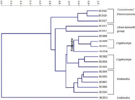
2.4.1 Cluster analysis of Brandy Creek morphotypes
clusters are separated is at 0.7. At this point most groups can be classified as Cunoniaceae/Elaeocarpaceae; Cryptocarya and Endiandra. The one group that falls outside of this is Litsea, which clusters between 0.6 and 0.7. The clusters are outlined below:
Cluster 1
BC 016, BC 017,and BC 018 aff. Cunoniaceae/Elaeocarpaceae. BC016 has undulate abaxial and adaxial anticlinal walls, with smooth perclinal walls. BC017 has angular to rounded anticlinal walls and striations on guard cells.BC018 has undulate anticlinal walls on abaxial surface only and perclinal walls are granular.
Cluster 2
BC011, Lauraceae Litsea bennetti group. BC 011 is highly papillate with angular to rounded epidermal cell walls.
Cluster 3
BC012. Litseabennettii group, BC 012 has undulate anticlinal walls on the adaxial surface and areoles on the abaxial surface.
Cluster 4
BC 009, BC 015, aff. Lauraceae Cryptocarya
The dendogram (Figure 2.4) shows that BC 009 and BC 015 are closely aligned based on the level of dissimilarity at less than 0.95. The features that distinguish the two
morphotypes from the other clusters, which include Cryptocarya, are the presence of areoles. BC 015 is further distinguished from BC009 by the presence of undulate anticlinal walls.
BC 008 and BC 014 aff. Lauraceae Endiandra.
anticlinal walls with a high degree of beading particularly on adaxial anticlinal walls of epidermal cells and vein course cells of abaxial surface.
All other Endiandra are either grouped together (cluster 6) or individually (cluster 7). At 0.7 dissimilarity, Endiandra is clustered with two Cryptocarya, but it is clear from the dendogram that as these groups are clustered between 0.8 and 0.9, the Endiandra is clearly different from the Cryptocarya in this cluster.
Cluster 5
BC 001, BC 002, BC 003 aff. Lauraceae Cryptocarya. The character that separates the three morphotypes of Cryptocarya (BC 001, BC 002 and BC003) from all others identified is the presence of the mesh- like appearance on the epidermal cell walls.
Cluster 6
BC 004, BC 005, BC 007, Lauraceae Endiandrais characterised by thickening and striations on guard cells.
BC 006, aff. Lauraceae Endiandra is grouped by the presence of mesh-like appearance on epidermal cells of adaxial surface. BC 006 is the only morphotype with an affinity to Endiandra to have this characteristic on the adaxial surface.
BC 010, Aff. Lauraceae Endiandra. BC 010 is distinguished by the absence of characters including thickened guard cells which is evident in BC 004, BC005 and BC007, presence of T pieces of thickened cuticle at poles of guards (BC008) and mesh like appearance on epidermal cells (BC 006).
Cluster 7
BC 013, Lauraceae aff. Endiandra has beaded and ridged anticlinal walls. A fine mesh of epidermal cells surrounds the stomata.
butterfly shaped cuticular scales, while Lauraceae affinity Endiandra have double
cuticular scales and Litsea has single cuticular scales. On a number of the Cryptocarya and Endiandra specimens there is unusual mesh structure on the epidermal cells. This feature has also been recorded from the Hotham Heights and Nerriga localities (Carpenter 2004; Hill 1986).
2.4.2 Taxonomic Descriptions of the Brandy Creek macroflora
Family: Lauraceae Jussieu
Genus: Laurophyllum Göppert
Morphotypes with affinity to Cryptocarya R. Brown
Morphotype BC – 001 Fig. 2.6 - 2.10.
Affinity/Identification Cryptocarya pleurosperma group Count/Specimens 11 BC 1390
Leaf symmetrical. Leaf base acute, cuneate. Leaf margin entire. Venation
eucamptodromous, spacing uniform, angle uniform. Tertiary venation random reticulate. Higher order veins regular polygonal reticulate. Areoles well developed, with 5 or more sides.
Adaxial surface epidermal cell walls isodiametric, angular to rounded, with mesh appearance of epidermal cells. Epidermal cells elongate over veins. Anticlinal walls smooth, thickened. Periclinal walls smooth to granular. Trichome bases present, with cuticular thickening around pore extending along radial walls of surrounding cells. Abaxial epidermal cell walls isodiametric, angular, with mesh-like appearance of epidermal cells. Epidermal cells elongate over veins. Anticlinal walls smooth, thickened. Periclinal walls smooth to granular. Trichome bases present, porous along vein course cells.Stomata paracytic, randomly orientated with butterfly-like cuticular scales.
Morphotype BC – 002 Fig. 2.11 – 2.15
Count/Specimens 45 BC 1608
Leaf symmetrical, elliptic. Leaf apex acute, acuminate. Base acute, cuneate. Leaf margin entire. Venation eucamptodromous, spacing uniform, angle uniform. Tertiary veins random reticulate. Higher order veins regular polygonal reticulate. Marginal venation looped. Areoles well developed, with five or more sides.
Adaxial epidermal cell walls isodiametric, angular to round with mesh-like appearance of epidermal cells. Epidermal cells elongate over veins. Anticlinal walls smooth, thickened. Periclinal walls smooth to granular. Trichomes bases rare. Abaxial cell walls isodiametric, angular to round with a mesh-like appearance of epidermal cells. Epidermal cells elongate over veins. Anticlinal walls beaded. Periclinal walls smooth to granular. Trichomes bases present. Hydathodes present, epidermal cells surrounding hydathodes cyclocytic. Stomata paracytic with butterfly-like cuticular scales.
Difference between BC001 and BC002 Hydathodes are absent from BC001
Morphotype BC – 003 Fig. 2.16 – 2.19
Affinity/Identification Cryptocarya Not assigned to a group.
Count/Specimens 4 BC 1205
Leaf symmetrical. Leaf apex acute, straight. Base acute, cuneate. Leaf margin entire. Venation pinnate.
Abaxial epidermal cell walls isodiametric, angular, rounded to undulate, Epidermal cells elongate over veins. Anticlinal walls smooth, ridged. Periclinal walls smooth to granular. Trichome bases rare. Adaxial cell walls isodiametric, angular, rounded, undulate to sinuous,. Epidermal cells elongate over veins. Anticlinal walls beaded, ridged. Periclinal walls smooth to granular. Trichome bases present. Stomata paracytic with butterfly-like cuticular scales.
Morphotype BC – 009 Fig. 2.20 -2.24
Count/Specimens 33 BC1613
Leaf symmetrical, elliptic. Leaf apex acute, acuminate. Base acute, cuneate. Leaf margin entire. Venation eucamptodromous, brochidodromous, spacing uniform, angle acute. Tertiary veins opposite percurrent, sinuous, uniform, with obtuse angle to primary vein. Higher order veins regular polygonal reticulate. Marginal ultimate looped. Areoles well developed, with five or more sides.
Adaxial epidermal cell walls isodiametric, angular to round. Epidermal cells elongate over veins. Anticlinal walls smooth, ridged. Periclinal walls smooth to granular. Trichomes bases rare. Abaxial epidermal cell walls isodiametric, angular to round. Epidermal cells elongate over veins. Anticlinal walls beaded, ridged. Periclinal walls smooth to granular. Trichome bases common. Areoles present. Stomata paracytic with butterfly-like cuticular scales.
Morphotype BC – 015 Fig. 2.25 - 2.29
Affinity/Identification aff. Cryptocarya Count/Specimens 6 BC 1580
Leaf microphyll, symmetrical, elliptic. Leaf apex acute, straight, rounded. Leaf margin entire. Venation weak brochidodromous, spacing irregular, angle uniform. Tertiary veins mixed opposite/percurrent, vein course straight, sinuous, angle to primary obtuse. Higher order veins regular polygonal reticulate, marginal ultimate looped. Areoles moderately developed.
Adaxial epidermal cell walls isodiametric, angular to round. Epidermal cells elongate over veins. Anticlinal walls ridged. Periclinal walls smooth to granular. Trichomes bases rare, mainly over vein course cells. Abaxial epidermal cell walls isodiametric angular, round to undulate. Epidermal cells elongate over veins. Anticlinal walls beaded, ridged. Periclinal walls smooth to granular. Trichome bases common, porous. Hydathodes present. Stomata didactic, paracytic, butterfly-like cuticular scales.
Morphotypes with affinity to Endiandra R. Brown
Affinity/Identification aff. Endiandra jonesii group Count/Specimens 11 BC 1335
Leaf symmetrical. Leaf apex acute. Base acute, cuneate. Leaf margin entire. Venation brochidodromous. Vein spacing uniform. Vein angle acute.
Adaxial epidermal cell walls isodiametric, angular to round. Epidermal cells elongate over veins. Anticlinal walls smooth. Periclinal walls smooth to granular. Trichome bases
present. Callimothalloid shields present. Abaxial epidermal cell walls isodiametric, angular to round. Epidermal cells elongate over veins. Anticlinal walls smooth. Periclinal walls smooth to granular. Trichome bases present, mainly over vein course cells. Callimothalloid shields present. Stomataparacytic with double cuticular scales. Guard cells striated.
Morphotype BC – 005 Fig.2.35 – 2.38
Affinity/Identification aff. Endiandra pubens group
Count/Specimens 8 BC 1401
Leaf architecture absent. Adaxial epidermal cell walls isodiametric, angular to round. Epidermal cells elongate over veins. Anticlinal walls ridged. Periclinal walls smooth to granular. Trichome bases present. Abaxial epidermal cell walls isodiametric,angular to round. Epidermal cells elongate over veins. Anticlinal walls smooth to beaded. Periclinal walls smooth to granular. Trichome bases very common, porous. Areoles present. Stomata paracytic, anomocytic with double cuticular scales. Guard cells striated, thickened.
Morphotype BC –006 Fig.2.39 – 2.42
Affinity/Identification aff. Endiandra pubens group Count/Specimens 13 BC1516
Adaxial epidermal cell walls isodiametric, angular to round with mesh-like appearance of epidermal cells. Epidermal cells elongate over veins. Anticlinal walls smooth, thickened. Periclinal walls smooth to granular. Trichome bases present. Abaxial epidermal cell walls isodiametric, angular to rounded. Epidermal cells elongate over veins. Anticlinal walls smooth, ridged, beaded. Periclinal walls smooth. Trichome bases common. Mesh-like appearance of epidermal cells surrounding stomata. Stomata paracytic, anomocytic with double cuticular scales. Guard cells thickened.
Morphotype BC – 007 Fig. 2.43 – 2.45 Affinity/Identification Not assigned to a group.
Count/Specimens 2 BC1526
Leaf architecture absent. Adaxial epidermal cell walls isodiametric, angular to round. Epidermal cells elongate over veins. Anticlinal cell walls smooth. Periclinal walls smooth to granular. Trichome bases very common. Abaxial epidermal cell walls isodiametric,angular to rounded. Epidermal cells elongate over veins. Anticlinal walls smooth to ridged.
Periclinal walls smooth to granular. Trichome bases very common. Stomata diacytic, paracytic with double cuticular scales. Guard cells striated, thickened.
Morphotype BC – 008 Fig. 2.46 -2.48
Affinity/Identification Not assigned to a group.
Count/Specimens 2 BC1260
Leaf architecture absent.
Morphotype BC – 010 Fig. 2.49 – 2.53
Affinity/Identification Not assigned to a group.
Count/ Specimens 206 BC1305
Leaf microphyll, symmetrical, elliptic. Leaf apex acute, straight, acuminate. Base acute, cuneate. Leaf margin entire. Venation eucamptodromous, spacing uniform, angle acute. Tertiary veins random reticulate. Higher order venation regular polygonal reticulate. Marginal ultimate veins looped. Areoles well developed, with five or more sides.
Adaxial epidermal cell walls isodiametric, angular to round. Epidermal cells elongate over veins. Anticlinal walls smooth, buttressed. Periclinal walls smooth to granular. Trichome bases present, glandular. Abaxial epidermal cell walls isodiametric, angular to round. Epidermal cells elongate over veins. Anticlinal walls granular. Periclinal walls smooth to granular. Trichomes bases common. Stomata paracytic with double cuticular scales.
Morphotype BC – 013 Fig 2.54 – 2.57
Affinity/Identification Not assigned to a group.
Count/Specimens 1 BC1051
Leaf architecture absent.
Adaxial absent. Abaxial epidermal cell walls isodiametric, angular to rounded with mesh-like appearance of the epidermal cell walls. Epidermal cells elongate over veins. Anticlinal walls beaded to ridged. Periclinal walls smooth to granular. Trichome bases common, porous. Stomata paracytic. Guard cells thickened on periclinal wall. Cuticular scales double, inner scale narrow, outer scale broad.
Morphotype BC – 014 Fig. 2.58 – 2.62
Affinity/Identification Not assigned to a group.
Count/Specimens 8 BC 1245
Adaxial epidermal cell walls isodiametric, angular, round to undulate. Epidermal cells elongate over veins. Anticlinal walls beaded. Periclinal walls smooth to granular. Trichome bases rare. Abaxial epidermal cell walls isodiametric, angular, round to undulate.
Epidermal cells elongate over veins. Anticlinal walls beaded. Periclinal walls smooth to granular. Trichome bases rare, mainly over vein course cells. Hydathodes present. Stomata paracytic, anomocytic. Guard cells thickened. Cuticular scales double.
Morphotypes with affinity to Litsea Lamarck
Morphotype BC – 011 Fig. 2.63 – 2.67
Affinity/Identification aff. Litsea bennetti group Count/Specimens 8 BC 1240
Leaf architecture absent
Adaxial epidermal cell walls isodiametric, angular to round. Epidermal cells elongate over veins. Anticlinal walls smooth, beaded and ridged. Periclinal walls smooth to granular. Trichome bases present, porous thickenings. Abaxialepidermal cell walls
isodiametric,angular to round. Epidermal cells elongate over veins. Anticlinal walls smooth. Periclinal walls papillate. Trichome bases rare. Subsidiary cells obscured by papillae surrounding stomata. Stomatal ledge straight, narrow, with cuticular collar.
Morphotype BC – 012 Fig. 2.68 - 2.70
Affinity/Identification aff. Litsea bennetti group Count/Specimens 4 BC 1336
Leaf architecture absent.
trichome base. Hydathodes present. Subsidiary cells obscured by papillae surrounding stomata. Stomatal ledge straight, narrow.
Family: aff.Cunoniaceae/Elaeocarpaceae
Morphotype BC – 016 Fig. 2.71 -2.75
Affinity/Identification
Count/Specimens 37 BC 1064
Leaf microphyll, symmetrical elliptic. Leaf apex acute, acuminate, straight. Base acute, cuneate. Leaf margin crenate. Venation brochidodromous, spacing increasing towards base, angle uniform. Tertiary veins mixed opposite, alternate, straight, angle variability inconsistent; angle to primary vein perpendicular. Higher order veins regular polygonal reticulate. Marginal ultimate looped-teeth. Areoles well developed, with five or more sides. Free ending veinlets 1- branched; 2 or more branches. Teeth spacing regular, sinus rounded, apex simple.
Adaxial epidermal cell walls isodiametric, angular. Epidermal cells elongate over veins. Anticlinal walls straight, rounded, undulate, sinuous, knobs. Abaxial epidermal cell walls isodiametric, angular. Epidermal cells elongate over veins. Anticlinal walls straight, rounded, undulate, sinuous, knobs. Stomata randomly orientated anomocytic. Anticlinal walls knobs. T- Pieces of thickened cutin at poles of guards.
Morphotype BC – 017 Fig. 2.76 -2.80
Affinity/Identification
Count/specimens 103 BC 1068
Adaxial epidermal cell walls isodiametric, angular. Epidermal cells elongate over veins. Anticlinal walls straight, rounded, sinuous, knobs, ridges. Periclinal walls striations.
Trichome bases present. Abaxial epidermal cell walls isodiametric, angular. Epidermal cells elongate over veins, anticlinal walls straight, rounded, undulate, sinuous, knobs, ridges. Periclinal walls striations, thickened. Trichomes bases present. Glandular/ secretory structure present. Stomata randomly orientated anomocytic. Anticlinal walls knobs, ridges. Periclinal walls of guard cells striate, thickened. T-pieces of thickened cuticle at poles of guards.
Morphotype BC – 018 Fig 2.81 -2.83
Affinity/Identification
Count/Specimens 13 BC 1183
Leaf microphyll, symmetrical elliptic. Leaf base acute, cuneate. Leaf margin crenate. Venation weak brochidodromous, spacing decreasing towards base, angle uniform. Tertiary veins random reticulate, straight, angle variability inconsistent, angle to primary vein obtuse. Higher order veins regular polygonal reticulate. Marginal ultimate loop-teeth present. Areoles well developed, with five sides. Teeth spacing regular, sinus angular, apex simple.
2.5 Discussion
2.5.1 Lauraceae
Fifteen of the 18 morphotypes 83% were assigned to Lauraceae based on cuticular characters. Figure 2.5. These findings strongly contrast with percentage representation as determined by palynomorh counts described in other parts of this study. Lauraceae, although present in high numbers in the macrofossil record at Brandy Creek, is not documented in the palynomorph count; this reflects the lack of preservation potential of Lauraceae pollen due to exine deterioration (Macphail 1980). The absence of Lauraceae in the Brandy Creek pollen record supports the notion that a holistic approach is required when looking at a fossil flora requiring both leaf and pollen/spores to provide a complete profile. The Brandy Creek palynomorphs are discussed in the following chapter.
For a leaf macrofossil to be assigned to Lauraceae the specimen generally needs to have the following characters; entire margin, hypostomatic, paracyctic stomata, with guard cells and over arching subsidiary cells (Hill 1986; Christophel and Rowett 1996). Individually, these characters are represented in a number of angiosperm families, however collectively they are diagnostics for Lauraceae (Doyle and Endress 2000). Using the key features of Lauraceae in cluster analyses, Lauraceae at Brandy Creek show affinity with three genera; Cryptocarya (5 morphotypes), featuring butterfly cuticular scales, Endiandra (8 morphotypes) with double cuticular scales, and Litsea (2 morphotypes) which have papilliae.
Variation of morphological characters is one of the challenges faced when using leaf cuticle for taxonomic assessment of a flora. For example, Stace (1965) and Wilkinson (1979) noted that there can be a high level of variation in epidermal anticlinal walls. Some highly variable morphotypes, having a combination of both angular and rounded