Original Research Article
Detection of inducible clindamycin resistance in Staphylococcus aureus
from various samples in a tertiary care hospital
Rohit Kumar*, Jagarti, Mrinmoy Sarma, Gautam Shalini
INTRODUCTION
Staphylococcus aureus is increasingly recognized as causing nosocomial and community acquired infections in every region of the world. The antimicrobial-resistance to Staphylococcus aureus is an increasing problem.1
Staphylococcus aureus can cause a range of illnesses, from minor skin infections, such as impetigo, boils, cellulitis, folliculitis, carbuncles, scalded skin syndrome, and abscesses, to life-threatening diseases such as
pneumonia, meningitis, osteomyelitis, endocarditis, toxic shock syndrome, bacteremia, and sepsis.2
Antibiotic resistance in Staphylococcus aureus was uncommon when penicillin was first introduced in 1943. By 1950, 40% of hospital Staphylococcus aureus isolates were penicillin-resistant; by 1960, this had risen to 80%.3
Methicillin was the first antibiotic in this class to be used (it was introduced in 1959), but, only two years later, the first case of MRSA was reported in England.4 Today,
Department of Microbiology, National Institute of Medical Sciences and Research, Jaipur, Rajasthan, India
Received: 24 September 2019
Accepted: 31 October 2019
*Correspondence:
Dr. Rohit Kumar,
E-mail: rk95rk@gmail.com
Copyright: © the author(s), publisher and licensee Medip Academy. This is an open-access article distributed under the terms of the Creative Commons Attribution Non-Commercial License, which permits unrestricted non-commercial use, distribution, and reproduction in any medium, provided the original work is properly cited.
ABSTRACT
Background: The increasing frequency of MRSA infections and rapidly changing patterns in antimicrobial resistance, led to renewed interest in the usage of Macrolides-Lincosamide-Streptogramin B (MLSB) antibiotics to treat Staphylococcus aureus infection. Clindamycin is an important drug used in the treatment of MRSA and MSSA infection. The aim of this study was to determine inducible and constitutive clindamycin resistance among clinical isolates of Staphylococcus aureus by D-test.
Methods: During a period of 6 months from July 2018 to December 2018, a total of 100 Staphylococcus aureus
isolated from different clinical samples were subjected to routine antibiotic sensitivity testing by Kirby Bauer’s disc diffusion method. Methicillin-resistance was determined by using the cefoxitin (30 µg) disc. Incidence of MLSBc and MLSBi in Staphylococcus aureus isolates by D-test as per CLSI guidelines.
Results: Out of 100 isolates of Staphylococcus aureus obtained from 350 clinical samples, 70(70%) were found to be MRSA and 30(30%) were MSSA. Among 100 Staphylococcus aureus isolates, 40% isolates showed MLSBi resistance, 28% isolates showed MLSBc resistance, 6% isolates showed MS phenotype and 26% isolates showed Sensitive phenotype. MLSBc and MLSBi were found to be higher in MRSA as compared to MSSA (21%, 27% and 7%, 10% respectively). All clinical isolates showed 100% sensitivity to Vancomycin and Linezolid in routine antibiotic susceptibility testing.
Conclusions: Continuous surveillance of the MLSB resistance is important and required before the prescription of clindamycin to treat MRSA infections.
Keywords: D-test, Inducible clindamycin resistance, Methicillin-resistant Staphylococcus aureus, Staphylococcus aureus
Staphylococcus aureus has become resistant to many commonly used antibiotics.4
The regular surveillance of hospital-acquired infections of MRSA may be helpful in formulating and monitoring the antibiotic policy. This may also help in preserving antibiotics like vancomycin, only for life-threatening staphylococcus diseases.5
The increasing prevalence of methicillin‑resistance among Staphylococci is a therapeutic threat.6 This has led
to renewed interest in the usage of Macrolide-Lincosamide-Streptogramin B (MLSB) antibiotics to treat Staphylococcus aureus infections, with clindamycin being the preferable agent due to its excellent pharmacokinetic properties.7
Clindamycin is a good alternative to treat soft tissue infections by both MRSA and MSSA infections.8 Its low
cost, fewer severe side effects, availability of oral and parenteral forms, lack of need for ren al
adjustments, good tissue penetration and ability to directly inhibit toxin production are its advantages. However, development of resistance especially inducible resistance is a major barrier in its usage.8,9
Resistance occurs by different mechanisms to these microbiologically related antibiotics. Resistance due to active efflux encoded by msr (A) gene confers resistance to macrolides and streptogramin B (MS phenotype) but not to clindamycin. Ribosomal target modification, another mechanism of resistance, confers resistance to macrolide, type B streptogramin and also to clindamycin (MLSB phenotype). MLSB resistance in Staphylococci is either constitutive (cMLSB), where rRNA methylase is always produced or inducible (iMLSB), where methylase is only produced in the presence of an inducer and is encoded by erm (A) or erm (C) gene.10, 11
Patients infected with iMLSB strains of Staphylococcus if treated with clindamycin can develop constitutive resistance during therapy and subsequently result in treatment failure. It has been demonstrated that clindamycin treatment in patients with iMLSB may lead to cMLSB and therapeutic failure.12
This study was aimed to find out the percentage of
Staphylococcus aureus having inducible clindamycin resistance (iMLSB) in the geographic area using D-test and to ascertain the relationship between Methicillin-resistant Staphylococcus aureus (MRSA) and inducible clindamycin resistance.
METHODS
The present study was conducted in the Department of Microbiology, National Institute of Medical Sciences and Research, Jaipur, Rajasthan for a period of 6 months from July 2018 to December 2018.
Study population
The study was done in 100 non-repeated isolates of
Staphylococcus aureus from various clinical specimens (Sputum, Blood, Urine, Ear swab, Pus, Pleural fluid and soft tissue) from both gender and all age groups from the OPDs, IPDs and ICU patients of NIMS Hospital, Jaipur.
Inclusion criteria
• Staphylococcus aureus isolated from various clinical samples were included in the study.
Exclusion criteria
• Three different organisms with no predominating organism and repeated isolate from same patient were excluded from study.
Methodology
Identification of isolated bacteria by the conventional microbiological methods including colony characteristics, Gram staining, catalase test, slide coagulase test, tube coagulase test and growth on mannitol salt agar.
Antibiotic susceptibility pattern of Staphylococcus aureus
was carried out by Kirby Bauer disc diffusion method on Mueller Hinton agar. Staphylococcus aureus ATCC 25923 was used for the purpose of quality control.
Methicillin resistance was determined by Cefoxitin (30 µg) disc diffusion test as per CLSI guidelines 2017. Clindamycin (2 µg) and Erythromycin (15 µg) discs were placed 15-20 mm apart from the center on Mueller Hinton Agar. Plates were analyzed after 18 hours of incubation at 37º C.
Interpretations of zone of Diameters were as follows-
• Inducible (MLSBi) phenotype: Staphylococcus aureus isolates showed D shape zone around the clindamycin disk while resistant to erythromycin (zone size ≤13 mm).
• Constitutive resistant (MLSBc) phenotype:
Staphylococcus aureus isolates were resistant to both drugs clindamycin (zone size ≤14 mm) and erythromycin (zone size ≤13 mm).
• MS phenotype: Staphylococcus aureus isolates exhibited resistance to erythromycin (zone size ≤13 mm) and sensitive to clindamycin (zone size ≥21 mm).
• Sensitive phenotype: Isolates of Staphylococcus aureus sensitive to erythromycin (zone size ≥23 mm) and clindamycin (zone size ≥21 mm).13, 8
Statistical analysis
significant) and student t-test was carried out for quantitative variables.
RESULTS
Out of 100 Staphylococcus aureus isolates, S. aureus was predominantly isolated from Pus samples 30% followed by Urine 23%, Blood samples 17%, Ear Swabs 15%, sputum prevalence was only 11%, Pleural fluid 2% and Soft tissue were 2% (Figure 1).
Figure 1: Distribution of Staphylococcus aureus among various samples.
Out of the 100 Staphylococcus aureus isolates, 70(70%) were MRSA and 30(30%) were MSSA (Table 1).
Table 1: Prevalence of MRSA and MSSA.
Total no of S.aureus N= 100
Organism
type Total no Percentage
MSSA 30 30% MRSA 70 70%
In between males and females no significant difference was observed. The rate of infection of S. aureus was 53% and 47% respectively. The rate of infection of S. aureus
was higher among the young age group 0-20 and 21- 40, with an infection rate of 28% and 39% respectively (Table 2).
Table 2: Age wise distribution of
Staphylococcus aureus.
S.no Age group Total
1. 0-20 28%
2. 21-40 39%
3. 41-60 17%
4. 60 above 16%
Of the 100 Staphylococcus aureus isolates among various OPD/IPD, MEDICINE unit were the most prevalent 24% followed by ENT unit 16%, Both Surgery and TB department were 11%, PICU were 10%, prevalence in
ICU was only 8%, Both NICU and Gynecology department were 7%, Orthopedics 4% and burns unit were 2% (Figure 2).
Figure 2: Area wise distribution of Staphylococcus
aureus among various OPD/IPD.
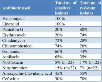
In the present study, the sensitive pattern of
Staphylococcus aureus isolates were Chloramphenicol (74%), Clindamycin (72%), Gentamicin (66%), Amikacin (65%), Amoxicillin - Clavulanic acid (45%), Cefoxitin (30%), Erythromycin (26%), Penicillin-G (20%) and less sensitive was shown to Nitrofurantoin (15%), and Norflaxacin (5%). And the resistant pattern of
Staphylococcus aureus isolates were Resistant isolates was higher in Penicillin-G (80%) followed by Erythromycin (74%), Cefoxitin (70%), Amoxicillin -Clavulanic acid (55%), Gentamicin (44%), Amikacin (35%), Clindamycin (28%), Chloramphenicol (26%) and less Resistant was shown to Norflaxacin (17%) and Nitrofurantion (7%). All Staphylococcus aureus isolates showed 100% sensitive to Vancomycin and Linezolid (Table 3).
Table 3: Antibiotic sensitivity pattern of
Staphylococcus aureus isolated from
clinical specimens.
Antibiotic used
Total no. of sensitive isolates
Total no. of resistant isolates
Vancomycin 100% - Linezolid 100% - Penicillin G 20% 80% Erythromycin 26% 74% Clindamycin 72% 28% Chloramphenicol 74% 26% Gentamicin 66% 44% Amikacin 65% 35% Norflaxacin 5% (n=22) 17% (n=22) Nitrofurantion 15% (n=22) 7% (n=22) Amoxycilin-Clavulanic acid 45% 55% Cefoxitin 30% 70%
SPUTUM 11%
BLOOD 17%
URINE 23% EAR
SWAB 15% PUS 30%
P. FLUID 2%
SOFT TISSUE 2%
BURNS 2%
MEDICINE 24%
ICU 8% NICU
7% PICU
10% SURGERY
11% ENT
16% TB 11%
GYNECOL OGY
7%
ORTHOPE DICS
Among 100 Staphylococcus aureus isolates, 40% isolates showed MLSBi resistance, 28% isolates showed MLSBc resistance, 6% isolates showed MS phenotype and 26% isolates showed Sensitive phenotype (Figure 3).
Figure 3: Prevalence of MLSBi, MLSBc, and MS phenotype in Staphylococcus aureus.
Out of 100 isolates, the prevalence of MLSBc and MLSBi were found to be higher in MRSA as compared to MSSA (21%, 27% and 7%, 10% respectively). 6% of total MS phenotype isolates were MRSA and 3% of MS phenotype isolates were MSSA (Table 4).
Table 4: Prevalence of MLSB among MRSA and MSSA.
S.no Zones MRSA MSSA
1 MLSBc E-R, CD-R 21% 7% 2 Sensitive Phenotype E-S,
CD-S 16% 10%
3 MLSBi D-zone positive 27% 10% 4 MS Phenotype E-R, CD-S 6% 3%
Total 70% 30%
DISCUSSION
In recent years, clindamycin has become an effective drug for some Staphylococcal infections especially skin and soft tissue infections and as a substitute for penicillin-allergic patients.14
However, clindamycin resistance can develop in staphylococcal isolates with inducible phenotype, and from such isolates, immediately constitutively resistant mutants have arisen among both in vitro testing and in vivo while doing clindamycin therapy.15
A total of 350 various clinical samples processed during the study period. Among 280 infected samples
Staphylococcus aureus were 100. The isolation rate of
Staphylococcus aureus was found to be 35.71%, it means above 1/4th of the total isolates were of Staphylococcus
aureus. This percentage reveals a higher infection rate of
Staphylococcus aureus than other bacteria.
In present study total of 70% MRSA were detected from various clinical samples by using cefoxitin disc diffusion technique, The High prevalence MRSA is in accordance with various studies like Dua’a Jarajreh, Amin Aqel el al, who reported 77.5% MRSA.16 Debasmita Dubey, Shakti
Rath et al, reported 83.57% MRSA, Venkata A, Rao AR, Kavita K et al, reported 75.27% in their study which is similar to this result that is 70%.17,18
The MRSA prevalence in various studies like Seifi N, Kahani N, Askari E et al, reported 41.7 % MRSA, Lt Col Mahina Lall, Brig A.K Sahni et al, reported 45.90%, Fahriye Eksi, Efgan Dogan Gayyurhan et al, reported 50.20% in their study which is lower when compared to this results.1,19,20
Results were reported in studies from Northern India such as New Delhi 44% MRSA by Rajaduraipandi K, Mani KR, Panneerselvam K et al, and 51% by Tyagi A, Kapil A, Singh P.21,22 In the year 2008 Tiwari HK, Sapkota D.
and Sen MR reported 38.44% MRSA which is lower when compared to this results.23,24
Prevalence rate of MRSA was observed in different countries of South Asia like Karachi 43% by Perwaiz S, Barakzi Q, Farooqi BJ et al, in, Nepal 38% and 40% by Tiwari HK, Das AK, Sapkota D et al, in and by Sanjana RK, Shah R, Chaudhary N et al, respectively which are lower when compared to the results.23,25,26
Tremendous increase in the methicillin resistant isolates in the hospital was observed, when compared with the study of Yogesh Kumar Gupta, Garima gupta et al.27 This
difference might be because they used oxacillin disc diffusion method for detection of MRSA on the other hand author used cefoxitin disc diffusion for the detection of MRSA.
In the present study, Overall prevalence of Inducible clindamycin resistance was 40%. Lt Col Mahina Lall, Brig A.K Sahni et al, reported 37.50%, Sreenivasulu RP. and Suresh R, reported 38.2%, Nilma R. Patil, Ulhas S. Mali et al, reported 36.95, Gaurav Dalela, Atul Vijay et al, reported 36.63% and Urmi JN, Summaiya MA, Latika SN et al, reported 43% inducible clindamycin resistance among S. aureus that is in accordance with this study.1,28-31
The lowest prevalence was reported 3.5% by Kalpana D, Mamta C, Vilas T at Nagpur district and highest resistance was reported 90% by Dizbay M, Gunal O, Ozkan Y et al. 32,33
However, low prevalence of inducible clindamycin resistant is also reported by various authors; Saderi H, Emadi B and Owlia P, reported 6.4%, Seifi N, Kahani N Askari E et al, reported 11.3%, R.P Adhikari, S Shrestha et al, reported 11.48% and Taruna Singh, Arvind B cMLSB
28%
Sensitive Phenotype
26% iMLSB
40%
MS Phenotype
Deshmukh et al, reported 14.8% inducible clindamycin resistance.19,34-36
In this study, Overall prevalence of constitutive resistant isolates was 28%, Gurdal Yilmaz, Kemalettin Aydin et al, reported 28.3% constitutive resistance among S. aureus that is similar with this study. Taruna Singh, Arvind B Deshmukh et al, reported 27%, Seifi N, Kahani N Askari E et al, reported 26% and R.P Adhikari, S Shrestha et al, reported 29.25% constitutive resistance.15,19,35,36
There is a higher variation for constitutive clindamycin resistance between various studies, because it depends on overuse of the drug and conversion of inducible phenotype to constitutive phenotype during treatment reported by Kalpana D, Mamta C, Vilas T i.e. 3.5% in iMLSB and 26.4% in cMLSB.32
However, low prevalence of constitute resistant isolates is also reported by various authors; Venkata A, Rao AR, Kavita K et al, reported 2.1%, Nilma R. Patil, Ulhas S. Mali et al, reported 8.69%, Urmi JN, Summaiya MA, Latika SN et al, reported 12% and Debasmita Dubey, Shakti Rath et al, reported 15.1%.17,18,29,31
Angel MR, Balaji V, Prakash J et al, didn’t report any constitutive resistance in their study and Saderi H, Emadi B and Owlia P reported about 93% constitutive clindamycin resistance in their study.34,37
In the present study, Chloramphenicol (74%) showed higher sensitivity pattern followed by Clindamycin (72%), where resistant isolates were higher in Penicillin-G (80%). All Staphylococcus aureus isolates showed 100% sensitive to Vancomycin and Linezolid.
Rajaduraipandi K, Mani KR, Panneerselvam K et al, (2006)21 depicted Almost all clinical MRSA strains (99.6%) were resistant to penicillin, 63.2% towards Gentamicin and erythromycin, where in present study it showed Penicillin-G 80% and Gentamicin 44% resistant lower to reported study.
Tiwari HK, Sapkota D and Sen MR et al, reported 100% MRSA strains were resistant to penicillin, 92.36% were resistant to chloramphenicol, 90.7% were resistant to norfloxacin, compared to this study Penicillin-G 80%, Chloramphenicol (26%) and Norflaxacin 17% resistant in
Staphylococcus aureus.23
CONCLUSION
Continuous monitoring of antibiotic susceptibility pattern of MRSA and use of a right and proper antimicrobial drug was helpful for minimizing the rate of MRSA infections. Judicial use of antibiotics may significantly decrease the further spread of MRSA in the community and as well as in the hospitals.
However, expression of inducible resistance to clindamycin could limit the effectiveness of this drug. In such cases, vancomycin and Linezolid are the drugs which are considered for therapy. There are reports of decreased vancomycin susceptibility amongst MRSA i.e. VISA (vancomycin-intermediate Staphylococcus aureus) and VRSA (vancomycin-resistant Staphylococcus aureus). Author did not find any isolate showing resistance to Vancomycin and Linezolid. Currently, VRSA is not widespread, but it could well be the next "superbug". .
Funding: No funding sources Conflict of interest: None declared
Ethical approval: The study was approved by the Institutional Ethics Committee
REFERENCES
1. Lall M, Sahni AK. Prevalence of inducible clindamycin resistance in Staphylococcus aureus
isolated from clinical samples. Medica J Armed Forces Ind. 2014 Jan 1;70(1):43-7.
2. Washington W, Allen S, Janda W, Koneman E, Procop G, Schrekenberger P. Taxanomy of Staphylococci and related Gram-Positive Cocci, clinical significance of Staphylococci and related Gram Positive Cocci. Koneman’s Color Atlas and Text book of practical Microbiology; 6th Ed. USA
2007.
3. Chambers HF. The changing epidemiology of
Staphylococcus aureus?. Emerging Infect Dis. 2001 Mar;7(2):178-82.
4. Jevons MP. “Celbenin”-resistant staphylococci. British Medica J. 1961 Jan 14;1(5219):124.
5. Pai V, Rao VI, Rao SP. Prevalence and antimicrobial susceptibility pattern on methicillin resistant Staphylococcous aureus [MRSA] isolates at a tertiary care hospital in Mangalore, South India, J Lab Phys. 2010;2:82-4.
6. Fasih N, Irfan S, Zafar A, Khan E, Hasan R. Inducible clindamycin resistance due to expression of erm genes in Staphylococcus aureus: report from a tertiary care hospital Karachi, Pakistan. J Pakistan Medica Assoc. 2010;60(9):750-3.
7. Kaur DC, Khare AS. Inducible clindamycin resistance in Staphylococcus aureus in a tertiary care rural hospital. Indian J Basic Appl Med Res. 2013;2:686‑93.
8. Fiebelkorn KR, Crawford SA, McElmeel ML, Jorgensen JH. Practical disk diffusion method for detection of inducible clindamycin resistance in
Staphylococcus aureus and coagulase-negative staphylococci. J Clin Microbiol. 2003;41:4740-4. 9. Kasten MJ. Clindamycin, metronidazole and
chloramphenicol. Mayo Clinc Proc. 1999;74:825-33. 10. Chelae S, Laohaprertthisarn V, Phengmak M,
by disc diffusion induction test. J Med Assoc Thai. 2009; 92(7):947-51.
11. Leclercq R. Mechanisms of resistance to macrolides and lincosamides: nature of the resistance elements and their clinical implications. Clini Infect Dis. 2002 Feb 15;34(4):482-92.
12. Siberry GK, Tekle T, Carroll K, Dick J. Failure of clindamycin treatment of methicillin-resistant
Staphylococcus aureus expressing inducible clindamycin resistance in vitro. Clini Infect Dis. 2003 Nov 1;37(9):1257-60.
13. Koneman’s. Color atlas and textbook of Diagnostic microbiology. 6th edition.983-987.
14. Drinkovic D, Fuller ER, Shore KP, Holland DJ, Ellis-Pegler R. Clindamycin treatment of
Staphylococcus aureus expressing inducible clindamycin resistance. J Antimicro Chemo. 2001 Aug 1;48(2):315-6.
15. Yilmaz G, Aydin K, Iskender S, Caylan R, Koksal I. Detection and prevalence of inducible clindamycin resistance in staphylococci. J Medica Microbiol. 2007 Mar 1;56(3):342-5.
16. Jarajreh DA, Aqel A, Alzoubi H, Al-Zereini W. Prevalence of inducible clindamycin resistance in methicillin-resistant Staphylococcus aureus: the first study in Jordan. J Infect Developing Countries. 2017 Apr 30;11(04):350-4.
17. Dubey D, Rath S, Sahu MC, Rout S, Debata NK, Padhy RN. A report on infection dynamics of inducible clindamycin resistance of Staphylococcus aureus isolated from a teaching hospital in India. Asian Pacific J Tropic Biomed. 2013 Feb 1;3(2):148-53.
18. Rao AVR, Kavitha A, Seetha KS. Prevalence of inducible clindamycin resistance among clinical isolates of staphylococci. Nat J Basic Medica Sci. 2012;3(1):68-71.
19. Seifi N, Kahani N, Askari E, Mahdipour S, Naderi Nasab M. Inducible clindamycin resistance in
Staphylococcus aureus isolates recovered from Mashhad, Iran. Iranian J Microbiol. 2012;4(2):82-6. 20. Eksi F, Gayyurhan ED, Bayram A, Karsligil T.
Determination of antimicrobial susceptibility patterns and inducible clindamycin resistance in
Staphylococcus aureus strains recovered from southeastern Turkey. J Microbiol, Immunol Infect. 2011 Feb 1;44(1):57-62.
21. Rajaduraipandi K, Mani KR, Panneerselvam K, Mani M, Bhaskar M, Manikandan P. Prevalence and antimicrobial susceptibility pattern of methicillin resistant Staphylococcus aureus: A multicentre study. Indian J Medica Microbiol. 2006 Jan 1;24(1):34-8.
22. Tyagi A, Kapil A, Singh P. Incidence of Methicillin Resistant Staphylococcus aureus [MRSA] in Pus Samples at a Tertiary Care Hospital, AIIMS, New Delhi. JIACM. 2008; 9:33-5.
23. Tiwari HK, Das AK, Sapkota D, Sivrajan K, Pahwa VK. Methicillin resistant Staphylococcus aureus: prevalence and antibiogram in a tertiary care
hospital in western Nepal. J Infect Developing Countries. 2009 Oct 22;3(09):681-4.
24. Tiwari HK, Sen MR. Emergence of vancomycin resistant Staphylococcus aureus (VRSA) from a tertiary care hospital from northern part of India. BMC Infect Dis. 2006 Dec;6(1):156.
25. Perwaiz S, Barakzi Q, Farooqi BJ, Khursheed N, Sabir N. Antimicrobial susceptibility pattern of clinical isolates of methicillin resistant
Staphylococcus aureus. J Pak Medica Assoc. 2007 Jan;57(1):2.
26. Sanjana RK, Shah R, Chaudhary N, Singh YI. Prevalence and antimicrobial susceptibility pattern of methicillin-resistant Staphylococcus aureus
(MRSA) in CMS-teaching hospital: a preliminary report. J College Medica Sci-Nepal. 2010;6(1):1-6. 27. Gupta YK, Gupta G, Garg SP, Nirwan PS.
Prevalence and antimicrobial susceptibility pattern of methicillin resistant Staphylococcus aureus
isolated at a tertiary care institute in northwest region of Rajasthan. Int. Res J Pharm. App Sci., 2013; 3(6):13-16.
28. Sreenivasulu RP, Suresh R. Phenotypic detection of Inducible Clindamycin resistance among the clinical isolates of Staphylococcus aureus by using the lower limit of inter disk space. J Microbiol Biotech Res. 2012, 2 (2):258-64.
29. Patil NR, Mali US, Kulkarni SA, Ghorpade MV, Vijay Mane. Detection of inducible clindamycin resistance among clinical isolates of Staphylococcus aureus in a tertiary care hospital. Int J Curr Microbiol App Sci. 2014;3(9):689-94.
30. Dalela G, Vijay A, Joshi M. Phenotypic Expression of erm Gene Among Staphylococcus aureus. Nat J Lab Med. 2016 Apr;5(2):25-9.
31. Jethwani UN, Mulla SA, Shah LN. Detection of inducible clindamycin resistance by an automated system in a tertiary care hospital. African J Microbiol Res. 2011 Sep 16;5(18):2870-2.
32. Date K, Choudhary M, Thombare V. Inducible clindamycin resistance in clinical isolates of staphylococci in a rural hospital. Int J Biol Med Res. 2012; 3(3): 1922-5.
33. Dizbay M, Günal O, Ozkan Y, Ozcan DK, Altunçekiç A, Arman D. Constitutive and inducible clindamycin resistance among nosocomially acquired staphylococci. Mikrobiyoloji bulteni. 2008 Apr;42(2):217-21.
34. Saderi H, Owlia P, Eslami M. Prevalence of Macrolide-Lincosamide-Streptogramin B (MLSB) resistance in S. aureus isolated from patients in Tehran, Iran. Iran J Pathol. 2009 Sep 1;4(4):161-6. 35. Adhikari RP, Shrestha S, Barakoti A, Amatya R.
Inducible clindamycin and methicillin resistant
Staphylococcus aureus in a tertiary care hospital, Kathmandu, Nepal. BMC Infect Dis. 2017 Dec;17(1):483.
hospital. Inter J Health Allied Sci. 2016 Apr 1;5(2):111-4.
37. Angel MR, Balaji V, Prakash JA, Brahmadathan KN, Mathews MS. Prevalence of inducible clindamycin resistance in gram positive organisms in a tertiary care centre. Ind J Medica Microbiol. 2008 Jul 1;26(3):262-4.