Manonmoney Jayaraman et al JMSCR Volume 07 Issue 05 May 2019 Page 887
GNB Causing Respiratory Infections in Pulmonary TB
Authors
Manonmoney Jayaraman*, Kalaivani Rajasekaran
Department of Microbiology, SRM Medical College Hospital and Research Centre, Kattankulathur, Chennai, Tamilnadu, India
Abstract
Introduction: Tuberculosisis caused by infection with members of Mycobacterium tuberculosis complex, which is rigorous and contagious disease. Bacterial infection is one of the most essential complications in the patients with pulmonary tuberculosis.
Aim and Objective: To study the prevalence of multidrug resistant Gram-negative bacteria causing
respiratory infections in pulmonary tuberculosis.
Materials and Methods: This Study was carried out at SRM MCH & RC, Tamil Nadu, India, from January
2017 to February 2018 after the Institutional Ethical committee approval. This was a Cross sectional study. The samples are collected under aseptic conditions. Gram negative bacilli isolation done by conventional culture method and antibiotic sensitivity done by disk diffusion method (Kirby-Bauer disc diffusion method).
Result: Out of 73 samples 10 (14.4%) samples were culture positive for gram negative bacilli. The isolates
obtained in positive culture are Klebsiella pneumoniae (5) 7.2%, Pseudomonas aeruginosa (3)4.3%, Acinetobacter baumannii (2) =2.8%.
Discussion: Pulmonary tuberculosis is one among the most common infectious diseases affecting humans worldwide. They are important causes of morbidity and mortality for all age groups, and each year approximately 7 million people die as a direct consequence of acute and chronic respiratory infections
Conclusion: In this study the prevalence of MDR-GNB is 14.4%.The predisposing factors of
Mycobacterium tuberculosis patients are nutrition deficiency and immune compromised status. Klebsiella pneumoniae followed by Pseudomonas aeruginosa and Acinetobacter baumannii were the most common gram-negative bacteria isolated in cultures from lower respiratory tract in pulmonary tuberculosis patients.
Keywords:Multidrug resistance, Gram negative bacteria, Mycobacterium tuberculosis.
Introduction
Tuberculosis (TB) is caused by infection with members of Mycobacterium tuberculosis complex (MTBC) which is rigorous and contagious disease. TB is transmitted by cough, with an infectious dose of less than 10 bacteria. The World Health Organization (WHO) estimated, 8.8 million people developed TB, of whom 3.9 million had so many tubercle bacilli in their
sputum that the bacilli could be identified by simple microscopy, and there were 1.7 million deaths due to TB.[1]
Primary tuberculosis is the comeback to the beginning infection in an individual not previously infected and sensitized to tuberculoprotein. The immunocompromised persons develop an effective immune response against M. tuberculosis and contain the primary
www.jmscr.igmpublication.org Index Copernicus Value: 79.54
Manonmoney Jayaraman et al JMSCR Volume 07 Issue 05 May 2019 Page 888 infection, series of M. tuberculosis infection to
primary or reactivation TB has in the past been interpreted as a function of the amount and efficiency of protective human immunity against
M.tuberculosis.[11].
The annual mortality is around three million (two million adults and one million children)following invasion of the secondary bacteria and fungi such as Beta – hemolytic Streptococci, Klebsiella pneumoniae, Pseudomonas aeruginosa, Candida albicans, Aspergillus niger and Aspergillus flavus resultant in the secondary infection which causes tissue destruction and ulceration which had already occurred as a result of primary infection.[2] In pulmonary tuberculosis it is this latter form of secondary infection which plays a vital role in blocking the direct admittance of air into the alveoli and produce tissue symbiosis producing a passive mixed infection leading to chronic obstructive lung disease ensuing in a fatal death. The mortality rate is 50-60 per cent in pulmonary tuberculosis is due to the invaders of secondary bacteria and fungi. Hence more attention is to be listening carefully for the simultaneous treatment for secondary infections along with the tuberculosis.[2]
The conventional diagnosis depends on the identification of M.tuberculosis bacilli, using conventional microbiological methods of sputum smear and culture or radiometric culture methods such as BACTEC or DNA probe based assays which can identify drug resistant as well. Smear – negative sputum may delay the diagnosis for 4-8 weeks or longer in the culture is also negative. A presumptive diagnosis based on the clinical and radiographic features should be made initiation of treatment after the barring of other possible causes of the radiographic findings, making the diagnosis of pulmonary tuberculosis is usually achieved by a combination of radiological and laboratory features in a patient with a compatible history.[2] Gram-negative bacteria grounds infections including pneumonia, bloodstream infections, wound or surgical site infections, and meningitis in healthcare settings. Gram negative bacteria are
resistant to multiple drugs and are ever more resistant to most available antibiotics. These bacteria have built-in abilities to find new ways to be resistant and can pass along genetic materials that allow other bacteria to become drug resistant as well[3].
Most common Gram-negative infections include in pulmonary tuberculosis patients are those
caused by Klebsiella, Acinetobacter,
Pseudomonas aeruginosa, and E. coli, as well as many other less common bacteria. Bacterial infection is one of the most essential complications in the patients with pulmonary tuberculosis[3].
The occurrence of nosocomial infection caused by Gram-negative bacilli has increased at an alarming rate Although previous studies have identified risk factors for MDR-GNB among pulmonary TB patients have not been elucidated. Most common bacteria agents of Lower Respiratory Tract Infection (LRTI) are
Pseudomonas, Acinetobacter, Klebsiella, Citrobacter, Escherichia coli.[4].
The superadded bacterial co infection can arise in TB patients. The dual infection with tuberculosis
and bacteria has been reported in
immunocompromised patients.[6]
In the present study finding the prevalence of multidrug gram-negative bacilli infections in pulmonary tuberculosis patients help in decreasing the added morbidity and mortality.
Aim and Objective
To study the prevalence of multidrug resistant Gram-negative bacteria causing respiratory infections in Pulmonary tuberculosis patients in a tertiary care centre.
Materials and Methods
Study Type: Cross sectional study.
Study Place: Tertiary care hospital, Tamilnadu, India.
Study period: Jan 2017-Feb 2018
Manonmoney Jayaraman et al JMSCR Volume 07 Issue 05 May 2019 Page 889
Exclusion Criteria: Patients with respiratory
infections apart from Tuberculosis infection. Specimen Collection:
Specimens: Sputum, Tracheal Aspirate, Bronchoalveolar lavage.
Specimens collected as per standard guidelines. Bartlett’s Grading system:
This technique has been applied to the evaluation of sputum samples. From the relative numbers of squamous epithelial cells and segmented neutrophils in direct Gram stain of sputum samples. Bartlett has devised a grading system for evaluating sputum samples. Using this system, negative numbers are assigned to a smear when squamous epithelial cells are observed to a smear when squamous epithelial cells are observed, indicating contamination with oropharyngeal secretions (saliva). Positive Numbers are assigned for the presence of segmented neutrophils, indicating the presence of active inflammation. A final score of 0 or less indicates either lack of inflammatory response or presence of significant salivary contamination, thus invalidating the specimen.[4]
Sputum collection Spot sample:
Give the patient labeled container, instruct the patient to go nearby to open space far away from other people and then instruct him by demonstrating with actual action:
Inhale deeply 2-3 times;
Cough out deeply from the chest;
Open the container, bring it close to the mouth and bring the sputum out into it;
Do Not give saliva or nasal secretions.
Close the container.
A good sputum sample is:
Thick (semi-solid), coughed out deeply from the lungs;
Purulent (yellowish, mucus);
Sufficient in amount (at least 2 ml).
A poor-quality sputum sample:
Contains only saliva (watery fluid) or nasal mucus;
Is small in quantity (less than 2 ml)[8].
The specimen is collected from deep cough; specimen should be examined. The patient should be instructed carefully on the proper collection of sputum, rather than saliva. Early morning sputum samples should be obtained because they contain overnight secretions in which pathogenic bacteria are more likely to be concentrated. Sputum is collected in wide-mouth container with a tightly fitted screw-cap lid can be used[4].
For diagnosis of tuberculosis, sputum examinations are performed according to Revised National Tuberculosis Control Programme (RNTCP) guidelines. If bronchoscopy is performed by pulmonologist to evaluate other diagnostic considerations, then an additional sputum sample collected immediately after bronchoscopy
In Pulmonary tuberculosis patient’s sputum, BAL and tracheal aspirate are collected in a sterile screw-top container the patients are instructed to the brush teeth and then rinse gargle with water before collection.
Tracheal aspirate collection
The lower respiratory tract may be sampled by introducing a catheter through the larynx into trachea .If an endotracheal tube is in place or there is a tracheotomy, aspirating tracheal secretions is simple which is collected by the pulmonologist and sample is transport as per standard guidelines in central laboratory.[4]
Bronchoalveolar lavage collection
Bronchoalveolar lavage involves the injection of 30 to 50µl of physiologic saline through a fibroptic bronchoscope which collected by the pulmonologist that has been threaded into the peripheral bronchiolar ramifications. The saline is then aspirated and submitted for smear and culture. Sample is transport as per standard guidelines in central laboratory. (4). bronchoscopy may have particularly high yield for Mycobacterium identification [9]
Direct microscopy
Manonmoney Jayaraman et al JMSCR Volume 07 Issue 05 May 2019 Page 890 performed in Class II A2 bio safety cabinet.
Smears are stained by the Ziehl-Neelsen procedure or one of its modifications, including the fluorescence staining method. Acid- fast bacilli in sputum are highly significant for mycobacterial infection[5]. Gram staining: To demonstrate and characterize bacteria.
Culture Methods
The samples are collected under aseptic conditions and it is processed under class II Bio safety cabinet level. Then it is cultured on nutrient agar plate, MacConkey agar plate, Blood agar plate and chocolate agar plate and it is incubated for 24 hours at 37 ˚C.
Preliminary identification by colony morphology and performing Gram staining ,hanging drop, catalase, oxidase and further identification by Indole, methyl red ,Voges-Proskauer Test, Citrate Utilization, Urease test, Triple Sugar Iron Agar (TSI), carbohydrate fermentation test, phenylalanine deaminase test (PPA).Antibiotic susceptibility test done by Kirby-Bauer Disk Diffusion method. All the tests were performed as per standard protocols.
Result
Out of 73 samples 10 (14.4%) samples were culture positive for gram negative bacilli. The isolates obtained in positive culture are Klebsiella pneumoniae (5) 7.2%, Pseudomonas aeruginosa
(3)4.3%, Acinetobacter baumannii (2) =2.8%. All Isolates were sensitive to all drugs except one strain of Acinetobacter which is resistance.
The strain of Acinetobacter baumanii was Multidrug resistance to Amikacin, Ampicillin, Amoxycillin/clavulanic acid, Ceftazidime, Ceftazidime/clavulanic acid, Cefotaxime, Ciprofloxacin, Cotrimoxazole Ertapenem,
Gentamicin, Meropenem, Piperacillin/
Tazobactum, Tigecycline ,Cefoxitin,
Cefaperazone/ sulbactum Ofloxacin,
Levofloxacin, Chloramphenicol.
Table 1 Number of samples (AFB smear positive)
S.no NO. of MALE/ FEMALE
TOTAL NO OF SAMPLES (n=73)
1. Male 62(85%)
2. Female 11(16%)
Total = 73
Total number of samples n=73
Male (62) 84% Female (11)5%
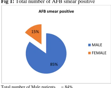
Fig 1: Total number of AFB smear positive
Total number of Male patients = 84% Total number of Female patients = 15%
Table 2: Age wise distribution of AFB positive
AGE MALE % FEMALE %
20-40 30 48.38% 5 45.45%
50-60 28 45.16% 5 45.45%
70-80 4 6.45% 1 9.09%
Total number of positive = 73 Positive male patients = 62 Positive female patients = 11
Fig 2: Number of Gram negative bacilli isolated in AFB positive sputum sample.
Total (n) =10
Klebsiella pneumonia 5( 7.2%)
Pseudomonas aeruginosa 3(4.3%)
Acinetobacter baumannii 2(3.2%)
85% 15%
AFB smear positive
MALE
Manonmoney Jayaraman et al JMSCR Volume 07 Issue 05 May 2019 Page 891 Discussion
Pulmonary tuberculosis is one among the most common infectious diseases affecting humans worldwide. They are important causes of morbidity and mortality for all age groups, and each year approximately 7 million people die as a direct consequence of acute and chronic respiratory infections.[8]
In this study there were 73 AFB positive samples out of which 10 were culture positive for gram negative bacteria. Number of Gram negative bacilli isolated are Klebsiella pneumoniae
5(7.2%), Pseudomonas aeruginosa are 3(4.30%) and Acinetobacter baumannii 2 (2.8%) Klebsiella pneumoniae was the predominant gram negative bacteria isolated in pulmonary tuberculosis patients.
According to the study conducted by Christopher et al[8]. Generally, Klebsiella pneumonia (30.16%) was the predominant isolate recovered, followed by Haemophilus influenza (17.05%),
Staphylococcus aureus (15.14%), and
Acinetobacter species (0.66%). K.pneumoniae
was also the predominant isolate in both genders. In this study also the major isolate is Klebsiella pneumoniae 5 (7.2%)
Another study by Egbagbe EE and Mordi RM(9)
K.pneumoniae was the most predominant isolate recovered from patients with LRTIs. β-lactams, fluoroquinolones and gentamicin were the most active antibacterial agents. Acinetobacter species have been associated with hospital-acquired pneumonia.
In another study 25% isolates of Acinetobacter spp, 22.8% iolates of P. aeruginosa, and 9%isolates of Klebsiella spp, were reistance to meropenem this was contrast to another study were 14.2%were resistance and 12-42.5% isolates of P.aeruginosa were resistant[10]. One strain of
Acinetobacter baumannii was Multidrug resistance to Amikacin, Ampicillin, Amoxycillin/ clavulanic acid, Ceftazidime, Ceftazidime/ clavulanic acid, Cefotaxime, Ciprofloxacin, Cotrimoxazole, Ertapenem
In this study Acinetobacter was resistant to was Multidrug resistance to Amikacin, Ampicillin, Amoxycillin/clavulanicacid,Ceftazidime,Ceftazidi me/clavulanicacid,Cefotaxime,Ciprofloxacin,Cotri moxazole, Ertapenem, Gentamicin, Meropenem, P iperacillin/ Tazobactum, Tigecycline, Cefoxitin,
Cefaperazone/sulbactum ,Ofloxacin,
Levofloxacin, Chloramphenicol.
The Acinetobacter isolated in mixed group showed lowered sensitivity to almost all the higher antibiotics (50% - ciprofloxacin, cefoperazone+sulbactum, 60% - ceftazidime, meropenem, amikacin, 70% - doxycyline, levofloxacin, piperacillin+tazobactum, imipenem, 80% - colistin) (Robert et al., 1980 and Leonid et al., 1998). The Pseudomonas isolatedin mixed group showed lowered sensitivity to almost all the higher antibiotics (50% - ciprofloxacin, amikacin, 62.5% -ceftazidime, meropenem, cefoperazone+ sulbactum, doxycyline, levofloxacin, 75% - piperacillin+tazobactum, imipenem, colistin)[3]. Futher Antibiotic susceptibility testing should be done to eliminate the co- infection of Gram negative bacilli in pulmonary tuberculosis patients which increases the morbidity and mortality.
Conclusion
In this study the prevalence of MDR-GNB is 14.4%. The males were more exposed to MDR-GNB infection. The predisposing factors of Mycobacterium tuberculosis patients are nutrition deficiency, immune compromised which are the various factors.
In pulmonary TB patients with longer duration of TB diagnostic delay create favorable conditions for the development of MDR-TB infections. The poor prescribing behavior inherent to the patients could predispose themselves to MDR-GNB infection. The advanced age, underlying disease, exposure to antimicrobial drugs are the known risk factors of harboring MDR bacteria.
Manonmoney Jayaraman et al JMSCR Volume 07 Issue 05 May 2019 Page 892 the resistant strain of gram-negative bacilli are
Acinetobacter baumannii.
GNB are among the most alarming antibiotic-resistant pathogens as they are often antibiotic-resistant to multiple antibiotic classes and capable of rapidly spreading resistance genes through mobile genetic elements.
This is a major public health problem and a cause for both substantial morbidity and mortality among hospitalized patients. Klebsiella pneumoniae followed by Pseudomonas aeruginosa and Acinetobacter baumannii were the most common GNB isolated in cultures from lower respiratory tract in pulmonary tuberculosis patients.
References
1. Woodhead M, Blasi F, Ewig, S, Garau J, Huchon G, Leven M. Guidelines for the management of adult lower respiratory tract infections-Full version. Clin Microbiol Infect 2011;17 Suppl 6:E1-59. 2. Aurora Epv, D’ Agata EMC. (2005) The
Rising Influx of Multidrug-Resistant Gram-Negative Bacilli into a Tertiary Care Hospital. Clinical Infectious Diseases. 3. Gandhi NR, Nunn p, Dheda K, Schaaf HS,
Zignol M, van Soolingen D, et,al (2010) Multiresistant and extensively drug-resistant tuberculosis: a threat to global control of tuberculosis. Lancet 375:1830-1843, doi.
4. Koneman’s Color Atlas and Textbook of Diagnostic Microbiology seventh edition Gary W. procop Deirdre L.Church.et.al 5. Robert WA, Catharine N, Ruth G, Sherwin
KA. Endemic aminoglycoside resistance in gram-negative bacilli: epidemiology and mechanisms. J Infect Dis 1980; 141:338-45.
6. Anshum Aneja Arora; Uma Maheswari
Krishnaswamy; and Mantha Satya
Padmaja; Tubercular and bacterial co infection: A case series
7. Christopher Aye Egbe1, Casimir
Ndiokwere1, Richard Omoregie1,2
Microbiology of Lower Respiratory Tract Infections in Benin City, Nigeria
8. Revised National Tuberculosis Control
Programme (RNTCP) manual for
laboratory techniciansCentral TB Division,
Directorate General of Health
ServicesMinistry of Health and Family Welfare, Nirman Bhavan, New Delhi 110 011.
9. Egbagbe EE, Mordi RM. Aetiology of lower respiratory tract infection in Benin City, Nigeria. J Med Biomed Res. 2006;5(2):22–27.
10. MARK H. LEPPER, M.D.**Chicago, Illinois Opportunistic Gram-Negative Rod Pulmonary Infections
11. Carroll KC. Laboratory Diagnosis of Lower Respiratory Tract Infections: Controversy and Conundrums. J Clin Microbiol 2002; 40:3115-3120
12. Shah NS, Wright A, Bai GH, et al. Worldwide emergence of extensively drugresistant tuberculosis. Emerg Infect Dis 2007; 13:380-7.
13. Okesola AO,Ige OM. Trends in bacterial pathogens of lower respiratory tract infections.Ind J Chest Dis All Sc 2008;50:269-272.
14. Kim H-r, Hwang S.S; Kim E—c; Lee S.M; Yang S-cYoo C-G; et al.(2011) Risk actors for multidrug-resistance bacterial infection among patients with tuberculosis. Journal of Hospital Infection.