i
Modulation of HIV-1 replication and T cell activation by FoxP3
by Derek A. Holmes
A dissertation submitted to the faculty of the University of North Carolina at Chapel Hill in partial fulfillment of the requirements for the degree of Doctor of Philosophy in the
Department of Microbiology and Immunology.
Chapel Hill 2008
Approved by:
Dr. Lishan Su, Advisor
Dr. Albert Baldwin, Reader
Dr. Blossom Damania, Reader
Dr. Ronald Swanstrom, Reader
iii Abstract
Derek A Holmes
FoxP3 modulation of HIV-1 Replication and T cell activation
(Under the direction of Dr. Lishan Su)
Regulatory T cells (Treg) are defined as the population of CD4+CD25+ that express
the forkhead transcription factor family member FoxP3. Treg cells are characterized by the
inability to produce IL-2 upon stimulation, defects in in vitro proliferation, and the ability to suppress effector T cell (Teff) activation. FoxP3 plays a crucial role in Treg function and is
thought to be a factor required for the development and maintenance of Tregs. We first set
out to describe the role of FoxP3 in the regulation of HIV LTR activation in Treg cells. Upon
primary Treg infection with HIV-1 NL4-luciferase reporter virus, we observed increased
transcription of the LTR. Similarly, primary T cells or Jurkat T cells transduced with
retrovirus expressing Foxp3 showed a similar enhancement of HIV-1 LTR. We demonstrate
that Foxp3 enhancement of LTR requires intact NF-κB binding sites, and FoxP3 expression is
associated with increased binding of NF-κB at the core enhancer of HIV-1 LTR and
differential histone acetylation of the LTR and IL-2 promoters.
We demonstrate that FoxP3 expression in Jurkat T cells and primary Tregs inhibits
iv
the proline-rich domain. We show that FoxP3 is present in a large molecular weight
complex with HDAC1, and interacts with HDAC1 in T cells. A point mutation in the forkhead
domain that inhibits Foxp3 function in Tregs in vivo is unable to associate with and inhibit HDAC1 activity, suggesting the importance of FoxP3/HDAC1 interaction for HDAC1
inhibition. Finally, we demonstrate knockdown of HDAC1 inhibits FoxP3 regulation of IL-2 expression and HIV-1 LTR activity in T cells.
Lastly, we report that FoxP3 interacts with the ATPase nucleosome remodeler Mi-2β
in T cells. This interaction requires the zinc-finger domain, and FoxP3 expression is
associated with a decreased binding of Mi-2β at the IL-2 promoter. We also demonstrate that knockdown of Mi-2β inhibits IL-2 expression, supporting a model for FoxP3-mediated repression of transcription through the removal of factors required for optimal gene
expression. Thus, FoxP3 regulates gene expression at multiple levels, from transcription
v
Acknowledgement
I would first like to acknowledge the people that got me this far in life. To my parents, even though I try desperately not to rely on you and go my own way, I always feel like you are close at hand and behind me in everything that I do. My close friends that I have made during my tenure at UNC, without you all I wouldn’t be able to say that my graduate school experience was by far the best adventure that I have embarked on. Anna gets special thanks for obvious reasons. To the lowest puppy that I know, thank you lowrider for the energy and constant attention.
vi
Table of Contents
List of Figures ... ix
List of Abbreviations ... xi
Chapter I ... 1
Introduction ... 2
Part I: HIV infection overview ... 2
Brief History and Emergence of HIV ... 2
Acute infection: A shift in the paradigm... 3
The chronic phase of infection ... 4
Part II: The Role of Regulatory T cells in immune-balance- Do Tregs hold the key? ... 5
Regulatory T cell in the Immune system: A brief history ... 5
FOXP3 gene in Tregs ... 7
Mechanisms of Treg Suppression ... 7
The Role of Tregs in infection ... 9
The Role of Tregs in HIV infection ... 11
PartIII: T cell activation ... 13
T cells in the immune system ... 13
The T cell Receptor (TCR) signaling cascade ... 14
Part IV: IL-2 promoter regulation- A model for T cell activation requirements ... 16
Transcriptional regulation at the IL-2 promoter: the promoter defined ... 16
Transcriptional regulation at the IL-2 promoter: Transcription factors. ... 17
The IL-2 enhanceosome ... 19
Transcriptional regulation of the HIV LTR: Lessons learned from IL-2? ... 20
Part V: Chromatin remodeling... 23
Chromatin remodeling at the IL-2 promoter ... 23
vii
The Mi-2/NuRD complex: Dynamic regulator of transcription. ... 25
Chromatin remodeling at the LTR ... 27
Part VI: Role of Histone deacetylases (HDACs) in T cell and HIV gene expression... 27
General overview of HDACs and HDAC inhibitors ... 27
Biological role of HDACi ... 29
HDACs and HDACi on the IL-2 promoter ... 30
HDACs and HDACi on HIV LTR ... 30
Regulation of HDACs... 31
Part VII: Foxp3 Regulation of gene transcription ... 32
Foxp3 gene expression signature- a ‘master regulator’? ... 32
Foxp3 protein structural analysis ... 34
Regulation of Foxp3 protein ... 35
Mechanisms of Foxp3 regulation of promoter activity ... 36
Scope of this dissertation ... 38
References ... 40
Chapter II ... 55
Abstract ... 56
Introduction ... 57
Experimental Procedures ... 61
Results ... 66
Discussion ... 71
Acknowledgements ... 76
References ... 85
Chapter III ... 90
Abstract ... 91
Introduction ... 92
Results ... 95
Discussion ... 101
Materials and Methods ... 106
Acknowledgements ... 110
References ... 121
viii
Abstract ... 125
Introduction ... 126
Results ... 130
Discussion ... 134
Materials and Methods ... 139
Acknowledgements ... 142
References ... 148
Chapter V ... 151
Perspective and Scope... 152
Chapter2: Foxp3 enhances HIV-1 LTR through an NF-κB-dependent mechanism. ... 153
Findings and Implications ... 153
Future Direction and Studies ... 156
Chapter 3: FoxP3 inhibits HDAC1 activity to modulate gene expression ... 158
Findings and Implications ... 158
Future Directions and Studies ... 160
Chapter 4: Foxp3 regulates IL-2 gene expression by inhibiting the ATPase chromatin remodeler Mi-2β. ... 163
Finding and Implications... 163
Future Directions and Studies ... 165
ix
List of Figures
Figure 2.1 Foxp3 inhibits IL-2, but enhances HIV-1 gene expression in T cells……….……..77
Figure 2.2 Foxp3 inhibits IL-2 promoter, but enhances HIV-1 LTR expression in T cells……….……..78
Figure 2.3 A critical activity of Foxp3 is required for LTR enhancement……….…..…..79
Figure 2.4 Foxp3-mediated LTR enhancement maps to the NF-κB sites………..………80
Figure 2.5 Foxp3 affects LTR expression via an NF-κB sequence-dependent and cell type dependent mechanism………..81
Figure 2.6 Foxp3 differentially affects the histone 3 acetylation level of the IL-2 and HIV-1 LTR promoters……….82
Figure 2.7 Foxp3 alters NF-κB occupancy at the LTR promoter……….…83
Figure S2.1………84
Figure 3.1 Foxp3 modulates gene expression similar to TSA……….…..110
Figure 3.2 Foxp3 inhibits global HDAC activity in human T cells and Tregs……….……111
Figure 3.3 Other forkhead family members inhibit HDAC activity……….………112
Figure 3.4 N-terminus and forkhead domains of Foxp3 are required to inhibit HDAC1 deacetylase activity………..113
Figure 3.5 Foxp3 interacts with HDAC1……….………114
Figure 3.6 HDAC1shRNA reduces Foxp3 activity at the IL-2 and HIV LTR promoters…..……….115
Figure S3.1 HDAC deacetylase activity in nuclear lysates of transduced Jurkat T cells…….………….…116
Figure S3.2 Foxp3 and HDAC1 co-fractionate in a high molecular weight complex……….………..……..117
Figure 4.1 Foxp3 co-fractionates with Mi-2β in a high molecular weight complex in 293T cells…….140
Figure 4.2 Foxp3 interacts with Mi-2β, and requires the zinc finger domain……….141
Figure 4.3 Foxp3 regulates Mi-2β association at the IL-2 promoter in Jurkat T cells………142
x
xi
List of Abbreviations
AIDS Acquired Immune-deficiency Syndrome
APC antigen presenting cells
ART anti-retroviral therapy
CMV Cytomegalovirus
ChIP chromatin Immunoprecipitation
Chip microarray
DAG diacylglycerol
DBD DNA-binding domain
DNase DNA nuclease
ER Endoplasmic reticulum
Foxp3 Forkhead box P3
Foxp3-C Foxp3 without N-terminus
Foxp3-LZFKH Foxp3 with Leucine zipper and forkhead
Foxp3-FKH Foxp3 with forkhead
GADS Grb2-related adapter downstream of Shc
GFP green fluorescent protein
GVHD graft-versus-host disease
HAT histone acetyl-transferase
HCV Hepatitis C virus
HDAC histone deacetylase
HDACi histone deacetylase inhibitor
xii
HSPG retrovirus vector
HSV Herpes Simplex Virus
H3 histone 3
H4 histone 4
IFN-γ Interferon gamma
IL-2 Interleukin-2
IL-4 Interleukin-4
Iono Ionomycin
IPEX immune dysregulation, polyendocrinopathy, enteropathy, X-linked
syndrome
IP3 Inositol-1,4,5-triphosphate
ISWI imitation SWI
ITAM immunoreceptor tyrosine based activation motifs
LAT linker for activation of T cells
LPS Lipopolysaccharide
LTR Long terminal repeat
MHC major histocompatibility complex
MNase micrococcal nuclease
NaBut sodium butyrate
NFAT nuclear factor of activated T cells
NF-κB nuclear factor-κB
NK Natural Killer
NuRD nucleosome remodeling and deacetylation
xiii
PLCγ1 phospholipase Cγ1
PMA phorbol 12-myristate 13-acetate
SAHA suberoylanalide hydroxamic acid
shRNA short hairpin RNA
siRNA small interfering RNA
SIV Simian Immunodeficiency virus
SOS son of sevenless
SWI/SNF switching defecting/sucrose non-fermenting
TCR T cell Receptor
Teff effector T cell
TGF-β transforming growth factor beta
TNF-α tumor necrosis factor alpha
TPX trapoxin
Treg regulatory T cell
TSA trichostatin-A
VPA valproic acid
1
Chapter I
2 Introduction
Introduction Forward: The primary goal of my graduate tenure and this introduction is to be as comprehensive and concise as possible, leaving no stone
unturned unless technical difficulties inhibit my progress. Therefore, I will not be giving
a complete review of the HIV lifecycle for fear of boredom and for the fact that it has
been extensively reviewed in the literature. Therefore I will refer you to a fantastic
review by Eric Freed (1) for a comprehensive look at HIV-1 replication. Thank you and
enjoy the following introduction to HIV, Foxp3 and Regulatory T cells.
Part I: HIV infection overview
Brief History and Emergence of HIV
The earliest documented case of HIV-1 infection came from a stored serum
sample collected in 1959 from an adult Bantu male in the Democratic Republic of Congo
(2). Similarly, HIV-2 ancestors have dated back to the 1940. Estimates by Korber et al. put the timing of HIV-1 M group divergence somewhere in the 1930’s (3). Currently, it is
proposed that HIV-1 came to humans from the chimpanzee, while HIV-2 has origins
from the sooty mangabey, which harbor SIVcpz and SIVsm respectively (4-6). In SIVsm’s
natural host, which includes chimpanzees, sooty mangabeys and African green
3
present in an “unnatural” host (Rhesus macaque and humans for SIVsm and HIV,
respectively) that disease progression is associated with chronic immune activation, T
cell depletion and subsequent immunodeficiency. This intriguing difference has formed
the foundation for studies to determine the mechanisms of immunopathogenesis in HIV
infection.
Acute infection: A shift in the paradigm
HIV disease progression can be separated into 3 distinct phases: (i) acute
infection occurring in the first 3-6 weeks in humans and 1-4 weeks in macaques, which is
associated with a spike in viral load and a subsequent decrease in viral load to the viral
set point; (ii) the chronic phase of infection lasting 6-10 years. This asymptomatic phase
coincides with a gradual increase in viral load and decrease in CD4+ T cell counts over
time; (iii) and the final phase lasting roughly 12-18 months and is associated with AIDS
and immune system failure (7). The focus of past research has been to determine the
mechanism of immune activation during the chronic phase and the resultant AIDS
progression. More recently, we have shifted our focus on what is happening during the
acute phase of infection as a predictor of disease progression. Now, HIV pathogenesis
can be divided into two major phases; the acute infection phase associated with a
dramatic loss of CD4+ T cells residing in mucosal tissue, and a chronic phase
characterized by immune activation and gradual loss of peripheral CD4+ T cells over
4
Looking more closely at the acute phase of infection, plasma viral load increase
coincides with CD8+ T cell increases and a drop in CD4+ T cell counts (9). This leads to
an inversion of the CD4+/CD8+ ratio. Until recently, the magnitude of CD4+ T cell
depletion and its consequence were not fully appreciated. Initial studies in 1998 using
an SIV model described a profound depletion of CD4+ T cells in both the gut and
gut-associated lymphoid tissue (GALT) (10, 11), and more recent studies have described a
similar depletion in the gut of HIV-1 infected individuals (12-14). The importance of
these findings is underscored by the fact that between 60-80% of the total CD4+ T cell
population resides in the gut and associated lymphoid tissue (15). The CD4+ T cell
population that is most affected and depleted by SIV and HIV has a resting memory
CD4+ phenotype, Ki-67- and CD69-, and CD45RA- (16). Of greater importance is that the
majority of mucosal CD4+ T cells are CD45RA-, and up to 75% express the HIV-1
coreceptor required for T cell infection, CCR5 (12, 17, 18). Thus, during the acute stage
of infection, the resting memory T cell is a major target of SIV and HIV infection and
depletion.
The chronic phase of infection
As previously stated, the chronic phase of HIV is associated with a steady decline
in CD4+ T cell numbers, hyper-immune activation, and a slow and steady rise in viral
load in patients not on ART. In the gut, the depletion of CD4+ memory T cells continues
5
infection (11, 12). As a consequence, chronic immune activation occurs due in part to
homeostatic proliferation of the T cell pool to replenish the HIV-depleted pool, and also
microbial translocation leading to LPS-mediated immune activation (19, 20).
Interestingly, not only does HIV affect the dynamics of T cell homeostasis, but the
function of CD4+ T cells is also impaired. HIV-1 specific T cells are preferentially infected
by HIV harbored in dendritic cells (21, 22) and have been shown to consequently lack
effector function. In general, disruption of immune homeostasis and subsequent overt
immune activation are hallmarks of this phase of HIV-1 infection, and defining and
characterizing the mechanisms contributing to immune activation is of great
importance.
Part II: The Role of Regulatory T cells in immune-balance- Do Tregs hold the key? In order to delineate the complicated mechanism behind HIV pathogenesis and
progression to AIDS, you must first understand mechanisms of immune control.
Therefore, the following section will be dedicated to describing and understanding a key
player in immune homeostasis, the Regulatory T cell (Treg).
Regulatory T cell in the Immune system: A brief history
The regulation of immune tolerance is a critical aspect of immunology. The
6
immune homeostasis. Excessive immune responses can have deleterious effects, as
seen in autoimmune disease or immune pathology from infection or allergic insult.
Similarly, understanding the mechanism of immune tolerance is beneficial in controlling
tissue rejection following transplantation, or reversing unresponsiveness to autologous
tumor cells. The idea of an intrinsic T cell population capable of suppressing the
immune system came to light in 1970 wherein the ability of T cells to both enhance and
suppress immune responses was first described (23). For several years following the
initial discovery, both CD8+ and CD4+ suppressor T cell populations were described, but
the inability to find a biological factor responsible for suppressor T cells led to a decline
in suppressor T cell interest. During the late 1980’s, T cell tolerance was discovered to
be associated with mechanisms of clonal T cell deletion in the thymus (24, 25), and the
discovery of secreted suppressive cytokines such as IL-10 further led to our thinking that
a specific T cell subset was not required for tolerance (26).
Simultaneously, work on natural self-tolerance mechanisms was in place with
the discovery in 1969 that removal of the thymus in neonatal mice resulted in ovarian
destruction due to autoimmunity (27). Importantly, removal of the thymus 3 days
following birth resulted in several autoimmune manifestations (28), and work by several
groups demonstrated that CD4+ T cells can be adoptively transferred into T
cell-deficient mice and cause disease (29). Further work by Sakaguchi et al. in 1995 demonstrated that a population of CD4+ CD25hi T cells was required for tolerance,
7
addition of CD4+CD25+ T cells could control autoimmunity (30, 31). Thus, there was a
specific T cell population required to control the balance of the immune system.
FOXP3 gene in Tregs
In 2001, the discovery of the role of a transcription factor mediating
autoimmune disease in mice can be considered the most important finding in Treg
biology. A member of the forkhead winged-helix family, Foxp3, when disrupted in the
scurfy mouse resulted in a fatal lymphoproliferative disorder (32). A disorder found in
humans, IPEX (immune dysregulation, polyendocrinopathy, enteropathy, X-linked
syndrome), was associated with mutation in the FOXP3 gene, and was characterized by autoimmune disease in multiple organs. In 2003, multiple groups were able to show
that Foxp3 protein localized to CD4+CD25+ T cells, Foxp3 expression was required for Treg development and function, and expression of Foxp3 by retroviral transduction in
CD4+CD25- T cells conferred a suppressive phenotype (33-35). Through this work it
became evident that Foxp3 is a critical factor in Treg function. A more comprehensive
look into the role of Foxp3 in Treg cells will be discussed in later sections.
Mechanisms of Treg Suppression
Following the discovery of CD4+CD25hi Treg subset, several groups were able to
isolation and further characterize the mechanism of Treg suppression activity. Several
8
cells for that matter) along with either APC-mediated stimulation or TCR stimulation
with CD3 antibody was able to repress proliferation (36, 37). Not only was it
demonstrated that Tregs were able to suppress T cells in vitro, but Tregs also were characterized by an inability to produce IL-2, were hypoproliferative in vitro upon TCR stimulation, and furthermore, the mechanism of Treg suppression was cell
contact-dependent. This lead to the current and ongoing research to discover the molecular
mechanisms involved in Treg suppression.
As previously described, ectopic expression of Foxp3 in CD4+ CD25- is able to
recapitulate the suppressive phenotype of Tregs almost completely. In terms of Treg
development in the thymus, Foxp3 expression is turned on 3 days following birth, correlating well with the previously described neonatal day 3 thymectomy-induced
autoimmune disease (38). Characteristic of Foxp3 expression was the increase in
activation marker CD25, and other cell surface markers associated with T cell
suppression such as CTLA-4 and GITR (33). Several studies have shown the ability of
Tregs to suppress several cell populations and cellular functions, including B cell
proliferation and immunoglobulin production, the function of mature dendritic cells, NK
and NKT cells, and importantly both naïve and memory CD4+ and CD8+ T cell
populations (39-44). Also of importance to note is the ability of Treg cells to suppress in
an antigen specific and non-specific manner, the latter resulting in bystander
9
To date, several mechanisms of suppression by Tregs have been demonstrated.
With respect to cell-to-cell contact, upregulated cell surface factors such as CTLA-4 and
LAG-3 have been shown to play a role in Treg suppression in vivo as demonstrated by administration of blocking antibodies for the specific factors in mice (45, 46). Others
have identified a role for cytokines in the suppressive activity of Tregs. IL-10 secreted
locally can control colitis and experimental allergic enchaphalomyelitis (EAE) (47, 48),
while TGF-β is expressed on the surface of both human and murine Tregs and is
required for suppression (40, 49). An exact mechanism of suppression still remains to
be resolved, although it is becoming clear that multiple mechanisms of suppression
occur that are heavily dependent on the microenvironment, location of action and cell
populations involved.
The Role of Tregs in infection
The concept of the study of regulatory T cells was rejuvenated through work on
autoreactive T cells and immune tolerance, but the impact of Tregs doesn’t stop there.
It was clear that Treg cells would play a major role in infection, and Belkaid et al.
provided the first evidence of the role of CD4+CD25+ T cells in the control of disease. In
this work, Leishmania major infection in mice when depleted of Treg cells was unable to establish a persistent infection and was cleared, due in part to a decrease in Treg cells
and thus a consequent increase in CD8+ T cell-mediated parasite clearance. This also
10
pathogens, and put Tregs in the forefront (50). Within a year of this finding, Treg cells
were also linked to control of the immune response to viral infection (44). In the
following sections, the role of Tregs in viral clearance and immunity will be discussed,
and more significantly, their role in HIV infection.
Since Tregs were now implicated in immune tolerance and pathogen clearance,
several groups set out to describe the role of Tregs in pathogen persistence.
Experiments with Herpes Simplex virus (HSV) infection in mice showed that depletion of
CD4+CD25+ T cells 3 days prior to infection resulted in elevated HSV-specific CD8+ T cell
response in vivo in the acute and memory phases, and elevated HSV-specific CD4+ T cell responses (44, 51). It became clear that the immune response to HSV infection was
increased by Treg removal, wherein better memory response was decreased in the
presence of Tregs, and protective immunity was compromised. In the case of a second
chronic disease, Hepatitis C virus (HCV), the study of Tregs is difficult due to a lack of an
animal model. Regardless, studies done in humans comparing chronic HCV-infected
individuals to HCV-infected and recovered persons demonstrated a significant
difference in the frequency of CD4+CD25+ T cells. Persistently HCV-infected patients
had an increased percentage of Tregs, and the Tregs were able to suppress HCV-specific
11 The Role of Tregs in HIV infection
Since the role of Tregs in establishing chronic versus acute diseases has been
established for several pathogens, the importance of this cell population in HIV-1
infection is of great importance. Since the hallmark of progression to AIDS is the
decrease in CD4+ T cells and hyper-immune activation, it is not unreasonable to
rationalize the importance of Tregs in chronic immune activation. The question remains
if depletion of Tregs in HIV-1-infected patients contributes to uncontrolled immune
activation. Thus determining the kinetics and functional response of Tregs is of great
importance. Characterization of the role of Treg cells in HIV-1 infection has been
controversial, most notably for the lack of consistent determination of a Treg phenotype
and technical methods for determining Treg numbers in HIV patients, and lack of
understanding of the dynamics of Tregs in peripheral blood over the time course of
disease. Thus, several groups have shown that Tregs numbers are either decreased
(53-57) or increased (58-61) in HIV-1 infection. A clearer understanding of Tregs in disease
progression has come to light due to the use of a monkey model of HIV infection. As
previously stated, SIV infection in African green monkeys and sooty mangabeys does not
result in AIDS-like disease, while several groups have shown that rhesus macaque
infection is an accelerated and consistent model for HIV disease progression (62-65).
Thus, using this model, Periera et al. demonstrated that Treg numbers in peripheral blood of SIV-infected sooty mangabeys did not change over the course of infection,
while there was a severe depletion in rhesus macaques. Interestingly, the function of
12
numerical or function loss of Tregs correlated with viral load (66). Similarly, Chase et al. described severe depletion of Treg cells in the gut of SIV-infected rhesus macaques
during the acute and chronic phase of the infection, consistent with the finding that the
majority of the CD4+CCR5+ T cell population is depleted in acute SIV infection (67).
Surprisingly, none of these studies determined the relative level of infection of Tregs
compared to memory T cells, although the Treg population was described in brief to
harbor genomic SIV.
Given the importance for Tregs in immune-homeostasis, depletion would likely
have a dramatic effect on T cell responses. Depletion of T cells by direct infection or
bystander apoptosis is well documented in HIV infection, and similarly Tregs in the
lymphoid tissue have been shown to be infected by SIV, with approximately 13% of Treg
cells being productively infected (58). To date there have been no studies showing
direct infection of Tregs in HIV infected individuals, although Tregs express the HIV
receptor CD4 and coreceptor CCR5 and are highly susceptible to infection in vitro (53). In fact, our group has shown that HIV replicates more efficiently in Tregs than Teff cells
(68), although others have demonstrated the opposite effect (69). More recently, our
group has shown that in humanized Rag2-/-γc-/- double knockout mice, HIV-1 is able to efficiently infect and deplete Treg cells (Jiang et al. manuscript submitted). In this study, they also determined that depletion of Tregs through ONTAK treatment diminished the
viral load and impaired T cell infection. Clearly, the role of Treg infection and depletion
requires further study to fully delineate their role in immune pathogenesis, viral
13 PartIII: T cell activation
T cells in the immune system
In vertebrates, the immune system is broken down into two separate divisions to
ward off external insults and invading pathogens. The innate immune response is an
ancient component of the immune repertoire found in both plants and animals, and is
vital in the recognition of infectious nonself from noninfectious self. This arm of
immunity is comprised of multiple cell types, including phagocytes such as dendritic
cells, macrophages and neutrophils, along with eosinophils, mast cells, basophils and
natural killer (NK) cells (70). What distinguishes these cells from the adaptive immune
arm is the recognition of nonself antigens with invariant receptors that do not require
DNA rearrangement events. Toll-like receptors are germline encoded and evolved to
recognize patterns produced by metabolic processes of microbial pathogens but not
host products. Conversely, the adaptive immune response uses an entirely different
receptor to mount an immune response to a specific pathogen. These T cell receptors
(TCR) are produced through somatic mutation and gene segment rearrangement to give
a staggering array (~108) of TCR specificity (71). This seemingly random recognition of
antigens provides us with a response capable of specificity to foreign antigens and
memory of infection, allowing for a faster and more efficient immune response.
However, an innate drawback of this type of system is the inability to differentiate self
from non-self, as seen in detrimental responses such as autoimmunity, allergy, and graft
14
Specific pathogen recognition by CD4+ T cells occurs through the TCR and an
interaction with a peptide/MHC class II complex. A tightly regulated event, TCR
activation results in a signaling cascade leading to proliferation, differentiation or even
apoptosis (72). The different subsets of CD4+ T cells include the interferon-gamma
(IFN-γ) producing Th1 cells, the IL-4 producing Th2 cells responsible for regulation of humoral
immunity, and the newly emerging Th17 “pathogenic” T cells (73). CD8+ T cells
recognize endogenous antigens presented on MHC class I molecules found on all
nucleated cells in the body, and are responsible for the clearance of infected cells.
The T cell Receptor (TCR) signaling cascade
The TCR on resting T cells is comprised of an αβ heterodimer and is associated
with the δ,γ,ε, and ζ chains to for the TCR/CD3 complex (74). Following engagement of
the TCR, a well defined signaling cascade is activated. The cytoplasmic domains of the
TCR contain immunoreceptor tyrosine-based activation motifs, or ITAMs, which are
activated by phosphorylation of specific tyrosine residues by the src family kinase
members Lck and Fyn (75). The Syk family member ZAP70 is recruited to the TCR/CD3
complex through interaction with the phosphorylated ITAM motifs, followed by
interaction with and activation of the membrane adaptor molecule LAT (linker for
activation of T cells) and SLP-76 (SH2-domian containing leukocyte protein of 76 kDa).
Together, these later components nucleate a complex that includes the proteins Grb2,
15
(phospholipase Cγ1) , and GADS (Grb2-related adapter downstream of Shc) (reviewed in
(76). The major downstream target of this multisubunit complex is IP3
(inositol-1,4,5-triphosphate) and DAG (diacylglycerol), which is hydrolyzed from PIP2
(phoshatidylinositol 4,5 bisphosphate) mediated by the actions of PLCγ-1. Immediately
following IP3 production, calcium stored in the endoplasmic reticulum (ER) is released
through the CRAC channels, and the increase in intracellular Ca2+ levels activates the
Ca2+/calmodulin-dependent serine/threonine phosphatase calcineurin. Activated
calcineurin then dephosphorylates the transcription factor NFAT (nuclear factor of
activated T cells) which localizes to the nucleus to regulate a variety of genes.
The other component of that cascade, DAG, requires PLCγ-1 to directly target
another serine/threonine kinase found associated with the TCR/CD3 complex, PKCθ
(protein kinase C). PKCθ when activated at the TCR interacts with a multisubunit
complex called the CBM which contains CARMA1, Bcl10, and MALT1 (77). This activated
CBM complex is required for IKK activation, which subsequently phosphorylates IκBα
leading to the release and nuclear import of NF-κB (78). These two pathways of
activating the transcription factors NFAT and NF-κB are the characteristic signaling
products as a result of T cell activation, but are among multiple factors that lead to, and
are required for, optimal T cell activation.
TCR stimulation alone is insufficient to activate a T cell to proliferate. In fact, TCR
stimulation without costimulation results in T cell unresponsiveness and anergy (79).
16
expression, and studies in vivo have shown that blockade of CD28 leads to T cell anergy (80). The ligands for CD28 are B7.1 and B7.2 (CD80 and CD86) and are expressed and
upregulated on APCs following CD40-CD40L interaction between T cell and APC. The
signaling events following CD28 ligation by B7.1 and B7.2 requires similar adaptor and
signaling molecules utilized by the TCR/CD3 pathway, thus the CD28-mediated signal is
an enhancing signal and a threshold requirement of T cell activation (81). In contrast,
CTLA-4 is an inhibitory molecule found on activated T cells that has homology to CD28
and a higher affinity for B7 receptors (82). Thus, CTLA-4 acts to block CD28 receptor
engagement and acts as an inhibitory receptor. Other TCR costimulatory (B7h/ICOS)(83)
and inhibitory (PDL-1/PD1)(84) receptor partners work together to regulate TCR
activation and subsequent gene expression in T cells.
Part IV: IL-2 promoter regulation- A model for T cell activation requirements
Transcriptional regulation at the IL-2 promoter: the promoter defined Initial work to determine the regulatory elements of the IL-2 promoter
characterized an approximately 300 base pair sequence upstream of the TATA box in
both murine and human T cells that was sufficient to drive the expression of a reporter
17
Therefore more distal regions of IL-2 are required for full promoter regulation (87, 88). But for all intents and purposes, the 300 base pair region (proximal promoter) originally
described encodes the transcription factor binding domains absolutely required for IL-2 expression. Since the early 1990’s, the transcription factors that bind to this minimal
promoter have been intensely studied and described, and the following section will go
into depth on the multiple factors and mechanisms of IL-2 gene expression with a focus on the proximal enhancer/promoter region.
Transcriptional regulation at the IL-2 promoter: Transcription factors.
NF-κB- The transcription factor NF-κB plays a large role in several aspects of T cell function, including development, homeostasis, activation, survival and function.
The importance of NF-κB is underscored by the effects of aberrant activities of NF-κB
resulting in autoimmunity, cancer, and inflammation (89, 90). NF-κB is a transcription
factor representing a conserved group of proteins in the Rel family, which includes the
members p65 (RelA), RelB, p50 (NF-κB1), p52 (NF-κB2) and c-Rel. The Rel family can be
separated into two distinct groups; (i) the RelA, RelB and c-Rel molecules are
characterized by the conserved rel-homology domain and contain a transactivation
domain; (ii) whereas the second group consists of the remaining p50 and p52 molecules
which lack a transcriptional activation domain and are processed from large precursor
proteins p105 and p100, respectively (91). NF-κB is found dimerized in the cytoplasm
18
IκBγ, Bcl3, p100 and p105 precursors (92). The predominant dimers of NF-κB found in T
cells includes the p50 homodimer, the p50/c-Rel heterodimer, the p65/p50
heterodimer, the latter two playing a substantial role in IL-2 transcriptional regulation (93). As stated previously, activatin of TCR leads to a signaling cascade resulting in the
activation of the IKK complex, proteosomal destruction of IκB followed by NF-κB
translocation to the nucleus and activation of IL-2. Although NF-κB is required for the optimal expression of IL-2, other factors play an equal if not more important role in transcriptional regulation.
NFAT/AP-1- The NFAT family of proteins plays a fundamental role in cytokine gene regulation in T cells. This family contains five members which include NFAT1
(NFATc2 or NFATp) which is found at high levels in resting T cells (94), NFAT2 (NFATc1 or
NFATc) is induced following activation, NFAT3 (NFATc4), NFAT4 (NFATc3) and NFAT5.
These members have a high sequence homology in the DNA-binding domains (DBD),
which interestingly also have homology to the Rel/NF-κB DBD (95, 96). In T cells and
most notably at the IL-2 promoter, NFAT requires the concomitant binding of AP-1 for activity. AP-1 binds the promoter in close proximity to at least three sites, with NFAT at
two distinct sites, with Oct-1, and potentially two additional NFAT sites (97-100). As
previously outline, the activation of NFAT and AP-1 are both calcium-dependent,
entering the nucleus following TCR stimulation. There are other transcription factors
necessary for complete IL-2 expression, such as Oct-1, NAB2, Bcl11B and others, but they are out of the scope of this introduction. Remarkably, the inhibition of any one of
19
still abundant. This interesting notion brings us to the concept of the enhanceosome in
IL-2 gene regulation.
The IL-2 enhanceosome
One interesting model generated from the work of Rothenberg et al. is the concept of an enhanceosome-like mechanism to regulate IL-2 (93). The concept of the enhanceosome was first modeled after the viral infection-inducible responsive gene IFN-β (101, 102). The concept follows the idea that an enhancer works as an integration platform, where specific factors triggered by specific responses must be available in an
all or nothing assembly. In the case of the INF-β enhanceosome, the high mobility group protein HMGI(Y) is required to bind the promoter/enhancer region, and any mutation in
this site or other surrounding transcription factor binding sites nearly ablates all in vivo activity (103). Similarly, at the IL-2 promoter, inhibition of one factor, such as NFAT, AP-1 or NF-κB, single handedly inhibits gene activation. A clear example of this occurs
during anergy induction, wherein TCR activation without CD28 costimulation results in T
cell unresponsiveness and no IL-2 induction. Even more striking, Rothenberg goes on to demonstrate through in vivo footprint analysis that a lack of DNA-protein interaction is evident at the proximal promoter in unstimulated T cells, and only under optimal
stimulation conditions does a DNA-protein footprint appear. Addition of a calcium
(NFAT) or NF-κB inhibitor, such as Cyclosporin A or forskolin, to an activated T cell
20
found at the IL-2 promoter is exceptionally unstable, requiring optimal signals and abundance of multiple transcription factors for activation. Later sections will focus on
the step preceding the binding of transcription factors to the IL-2 promoter, that is to say the role of chromatin remodeling required for gene expression.
Transcriptional regulation of the HIV LTR: Lessons learned from IL-2?
HIV infects multiple cell types, from macrophages to lymphocytes, thymocytes,
dendritic cells among others. The ability of HIV to adapt to each of these cell types is
apparent in studies comparing the capacity of HIV gene transcription in different cell
types. Unlike the above description of the enhanceosome-like complexity of the IL-2 promoter, the HIV promoter only requires a few specific factors for optimal
transcription. Importantly, the transcription of HIV-1 LTR following integration into the
genome occurs in two steps, (i) the early phase requires host transcription factors for
initiation of transcription, and (ii) the late phase transcription which requires the
HIV-1-encoded viral protein Tat (104). The basic architecture of the HIV LTR is divided into 3
regions, the U3 (nucleotides -453 to -1), R (+1 to +98) and U5 (+99 to +180), which
contains the regulatory element (-453 to -104), enhancer (-105 to -79) and core domains
(-78 to -1) (all found in the U3) along with the TAR regulatory domain (+1 to +60) found
in the R region (105). The basal transcription elements of HIV LTR required in all cell
types resides in the core promoter region, and includes the TATA box for binding of host
21
107). Sp1 is able to recruit or synergize with activation factors such as COUP-TF (108),
NF-κB (109), and Tat (110), and has been reported to recruit CyclinT1 for
Tat-independent transcription (111). The importance of this region is underscored by the
fact that deletion of the core promoter ablates LTR transcription in all cells types.
The enhancer region described previously contains two adjacent NF-κB binding
sites with an overlapping NFAT binding site. The NF-κB sites are critical for optimal LTR
activity. Similar to the IL-2 promoter, the most abundant NF-κB factor found in activated T cells, the p65/p50 heterodimer, is largely responsible for binding to and
activating the LTR. Deletion of the NF-κB binding sites greatly reduces the ability of viral
replication in CD4+ T cells and other cell types as well. An interesting and not fully
understood interplay also occurs in this region. The NFAT binding site overlaps the
NF-κB binding site, and therefore based on stearic hindrance, binding of these two factors
are mutually exclusive at this site. The importance of NFAT in LTR transcription is
supported by the observation that NF-κB and Sp-1 mutant viruses replicate in CD4+ T
cells upon phorbol ester and Ionomycin stimulation. NFAT has been shown to be both a
positive and a negative regulator of LTR transcription. Specifically, NFAT1 has been
shown to be a transcriptional activator and repressor in CD4+ T cells (112-114), while
NFAT2 has been shown to only activate the LTR (115). It is not all together clear the role
that the different NFAT and NF-κB factors play in binding to the LTR, but differential
abundance of each factor in unstimulated CD4+ T cells and stimulated cells is intriguing.
As noted earlier, there is an abundance of NFAT1 in unstimulated cells, and NFAT2 levels
22
NFAT1 would be inhibitory to LTR expression in naïve T cells, whereas NFAT2 might be a
more potent activator of LTR and is abundant in stimulated T cells. Thus it is intriguing
to speculate that HIV-1 LTR has adapted to use most efficiently the transcription factors
that are available under conditions favorable for replication. This theory is supported by
Mouzaki et al. when they described a repression-derepression mechanism in primary T cells that is NFAT dependent (116). Although NFAT and NF-κB are the two key enhancer
molecules that have been heavily studied and described, a region just upstream of the
enhancer contains a regulatory element capable of binding multiple factors in a cell and
environment specific way, and a comprehensive review can be found elsewhere and
thus is not a focus of this review (104).
One keys aspect of HIV replication is the innate ability of the virus to adapt to
multiple cellular environments, both as an efficient way of replication and evasion of
immune responses. Not so surprisingly, the LTR has also evolved to exploit the
mechanisms of the host cell, and thus has an exquisite ability to respond to host cell
stimuli. With respect to the HIV LTR, the IL-2 promoter acts as a model of activation-dependent regulation of gene transcription. The following chapters will focus on
another important method of promoter regulation, focusing on chromatin structure and
23 Part V: Chromatin remodeling
Chromatin remodeling at the IL-2 promoter
Insights into the regulation of the IL-2 promoter were provided by studies determining the tissue specificity of IL-2 expression. So far, tissue-specific transcription factors have yet to be revealed, leading investigators to search for the possible
mechanism of promoter regulation by chromatin modification. Early studies by
Rothenberg’s group demonstrated the dynamics of cis-regulatory regions in the first 600
base pairs upstream of the transcription start site. In the 5’ region flanking the proximal
promoter, approximately -510 to -313, is maintained in an open chromatin
conformation in both unstimulated and stimulated CD4+ T cells (117). This is in stark
contrast to the proximal promoter, -300 to -45, which is restructured within 1-2 hours
following antigenic stimulation (118). More importantly, the distal promoter region
maintains DNA/protein interactions, suggesting a role of this region in nucleosome
remodeling. In 2001, Attema et al. described a positioned nucleosome located from -200 to -60, which following activation, is remodeled and associated with loss of histones
H3 and H4 and a decrease in histone acetylation (119, 120). Thus, the current model of
histone dynamics and chromatin remodeling at the IL-2 gene is a regulatory complex positioned just upstream of the proximal promoter that contains bound factors in
unstimulated T cells. Upon stimulation, these factors facilitate the destabilization of the
nucleosome positioned at the proximal promoter, followed by binding of transcription
24
contribute to or are required for chromatin accessibility at the IL-2 promoter, but the role of ATP-dependent chromatin remodeling factors is likely required.
ATP-dependent chromatin remodeling complexes
In eukaryotic cells, histone modifying enzymes and also ATP-dependent
chromatin modifying complexes play a crucial role in unraveling or relaxing tightly
bound chromatin to allow access to transcription factors (121, 122). Enzymes such as
histone acetyltransferases (HAT) and histone deacetylases (HDAC) modify specific
residues on histone tails, and other molecules are known to regulate the
phosphorylation, methylation and ubiquitination as ways of tightly regulating histone
dynamics (122-124). While covalent modifications actively repress or activate
transcription of various promoters, other mechanisms of chromatin remodeling are in
place, including DNA methylation, the use of histone variants, and histone displacement.
The latter will be the focus of this section, wherein we discuss the mechanism of histone
displacement and the requirement of ATP-dependent chromatin remodeling complexes.
The nucleosome consists of 146 base pairs of DNA wrapped twice around a
histone octamer containing two copies of H2A-H2B and H3-H4 dimer pairs, which can be
further packaged with linker histones (H1) to create higher order structures. The exact
mechanism of nucleosome remodeling remains elusive, and “remodeling” refers to the
process of altering the DNA-histone interaction in a process requirimg ATP hydrolysis.
25
and these are reviewed in Cosgrove et al. (125). The result is chromatin that has been freed from nucleosomal interaction, allowing for other factors to bind. Four members
of ATP-dependent chromatin remodelers responsible for maintaining this chromatin
fluidity includes SWI/SNF (switching defecting/sucrose non-fermenting), ISWI (imitation
SWI), the INO80 (inositol requiring 80) and the Mi-2/NuRD (nucleosome remodeling and
deacetylation) complexes. We will focus on the latter in the following section.
The Mi-2/NuRD complex: Dynamic regulator of transcription.
The ATPase containing molecule of the Mi-2/NuRD complex is Mi-2β (CHD4).
The CHD family, a subclass of the SWI/SNF family, contains CHD1-5 and are
characterized by conserved ATPase domain, PHD finger domain, chromodomains and a
putative DNA-binding domain (126). The two most widely studied CHD members, CHD3
and CDH4 (Mi-2α/β) were initially identified as autoantigens found in dermatomyositis,
a disease of the connective tissue (127, 128). As its name states, the NuRD complex is
high molecular weight complex that bridges two fundamental actions of chromatin
remodeling, ATPase activity and histone deacetylation. Associated with the Mi-2/NuRD
complex are HDAC1 and HDAC2 which are responsible for the main deacetylase
function, the H4 interacting proteins RbAp46/48, methyl-CpG binding molecules
MBD2/3, and the metastasis associated (MTA) family members MTA1-3 (129-132).
Mi-2β is the main subunit containing the ATPase activity. By itself it can interact
26
nucleosome displacement (133, 134). As stated previously, the reported HDAC function
of the Mi-2/NuRD complex has been shown to be critical for the repression of a
multitude of promoters. Even though Mi-2β contains a putative DNA binding domain, it
can be targeted to promoters through the interaction with transcription factors such as
Bcl6, ikaros and Bcl11B (135-137). Interestingly, the first loss-of-function study
described a role for Mi-2β in the activation of the CD4 gene in developing thymocytes. This was the first study to show that Mi-2β was able to act as an activating complex
through its association and recruitment of p300 HAT (138). Other groups have shown
that Mi-2β can act as an activator or repressor of transcription depending on the
association of Mi-2β with RET Finger Protein (RFP) and Brg1 biochemically (139). Thus,
ATP-dependent chromatin remodeling factors are required for differential regulation of
gene transcription by multiple mechanisms.
So far, the molecules responsible for increasing the chromatin accessibility at the
proximal IL-2 promoter following activation have not been described. The role Mi-2β at the IL-2 promoter has not been previously determined, although several factors that are known to associate with the Mi-2/NuRD complex, such as NAB2 and Bcl11B, are factors
required for the activation of the IL-2 promoter in vivo (137, 140-142). More
importantly, these ATP-dependent chromatin remodeling factors are important in the
27 Chromatin remodeling at the LTR
Transcription of the HIV LTR follows integration into the target chromosome,
with a preference for regions of active replication (144, 145). The chromatin structure
of the LTR has been extensively studied for the prospect of understanding how viral
latency is regulated. Two nucleosomes are positioned on the LTR, nuc-0 is bound to the
Regulatory Element in the U3 region encompassing -415 to -255, while nuc-1 is found in
the Tar Element located in the +10 to +155 region of the R domain (146-148). Upon T
cell stimulation, nuc-1 is known to actively remodel, and histone acetylation is known to
regulate LTR transcription (149, 150). The HAT p300 is recruited by p65 interaction to
acetylate histones and activate the LTR (151). Under conditions of viral latency, such as
resting or unstimulated cells, the LTR is occupied by a p50 homodimer associated with
HDAC1 (152). Similarly, YY1 and LSF have also been shown to recruit HDACs to maintain
a hypoacetylation state of histones (153). It is clear that there are multiple levels of
regulation of both the IL-2 and LTR promoters including both transcription factors and chromatin remodeling. The next section will be devoted to the role of HDACs in
promoter regulation.
Part VI: Role of Histone deacetylases (HDACs) in T cell and HIV gene expression
28
HDACs are the enzymes that mediate the removal of acetyl groups from the
ε-amino groups of lysine residues. Originally named for their role in the removal of acetyl
groups from histones, it is well known that other proteins are also deacetylated by
HDACs. The HDAC family has 18 total members separated into groups based on
phylogenetic analysis and homology with yeast HDACs (154). Class I HDACs includes
HDAC1-3 and 8, class II is comprised of group IIa which includes HDAC4,5,7 and 9, and
IIb containing HDAC6 and 10. Class III contains SIRT1-7, and class IV contains HDAC11.
In general, class I HDACs remain nuclear while the class II HDACs are able to shuttle
between the nucleus and cytoplasm. HDACs have become extremely promising
therapeutic targets due to their aberrant expression and association with cancer
(reviewed in (155)), and HDAC inhibitors (HDACi) have shown therapeutic potential in
several hematologic malignancies (156-158).
The most extensively studied HDACs are the class I and II proteins. All HDACs in
these two classes contain similar catalytic domains, the HDAC domain. Unlike the class I
HDACs, class II HDAC contain an extended amino-terminal adapter domain that has
been shown to interact with several transcription factors (reviewed in (159)). The
contribution of the catalytic domain of the class II HDACs in transcriptional repression
has been disputed. Attempts to successfully isolate recombinant forms of class II HDACs
with enzymatic activity has been elusive (160, 161), and natural splice variants of HDAC9
lacking the HDAC catalytic domain or overexpression of the N-terminus alone is able to
29
different from class I HDACs, which instead contain a tyrosine at this site. Swapping the
histidine 976 for a tyrosine conferred class I enzymatic levels to class II enzymes, and
vice versa. Thus, class II HDACs intrinsically contain low deacetylase activity and require
associated class I molecules in vivo (164).
Biological role of HDACi
HDACi in treatment of cancer has been shown to selectively kill tumor cells,
which are approximately tenfold more sensitive to HDACi induced apoptosis (165).
Though the mechanism is not completely understood, evidence of apoptosis induction
through death receptor extrinsic pathways, mitochondrial pathways, reactive oxygen
species (ROS) production, and cell cycle arrest have all been described (reviewed in
(166)). There is increasing evidence for a role of HDACi in immunomodulation, the
enhancement of anti-tumor immune targeting. Groups have shown that HDACi
upregulates MHC class I and II molecules, costimulatory molecules such as CD40, CD80,
CD86 and ICAM1 to augment the immunogenicity of tumor cells (167, 168). The HDACi
suberoylanalide hydroxamic acid (SAHA) can suppress acute graft-versus-host disease
(GVHD) through modulation of cytokine production (169). Recently, the Hancock group
determined that in vivo administration of the HDACi TSA along with rapamycin was able to enhance Treg-mediated suppression of inflammatory bowel disease, induce islet
allograft survival and donor-specific allograft tolerance. In this study, TSA enhanced the
30
activity on a per cell basis (170). Taken together, HDACs play a large role in immune
regulation and cellular activity.
HDACs and HDACi on the IL-2 promoter
As stated in the previous sections, HDACs play an extensive role in gene
regulation in immune cells. Approximately 2-8% of gene are enhanced or repressed by
HDACi (171-174), and in a recent study of HDAC1-specific knockdown in embryonic stem
cells, approximately 7% of the genes were dysregulated (175). Surprisingly, a number of
genes were enhanced or inhibited by HDAC1 knockdown or HDACi, due to both direct
effects and indirect effects. In T cells, IL-2 transcription has been shown to be
downregulated upon HDACi treatment, although the specific HDAC targeted by HDACi
administration was not investigated (176-178). One of the downstream targets of
HDACi is NF-κB. Specifically, it has been shown that HDACi can alter the transactivation
activity of NF-κB without altering nuclear localization or DNA binding activity. Still,
others have shown that HDACi can enhance NF-κB nuclear localization, and there is
evidence that HDACi can differentially regulate NF-κB in a cell-dependent manner.
Recently, Matsuoka et al. demonstrated HDACi disrupts the HDAC4/N-CoR complex as a mechanism of IL-2 repression (179), thus class II HDACs can regulate IL-2. It remains to be examined if class I HDACs can regulate IL-2 transcription.
31
LTR regulation by HDACi has been described by multiple groups using TSA,
trapoxin (TPX), valproic acid (VPA) and sodium butyrate (NaBut). The addition of these
inhibitors leads to an increase in nuc-1 acetylation and thus transcriptional activation of
LTR that are transiently transfected, stably integrated and in the context of
reconstituted chromatin templates (150, 180-186). The viral protein Tat is
hyperacetylated upon HDACi treatment leading to more efficient transcriptional activity.
Similar to IL-2, regulation of p65 by deacetylation affects the activity of HIV LTR. Removal of the NF-κB elements inhibited the effects of HDACi on the LTR (183).
Interestingly, addition of TSA or NaBut resulted in the prolonged nuclear localization of
p65/p50, and also delayed the reappearance of cytoplasmic IκB through an
IKK-dependent activation. Therefore, HDACi is able to affect the activity of NF-κB not only
through regulation of acetylation, but also through upstream mediators of the NF-κB
pathway.
Regulation of HDACs
HDACs play important roles in multiple processes of transcriptional regulation
and are therefore tightly regulated. Several HDACs are found in large multiprotein
complexes and are thus regulated by association with cofactors. As previously stated,
32
section), Sin3 and CoREST complexes (129, 132, 187-189). Separate from complex
formation, HDACs can also be covalently modified in vivo. Several groups have
demonstrated phosphorylation of HDAC1 and HDAC2 at multiple sites, and it appears as
though the extent of phosphorylation results in differential HDAC enzymatic activity
(190-192). Others have shown that HDAC1 can be modified by sumoylation at lysine’s
444 and 476, although the functional activity following modification remains to be
clarified (193, 194). Interestingly, there have been multiple reports of viral proteins
regulating HDACs through association in vivo. The Adenoviral gene product Gam1 inhibits HDAC1 deacetylase activity through changes in sumoylation (194), and CMV
immediate early 1 (IE1) and IE2 proteins are able to inhibit HDAC3 through unknown
mechanisms (195).
Part VII: Foxp3 Regulation of gene transcription
Foxp3 gene expression signature- a ‘master regulator’?
The notion of Foxp3 as a “master regulator” or “lineage commitment” factor has
been scrutinized and the role of Foxp3 in gene regulation has been extensively studied.
The hallmark of a lineage factor is that the presence or absence of the factor is sufficient
to confer or preclude a particular phenotype. Such a role for Foxp3 was identified on
the basis that loss of function results in severe immunodeficiency, and that retroviral
33
Foxp3 might not be absolutely required for Treg function exists. Nearly all human T cells
contain some level of Foxp3 following activation without any obvious suppressive
activity (196, 197). In human IPEX patients, a select population of T cells with regulatory
activity was able to persist in the absence of Foxp3 gene expression (198). Consistent with this notion, several groups have also shown T cells with characteristics of Treg cells
in the periphery of mice in the absence of Foxp3 (199, 200). Hill et al. extensively looked at the gene signature of Treg cells, both natural and induced under several
conditions, and concluded that Foxp3 is perhaps one of several factors required for the
full phenotype of a Treg cell (201). Studies utilizing transgenic mice with GFP coding
sequence inserted into the Foxp3 locus, GFP+ Foxp3-less mice, showed that several features of Treg cells are maintained in GFP+ Foxp3-less cells (199). The current
hypothesis is that Foxp3 stabilizes or amplifies features of chronic TCR stimulation,
although several features of Treg function, such as suppression and cell cycle
progression, are highly dependent on Foxp3 (201, 202). In this regard, genes such as
Ctla4, GzmB and others required for negative regulation of T cell signaling are further amplified by Foxp3 expression or reduced in GFP+ Foxp3-less cells. Foxp3 is also able to override other developmental T cell pathways, such as Th17, Th1 and Th2 programs,
demonstrated by the increased expression of related cytokines in the absence of Foxp3
(199, 203-205). Importantly, the level of Foxp3 expression is crucial to Treg function.
FILIG mice, which harbor a mutation resulting in low expression levels of Foxp3, show a
accelerated onset of autoimmune disease (204). Similarly, transient expression of Foxp3
34
Regardless, Foxp3 is required for a set of critical functions of Treg cells, and the
mechanisms of Foxp3-mediated gene regulation will be reviewed in the following
sections.
Foxp3 protein structural analysis
In order to understand the function of Foxp3 in transcriptional regulation, the
organization, domains and binding partners must be identified and characterized. The
Foxp3 protein is approximately a 47kDa protein containing 431 amino acids in humans
and 429 in mice with approximately 86% homology. Foxp3 protein is characterized by 4
functional domains; the N-terminus contains a proline-rich region contained in amino
acids 1-193, a C2H2 zinc-finger motif (200-223), a leucine-zipper-like motif (240-261)
and a carboxy-terminal forkhead domain (338-421). Insight into the functional role of
these domains is demonstrated in IPEX patients, where natural mutations of FOXP3 have been described for every domain except the zinc-finger. To date, 13 mutations
have been described in IPEX patients. A mutation in the proline-rich region, del 249 (T)
in exon 2, results in a frameshift and early termination (206). Two mutations are found
in the leucine zipper region, both of which are codon deletions (del770-772 and
del772-774) in exon 7 (207, 208). The Forkhead domain contains 5 separate missense
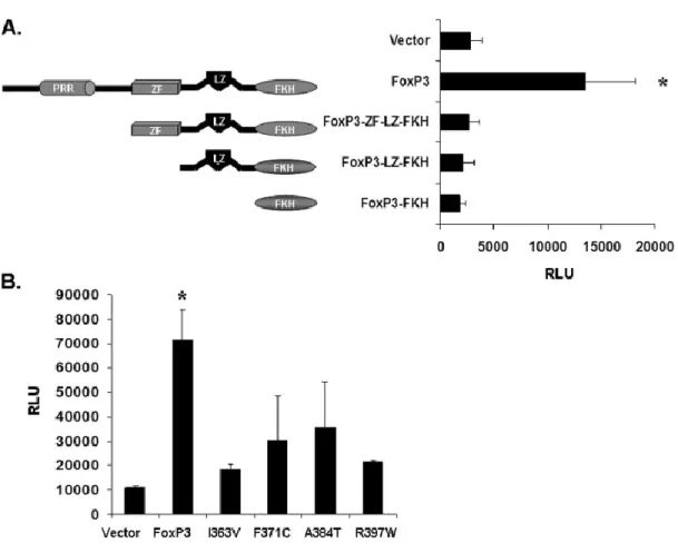
mutations, R347H, I363V, F371C, A384T and R397W (206, 208-210). Other mutations
include mutations in the stop codon, resulting in C-terminal extensions, a splice site
35
causing the deletion of exon 9 (207, 209-211). Studies with various IPEX mutants have
demonstrated multiple roles of these domains in Foxp3 function. The leucine zipper has
been show to be required for oligomerization in vivo, wherein deletion of amino acids 250 or 251 results in a loss of Foxp3 homotetramerization or Foxp1/Foxp3
heteromerization (212-214). Interestingly, mutations in the leucine zipper domain
disrupt binding and transcriptional function, but does not impair association of Foxp3
with a multiprotein complex. Analysis by the Ziegler group demonstrated that the
N-terminus and forkhead domain are required for repression of targeted promoters, with
the proline-rich region alone capable of repression, and the forkhead domain required
for DNA binding and nuclear localization. Also, while disruption of the leucine zipper
was necessary and sufficient to abrogate multimerization, a mutation disrupting the zinc
finger of Foxp3 (C204S) had no effect on Foxp3 multimerization and had only a mild
effect on repression of a targeted promoter (214).
Regulation of Foxp3 protein
Currently two isoforms of Foxp3 are found in human Treg cells, a full length
transcript and an exon 2 deleted form (215, 216). Murine Foxp3 protein is only
expressed in the full length form and found to be completely nuclear (217). Conversely,
the full length human Foxp3 (Foxp3a) is found both in the cytoplasm and nucleus while
the truncated isoform (Foxp3b) is nuclear only due to a lack of a putative nuclear export
36
cells is currently being studied. Several groups have documented the ability of TGF-β to
convert T cells into Treg cells (219, 220), and recent investigation of the Foxp3 promoter
has demonstrated a dependence of NFAT, Sp1, AP-1, STAT5 and, more recently, Smad3
for Foxp3 activation (221-223). While the factors regulating Foxp3 expression are under intense investigation, a more thorough look into the temporal regulation of Foxp3 gene
expression and the transcription factors required will give insight into the requirements
for Treg development, function and maintenance.
Mechanisms of Foxp3 regulation of promoter activity
Extensive studies on the gene profile of Treg cells along with ChIP-Chip
(microarray on immunoprecipitated chromatin) experiments determining the direct
targets of Foxp3 protein has produced insight into the requirement of Foxp3 protein for
the Treg signature. It is now evident that multiple signals, including TGF-β signaling, IL-2
receptor signaling and Foxp3 converge to impart this Treg signature. Nevertheless,
there are multiple characteristic promoters that are directly regulated by Foxp3,
including IL-2, Ctla4, Tnfrsf18 (GITR) among others. The mechanisms of Foxp3 regulation of these promoters will be discussed in the following paragraph.
Foxp3 was initially described by Bettelli et al. to functionally interact with and inhibit the transactivation activity of the transcription factors NFAT and NF-κB (224).
More biochemical and structural analysis of the interaction of Foxp3 and NFAT was
37
Foxp3 interaction was able to inhibit Foxp3 promoter occupancy and regulation of
several characteristic genes. Thus, a model was proposed where Foxp3 was able to
inhibit an activating NFAT:AP-1 complex by promoting a repressive NFAT:Foxp3 complex
at the IL-2 promoter (225). More recently, Foxp3 was also demonstrated to inhibit AP-1 DNA binding activity to further inhibit AP-1-dependent genes such as IL-2 (226). Reports from Grant et al. and our published data point to a role of Foxp3 in transcriptional regulation of retroviral promoters. In both cases, the HIV-1 promoter was regulated by
Foxp3-mediated modulation of NF-κB, although with disparate results (68, 69).
Interestingly, two studies showed an increase or ‘stabilization’ of transcription factors at
various promoters regulated by Foxp3, correlating with the notion that Foxp3 might play
a role of enhancing or stabilizing a TCR-mediated signal (68, 225). Factors involved in T
cell development also interact with Foxp3 to regulate targeted promoters. The
Sakaguchi group described an interaction with Foxp3 and AML1/Runx1 transcription
factor. In this study, AML1/Runx1 bound to a region upstream of the core enhancer
region of the IL-2 promoter and enhanced IL-2 expression. A Foxp3/AML1 complex was able to bind this region and was shown to be required for Foxp3 repression of IL-2 (227).
Foxp3 regulates the expression of various promoters at multiple levels. Several
groups have demonstrated an increase or decrease in histone acetylation of promoters
regulated by Foxp3 (68, 225, 228). In accordance with this notion, factors modulating
histone acetylation, such as HATs and HDACs, have been shown to interact with and be