Original Research Article
HER2/neu immunohistochemical expression in gastric carcinoma
Anjali Sadanandan
1, Arunraj C. N.
2*
INTRODUCTION
Gastric cancer remains one of the deadly diseases with poor prognosis and is the fifth most common cancer and the third most common cause of cancer-related deaths worldwide.1 In
India, it is the fifth most common cancer and the fifth most common cause of cancer-related deaths.
Gastric carcinogenesis is a multistep and multifactorial process.2,3 H. pylori infection, atrophic gastritis, intestinal
metaplasia, and dysplasia are related to gastric adenocarcinoma.3 Reflux oesophagitis, Barrett’s esophagus,
and dysplasia are associated with the development of
proximal gastric adenocarcinomas.4 Gastric adenocarcinoma
is the most common malignancy of the stomach, comprising over 90% of all gastric malignancies. The stage is the most important prognostic factor for gastric carcinoma followed by histologic type.4 However patients in the same stage and
histological type may have varied prognosis, therefore additional parameters have to be identified in addition to stage and tumor type in order to better identify the biological subsets of this disease.4 The only proven, potentially curative
treatment for gastric cancer is surgical resection of all gross and microscopic disease. Even after what is felt to be a curative gastrectomy, the disease recurs in both regional and /or distant sites in the majority of patients.5
1Department of Pathology, 2Department of General Medicine, Travancore Medical College, Kollam, Kerala, India
Received: 03 December 2019
Revised: 27 December 2019
Accepted: 31 December 2019
*Correspondence:
Dr. Arunraj C. N.,
E-mail: drarunrajcn@gmail.com
Copyright: © the author(s), publisher and licensee Medip Academy. This is an open-access article distributed under the terms of the Creative Commons Attribution Non-Commercial License, which permits unrestricted non-commercial use, distribution, and reproduction in any medium, provided the original work is properly cited.
ABSTRACT
Background: Gastric carcinoma is a deadly disease with high mortality. A better understanding of the molecular basis of gastric cancer has contributed to the development of rationally designed molecular targeted therapies which will improve the survival rate. A genetic alteration that could help in targeted therapy and prognostication includes Human Epidermal Growth Factor Receptor 2 (HER2/neu) overexpression in gastric carcinoma. The objective of the present study was to identify and evaluate the HER2/neu protein immunohistochemical expression in gastric cancer from biopsies and surgical resection specimens and to evaluate their correlation with histopathological features.
Methods: Total/subtotal gastrectomy specimens and gastric biopsies from a tertiary care center in South India were included in the study and assessed by light microscopy and Immunohistochemistry (IHC).
Results: HER2/neu overexpression was seen in 28.6% of gastric adenocarcinoma. HER2/neu overexpression was seen in 44.2% of intestinal-type and 20% of mixed type with none of the diffuse type exhibited HER2/neu positivity and this was statistically highly significant with p value of <0.01. HER2/neu positivity was found in 50% well-differentiated and 36.4% moderately well-differentiated tumors with none of the poorly well-differentiated tumors exhibiting HER2/neu positivity and this was statistically highly significant with p value of <0.01.
Conclusions: This study highlights the importance of the identification of HER2/neu overexpression in gastric adenocarcinoma. This will help in prognostication and identifying patients suitable for novel therapeutic interventions which will help in prolongation of survival of patients with this deadly disease.
Keywords: Gastric adenocarcinoma, Human epidermal growth factor receptor 2, Immunohistochemistry
Currently available agents are not very effective resulting in a high recurrence rate, low survival rate, poor prognosis of advanced gastric cancer patients.5 A better
understanding of the molecular basis of cancer has contributed to the development of rationally designed molecular targeted therapies that interfere with signaling cascades involved in cell differentiation, proliferation, and survival.4 Current targeted therapy for advanced
gastric carcinoma depends on the evaluation of target gene status.5 The combination of various therapeutic
agents has modestly improved the overall survival rates. Hence there is a need to study newer targeted therapeutic agents that improve the survival rates.6
The HER2/neu protein is 185-kDa transmembrane Tyrosine Kinase (TK) receptor and a member of the Epidermal Growth Factor Receptors (EGFRs) family which can influence cell proliferation, apoptosis, adhesion, migration, and differentiation.4,7 HER2/neu is
encoded by a gene located on chromosome 17q21. In carcinomas, HER2/neu acts as an oncogene, mainly because high-level amplification of the gene induces protein overexpression in the cellular membrane and subsequent acquisition of advantageous properties for a malignant cell. HER2/neu overexpression and /or amplification have been detected in 10-34% of invasive breast cancers and with 13-44% gastric cancer and correlates with poor outcome and more aggressive disease. Overexpression of HER2/neu protein in gastric cancer, using IHC, was first described in 1986. In contrast to breast, basolateral membrane staining instead of circumferential membranous staining is sufficient to qualify for positive immunostaining. Incomplete basolateral membrane HER2/neu staining is more common in gastric cancer than in breast cancer due to the higher frequency of glandular formations that occur in gastric tissue.4,8,9
Trastuzumab (Herceptin) is a monoclonal antibody that specifically targets HER2 protein by directly binding to the extracellular domain of the receptor. The antitumor mechanism of Herceptin is not fully understood,
however, some mechanisms postulated are the
interruption of HER2/neu mediated cell signaling pathways and cell cycle progression, induction of antibody-dependent cell-mediated cytotoxicity and apoptosis; induction of anti-angiogenesis effects and increasing receptor turn over by endocytosis.10
Trastuzumab was approved in combination with cisplatin and a fluoropyrimidine, for the treatment of patients with
HER2/neu overexpressing metastatic gastric or
gastroesophageal junction adenocarcinoma who have not received prior treatment.8
The pivotal ToGA (Trastuzumab for gastric cancer) trial 2010 demonstrated a median survival of 13.1 months for patients receiving Trastuzumab and chemotherapy and 11.7 months for patients receiving chemotherapy alone thereby putting forth the advantage of concurrent use of Trastuzumab along with chemotherapy.8
The objective of the present study was to identify and evaluate the HER2/neu protein immunohistochemical expression in gastric cancer from biopsies and surgical resection specimens and to evaluate their correlation with histological subtypes and histological grading. As there is currently limited data available on immunohistochemical expression of HER2/neu in gastric carcinoma in the Indian population, authors proposed to conduct this study to evaluate the same.
METHODS
The present study “HER2/neu immunohistochemical expression in gastric carcinoma” is a descriptive study conducted in the Department of Pathology in a tertiary care hospital in South India with the cooperation of
Departments of General Surgery, Medical
Gastroenterology, and Surgical Gastroenterology over a period of two years from February 2017 to January 2019. A total of 70 cases including total/subtotal gastrectomy specimens and gastric biopsies confirmed as gastric adenocarcinoma by histopathological examination were included in the study. Approval for the study was obtained from the Institutional Ethics Committee.
Inclusion criteria
• Total or subtotal gastrectomy specimens, gastric biopsies from patients with gastric adenocarcinoma who have not received any previous treatment were included in the study.
Exclusion criteria
• Cases where there is extensive tumor necrosis without sufficient viable tumor cells for an accurate evaluation of the immunohistochemical results.
The detailed clinical history including age, gender, and results of relevant investigations done was collected or abstracted from the patient’s case files. For prospective cases total, subtotal gastrectomies and small biopsy specimens were received in the Pathology Department in 10% formalin. In every case, the standard protocol for surgical grossing of resected specimens was followed. After a detailed specimen description, multiple sections were taken from the tumor, surgical margins, omentum, mesentery, and all the lymph nodes. For retrospective cases, the histopathology reports, slides and paraffin blocks were retrieved from the archives. After conventional processing, paraffin sections of 5 µm thickness were stained by hematoxylin and eosin (H and E) for histopathological study.
classified into intestinal and diffuse according to Lauren’s classification. p TNM staging was done in gastrectomy specimens.
The technique of IHC included antigen retrieval in tris EDTA buffer in a microwave oven, blocking endogenous peroxidase with 3% hydrogen peroxide, incubating with primary mouse monoclonal antibody against HER2/neu protein (PATH INSITU) and linking with rabbit anti-mouse secondary antibody, enzyme linking with streptavidin- horseradish peroxidase, developing
chromogen with Diaminobenzidine (DAB) and
counterstaining with hematoxylin. Positive and negative controls were run with each batch of slides. The immunostained slides have been evaluated using the HERCEP Test (Dako Denmark A/S, Glostrup, Denmark) that has been frequently used to evaluate the patterns of membranous immunoreactivity on the tumor cells for HER2/neu overexpression. This scoring system is based on the intensity of reactivity, whether complete or incomplete and the percentage of reactive cells.11,12 IHC score of 0 and
1+ was considered negative, while an IHC score of 2+ and 3+ was considered positive (Table 1).11,13,14
Table 1: HER2/neu scoring for gastric carcinoma.13
Score Surgical specimen staining pattern Biopsy specimen staining pattern
HER2 /neu over expression assessment
0 No reactivity or membranous reactivity in
<10% of tumor cells
No reactivity or membranous reactivity in
any tumor cells Negative
1+
Faint/barely perceptible membranous reactivity in ≥10% of tumor cells, cells are reactive in only part of their membrane
Tumor cell cluster (5 tumor cells) with faint/barely perceptible membranous reactivity irrespective of the percentage of tumor cells stained.
Negative
2+
Weak to moderate complete, basolateral, or lateral membranous reactivity in ≥10% of tumor cells.
Tumor cell cluster with weak to moderate complete, basolateral, or lateral membranous reactivity irrespective of the percentage of tumor cells stained.
Positive
3+
Strong complete, basolateral, or lateral membranous reactivity in ≥10% of tumor cells.
Tumor cell clusters with strong complete, basolateral or lateral membranous reactivity irrespective of percentage of tumor cells stained.
Positive
The relationship between various parameters such as age, gender, anatomic site of the tumor, histologic type and grade, with overexpression of HER2/neu were studied in all biopsy specimens. In addition, in gastrectomy specimens, the pathologic stage, lymph node status was also studied.
Statistical analysis
Data were analyzed using computer software, Statistical Package for Social Sciences (SPSS) version 16. Data are expressed in their frequency and percentages. To elucidate the associations and comparisons between different parameters, Chi-square test was used as the nonparametric test. For all statistical evaluations, a two-tailed probability of value <0.05 was considered significant.
RESULTS
A total of 70 cases which were confirmed as gastric adenocarcinoma were included for the study. Among the total 70 specimens, 50(71.4%) were gastric biopsies, 17(24.3%) were total gastrectomies and 3(4.3%) were subtotal gastrectomies. The age group of patients in the
study ranged from 30-80 yrs. The maximum incidence was at the 50-59 yr age group (34.3%). Around 57.2% occurred in the age group 40-59 yrs. Of 70 cases studied, males accounted for the majority - 54(77.1%). Among 70 cases, 43(61.4%) were intestinal type, 22(31.4%) were diffuse type and 5(7.2%) were mixed type. Among 70 cases, 33 cases (47.1%) were moderately differentiated, 21 cases (30%) were poorly differentiated and 16 cases (22.9%) were well differentiated. Antrum was the commonest site (40%) followed by antropyloric (21.4%), body (17.2%), OGJ (14.3%) and Cardia and Fundus (7.1%). Among the 20 gastrectomy specimens, 15(75%) had tumor size <5 cm and 5(25%) had a tumor size of >5 cm. Among 20 gastrectomy specimens, 11(55%) had lymph node involvement. Among the 20 gastrectomy specimens, 40% exhibited T3 and 30% exhibited T4 stage. T1 and T2 were 15% each.
Type and grade of tumor
Among the 33 moderately differentiated cases, 27(81.8%) were of the intestinal type, 5(15.2%) were of mixed type and 1(3%) was of diffuse type. All these findings were statistically highly significant (p value <0.001) (Table 2).
Table 2: Type and grade of tumor.
Grade
Type
Total
Intestinal Type
Diffuse Type
Mixed Type
Poorly differentiated
0 21 0 21
100.00% 100.00%
Moderately differentiated
27 1 5 33
81.80% 3.00% 15.20% 100.00%
Well
differentiated
16 0 0 16
100.00% 100.00%
Total 43 22 5 70
Chi square: 69.529; p <0.001
Frequency of HER2/neu overexpression among the gastric adenocarcinoma cases
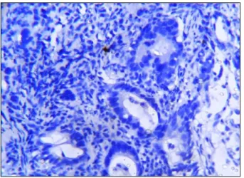
Figure 1: Photomicrograph of moderately differentiated intestinal type of gastric
adenocarcinoma showing HER2/neu 0 staining (IHC, 400x).
Table 3: Frequency of HER2/neu positive and negative cases.
HER2 / neu Frequency Percent
Negative 50 71.4
Positive 20 28.6
Total 70 100
In HER2/neu IHC staining, among the total 70 cases, 12 cases (17.2%) showed 3+, 8 cases (11.4%) showed 2+, 18 cases (25.7%) showed 1+ and 32 cases (45.7%) showed no staining (Figures 1,2,3,4). 0 and 1+ staining is taken as HER-2/neu Negative and 2+ and 3+ are HER- 2/neu Positive. 20 specimens (28.6%) were HER-2/neu Positive (Table 3).
Figure 2: Photomicrograph of poorly differentiated diffuse type of gastric adenocarcinoma showing
HER2/neu 1+ staining (IHC, 400x).
Figure 3: Photomicrograph of well-differentiated intestinal type of gastric adenocarcinoma showing
HER2/neu 2+ positivity in >10% tumor cells (IHC, 400x).
Figure 4: Photomicrograph of well-differentiated intestinal type of gastric adenocarcinoma showing
HER2/neu and type of tumor
Of the 43 intestinal types, 19(44.2%) were HER2/neu positive, 24(55.8%) were HER2/neu negative. Of the 22 diffuse types, all were HER2/neu negative. Of the 5 mixed types, 1(20%) was HER2/neu positive and 4(80%) were HER2/neu negative. Among the 20 HER2/neu positive cases, 19(95%) were of intestinal-type and 1(5%) was of mixed type, none were of diffuse type. Among the 50 HER2/neu negative cases, 24(48%) were intestinal type, 22(44%) were of diffuse-type and 4(8%) were of mixed type. All these findings were statistically highly significant (p value <0.01) (Table 4).
Table 4: HER2/neu and type of tumor.
Type HER2 / neu Total
Negative Positive
Intestinal type 24 19 43
55.80% 44.20% 100.00%
Diffuse type 22 0 22
100.00% 100.00%
Mixed type 4 1 5
80.00% 20.00% 100.00%
Total 50 20 70
Chi square: 14.117; p <0.01
HER2/neu and grade of tumor
Among the 21 poorly differentiated cases, all (100%) were HER2/neu negative. Among the 33 moderately differentiated cases, 21(63.6%) were HER2/neu negative and 12(36.4%) were HER2/neu positive. Among the 16 well-differentiated cases, 8(50%) each were HER2/neu positive and negative. Among the 20 HER2/neu positive cases, 12(60%) were moderately differentiated and 8(40%) were well-differentiated and none were poorly differentiated. Among the 50 HER2/neu negative cases, 21(42%) each were moderately differentiated and poorly differentiated and 8(16%) were well differentiated. All these findings were statistically highly significant (p value <0.01) (Table 5).
Table 5: HER2/neu and grade of tumor.
Grade HER2 / neu Total
Negative Positive
Poorly differentiated
21 0 21
100.00% 100.00%
Moderately differentiated
21 12 33
63.60% 36.40% 100.00%
Well
differentiated
8 8 16
50.00% 50.00% 100.00%
Total 50 20 70
Chi square: 12.982; p <0.01
In this study, there was no significant association between HER2/neu with age, gender, nature of the
specimen, site of the tumor, size of the tumor, lymph node status and tumor invasion.
DISCUSSION
Gastric carcinoma is a deadly disease with high mortality. Even after a potentially curative surgical resection for gross and microscopic disease, it recurs in both regional and distant sites in the majority of patients.5 This
emphasizes the importance of early detection,
prognostication, and novel individualized targeted therapy. A genetic alteration which could help in prognostication and targeted therapy include HER2/neu overexpression in gastric carcinoma.
In the present study, of the 70 specimens, 50(71.4%) were gastric biopsies and 20(28.6) were gastrectomies (17(24.3%) total gastrectomies and 3(4.3%) subtotal gastrectomies). The relative percentage of gastric biopsy and gastrectomy specimens in other studies were 73% and 27% - Rajagopal I et al, 73% and 27 % - Lakshmi V et al.15,16
In the present study, of the 70 cases, 20(28.6%) showed HER2/neu positivity. Previous studies have reported the prevalence of overexpression of HER2/neu in gastric carcinoma ranging from 13-44% (Table 6). Previous studies from India are showing HER2/neu positivity in the range of 21.4-44.2%. The result from South India is in concordance with this study. Authors included both IHC 3+ and 2+ as HER2/neu positivity. This might have caused the inclusion of more cases into HER2/neu positivity because IHC 2+ has only around 58% concordance with FISH.17 Authors could not do FISH
studies for IHC 2+ (equivocal) cases due to financial constraints. IHC 2+ cases were subjected to FISH confirmation in studies done by Gravalos et al, He C et al, and Sekaran et al, but was not done in studies conducted by Rajagopal I et al, Tewari M et al, Lakshmi V et al, and Lordick et al.4,10,15,16,18-20
Table 6: Comparison of HER2/neu overexpression in various studies.
Study Geographic
zone
No of cases
Her2/neu positivity (%) Gravalos et al,4 Europe 166 13
He C et al,10 China 197 18.3
Lordick et al,20
Europe, Asia, Latin America
1527 22 Rajagopal I et al,15 South India 60 26.7
Lakshmi V et al,16 South India 78 35.9
Sekaran A et al,18 India 52 44.2
Tewari M et al,19 North India 70 21.4
Present study South India 70 28.6
was statistically highly significant with p value <0.01. This is in concordance with other studies which show a
disproportionate overexpression of HER2/neu in
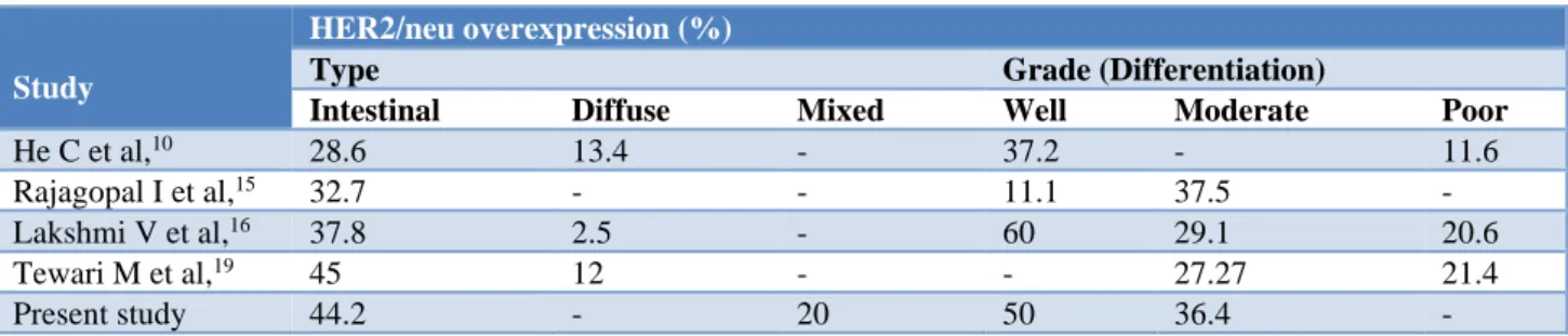
intestinal compared to diffuse type of gastric carcinoma (Table 7).
The evolution of intestinal and diffuse types of gastric carcinomas along different molecular pathways could explain the association of HER2/neu with intestinal type.
A review of the literature shows a decreased percentage of HER2/neu overexpression in the diffuse types of gastric cancers rather than absent expression. The present study and study by Rajagopal I et al, shows the total absence of HER2/neu expression in diffuse type.15
This could probably be because of the smaller number of diffuse types of gastric cancers included in these two studies. Studies by He C et al, Rajagopal I et al, Tewari M et al, and Lakshmi V et al, had not categorized any of the cases to mixed type and hence no HER2/neu positivity in the mixed type category.10,15,16,19
In the present study as per CAP grading, HER2/neu positivity was found in 50% of well-differentiated and 36.4% of moderately differentiated tumors with none of the poorly differentiated tumors expressing HER2/neu and this was statistically highly significant with p value <0.01 and indicating that better-differentiated tumors showed HER2/neu overexpression. The findings of the present study are in concordance with the other studies in that HER2/neu expression is more in well and moderately
differentiated tumors in comparison to poorly
differentiated tumors (Table 7). An exception to this is the study by Tewari M et al, which shows the total absence of HER2/neu expression in well-differentiated tumors.19 This could probably be explained by the fact
that only 3 out of 70 cases were well-differentiated tumors in that study. Similar to the present study another study from South India by Rajagopal I et al, shows a complete absence of HER2/neu expression in poorly differentiated tumors which could probably be explained by a geographical variation in gastric cancer epidemiology within India.15 HER2/neu overexpression is
commoner in better-differentiated tumors. None of the diffuse types showed HER2/neu overexpression.
Table 7: Comparison of HER2/neu overexpression with respect to histologic type and grade in various studies.
Study
HER2/neu overexpression (%)
Type Grade (Differentiation)
Intestinal Diffuse Mixed Well Moderate Poor
He C et al,10 28.6 13.4 - 37.2 - 11.6
Rajagopal I et al,15 32.7 - - 11.1 37.5 -
Lakshmi V et al,16 37.8 2.5 - 60 29.1 20.6
Tewari M et al,19 45 12 - - 27.27 21.4
Present study 44.2 - 20 50 36.4 -
In this study, there was no significant association between HER2/neu with age, gender, nature of the specimen, site of the tumor, size of the tumor, lymph node status and tumor invasion. Few of the previous studies have noticed a higher rate of HER2/neu overexpression in gastric biopsies compared to gastrectomies.15,17,20 Also, few studies have noticed a
higher rate of HER2/neu positivity in gastro-esophageal junction tumors compared to other locations.4,15,20 The
present study also showed a higher rate of HER2/neu overexpression in gastric biopsies compared to gastrectomies and also noticed a higher rate of HER2/neu positivity in gastro-esophageal junction tumors but did not achieve statistical significance.
The present study has a few limitations. The proportion of gastrectomy specimens was less compared to gastric biopsies which could have influenced the tumor grading, typing and also IHC staining because of the relatively small representative tissue in the gastric biopsy. Another limitation is inadequate patient follow up to allow comparison of
prognosis and survival rate between tumor subgroups and their IHC profiles.
Also, various study groups have used different protocols for HER2/neu overexpression which could affect the comparison between various studies. Regarding HER2/neu testing, IHC 2+ (equivocal) cases should have been confirmed with gene amplification studies which were not done in the present study because of financial constraints. This could have spuriously increased the IHC positive cases. Due to the relatively smaller sample size, findings of this study have to be confirmed with future large-scale studies.
These results show that in patients with gastric adenocarcinoma, identification of genetic alterations will help in the prognostication and identifying patients who might benefit from targeted therapy. Trastuzumab is a monoclonal antibody that specifically targets HER2/neu
receptor protein. Trastuzumab combined with
advanced gastric carcinoma as stated in ToGA (Trastuzumab for gastric cancer) trial 2010.
CONCLUSION
This study highlights the importance of identification of HER2/neu overexpression in gastric adenocarcinoma which helps in prognostication and identifying patients suitable for novel therapeutic interventions. HER2/neu overexpression was seen in 28.6% cases of gastric adenocarcinoma. HER2/neu positivity was seen in better-differentiated tumors with none of the poorly differentiated tumors showing positivity.
Also, none of the diffuse types showed HER2/neu overexpression. There is no significant association of HER2/neu with age, gender, nature of the specimen, site of the tumor, size of the tumor, lymph node status and tumor invasion. The identification of these genetic alterations will help in prognostication, prediction of progression and identification of patients suitable for novel therapeutic agents which will help in prolongation of survival of patients with this deadly disease.
Funding: No funding sources Conflict of interest: None declared
Ethical approval: The study was approved by the Institutional Ethics Committee
REFERENCES
1. Bray F, Ferlay J, Soerjomataram I, Siegel RL, Torre LA, Jemal A. Global cancer statistics 2018: GLOBOCAN estimates of incidence and mortality worldwide for 36 cancers in 185 countries. CA Cancer J Clin. 2018 Nov;68(6):394-424.
2. Hu B, El Hajj N, Sittler S, Lammert N, Barnes R, Meloni-Ehrig A. Gastric cancer: Classification, histology and application of molecular pathology. J Gastro Oncol. 2012 Sep;3(3):251.
3. Saghier AA, Kabanja JH, Afreen S, Sagar M. Gastric cancer: environmental risk factors, treatment and prevention. J Carcinogene Mutagene. 2013; S14:2157-2518.
4. Gravalos C, Jimeno A. HER2 in gastric cancer: a new prognostic factor and a novel therapeutic target. Annal Oncol. 2008 Apr 25;19(9):1523-9.
5. Yan SY, Hu Y, Fan JG, Tao GQ, Lu YM, Cai X, et
al. Clinicopathologic significance of HER-2/neu protein expression and gene amplification in gastric carcinoma. World J Gastroenterol: WJG. 2011 Mar 21;17(11):1501-06.
6. Gómez-Martin C, Garralda E, Echarri MJ,
Ballesteros A, Arcediano A, Rodríguez-Peralto JL, et al. HER2/neu testing for anti-HER2-based therapies in patients with unresectable and/or metastatic gastric cancer. J Clin Pathol. 2012 Aug 1;65(8):751-7.
7. Jung JE, Ioshii SO. Immunohistochemical
assessment of HER2 expression in gastric cancer in
a cohort of 118 Brazilian patients. J Brasileiro Patol Med Lab. 2013 Oct;49(5):361-7.
8. Iqbal N, Iqbal N. Human epidermal growth factor receptor 2 (HER2) in cancers: overexpression and therapeutic implications. Mol Biol Inter. 2014; September 7: Article ID 852748:1-9.
9. Xie SD, Xu CY, Shen JG, Jiang ZN, Wang LB. HER 2/neu protein expression in gastric cancer is
associated with poor survival. Erratum
in/mmr/12/3/4794. Mol Med Report. 2009 Nov 1;2(6):943-6.
10. He C, Bian XY, Ni XZ, Shen DP, Shen YY, Liu H,
et al. Correlation of human epidermal growth factor receptor 2 expression with clinicopathological characteristics and prognosis in gastric cancer.
World J Gastroenterol: WJG. 2013 Apr
14;19(14):2171-8.
11. Rakhshani N, Kalantari E, Bakhti H, Sohrabi MR,
Mehrazma M. Evaluation of HER-2/neu
overexpression in gastric carcinoma using a tissue microarray. Asian Pac J Cancer Prev. 2014 Jan 1;15(18):7597-602.
12. Mrklic I, Bendic A, Kunac N, Bezic J, Forempoher G, Durdov MG, et al. Her-2/neu assessment for gastric carcinoma: validation of scoring system. Hepato-Gastroenterol. 2012;59(113):300-3.
13. Hofmann M, Stoss O, Shi D, Büttner R, Van De Vijver M, Kim W, et al. Assessment of a HER2 scoring system for gastric cancer: results from a validation study. Histopathol. 2008 Jun;52(7):797-805.
14. Rüschoff J, Dietel M, Baretton G, Arbogast S, Walch A, Monges G, et al. HER2 diagnostics in gastric cancer-guideline validation and development of standardized immunohistochemical testing. Virchows Archiv. 2010 Sep 1;457(3):299-307.
15. Rajagopal I, Niveditha SR, Sahadev R, Nagappa PK, Rajendra SG. HER 2 expression in gastric and gastro-esophageal junction (GEJ) adenocarcinomas. J Clin Diag Res: JCDR. 2015 Mar;9(3):EC06-EC10. 16. Lakshmi V, Valluru VR, Madhavi J, Valluru N. Role of her 2 neu in gastric carcinoma-3 year study in a medical college hospital. Ind J Appl Res. 2014;4(11)47-50.
17. Yano T, Ochiai A, Doi T, Hashizume K, Nakanishi M, Ouchi K, et al. Expression of HER2/neu in gastric cancer: comparison between protein expression and gene amplification using a new commercial kit. J Clin Oncol. 2004;22:14S:4053.
18. Sekaran A, Kandagaddala RS, Darisetty S,
Lakhtakia S, Ayyagari S, Rao GV, et al. HER2 expression in gastric cancer in Indian population-an immunohistochemistry and fluorescence in situ hybridization study. Ind J Gastroenterol. 2012 Jun 1;31(3):106-10.
19. Tewari M, Kumar A, Mishra RR, Kumar M, Shukla
HS. HER2 expression in gastric and
20. Lordick F, Bang YJ, Kang YK, Reyes DO, Manikhas GM, Shen L, et al. 3541 POSTER positive advanced gastric cancer: similar HER2-positivity levels to breast cancer. EJC Supplements. 2007 Sep 1;5(4):272.