LESSON ASSIGNMENT
LESSON 3 Collection of Blood and Preparation of Blood Smears. TEXT ASSIGNMENT Paragraphs 3-1 through 3-8.
LESSON OBJECTIVES After completing this lesson, you should be able to: 3-1. Select the statement which best describes the
requirements for selection and care of blood
collection equipment.
3-2. Select the correct steps in collecting,
processing, and recording blood specimens. 3-3. Select the names and functions of commonly
used anticoagulants
3-4. Select the correct method and use the proper procedures in staining blood films for the type of
blood cells found.
3-5. Select the correct function for each of Wright's component stain and buffer solutions.
3-6. Select the correct steps to prepare a blood
smears.
3-7. Select the factors that affect the quality of stained blood smears.
SUGGESTION After completing the assignment, complete the exercises at the end of this lesson. These exercises will help you to achieve the lesson objectives.
LESSON 3
COLLECTION OF BLOOD AND PREPARATION OF BLOOD SMEARS Section I. COLLECTION OF BLOOD SPECIMENS
3-1. INTRODUCTION
a. Hematological laboratory procedures are based upon the examination of blood specimens. To obtain valid test results, specimens must be properly collected, processed, and recorded. Blood specimens are usually obtained by either venous or capillary puncture. The source of the specimen is determined chiefly by the quantity of blood required to perform the laboratory procedures and the age and condition of the patient.
b. There is generally little difference in blood counts performed on venous or capillary blood if a free-flowing capillary blood specimen is obtained. Valid blood counts cannot be made when capillary specimens are not taken from a free-flowing sample or when they are obtained from cyanotic or calloused areas or areas of local stasis. White blood cell counts made on blood obtained from such sources can vary as much as 1000 to 1500 cells per cu mm from their real value. For general purposes, however, venous samples are preferable since they allow for multiple and repeated hematological
examinations and provide a sufficient quantity of blood for performing any other required laboratory procedure. Further, with venous blood the chances of error are reduced because operations are made under ideal conditions and repeat operations are possible. In situations where there are limitations on the quantity of blood that can be obtained, that is, in small infants or extensive burn cases, microquantitative methods are satisfactory for performing an analysis on a specimen obtained by capillary puncture.
3-2. VENIPUNCTURE
a. Site. To obtain blood by venipuncture, draw the specimen directly from a patient's vein with a sterile hypodermic needle and syringe or a vacuum blood sample device.
(1) In adults use the veins located in the proximal forearm or antecubital space as illustrated in figure 3-1. In infants employ the jugular or femoral vein for the venipuncture. The vein selected should be large, readily accessible, and sufficiently close to the surface to be seen and palpated. If venipuncture poses a problem due to the age of the patient, sclerotization due to repeated venipuncture, or any other unusual circumstance, the technician should consult a physician concerning the procedure. UNDER NO CIRCUMSTANCES SHOULD A TECHNICIAN WITHDRAW BLOOD
FROM A SAGITTAL SINUS, JUGULAR VEIN, OR FEMORAL VEIN. This should be left to the discretion of the physician in charge.
Figure 3-1. Site of venipuncture.
(2) Occasionally, the best vein is found on the hand, leg, or foot. These areas are more sensitive, and the veins are not as firmly anchored as those of the arm. Veins can become distended and easier to enter by allowing the arm to hang down for 2 or 3 minutes, by massaging the blood vessel toward the body, or by gently slapping the site of puncture. Young and vigorous persons usually have elastic veins well filled with blood. Elderly or debilitated persons can have sclerosed or fragile veins, which are hard to enter or which collapse easily.
b. Equipment. All syringes, needles, lancets, and other instruments used for the collection of blood specimens must be sterile. Disposable syringes or blood
collection sets with vacuum tubes are available through normal supply channels. These should be used whenever possible. Aseptic technique is necessary to prevent the possible transmission of homologous serum hepatitis. The following are necessary to perform a venipuncture.
(1) Isopropyl alcohol, 70 percent, prep pads. (2) Tourniquet.
(3) Sterile syringes or vacuum blood sample devices. (4) Gauze pad, 2 x 2 inches.
(5) Needle, 1 to 1 ½ inches long, 19–23 gauge. (6) Suitable blood collection tubes and labels. (7) Gloves, latex.
c. Preparation.
(1) Cleanse hands thoroughly with soap and water. (2) Place an identifying label on the blood collecting tube.
(3) Assemble the sterile needle and syringe. If a vacuum system is used, screw the needle into the plastic holder. Always leave the cap over the needle when not in use.
(4) Check to make sure that the syringe works smoothly. The syringe must be dry to avoid hemolysis of the red cells. The plunger must match the syringe and must be pushed firmly to the bottom of the cylinder to prevent injection of air into the vein. This can be fatal.
d. Syringe Procedure.
(1) Place a tourniquet around the patient’s arm above the elbow tightly enough to check venous circulation, but not so tightly as to stop arterial flow. (If latex tubing is used, place it approximately 2 inches above he proposed venipuncture site). Form a loop with the longer end and draw the loop under the shorter end so that the tails of the tubing are turned away from the proposed site (see figure 3-2a).
CAUTION: Do not allow the tourniquet to remain in place for more than 1 minute. Check the pulse at the wrist to make sure that arterial circulation is not
cut off.
(2) Instruct the patient to make a tight fist.
(3) By inspection and palpation locate the desired vein, determine the direction of its course, and estimate its size and depth (see figure 3-2a venipuncture procedure, a through h).
(4) Release tourniquet.
(5) Cleanse the skin over the selected vein with prep pads in 70 percent isopropyl alcohol in a circular motion starting from the center and working your way out. Allow the area to air dry for 30 seconds to 1 minute. Do not contaminate the area after cleaning (see figure 3-2b).
Figure 3-2b. Venipuncture procedure: Clean the puncture site. (6) Put on gloves.
(7) Replace tourniquet on arm and have the patient straighten out the arm and make a fist.
(8) Grasp the syringe in the right hand and place forefinger on the hub of the needle to guide it. Grasp the forearm with the left hand about 2 inches below the area to be punctured and hold the skin taut with the thumb (see figure 3-2c).
(9) With the needle bevel up, parallel to, and alongside the vein, insert the needle quickly under the skin and then into the vein. The insertion into the skin and vein can be performed in one complete motion (see figure 3-2d). After entry into the vein, blood will appear in the needle hub. Do not probe or move the needle horizontally, as discomfort and possible nerve damage may result.
Figure 3-2d. Venipuncture procedure: Insert needle into the vein. (10) Aspiration of the blood is accomplished by gently pulling upon the syringe plunger (see figure 3-2e). The syringe barrel should be held steady during this process. Withdraw the desired quantity.
Figure 3-2e. Venipuncture procedure: Aspirate the blood.
(11) Remove the tourniquet by pulling on the long, looped end of the tubing only after blood is drawn into the syringe (see figure 3-2f).
CAUTION: Do not remove the needle now. If the needle were remove prior to the Tourniquet being removed, blood would be forced out of the
venipuncture site, resulting in blood loss and/or hematoma formation (Pooling of blood under the skin).
(12) Place a sterile gauze pad over the point where the needle entered the skin and deftly withdraw the needle simultaneously putting pressure on the site (see figure 3-2g).
Figure 3-2g. Venipuncture procedure: Place a sterile pad over the site and withdraw the needle.
(13) Have the patient extend the arm and maintain light pressure on the gauze pad over venipuncture site (see figure 3-2h).
Figure 3-2h. Venipuncture procedure: Have the patient extend the arm and maintain light pressure on the site.
e. Vacutainer Procedure.
(1) Place the Vacutainer tube in the holder until the rubber stopper reaches the guideline. The short needle should be embedded in the stopper, but the needle must not break the vacuum (see figure 3-3).
Figure 3-3. Vacutainer system. (2) Follow steps 1-8 in syringe procedure.
(3) Enter the vein with the needle parallel to and alongside the vein. Probing or horizontal movement of the needle while under the skin must be avoided.
(4) After entry into the vein push the tube all the way into the holder; vacuum is broken, and blood flows freely into the tube. Release the tourniquet at this time by pulling the long, looped end of the tube.
(5) If the multiple needle is used or more than one tube is required, release the tourniquet after the first tube is filled; remove the filled tube and insert the next one. CAUTION: Ensure the needle is not moved while tubes are being changed.
(6) Place a sterile gauze pad over the point where the needle enters the skin and deftly withdraw the needle, placing pressure on the site.
(7) Have the patient extend the arm and maintain light pressure on the gauze pad over the venipuncture site.
f. Discussion.
(1) Cleanliness is essential when performing a venipuncture.
(2) It is most important that correct technique be practiced in order to avoid unnecessary pain to the patient, prevent tissue damage, secure a good representative blood specimen, and prevent contamination of the specimen or infection of the patient.
(3) Syringes and needles must be thoroughly inspected for damage or malfunction.
(4) If difficulty is experienced in entering the vein or a hematoma begins to form, release the tourniquet and promptly withdraw the needle and apply pressure to the wound.
(5) Vigorous pulling on the plunger of the syringe can collapse the vein, produce hemolysis of the blood specimen, or cause air to enter the syringe.
(6) When repeated venipunctures have to be performed on one patient, it is advisable to select different sites for blood withdrawal.
(7) Remove the tourniquet as early as possible once a good flow of blood has been established. Prolonged application of the tourniquet results in partial stasis of blood and changes many quantitative values of blood components.
(8) Blood drawn by venipuncture is often stored for a period of time before it is analyzed. For this reason, certain general precautions must be followed in order to ensure a valid analysis. Before withdrawing blood from its container, make sure the anticoagulated blood sample is thoroughly but gently mixed. Blood containers should be tightly stoppered at all times to prevent drying or contamination. Store the blood specimen in the refrigerator. Blood count must be done within 3 hours of collection. Under no circumstances should blood taken for hematological examinations be stored overnight.
3-3. CAPILLARY PUNCTURE
a. Site. Several different sites are suitable for capillary puncture. Because it is the most accessible, the palmer or lateral surface of the tip of the finger (preferably ring finger) is the most common site in adults. However, certain problems can be
encountered such as heavy calloused areas or excessive tissue fluids (edema) that tend to result in non-representative samples. The lobe of the ear can be used for capillary puncture. However, differences in cell concentration do occur when blood is obtained from this site, primarily because of higher lymphocyte concentrations in the ear lobe. Because of the small amount of tissue on the fingers of infants, preferred site is the heel or big toe. A modification of the normal technique that has proven quite satisfactory when working with the heel of infants is to make two incisions in a crisscross fashion or “T”.
NOTE: To be a valid report, work done on capillary blood must be from a FREE- FLOWING puncture wound.
b. Equipment.
(1) Gauze pads 2 x 2 inches. (2) Blood lancet.
(3) Glass slides, heparinized capillary tubes, and other devices to receive the specimen.
(4) Isopropyl alcohol, 70 percent, prep pads. c. Procedure.
(1) The puncture site should be warm to assure good circulation of blood. If it is cold, apply warm water (38º to 40º C) for a few minutes. If blood is to be drawn from the ear, the edge of the lobe, not the flat side, should be punctured.
(2) The site to be punctured is first rubbed with alcohol prep pads to remove dirt and epithelial debris, increase circulation, and render the area reasonably
disinfected (see figure 3-4 capillary puncture procedure, a through d).
Figure 3-4a. Capillary puncture procedure: Clean the puncture site. (3) Allow sufficient time for the circulation to equalize.
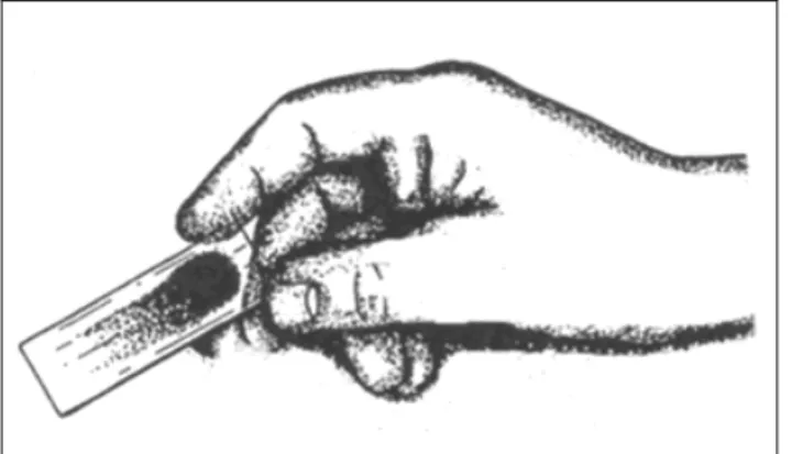
(4) While making a finger puncture, apply gentle pressure to the finger to hold the skin taut. Hold the finger in one hand and the lancet in the other. The puncture is made perpendicular to the lines of the fingerprints, which results in a more
free-flowing wound (see figure 3-4b).
(5) The first drop of blood that appears is wiped away before specimens are taken (see figure 3-4c).
Figure 3-4c. Capillary puncture procedure: Wipe away first drop of blood. (6) The blood must not be squeezed out since this dilutes it with fluid from the tissues, thus altering the ratio of cellular elements to fluid, as well as the ratio of cellular elements to each other.
(7) After the desired specimens have been collected, have the patient hold a sterile dry gauze pad over the wound until bleeding stops (see figure 3-4d).
Figure 3-4d. Capillary puncture procedure: Apply pressure to the site. d. Discussion.
(1) Us a disposable lancet for puncture of the skin.
(2) Do not use the finger on a hand which has been hanging over the side of the bed as it is likely to be congested. Edematous or cyanotic areas should not be used.
(3) The finger should be thoroughly dry prior to puncture; blood will not well up on a finger that is moist. Furthermore, the alcohol or other antiseptic used can coagulate the blood proteins causing cell clumping and erroneous values as well as dilute cell volumes. This will result in incorrect counts and differentials.
(4) Finger punctures should be made along the lateral aspect of the fingertip. More nerve endings are located on the fingerprint area of the fingers;
therefore, more pain results from punctures in this area. Scars can also form in these sensitive areas, and difficulty may be encountered in puncturing a callous. All of these difficulties are eliminated by drawing the blood from the lateral rather than the ventral aspect of the finger.
3-4. ANTICOAGULANTS
a. Anticoagulants are used to prevent the clotting of the blood specimens and the reagent employed should not bring about alteration of blood components.
Unfortunately, many anticoagulants can alter cell structures as well as coagulation. The anticoagulants most often used are ethylene-diamine-tetra-acetate (EDTA), ammonium-potassium oxalate and heparin.
b. The choice of anticoagulant will depend on the analysis to be made. Ethylene-diamine-tetra-acetate (EDTA) is the anticoagulant of choice for most
hematological analyses. This anticoagulant causes a minimum of distortion to the cells and platelets. It does not dissolve quickly in blood, however, so the tube must be inverted four or five times after blood is added. The dipotassium salt is prepared as a 1 percent solution in distilled water, and a final concentration of 0.5 ml of anticoagulant for each 5 ml blood is used. Another common anticoagulant is arnmonium-potassium oxalate. This combination of oxalates does not shrink or enlarge the red blood cells appreciably. It is essential, however, to add an optimal volume of blood to the oxalate, no less than 3.5 nor more than 6.0 mI.
c. Heparin does not alter the size of cellular components. It is, in fact, the standard for comparison of anticoagulant distortion. Heparin is more expensive and dissolves less readily than double oxalate salts. Approximately 0.5 to 1.0 mg is required to anticoagulate 5 ml of blood for 72 hours. The quantity of anticoagulant noted above in each case is sufficient to prevent clotting of the blood specimen. On the other hand, an excess of anticoagulant should be avoided because too much will result in distortion of cells and hemolysis. Ideally, differential blood smears should not be prepared from blood that contains an anticoagulant.
d. If oxalate is added to vials and dried in an oven, take great care to avoid temperatures above 80oC. Oxalates are converted to carbonates by prolonged exposure to elevated temperatures. Under normal circumstances, it should not be necessary to prepare your own oxalate solutions since prepared anticoagulant vacuum tubes are available from Federal medical supply sources.
e. Sodium citrate is the anticoagulant of choice for coagulation studies. It is used in a concentration of 1 part 0.11 M sodium citrate to 9 parts whole blood. It prevents coagulation by binding the calcium of the blood in a soluble complex.
f. Sodium oxalate is another anticoagulant widely used in coagulation studies. It is used in a concentration of 1 part 0.1 M sodium oxalate to 9 parts whole blood. The sodium oxalate combines with calcium in the blood to form insoluble calcium oxalate, thereby, preventing coagulation.
NOTE: Under normal circumstances, it should not be necessary to prepare an anticoagulant since prepared anticoagulant tubes are available through the Federal supply system.
g. A correctly anticoagulated blood sample is essential to the proper
performance of a blood cell count. The cellular constituents must remain free in the plasma and should be as similar as is possible to those remaining in the patient's circulation.
Section II. PREPARATION AND STAINING OF BLOOD SMEARS 3-5. INTRODUCTION
a. The type of blood cells found in the peripheral blood smears may be of diagnostic and prognostic importance. For this reason proper preparation and staining of blood films is essential for the identification and study of different kinds of leukocytes. The appearance of erythrocytes and thrombocytes will often give important clues that help distinguish between different types of diseases or other physical changes.
b. There are two basic methods for the preparation of blood smears: the cover slip and the slide methods. The cover slip method has certain advantages over the slide method; distribution of cells is like that of the in vivo circulation. The principal disadvantage of the latter method is that cover slips are very fragile and easily broken during processing.
c. The slides and cover glasses must be chemically clean and dry.
d. The foundation for the morphological study of blood was based on Ehrlich's investigations of the aniline dyes, dating back to 1877, while he was still a student. Originally, simple dyes were used in the clinical laboratory and tissues were stained successively if more than one color was desired. The majority of the aniline dyes are in the form of salts of acids and bases. During the process of staining, compounds are probably formed between the basic dyes and the acid nuclear substances of cells and between the acid dyes (so called "neutral" dyes). In this way, the staining principles of the original components were preserved; and, in addition, new staining properties dependent upon the union of the component dyes were developed. These were, therefore, termed polychromic dyes.
e. One modification of these polychromic stains is Wright's stain. This is the stain most used in Army laboratories today. Wright's stain is a methyl alcohol solution of an acid dye and a basic dye. The acid dye is known as eosin, which is red in color. The basic dye, methylene blue, is blue in color. The white cells are mostly identified by their preference for these dyes. In some cases the cells are even named for the dye that they prefer. For example, cells that prefer a mixture of the acid and basic dye are called neutrophils. In the staining process, a buffer solution is used to control the acid-base balance of the stain. This is a most important function. If the buffer solution is too acid it makes the acid dye too bright and the basic dye too faint. On the other hand, if the buffer solution is too basic it makes the basic dye too bright and the acid dye too faint. In either case, the result is a poorly stained slide. The acid-base balance of a solution is measured by its pH value. A buffer solution should have a pH value between 6.4 and 6.8. This allows the best color contrast between acid and basic dyes.
f. When optimal staining conditions exist, Wright’s stain is very satisfactory and easily differentiates cells. The eosin component stains cell cytoplasm, and the
methylene blue component stains nuclear material, granules, and inclusions. Both stains oxidize rapidly because they are in alkaline solution. Giemsa, a purified
polychrome stain, is added to compensate for this defect by maintaining the azurophilic staining property of the mixture.
3-6. SLIDE METHOD FOR PREPARING BLOOD SMEARS
a. Principle. A small drop of blood is placed near one end of a clean glass slide. Using a second slide as the spreader, the blood is streaked into a thin film and allowed to dry. It is then fixed and stained with modified Wright's stain.
b. Equipment.
(1) Venipuncture or finger puncture material. (2) Clean glass slides.
c. Reagents.
(1) Methanol Fixative. (2) Eosinate stain (orange). (3) Polychrome stain (purple). (4) Water, deionized.
3-7. SLIDE PREPARATION
a. Make a finger puncture or venipuncture in the usual manner.
b. Touch a drop of blood to a clean glass slide at a point midway between the sides of the slide and a short distance from one end. If a venipuncture is made, use a capillary tube to transfer a drop of blood from the tube to the slide. If a finger puncture is made, dispense the drop of blood from the puncture site after discarding the first drop.
NOTE: The drop of blood should be no larger than 1/8 to 3/16 inch in diameter (see figure 3-5, side method for preparation of blood films, a through c.
c. Lay the specimen slide on a flat surface and hold it securely. Place a smooth, clean edge of the spreader slide on the specimen slide at an angle of about 300 from the horizontal (see figure 3-5a).
Figure 3-5a. Side method for preparation of blood films: Place spreader slide at an angle of about 300 from the horizontal.
d. Pull the spreader slide toward the drop of blood until contact is made within the acute angle formed by the two slides as shown in figure 3-5b.
Figure 3-5b. Side method for preparation of blood films: Contact blood with spreader.
e. Allow the blood to spread toward the sides of the slide.
f. Push the spreader slide smoothly and lightly toward the opposite end of the specimen slide, drawing the blood behind it into a thin film (see figure 3-5c).
Figure 3-5c, Side method for preparation of blood films: Finished slide.
g. Allow the blood film to air-dry completely. Do not blow on the slide in an effort to enhance drying.
h. Using a lead pencil, write the name (or identification) of the patient on the frosted end of the slide. Do not use a wax pencil or marker as it dissolves during the staining process.
3-8. SLIDE STAINING
a. Place stains in four separate containers in the following order: (1) Methanol fixative.
(2) Eosinate stain (orange). (3) Polychrome stain (purple). (4) Water, deionized.
b. Dip the air dried blood smear in Methanol Fixative (up and down motion) for 30 seconds.
c. Dip the smear in orange Eosin stain (up and down motion) for 30 seconds. d. Rinse slide with distilled or deionized water (tap water has chlorine which bleaches the stain.
e. Dip the smear in purple Polychrome stain (up and down motion) for 30 seconds.
f. Let slide air dry in the vertical position – do not blot. g. Cover solutions to prevent evaporation.
NOTE: Touch off excess liquid at the container edge to reduce carryover from one solution to another. - Stain smears within one hour of collection – WBC’s degrade in stored samples. Staining time vary between manufacturers h. Discussion.
(1) A properly prepared blood smear is margin-free; has no lines, ridges, or holes; is placed centrally on the slide; has an adequate thin area; and has a uniform distribution of leukocytes.
(2) It is preferable that blood smears not be made from blood containing anticoagulants since the leukocytes change their staining characteristics, develop vacuoles, engulf oxalate crystals, and show nuclear deformities. However, satisfactory slides are made with blood anticoagulated with EDTA.
(3) Avoid the following errors:
(a) Thick films made from an excess amount of blood placed on the slide.
(b) Delay in transferring the blood to the slide.
(c) A spreader slide that has damaged or unpolished ends. (d) The use of dirty, dusty, greasy, or scratched slides.
(4) All slides most by fixed in methanol 30 minutes before staining.
(5) In cases of marked leukopenia, smears can be prepared from the white cell layer ("huffy coat") obtained by centrifuging the blood slowly in a Wintrobe
hematocrit tube at 500-800 rpm for 5 minutes.
(6) It is important that the blood film be completely dried before staining; otherwise the wet areas will wash off the slide.
(7) Protect blood slides from insects such as flies, cockroaches, etc. They can "clean" raw blood slides very rapidly.
(8) Protect slides from areas of high humidity. Excessive moisture tends to hemolyze red blood cells.
(9) Slides should be stained as soon as possible after preparation. White cells tend to become distorted and disintegrate very rapidly, thus causing considerable difficulty in identification.
(10) After the staining is complete, do not blot the smear but air-dry it. To speed up the drying process, the smear can be placed in the heat of the substage light. It is important that the slide not be heated too intensely or too long since overheating tends to darken the staining reaction.
(11) A good quality smear should macroscopically pinkish-gray in hue. It should not be blue, green, or red. Microscopically, the red blood cells should be pink to orange and the white blood cells bluish if they display their true staining color.
(12) If the RBCs are bluish or green, this indicates that the stain is too alkaline. With an alkaline stain, the WBCs stain heavily and generally display fair
distinguishing characteristics. However, the heavy stain masks any abnormalities of the RBCs. Heavy staining can be caused by:
(a) Blood smears which are too thick. (b) Over-staining (prolonged buffer action). (c) Evaporation of the methanol in the stain. (d) Stain or diluent which is alkaline.
(e) Alkaline fumes.
(13) If the red blood cells are bright red, the stain is too acid. In this condition they stain well but the white blood cells (except eosinophilic granules) stain very poorly if at all. Thus, the stain is of no value for differential studies. ''Tendency toward acid staining is caused by:
(a) Incomplete drying before staining.
(b) Insufficient staining (insufficient buffer action). (c) Overdilution of the stain with buffer.
(d) Prolonged washing of the slide after staining. (e) Stain or buffer which is acid.
(14) The staining reactions of blood are as given in table 3-1. Type of blood cell
or component Good stain Acid stain Alkaline stain
Erythrocytes All nuclei
Eosinophilic granules
Pink to orange Purple-blue Granules red
Bright red Pale blue
Brilliant red, distinct
Blue or green Dark blue
Deep gray or blue
Neutrophilic Granules Lymphocyte
Cytoplasm
Violet-pink Blue
Pale Pale blue
Dark, prominent
Table 3-1. Staining reactions.
(15) A poorly stained smear can sometimes be saved by washing rapidly with 95 percent alcohol, washing quickly in water, then restraining.
EXERCISES, LESSON 3
INSTRUCTIONS: Answer the following exercises by marking the lettered response that best answers the exercise, by completing the incomplete statement, or by writing the answer in the space provided at the end of the exercise.
After you have completed all of these items, turn to "Solutions to Exercises" at the end of the lesson and check your answers. For each exercise answered incorrectly, reread the material referenced with the solution. Some questions have more than one answer, so read them carefully.
1. To obtain valid blood test results, specimens must be properly: a. Collected.
b. Processed. c. Recorded.
d. a and c. e. a, b, and c.
2. Blood counts on venous and capillary blood are nearly the same if the capillary puncture is:
a. Shallow. b. Sterile. c. Free-flowing.
d. Located on the finger.
3. Valid blood counts cannot be made when:
a. Capillary specimens are not taken from a free-flowing sample.
b. When capillary specimens are obtained from cyanotic or calloused areas. c. When sources vary as much as 150 to 1550 cells per cu mm from the real
value.
4. Venous samples are preferred over capillary samples because they: a. Allow for several and repeated hematological examinations.
b. Provide a sufficient amount of blood to perform the various laboratory tests
needed.
c. Provide for less chance of error because operations are made under better conditions and repeated operations are possible.
d. a, b, and c.
5. Which blood count method would be performed if blood from an extensive burn victim was needed?
a. Venous vacutainer collection.
b. Capillary (micro quantitative) collection method. c. Syringe method.
d. Arterial blood collection.
6. When collecting blood for white blood cell counts, blood obtained from free-flowing areas or areas of local stasis sources can vary as much as:
a. 400-1000 cells per cu mm from their real value. b. 800-1200 cells per cu mm from their real value. c. 1000-1300 cells per cu mm from their real value. d. 1000-1500 cells per cu mm from their real value.
7. For adults, which veins should be used for venipuncture? a. Veins located in the distal forearm or antecubital space. b. Veins located in the proximal forearm or antecubital space. c. The jugular vein.
8. For the elderly or debilitated persons, or those who may have sclerosed or fragile veins, what should you do for the venipuncture?
a. Consult with a physician concerning the procedure.
b. Take blood from the veins located in the proximal forearm or antecubital
space.
c. Use the jugular vein. d. Select the femoral vein.
9. If blood is needed from infants, which veins should be used for the venipuncture? a. Sagittal sinus area.
b. Veins located in the proximal forearm or antecubital space. c. The collapsed vein.
d. The jugular or femoral vein. The vein selected should be large, readily accessible, and sufficiently close to the surface to be seen and palpated.
10. If venipuncture poses a problem due to the age of the patient, sclerotization due to repeated venipuncture, or any other unusual circumstance, what procedure is to be followed?
a. Under some circumstances the technician should withdraw blood from a sagittal sinus, jugular vein, or femoral vein.
b. Under no circumstances should a technician withdraw blood from a sagittal sinus, jugular vein, or femoral vein.
c. The technician may withdraw blood from a sagittal sinus, jugular vein, or femoral vein.
11. Which of the following is normally used for the collection of blood specimens? a. Isopropyl alcohol, 40 percent.
b. Gauze pads, 6 x 6 inches.
c. Needle, 1 to 1 1/2 inches, 19-23 gauge. d. Needle, large bevel.
12. Veins are made easier to enter if:
a. The site of puncture is gently slapped. b. The vein is massaged toward the heart. c. a and b.
d. The arm hangs down for 4 to 6 minutes.
13. Generally speaking, veins from which group of people tend to collapse more easily; and, therefore, greater care may be needed to select and puncture the vein?
a. Children.
b. Middle-aged adult. c. Elderly.
d. Hypertensive people.
14. Blood collection instruments should be: a. Glass and disposable.
b. Plastic and calibrated. c. Sterile and disposable. d. Aseptic and anticoagulated.
15. The syringe and needle for venipuncture must be dry to avoid ________________ of the red blood cells.
a. Hemolysis. b. Coagulation. c. Contamination.
d. Hemoglobin reduction.
16. What must the technician do to prevent an possibly fatal injection of air into the vein,?
a. Use a longer plunger than the syringe. b. Use a shorter plunger than the syringe. c. It makes no difference.
d. The plunger must match the syringe.
17. If latex tubing is used as a tourniquet, how far above the venipuncture site should it be secured?
a. 1 inch. b. 2 inches. c. 3 inches. d. 4 inches.
18. Prolonged application of a tourniquet may change the concentration of many blood components. The maximum period over which a tourniquet should be applied for a venipuncture is:
a. 1 minute. b. 2 minutes. c. 4 minutes. d. 6 minutes.
19. Besides inspecting and palpating to locate the desired vein for venipuncture, on what other items should you focus?
a. Direction of vein course and estimate its size and depth. b. Direction of vein course and estimate its length and color. c. Direction of vein course and estimate its position and elasticity. d. The vein’s thickness, length, and size.
20. When preparing for the venipuncture, what should be done with the needle? a. Keep the cap on until ready to stick.
b. Place it on a sterile pad. c. Dispose in sharps container. d. Sterilize it with alcohol.
21. What are the reasons for inspecting a possible puncture site?
a. Estimate the size and depth of the vein (some may be too small or shallow). b. Determine the direction of the vein’s course (puncture with the grain, so to
speak).
c. Palpate the vein (for resiliency). d. All of the above.
22. What may NOT be done once the puncture area is cleansed and excess alcohol wiped off?
a. Grasp the forearm with the left hand. b. Straighten the arm.
c. Contaminate the area.
d. Have the patient make a clenched fist.
23. Puncture of the Vacutainer stopper is completed immediately: a. Before the needle enters the vein.
b. After the needle enters the vein. c. Before withdrawal of the needl. d. After withdrawal of the needle.
24. Which way is the needle bevel supposed to be and how is it to be situated at time of entry?
a. Bevel side down; parallel with and alongside the vein. b. Bevel side up; adjunct with and alongside the vein.
c. Bevel side perpendicular; perpendicular to and close to the vein. d. Bevel side up; parallel with, and alongside the vein.
25. After the needle for a venipuncture is withdrawn, what must the patient do? a. Take an iron compound.
b. Lie down for 10 minutes.
c. Keep his fist clenched for 5 minutes.
26. Which of the following is an important vacutainer procedure?
a. The short needle should be embedded in the stopper, but the needle must not break the vacuum.
b. Any needle should be embedded in the stopper, but the needle must not break
the vacuum.
c. The long needle should be embedded in the stopper, but the needle must not break the vacuum.
d. The first needle should be embedded in the stopper, but the needle must not break the vacuum.
27. If the multiple needle is used or more than one tube is required for venipuncture, which of the following is to be followed?
a. Tighten the tourniquet after the first tube is filled; remove the filled tube and insert the next one.
b. Loosen the tourniquet after the first tube is filled; remove the filled tube. c. Release the tourniquet after the first tube is filled; remove the filled tube and
insert the next one.
d. Tighten the tourniquet after the first tube is filled and insert the next one.
28. Why must you be careful not to remove the needle while tubes are being changed?
a. The blood will continue to flow. b. The skin may rip.
c. a and b may occur separately or at one tine. d. All the above.
29. Why is it most important that correct venipuncture technique be practiced? a. Avoid unnecessary pain to the patient.
b. Prevent tissue damage.
c. Secure a good representative blood specimen.
d. Prevent contamination of the specimen or infection of the patient. e. All of the above.
30. What may occur to the donor if the tourniquet is not removed as early as possible once the blood starts flowing well?
a. Coagulation of the blood.
b. Changes in the quantitative values of the blood components. c. Hemolysis of the blood specimen.
d. All of the above.
31. The blood count should be performed within _________________ once the blood is collected.
a. 30 minutes. b. 3 hours. c. 24 hours. d. 48 hours.
32. The site of a capillary puncture should be:
a. Warm.
b. Cold.
c. Hot.
33. When is the tourniquet released and removed?
a. When a hematoma begins to form, the first drop of blood appears, or when it is hard to enter the vein.
b. When it is hard to enter the vein, a hematoma begins to form, or the first drop of blood appears, aspiration occurs.
c. When a hematoma begins to form, or it is hard to enter the vein. d. All of the above.
34. What will occur if blood is squeezed from a capillary puncture? a. Infections.
b. Unnecessary pain. c. Free- flowing punctures. d. Inaccurate test results.
35. Which aspect of the fingertip should be used as the site for a capillary puncture? a. Dorsal.
b. Ventral. c. Frontal. d. Lateral.
36. Sodium citrate is a good anticoagulant for coagulation studies because:
a. A concentration of one part 0.2 M sodium citrate is used to 9 parts of whole
blood.
b. It binds the calcium of the blood into a soluble complex to prevent coagulation. c. It combines cellular constituents in the plasma.
d. A concentration of one part 0.1 M sodium citrate is used to 15 parts of whole
37. Which of the following is true of heparin?
a. It does not alter the size of cellular components. b. It dissolves more rapidly than double oxalate salts. c. It is least expensive.
d. It can be used in excessive amounts.
38. EDTA ammonium-potassium oxalate, and heparin are commonly used: a. Stains.
b. Buffers. c. Fixatives. d. Anticoagulants.
39. What are the two basic methods for the preparation of blood smears? a. Cover slip and polychromic stains.
b. Slide and acid dye. c. Slide and cover slip. d. Methylene blue and slide.
40. How many solutions are needed to perform a Wright's stain buffer?
a. 1.
b. 2.
c. 3.
41. Before staining, what should be done? a. Dip in Esoin stain for 30 seconds. b. Fix in methanol for 30 seconds. c. Rinse in deionized water. d. Dip in Polychrome stain.
42. On a dried blood smear, where is the name or identification of the patient written?
a. Side.
b. Middle. c. Thin area. d. Frosted end.
43. When smears for a differential leukocyte count contain a low concentration of white blood cells, but marked leukopenia, they can be prepared from the
_______________________ layer by slowly centrifuging the blood specimen in a _______________________ tube.
a. Top; volumetric.
b. Buffy coat; Wintrobe hematocrit. c. Plasma layer; test.
d. Red blood cell layer; Vacutainer.
44. If areas of a blood smear are still wet when staining is to begin, they will: a. Hemolyze.
b. Wash away. c. Stain well.
45. What is indicated if, when staining the slide, the RBCs are bluish or green? a. The stain is too acidic.
b. The WOC stains very lightly. c. Insufficient staining.
d. The film is too thick.
46. Why should slides be stained quickly after preparation? a. So buffers will appear unequal.
b. WBC distort and disintegrate quickly. c. Lines and ridges will appear.
d. Acid fumes will develop.
47. Which is an error that should be avoided when staining slides? a. Routinely transferring of blood to the slide.
b. Using an oil cover slide.
c. Using clean, dust free, and smooth slides. d. Using thin films of blood and placing on slides.
SOLUTIONS TO EXECISES, LESSON 3 1. e (para 3-1a)
2. c (para 3-1b) 3. d (para 3-1b) 4. d (para 3-1b) 5. b (para 3-1b) 6. d (para 3-1b) 7. b (para 3-2a(1)) 8. a (para 3-2a(1)) 9. d (para 3-2a(1)) 10. b (para 3-2a(1)) 11. c (para 3-2b) 12. c (para 3-2a(2)) 13. c (para 3-2a(2)) 14. c (para 3-2b) 15. a (para 3-2c(4)) 16. d (para 3-2c(4)) 17. b (para 3-2d(1))
18. b (para 3-2d(1), CAUTION} 19. a (para 3-2d(3))
21. d (para 3-2d(3)) 22. c (para 3-2d(5) 23. b (para 3-2e(4)) 24. d (para 3-2c(9)) 25. d (paras 3-2d(11), e(7)) 26. a (para 3-2e(1)) 27. c (para 3-2e(5)) 28. d (para 3-2d) 29. e (para 3-2f(2)) 30. b (para 3-2f(7)) 31. b (para 3-2f(8)) 32. a (para 3-3c(1)) 33. c (para 3-2f(4))
34. d (paras 3-3a, c(6), d(3) 35. d (para 3-3d(4))
36. b (para 3-4e) 37. a (para 3-4c) 38. d (para 3-4a) 39. c (para 3-5b) 40. d (para 3-6c(1)
41. b (para 3-6c(1)(c) 42. d (para 3-7h) 43. b (para 3-8e(5)) 44. b (para 3-8e(6)) 45. d (para 3-8e(14)) 46. b (para 3-8e(9)) 47. b (para 3-8e(3))