REVIEW
MRI for the preoperative evaluation
of femoroacetabular impingement
Angela E. Li
1 &Shari T. Jawetz
1&Harry G. Greditzer IV
1&Alissa J. Burge
1&Danyal H. Nawabi
2&Hollis G. Potter
1Received: 4 October 2015 / Revised: 24 November 2015 / Accepted: 10 December 2015 / Published online: 29 December 2015
#The Author(s) 2015. This article is published with open access at Springerlink.com
Abstract
Femoroacetabular impingement (FAI) refers to a
condition characterized by impingement of the femoral head
–
neck junction against the acetabular rim, often due to
underly-ing osseous and/or soft tissue morphological abnormalities. It is
a common cause of hip pain and limited range of motion in
young and middle-aged adults. Hip preservation surgery aims
to correct the morphological variants seen in FAI, thereby
re-lieving pain and improving function, and potentially preventing
early osteoarthritis. The purpose of this article is to review the
mechanisms of chondral and labral injury in FAI to facilitate an
understanding of patterns of chondrolabral injury seen on MRI.
Preoperative MRI evaluation of FAI should include assessment
of osseous morphologic abnormalities, labral tears, cartilage
status, and other associated compensatory injuries of the pelvis.
As advanced chondral wear is the major relative
contraindica-tion for hip preservacontraindica-tion surgery, MRI is useful in the seleccontraindica-tion
of patients likely to benefit from surgery.
Teaching points
•
The most common anatomical osseous abnormalities
predis-posing to FAI include cam and pincer lesions.
•
Morphological abnormalities, labral lesions, and cartilage
status should be assessed.
•
In cam impingement, chondral wear most commonly occurs
anterosuperiorly
.
•
Pre-existing advanced osteoarthritis is the strongest
predic-tor of poor outcomes after FAI surgery.
•
Injury to muscles and tendons or other pelvic structures can
coexist with FAI.
Keywords
Magnetic resonance imaging . Femoroacetabular
impingement . Cartilage . Arthroscopy . Preoperative
Introduction
Femoroacetabular impingement (FAI) refers to
pathologi-cal contact between an abnormally shaped femoral head
and acetabulum, which can result in early labral and
chondral damage. It is an important cause of hip pain
and restricted range of motion in young adults.
Symptom-atic FAI ultimately requiring surgery is more prevalent in
high-level athletes than in individuals who participate in
recreational sports activity [1]. FAI is the most common
cause of labral tears; osseous morphological changes
as-sociated with FAI have been found in up to 79 % of
patients with symptomatic tears [2]. FAI predisposes
pa-tients to premature chondral wear and osteoarthritis [3
–
5].
Initial treatment of FAI is typically conservative in
na-ture, and can include activity modification, non-steroidal
anti-inflammatory medications, and physical therapy. If
the pain causing limitation of activity persists, surgery
can be considered. Hip preservation surgery aims to treat
* Angela E. Li
angela.li.rad@gmail.com
Shari T. Jawetz jawetzs@hss.edu
Harry G. Greditzer, IV greditzerh@hss.edu
Alissa J. Burge burgea@hss.edu
Danyal H. Nawabi nawabid@hss.edu
Hollis G. Potter potterh@hss.edu
1
Department of Radiology and Imaging, Hospital for Special Surgery, 535 E 70th Street, New York, NY 10021, USA
2 Sports Medicine and Shoulder Service, Hospital for Special Surgery,
the structural lesions causing FAI [6], thereby relieving
pain and improving function. Whilst it has been proven
to improve symptoms, and some studies have shown
promising results in the prevention of osteoarthritis, there
is insufficient data to conclude whether it prevents the
onset of early osteoarthritis [7
–
9]. Hip preservation
sur-gery can be arthroscopic or open, and may include
ace-tabular rim resection, osteochondroplasty, and labral or
cartilage debridement or repair techniques. Pre-existing
advanced osteoarthritis is the strongest predictor of poor
outcomes following FAI surgery [10
–
12], and thus the
major relative contraindication to hip preservation surgery
is severe chondral wear.
The most common anatomical osseous abnormalities
pre-disposing to FAI include cam or pincer lesions; however,
mixed cam and pincer impingement is more common than
either one in isolation [7,
8]. Cam and pincer structural lesions
are common, and are found in up to 25 % of asymptomatic
individuals [3,
13]. As such, a diagnosis of FAI should be
made only in the correct clinical context. The most common
presenting symptom is activity-related groin pain which is
exacerbated by hip flexion and internal rotation. Patients often
have a positive impingement test, where internal rotation and
adduction with the hip in 90° of flexion reproduces the pain [5,
14].
Multiple imaging modalities, including radiographs
and computed tomography (CT), are used in the
preop-erative evaluation of FAI [15]. Radiographs often serve
as an initial screening tool to assess for pincer or cam
lesions, hip dysplasia, or advanced osteoarthritis [16]. In
addition to findings on radiographs, CT with 3D
reconstructions allows detailed assessment of osseous
morphology in preoperative planning [17].
The superior soft tissue contrast of MRI [18] allows for
direct evaluation of the labrum and cartilage, and can
guide the selection of patients who will benefit from this
surgery. MRI can be performed without or with injection
of intra-articular contrast, and does not employ ionizing
radiation, which is of critical importance in this young
patient population. The purpose of this article is to outline
the pathophysiology of FAI and to provide an approach
for preoperative MRI evaluation.
Etiology and pathophysiology
Various static and dynamic factors can contribute to abnormal
biomechanics which predispose to femoroacetabular
impinge-ment [19]. Static factors include incongruence between the
femoral head and acetabulum, leading to asymmetric load
and mechanical stress on the chondral surfaces of the hip joint.
Dynamic factors include abnormal engagement of the femoral
head and acetabulum at the extremes of motion, typically in
full flexion, also resulting in asymmetric load and stress on
chondral surfaces [20].
Cam morphology refers to asphericity of the femoral head,
with loss of offset of the femoral head
–
neck junction, likened
to a
“
pistol grip
”
deformity as seen on anteroposterior
radio-graph images. The physis extends lateral to the circular region
of the femoral head. One hypothesis for the development of
the aspherical femoral head is abnormal growth and lateral
extension of the femoral head physis, with eccentric closure
[21]. It has been posited that intense physical activity in
child-hood can result in premature physeal arrest and the
develop-ment of cam lesions [22].
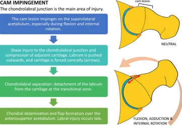
C a m i m p i n g e m e n t c a u s e s s h e a r f o r c e s a t t h e
chondrolabral junction. With hip flexion and internal
ro-tation, the labrum is pushed outward and the cartilage is
compressed and pushed centrally into the joint, initially
resulting in separation of the labrum and cartilage at the
transitional zone (defined as the region between the
la-brum and cartilage), termed chondrolabral separation or
labral detachment [3,
5]. This is followed by adjacent
chondral delamination near the transitional zone [3,
10]
(Fig.
1). The main focus of injury, therefore, is the
chondrolabral junction; intrasubstance labral tears tend
to occur later in the disease process. Due to the
predom-inantly peripheral (outer) location of labral tears in cam
impingement and blood supply from the capsule, healing
rates are more favourable than for labral tears in pincer
impingement [4].
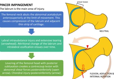
Pincer-type lesions are related to overcoverage of the
fem-oral head. These are most commonly due to focal superior
acetabular retroversion, but global acetabular overcoverage
may also cause pincer-type impingement. Repetitive contact
between the prominent acetabular rim and femoral neck
dur-ing flexion and internal rotation results in labral compression.
The labrum is the first structure to sustain injury, with labral
degeneration and intrasubstance tears most commonly found
anterosuperiorly [3,
5] (Fig.
2). Only a thin strip of cartilage
adjacent to the labrum is compressed, resulting in limited
chondral wear, compared to the more extensive chondral
de-lamination or deep chondral wear seen with cam impingement
[5]. Levering of the femoral head results in contrecoup injury
during hip flexion and internal rotation, with chondral wear
over the posteroinferior acetabulum [3].
Fig. 2 Diagram outlining the mechanisms of chondral and labral injury in pincer
impingement. The labrum is the main focus of damage. Chondral injury is initially limited to a relatively small strip of cartilage at the transition zone
Table 1 Etiology of cam and
pincer lesions Cam lesions Pincer lesions
Primary Idiopathic Idiopathic
Secondary •Developmental •Developmental
•Coxa vara •Coxa profunda
•Perthes disease •Protrusio acetabuli
•SCFE •Traumatic
•Traumatic •Post-traumatic deformity of acetabulum •Malunited femoral neck fracture •Iatrogenic
•Iatrogenic •Overcorrection of hip dysplasia •Femoral head osteotomy
Cam and pincer lesions are usually idiopathic in etiology,
although they can be secondary to developmental
abnormali-ties. Perthes disease and slipped capital femoral epiphysis can
lead to the development of cam lesions (Table
1). Prior trauma
or hip surgery can result in either cam or pincer morphology.
Occurrence of pincer FAI has been reported after iatrogenic
overcorrection of acetabular dysplasia with periacetabular
osteotomy [23].
There are gender-specific differences in FAI, with large
cam lesions more prevalent in young males and pincer lesions
more common in middle-aged females [5,
24,
25]. The
asso-ciation between hip dysplasia and FAI is well known, with 75
% of patients with hip dysplasia having an aspherical femoral
head or insufficient offset of the femoral head
–
neck junction,
predisposing to symptomatic impingement [26].
MR imaging technique
Variable accuracy has been reported for non-contrast MRI
for the detection of labral tears, depending on the
mag-netic field strength, slice thickness, and field of view [27].
Three studies using high-resolution MRI at 1.5T and a
small field of view demonstrated sensitivity of 77
–
97 %
for the detection of labral tears compared with a surgical
reference standard [28
–
30].
Direct MR arthrography involves intra-articular injection
of diluted gadolinium contrast into the hip joint, and has the
benefit of joint distention and improved contrast-to-noise
ra-tio. A recent review of 19 studies evaluating MR arthrography
demonstrated sensitivity of 69
–
100 % for the detection of
labral tears with a surgical reference standard, with 12 of these
studies showing sensitivity greater than 90 % [31].
A meta-analysis found that MR arthrography had
higher sensitivity but lower specificity than conventional
MRI for the detection of labral tears [27]. For
evalua-tion of chondral lesions, MRI has shown higher
diag-nostic accuracy, with another meta-analysis
demonstrat-ing pooled sensitivity and specificity of 59 % and 94 %
for MRI, compared to 62 % and 86 % for MR
arthrography [32]. MR arthrography has the
disadvan-tage of being more time consuming and invasive, with
some patients experiencing post-procedural pain [33,
34]. The choice between conventional MRI and MRA
will vary by institution and will depend on the ability to
optimize the MRI scanning protocol with each
technique.
At the authors
’
institution, MRI of the hip is
per-formed using a cardiac or small body coil on a 1.5T
or 3.0T scanner. Dedicated non-contrast high-resolution
fast spin-echo sequences of the affected hip are
per-formed in the sagittal, coronal, and axial oblique planes.
The axial oblique plane (Swiss axial) is obtained along
the long axis of the femoral neck (Fig.
3). Larger
field-of-view coronal inversion recovery and axial fast
spin-echo sequences of the pelvis are performed to assess for
associated injuries and other causes of hip pain,
includ-ing tendinopathy and the athletic pubalgia spectrum of
injuries. An axial sequence of the femoral condyles is
also performed to correct for distal femoral rotation in
the calculation of femoral anteversion. A suggested
standard MRI pulse sequence protocol is outlined in
Table
2.
Radial imaging can also be performed with image
slices obtained perpendicular to the hip joint. This allows
evaluation of osseous abnormalities, labral and chondral
lesions, and measurement of alpha angles in different
Fig. 3 Obtaining Swiss axial images and calculating the alpha angle.a
The Swiss axial (axial oblique) images are obtained along the long axis of the femoral neck.bTo calculate the alpha angle, the axial oblique image through the midportion of the femoral neck (red line) is chosen. A circle is drawn over the femoral head cortex (blue circle). A line is drawn through the long axis of the femoral neck at its narrowest point (purple line),
clock-face locations [35]. By convention, 12 o'clock refers
to the superior hip joint, and 3 o'clock refers to the
ante-rior aspect of the hip joint bilaterally.
Preoperative evaluation
Preoperative evaluation of FAI on MRI should include
assessment of osseous morphological abnormalities, labral
lesions, cartilage status, and associated soft tissue injuries.
A checklist for preoperative evaluation of FAI is given in
Table
3.
Osseous morphology
The identification of osseous morphology predisposing to
FAI is important, as surgical treatment of labral tears
without addressing the bony impingement is a common
cause for symptom recurrence [20]. Cam morphology
ap-pears on MRI as insufficient offset between the femoral
head and neck, with a focal osseous bump or protuberance
at the femoral head
–
neck junction, which can be assessed
on axial or coronal images (Fig.
4). This is often
associ-ated with fibrocystic change at the femoral neck
anterosuperiorly from chronic impingement, which can
be seen on MRI as small cysts varying in diameter [36].
Table 2 Suggested MRI pulse sequence protocol at 3T and (1.5T)
3T (1.5T) Coronal STIR wide FOV
Axial PD wide FOV
Sagittal PD Axial oblique PD
Coronal PD
TR/TE (ms) 4000/19 (4000/17)
4800/40 (4000/30)
4000/30 (3500/26)
4000/40 (4000/26)
4000/32 (4000/26) Flip angle 180 (180) 180 (180) 180 (180) 180 (180) 180 (180)
ETL 12 (7) 12 (7) 12 (8) 12 (9) 12 (7)
RBW (kHz) 50 (32) 50 (32) 50 (32) 50 (32) 50 (32)
NEX 2 (2) 1 (2) 2 (3) 2 (3) 2 (3)
Matrix 288 × 288 (256 × 192)
512 × 256 (512 × 256)
512 × 384 (512 × 384)
512 × 320 (512 × 256)
512 × 384 (512 × 384) FOV (cm) 38 (34) 36 (36) 19 (20) 19 (20) 19 (20) Slice thickness
(mm)/gap
5.5/0 (5.0/0) 5.0/0 (5.0/0) 2.5/0 (2.5–2.8/0) 3.0/0 (3.0/0) 3.0 (3.0/0)
STIRshort tau inversion recovery,PDproton density,TRrepetition time,TEecho time,ETLecho train length,
RBWreceiver bandwidth,NEXnumber of excitations,FOVfield of view
Table 3 MRI reporting checklist
for preoperative FAI evaluation Preoperative FAI reporting checklist
Osseous morphological abnormalities Cam lesion Pincer lesion
- Superior acetabular retroversion - True acetabular retroversion
- Global acetabular overcoverage: coxa profunda or protrusio Mixed cam/pincer deformity
Labral lesions Chondrolabral separation Labral tear or degeneration Intralabral ossification Cartilage Chondral delamination
Chondral loss
Other signs of osteoarthritis: subchondral cysts, sclerosis, osteophytes Measurements Alpha angle
Femoral anteversion
Associated injuries Pubic symphysis stress reaction Adductor aponeurosis injury
Pincer impingement can occur due to focal or global
acetabular overcoverage. Focal acetabular overcoverage
o r c r a n i a l a c e t a b u l a r r e t r o v e r s i o n i s d u e t o a n
anterosuperior rim lesion, resulting in anterior acetabular
overcoverage only at the superior acetabulum. The
“
cross-over
”
sign may be seen on radiographs, where the anterior
acetabular rim projects lateral to the posterior acetabular
rim. However, a false crossover sign can appear,
depend-ing on the degree of pelvic tilt and inclination, and the
tilting of the x-ray tube. Despite well-positioned
radio-graphs, the
“
crossover
”
sign can overestimate the
inci-dence of cranial acetabular retroversion [37]. On MRI,
the degree of acetabular retroversion is determined by
drawing a line between the lateral margins of the
acetab-ulum on the cranial-most axial slices (Fig.
5).
Global acetabular overcoverage can occur due to coxa
profunda, protrusio, or true acetabular retroversion, which
can result in pincer impingement. True acetabular
retro-version refers to posterior wall undercoverage both
supe-riorly and infesupe-riorly, with relative anterior acetabular
overcoverage. Coxa profunda is characterized by a deep
acetabulum with circumferential medial joint space loss.
On radiographs, the wall of the acetabulum projects
medial to the ilioischial line [38]. Acetabular depth can be
calculated by measuring the distance between a line drawn
through the center of the femoral head and a line joining the
anterior and posterior acetabular rims [39]. Protrusio acetabuli
is diagnosed when the femoral head projects medial to the
ilioischial line on an AP pelvis radiograph [40].
Labral tears and chondrolabral separation
The normal acetabular labrum appears as a low-signal triangle
with smooth margins [41]. A common pitfall in MRI
evalua-tion of the labrum is the presence of sublabral recesses. These
are normal variants that typically do not extend the full
thick-ness of the labrum, and are more often seen anteroinferiorly or
posteroinferiorly [42,
43]. A hyperintense cleft in the
anterosuperior labrum should be viewed with high suspicion
for a labral tear [44]. On MRI, chondrolabral separation
appears as a fluid signal intensity cleft undermining the
labrum at the chondrolabral junction, with or without labral
detachment (Figs.
6,
7,
and
8) [15,
41].
The labral degeneration of pincer impingement appears
as increased signal intensity in the labrum on
fluid-Fig. 4 Oblique axial (a) and coronal (b) proton density (PD)-weighted MRI images in a 30-year-old man with a cam deformity. There is an osseous protuberance (arrows) at the femoral head–neck junction
anterolaterally, with loss of offset of the femoral head–neck junction. The physeal scar extends lateral to the circular region of the femoral head (dashed circle) on the coronal image
Fig. 5 An 18-year-old girl with pincer deformity. (a) On an axial oblique PD-weighted MRI image superiorly, the anterior rim of the acetabulum (arrow) is located lateral to the posterior rim (arrowhead), indicative of superior acetabular retroversion (blue line). Superior acetabular
sensitive MRI sequences, and it may be associated with
hypertrophy of the labrum [41]. Labral tears are
manifest-ed as linear fluid signal intensity extending from the labral
surface into the substance of the labrum (Fig.
8). As the
disease progresses, the labrum gradually becomes thinner
and increasingly attenuated until it is finally no longer
visible [4]. Associated paralabral cysts are frequently
demonstrated on MRI [45].
Chronic microtrauma and degeneration of the labrum
results in osseous metaplasia and intralabral ossification,
which is more commonly seen in pincer impingement [3].
I n t r a l a b r a l o s s i f i c a t i o n c a n r e s u l t i n a c e t a b u l a r
overcoverage and cause further impingement [4]. On
MRI, intralabral ossification appears as small foci of
sig-nal intensity similar to bone marrow (Fig.
8) [46].
Chondral wear and other signs of osteoarthritis
In cam impingement, chondral wear or delamination
most commonly occurs anterosuperiorly, maximally at
1 o'clock [3,
39] (Fig.
6), appearing on the acetabular
side initially, with involvement of the femoral side in
more advanced cases. Pincer lesions result in greater
circumferential chondral wear, which is most marked
superiorly at 11 to 1 o'clock [3], or posteroinferiorly
due to contrecoup forces [39]. In advanced
osteoarthri-tis, MRI may demonstrate subchondral sclerosis,
subchondral cyst formation, marginal osteophytes, or
bone-on-bone contact (Fig.
9).
Advanced cartilage imaging techniques
Assessment of cartilage on standard MRI pulse sequences
is challenging due to the relatively thin layer of cartilage
in the hip. Additionally, the curved surfaces of the femoral
head and acetabulum result in partial volume effects,
making evaluation difficult. Quantitative MRI of cartilage
can detect early changes associated with chondral
degen-eration, and can thus provide additional information for
consideration of arthroscopy or for longitudinal follow-up
of patients following FAI surgery. Various quantitative
MRI techniques are available, including delayed
gadolinium-enhanced MRI of cartilage (dGEMRIC), and
T1rho (T1
ρ), and T2 mapping.
Fig. 6 A 26-year-old man with FAI.aCoronal PD-weighted image shows a cam lesion at the femoral head–neck junction (open arrow). There is chondrolabral separation, with a cleft between the labrum and cartilage (arrow). A paralabral cyst is also seen (black arrowhead).bSagittal PD-weighted image shows chondral delamination near the transition zone anterosuperiorly (white arrowhead)
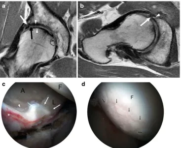
Fig. 7 A 46-year-old woman with combined cam and pincer impingement.aSagittal PD-weighted MRI demonstrates separation at the chondrolabral junction (black arrow).bArthroscopic photo in the same patient demonstrates acetabular cartilage (A), labrum (L), and the
dGEMRIC involves intravenous administration of
gad-olinium contrast to assesses T1 relaxation of cartilage,
requiring an injection followed by brief exercise and then
scanning after 60
–
90 minutes. The distribution of
gado-linium in the cartilage is inversely proportional to its
gly-cosaminoglycan content. Areas of chondral degeneration
Fig. 8 A 48-year-old man with combined cam and pincer impingement.
aCoronal PD-weighted MRI demonstrates labral degeneration with intralabral ossification (white arrow). Separation is seen at the chondrolabral junction (black arrow). There is moderate to high-grade chondral wear over the superior femoral head and anterosuperior dome (arrowheads).bAxial oblique PD-weighted image shows a non-displaced tear of the anterior and anterosuperior labrum (arrow), with
an associated intralabral cyst (arrowhead). c Arthroscopy image in the same patient demonstrates the labrum (L), acetabular (A), and femoral head (F) articular surfaces. An intrasubstance labral tear between 12 and 4 o'clock (white arrows), impaction erythema (asterisk), and chondral delamination at the transition zone (arrowhead) are seen.dThere is moderate chondral wear (black arrows) over the femoral head, and normal cartilage is seen adjacent to this area (F)
Fig. 9 A 36-year-old man with cam-type FAI and severe osteoarthritis. Coronal (a) and sagittal (b) PD-weighted MR images of the right hip demonstrate chronic degeneration of the labrum (arrowhead). Chondral loss with extensive bone-on-bone contact is seen over the superior
with decreased glycosaminoglycan will have increased
gadolinium concentration, and therefore reduced T1
relax-ation times. Studies have shown that subjects with FAI
have significantly lower T1 relaxation values in hip
carti-lage compared to asymptomatic individuals [47].
T1
ρrelaxation times have been correlated with
pro-teoglycan content, and increased values of T1
ρreflect
proteoglycan loss [48]. T2 mapping reveals changes in
collagen orientation. Disorganization of collagen occurs
with cartilage degeneration, resulting in prolonged T2
relaxation times [49]. Patients with FAI have
significant-ly higher T1rho and T2 relaxation times [50],
corre-sponding to changes in proteoglycan and collagen
con-tent and structure (Fig.
10).
Measurements
Alpha angles
Alpha angles are measured utilizing oblique axial images,
obtained parallel to the long axis of the femoral neck,
through the midportion of the femoral neck (Fig.
3). An
alpha angle of >55° is considered a risk factor for FAI
[51], although there is considerable overlap between the
r an g e o f a l p h a v al u es i n s y m p t om a t i c a n d no n
-Fig. 10 Sagittal PD-weighted MRI of the hip in a 30-year-old woman demonstrates mild chondral hyperintensity over the anterosuperior acetabular dome (arrowhead), with corresponding prolongation of relaxation times on T2 mapping and T1rho images (white arrows)
Fig. 11 Calculation of femoral version corrected for distal femoral rotation. On the straight axial image(s) of the hip/pelvis covering the femoral head and neck, a line is drawn between the center of the femoral head and center of the femoral neck at its narrowest point to calculate the uncorrected femoral anteversion angle (A; A is a negative value when the femur is retroverted). To correct for distal femoral rotation, another line is drawn along the posterior border of the femoral condyles to calculate the angle (B; B is a negative value when the knee is internally rotated). Femoral version = A−B. A positive value indicates femoral anteversion. A negative value indicates femoral retroversion. Normal femoral anteversion is approximately 12–13°
symptomatic subjects, the measured alpha angle should be
considered in the clinical context of the patient
’
s
symp-toms [52].
Femoral anteversion
Femoral anteversion (antetorsion) is the angle between the
femoral neck and the femoral condyles. This can be
cal-culated by measuring angles on the straight or oblique
axial images of the femoral neck using a correction factor,
taking into account the relative anteversion or retroversion
of the femoral condyles (Fig.
11) [53]. Normal femoral
anteversion is approximately 12
–
13° [54,
55]. Femoral
retroversion or a relative decrease in femoral anteversion
exacerbates the effect of a cam or pincer lesion, as
im-pingement may occur with only minimal internal rotation
and hip flexion. Increased anteversion results in reduced
external rotation, with the potential for impaction of the
femur on the posterior acetabulum.
Compensatory injuries
Biomechanical alterations in the hip joint can result in
abnormal forces across the pelvis and strain of other
muscles and tendons [20] (Fig.
12). The athletic
pubalgia spectrum of injuries often coexists with FAI.
Consequently, overlapping symptomatology can make
the diagnosis challenging for the clinician [56,
57].
Thus, on MRI, readers should also evaluate for features
of athletic pubalgia, such as tears of the rectus
abdominis-adductor aponeurosis, bone marrow edema
adjacent to the pubic symphysis, or a cleft of signal
hyperintensity extending from the interpubic disk along
the inferomedial margin of the pubis (Fig.
13) [58]. The
iliopsoas, hip abductor, and hamstring tendons should
also be evaluated for concomitant tendinopathy and/or
tears. The presence or absence of bursitis should be
noted. Sacroiliac and lumbar spine pathology should
al-so be excluded.
Differential diagnoses
MRI can be used for the differential diagnosis of hip
pain, including stress fractures and avascular necrosis.
Other extra-articular forms of hip impingement can
mimic FAI, including subspinous, ischiofemoral, and
iliopsoas impingement.
Subspinous impingement occurs where a prominent
ante-rior infeante-rior iliac spine (AIIS) impinges against the femoral
neck during flexion [59]. Enlargement or overhang of the
AIIS can be developmental or due to prior avulsion or pelvic
osteotomy, and can be successfully treated with arthroscopic
AIIS decompression [60].
Ischiofemoral impingement involves impingement of the
ischial tuberosity and the lesser trochanter, resulting in
com-pression of the intervening quadratus femoris muscle. This is
seen on MRI as narrowing of the space between the lesser
trochanter and ischial tuberosity, with edema, tear, or fatty
atrophy of the quadratus femoris muscle [61].
Conclusion
Knowledge of the mechanisms of injury in FAI can
facilitate an understanding of patterns of chondrolabral
injury seen on MRI. As advanced chondral wear is a
relative contraindication to hip preservation surgery,
MRI assessment of the integrity of hip joint cartilage
can assist in the selection of patients most likely to
benefit from surgery. A description of the underlying
osseous morphology and the presence of compensatory
soft tissue injuries should also be included in the
pre-operative evaluation of FAI.
Compliance with ethical standards
Disclosures H.G. Potter receives institutional research support from GE Healthcare.
Funding None
Open AccessThis article is distributed under the terms of the Creative C o m m o n s A t t r i b u t i o n 4 . 0 I n t e r n a t i o n a l L i c e n s e ( h t t p : / / creativecommons.org/licenses/by/4.0/), which permits unrestricted use, distribution, and reproduction in any medium, provided you give appro-priate credit to the original author(s) and the source, provide a link to the Creative Commons license, and indicate if changes were made.
References
1. Nawabi DH, Bedi A, Tibor LM, Magennis E, Kelly BT (2014) The demographic characteristics of high-level and recreational athletes undergoing hip arthroscopy for femoroacetabular impingement: a sports-specific analysis. Arthroscopy: J Arthroscopic Related Surg: Off Publ Arthroscopy Assoc North Am Int Arthroscopy Assoc 30(3):398–405
2. Guevara CJ, Pietrobon R, Carothers JT, Olson SA, Vail TP (2006) Comprehensive morphologic evaluation of the hip in patients with symptomatic labral tear. Clin Orthop Relat Res 453:277–285 3. Beck M, Kalhor M, Leunig M, Ganz R (2005) Hip morphology
influences the pattern of damage to the acetabular cartilage: femoroacetabular impingement as a cause of early osteoarthritis of the hip. J Bone Joint Surg British Vol 87(7):1012–1018
4. Ganz R, Leunig M, Leunig-Ganz K, Harris WH (2008) The etiol-ogy of osteoarthritis of the hip: an integrated mechanical concept. Clin Orthop Relat Res 466(2):264–272
5. Ganz R, Parvizi J, Beck M, Leunig M, Notzli H, Siebenrock KA (2003) Femoroacetabular impingement: a cause for osteoarthritis of the hip. Clin Orthop Relat Res 417:112–120
6. Bedi A, Kelly BT, Khanduja V (2013) Arthroscopic hip preserva-tion surgery: current concepts and perspective. Bone Joint J 95-b(1):10–19
7. Ng VY, Arora N, Best TM, Pan X, Ellis TJ (2010) Efficacy of surgery for femoroacetabular impingement: a systematic review. Am J Sports Med 38(11):2337–2345
8. Malviya A, Stafford GH, Villar RN (2012) Impact of arthroscopy of the hip for femoroacetabular impingement on quality of life at a mean follow-up of 3.2 years. J Bone Joint Surg British Vol 94(4): 466–470
9. Freeman CR, Azzam MG, Leunig M (2014) Hip preservation sur-gery: surgical care for femoroacetabular impingement and the pos-sibility of preventing hip osteoarthritis. J Hip Preserv Surg 1(2):46– 55
10. Beck M, Leunig M, Parvizi J, Boutier V, Wyss D, Ganz R (2004) Anterior femoroacetabular impingement: part II. Midterm results of surgical treatment. Clin Orthop Relat Res 418:67–73
11. Bedi A, Chen N, Robertson W, Kelly BT (2008) The management of labral tears and femoroacetabular impingement of the hip in the young, active patient. Arthroscopy: J Arthroscopic Related Surg: Off Publ Arthroscopy Assoc North Am Int Arthroscopy Assoc 24(10):1135–1145
12. Meftah M, Rodriguez JA, Panagopoulos G, Alexiades MM (2011) Long-term results of arthroscopic labral debridement: predictors of outcomes. Orthopedics 34(10):e588–592
13. Frank JM, Harris JD, Erickson BJ et al (2015) Prevalence of F e m o r o a c e t a b u l a r I m p i n g e m e n t I m a g i n g F i n d i n g s i n Asymptomatic Volunteers: A Systematic Review. Arthroscopy: J Arthroscopic Related Surg: Off Publ Arthroscopy Assoc N Am Int Arthroscopy Assoc. doi:10.1016/j.arthro.2014.11.042
14. Beaule PE, Allen DJ, Clohisy JC, Schoenecker P, Leunig M (2009) The young adult with hip impingement: deciding on the optimal intervention. J bone Joint Surg Am Vol 91(1): 210–221
15. Bredella MA, Ulbrich EJ, Stoller DW, Anderson SE (2013) Femoroacetabular impingement. Magn Reson Imaging Clin N Am 21(1):45–64
1 6 . Ta n n a s t M , S i e b e n r o c k K A , A n d e r s o n S E ( 2 0 0 7 ) Femoroacetabular impingement: radiographic diagnosis–what the radiologist should know. AJR Am J Roentgenol 188(6):1540– 1552
17. Bedi A, Dolan M, Magennis E, Lipman J, Buly R, Kelly BT (2012) Computer-assisted modeling of osseous impingement and resection in femoroacetabular impingement. Arthroscopy: J Arthroscopic Related Surg: Off Publ Arthroscopy Assoc N Am Int Arthroscopy Assoc 28(2):204–210
18. Gold SL, Burge AJ, Potter HG (2012) MRI of hip cartilage: joint morphology, structure, and composition. Clin Orthop Relat Res 470(12):3321–3331
19. Bedi A, Kelly BT (2013) Femoroacetabular impingement. J Bone Joint Surg Am Vol 95(1):82–92
20. Bedi A, Dolan M, Leunig M, Kelly BT (2011) Static and dynamic mechanical causes of hip pain. Arthroscopy: J Arthroscopic Related Surg: Off Publ Arthroscopy Assoc N Am Int Arthroscopy Assoc 27(2):235–251
21. Siebenrock KA, Wahab KH, Werlen S, Kalhor M, Leunig M, Ganz R (2004) Abnormal extension of the femoral head epiphysis as a cause of cam impingement. Clin Orthop Relat Res 418:54–60 22. Byrd JW (2014) Femoroacetabular impingement in athletes: current
concepts. Am J Sports Med 42(3):737–751
23. Myers SR, Eijer H, Ganz R (1999) Anterior femoroacetabular im-pingement after periacetabular osteotomy. Clin Orthopaedics Related Res (363):93-99
24. Domb BG, Philippon MJ, Giordano BD (2013) Arthroscopic capsulotomy, capsular repair, and capsular plication of the hip: relation to atraumatic instability. Arthroscopy: J Arthroscopic Related Surgery: Off Publ Arthroscopy Assoc N Am Int Arthroscopy Assoc 29(1):162–173
25. Hetsroni I, Dela Torre K, Duke G, Lyman S, Kelly BT (2013) Sex differences of hip morphology in young adults with hip pain and labral tears. Arthroscopy: J Arthroscopic Related Surg: Off Publ Arthroscopy Assoc N Am Int Arthroscopy Assoc 29(1):54–63 26. Clohisy JC, Nunley RM, Carlisle JC, Schoenecker PL (2009)
Incidence and characteristics of femoral deformities in the dysplas-tic hip. Clin Orthop Relat Res 467(1):128–134
27. Smith TO, Hilton G, Toms AP, Donell ST, Hing CB (2011) The diagnostic accuracy of acetabular labral tears using magnetic reso-nance imaging and magnetic resoreso-nance arthrography: a meta-anal-ysis. Eur Radiol 21(4):863–874. doi:10.1007/s00330-010-1956-7
28. Sutter R, Zubler V, Hoffmann A et al (2014) Hip MRI: how useful is intraarticular contrast material for evaluating surgically proven lesions of the labrum and articular cartilage? AJR. Am J Roentgenol 202(1):160–169
29. Zlatkin MB, Pevsner D, Sanders TG, Hancock CR, Ceballos CE, Herrera MF (2010) Acetabular labral tears and cartilage lesions of the hip: indirect MR arthrographic correlation with arthroscopy–a preliminary study. AJR Am J Roentgenol 194(3):709–714 30. Mintz DN, Hooper T, Connell D, Buly R, Padgett DE, Potter HG
(2005) Magnetic resonance imaging of the hip: detection of labral and chondral abnormalities using noncontrast imaging. Arthroscopy: J Arthroscopic Related Surg: Off Publ Arthroscopy Assoc N Am Int Arthroscopy Assoc 21(4):385–393
31. Naraghi A, White LM (2015) MRI of Labral and Chondral Lesions of the Hip. Am J Roentgenol 205(3):479–490
33. Giaconi JC, Link TM, Vail TP et al (2011) Morbidity of direct MR arthrography. AJR Am J Roentgenol 196(4):868–874
34. Saupe N, Zanetti M, Pfirrmann CW, Wels T, Schwenke C, Hodler J (2009) Pain and other side effects after MR arthrography: prospec-tive evaluation in 1085 patients. Radiology 250(3):830–838 35. Petchprapa CN, Dunham KS, Lattanzi R, Recht MP (2013)
Demystifying radial imaging of the hip. Radiographics: Rev Publ Radiol Soc N Am, Inc 33(3):E97–e112
36. Leunig M, Beck M, Kalhor M, Kim YJ, Werlen S, Ganz R (2005) Fibrocystic changes at anterosuperior femoral neck: prevalence in hips with femoroacetabular impingement. Radiology 236(1):237– 246
37. Zaltz I, Kelly BT, Hetsroni I, Bedi A (2013) The crossover sign overestimates acetabular retroversion. Clin Orthop Relat Res 471(8):2463–2470
38. Anderson LA, Kapron AL, Aoki SK, Peters CL (2012) Coxa profunda: is the deep acetabulum overcovered? Clin Orthop Relat Res 470(12):3375–3382
39. Pfirrmann CW, Mengiardi B, Dora C, Kalberer F, Zanetti M, Hodler J (2006) Cam and pincer femoroacetabular impingement: charac-teristic MR arthrographic findings in 50 patients. Radiology 240(3): 778–785
40. Leunig M, Nho SJ, Turchetto L, Ganz R (2009) Protrusio acetabuli: new insights and experience with joint preservation. Clin Orthop Relat Res 467(9):2241–2250
41. Kassarjian A, Brisson M, Palmer WE (2007) Femoroacetabular impingement. Eur J Radiol 63(1):29–35
42. Studler U, Kalberer F, Leunig M et al (2008) MR arthrography of the hip: differentiation between an anterior sublabral recess as a normal variant and a labral tear. Radiology 249(3):947–954 43. Dinauer PA, Murphy KP, Carroll JF (2004) Sublabral sulcus at the
posteroinferior acetabulum: a potential pitfall in MR arthrography diagnosis of acetabular labral tears. AJR Am J Roentgenol 183(6): 1745–1753
44. DuBois DF, Omar IM (2010) MR imaging of the hip: normal ana-tomic variants and imaging pitfalls. Magn Reson Imaging Clin N Am 18(4):663–674
45. Kassarjian A, Yoon LS, Belzile E, Connolly SA, Millis MB, Palmer WE (2005) Triad of MR arthrographic findings in patients with cam-type femoroacetabular impingement. Radiology 236(2):588– 592
46. Werlen S, Leunig M, Ganz R Magnetic Resonance Arthrography of the Hip in Femoroacetabular Impingement: Technique and Findings. Operativ Tech Orthopaedics, 15(3):191-203
47. Bittersohl B, Steppacher S, Haamberg T et al (2009) Cartilage dam-age in femoroacetabular impingement (FAI): preliminary results on comparison of standard diagnostic vs delayed gadolinium-enhanced magnetic resonance imaging of cartilage (dGEMRIC). Osteoarthritis Cartil / OARS, Osteoarthritis Res Soc 17(10):1297– 1306
48. Wheaton AJ, Casey FL, Gougoutas AJ et al (2004) Correlation of T1rho with fixed charge density in cartilage. J Magnet Resonance Imaging: JMRI 20(3):519–525
49. Riley GM, McWalter EJ, Stevens KJ, Safran MR, Lattanzi R, Gold GE (2015) MRI of the Hip for the evaluation of femoroacetabular impingement; past, present, and future. J Magnet Resonance Imaging: JMRI 41(3):558–572
50. Subburaj K, Valentinitsch A, Dillon AB et al (2013) Regional var-iations in MR relaxation of hip joint cartilage in subjects with and without femoralacetabular impingement. Magn Reson Imaging 31(7):1129–1136
51. Notzli HP, Wyss TF, Stoecklin CH, Schmid MR, Treiber K, Hodler J (2002) The contour of the femoral head-neck junction as a pre-dictor for the risk of anterior impingement. J Bone Joint Surg British Vol 84(4):556–560
52. Sutter R, Dietrich TJ, Zingg PO, Pfirrmann CW (2012) How useful is the alpha angle for discriminating between symptomatic patients with cam-type femoroacetabular impingement and asymptomatic volunteers? Radiology 264(2):514–521. doi:10.1148/radiol. 12112479
53. Sutter R, Dietrich TJ, Zingg PO, Pfirrmann CWA (2015) Assessment of Femoral Antetorsion With MRI: Comparison of Oblique Measurements to Standard Transverse Measurements. Am J Roentgenol 205(1):130–135. doi:10.2214/AJR.14.13617
54. Sutter R, Dietrich TJ, Zingg PO, Pfirrmann CW (2012) Femoral antetorsion: comparing asymptomatic volunteers and patients with femoroacetabular impingement. Radiology 263(2):475–483 55. Prasad R, Vettivel S, Isaac B, Jeyaseelan L, Chandi G (1996) Angle
of torsion of the femur and its correlates. Clin Anatomy (New York, NY) 9(2):109–117
56. Hammoud S, Bedi A, Magennis E, Meyers WC, Kelly BT (2012) High incidence of athletic pubalgia symptoms in professional ath-letes with symptomatic femoroacetabular impingement. Arthroscopy: J Arthroscopic Related Surg: Off Publ Arthroscopy Assoc N Am Int Arthroscopy Assoc 28(10):1388–1395
57. Economopoulos KJ, Milewski MD, Hanks JB, Hart JM, Diduch DR (2014) Radiographic evidence of femoroacetabular impinge-ment in athletes with athletic pubalgia. Sports Health 6(2):171–177 58. Omar IM, Zoga AC, Kavanagh EC et al (2008) Athletic pubalgia and "sports hernia": optimal MR imaging technique and findings. Radiographics : Rev Publ Radiol Soc N Am, Inc 28(5):1415–1438 59. Larson CM, Kelly BT, Stone RM (2011) Making a case for anterior inferior iliac spine/subspine hip impingement: three representative case reports and proposed concept. Arthroscopy: J Arthroscopic Related Surg: Off Publ Arthroscopy Assoc N Am Int Arthroscopy Assoc 27(12):1732–1737
60. Hetsroni I, Larson CM, Dela Torre K, Zbeda RM, Magennis E, Kelly BT (2012) Anterior inferior iliac spine deformity as an extra-articular source for hip impingement: a series of 10 patients treated with arthroscopic decompression. Arthroscopy: J Arthroscopic Related Surg: Off Publ Arthroscopy Assoc N Am Int Arthroscopy Assoc 28(11):1644–1653