233
© 2018 by the Serbian Biological Society
Therapeutic effects of polysaccharides extracted from
Porphyra yezoensis
in rats with cerebral ischemia/reperfusion injury
Changjiang Sun1, Feng Wu2, Dandan Chen1 and Jianbin Ge1,*
1Department of Pharmacy, The Second People’s Hospital of Nantong, Nantong 226002, Jiangsu, China
2Department of Pharmacology, Nantong University School of Medicine, Nantong 226001, Jiangsu, China
*Corresponding author: [email protected]
Received: June 21, 2017; Revised: October 8, 2017; Accepted: October 8, 2017; Published online: October 16, 2017
Abstract: The polysaccharides of Porphyra yezoensis (PPY), porphyrans, are recognized as the major active components as they have several biological properties. This study was performed to assess the effect of PPY administration against cerebral ischemia-reperfusion injury (IRI). Thirty-two adult Sprague-Dawley (SD) rats were chosen and divided into 4 groups as follows: group I – rats received only saline; group II – subjected to middle cerebral artery occlusion (MCAO) for 60 min followed by 24 h of reperfusion (IRI-induced); group III – pretreated with PPY (100 mg/kg) for 7 days, followed by IRI induction; group IV – treated with PPY (100 mg/kg) for 7 days without IRI induction. All the data were analyzed by Dunnett’s (multiple
com-parisons) test using SPSS software. Pretreatment with PPY significantly (p<0.01) lowered the neurological deficit and cerebral
infarct volume in comparison with IRI-induced rats. A pronounced (p<0.01) increase in the levels of antioxidant components
(superoxide dismutase (SOD), catalase (CAT), reduced glutathione (GSH)) was observed in PPY-supplemented rats. Also, the proinflammatory markers: interleukin-1β (IL-1β), interleukin-6 (IL-6), tumor necrosis factor alpha (TNF-α) and the nuclear
factor-kappa B protein, subunit p65 (NF-κB p65) were substantially (p<0.01) suppressed in PPY-administered rats. Moreover,
the protein levels of TNF-α and NF-κB p65 were considerably (p<0.01) downregulated upon pretreatment with PPY. Our
data suggest that PPY could exhibit neuroprotective activity by attenuating oxidative stress and the inflammatory response.
Key words: Porphyra yezoensis; neurological deficit; cerebral infarct; oxidative stress
How to cite this article: Sun C, Wu F, Chen D, Ge J. Therapeutic effects of polysaccharides extracted from Porphyra yezoensis in rats with cerebral ischemia/ reperfusion injury. Arch Biol Sci. 2018;70(2):233-9.
INTRODUCTION
Ischemic stroke accounts for almost all strokes (88%) and is caused by the occlusion of brain blood vessels, especially the middle cerebral artery, which results in brain damage or deformities that are associated with high mortality and morbidity [1,2]. Normally, after an ischemic stroke, the blood flow must be restored as quickly as possible (reperfusion) to enable the survival of brain cells. However, reperfusion prob-ably alters the biochemical and molecular signaling pathways and thereby worsens the brain damage process by increasing oxidative stress, inflammation, apoptosis, edema and hemorrhage [3,4]. The best treatment strategy for stroke is with recombinant tis-sue-plasminogen activator (rtPA) to resolve the clot (thrombolysis), but the usage of this method is lim-ited (short-term), and hence there is a high demand for an effective drug for abolishing stroke [5].
Multiple lines of evidence have shown that in-flammation and oxidative stress (imbalance between antioxidants and oxidants) are the major pathophys-iological events that contribute to ischemic stroke or brain injury [1,4]. The inflammatory cascade is trig-gered during the early phase of IR by the excessive production of reactive oxygen species (ROS) via mi-croglial activation and neutrophil infiltration. This subsequently upregulates the production of proin-flammatory cytokines such as TNF-α, IL-1β, IL-6, and thus damages cerebrovascular endothelial cells by activating the apoptotic cascade [5,6]. Therefore, to combat ischemic brain stroke or IR brain injury, a drug with antiinflammatory activity would help in alleviating the different IRI-induced disorders.
Porphyra yezoensis is, a popular sea weed (red
marine algae), belongs to the Bangiaceae family. P.
and Japan [7].Itishighly consumed due to its special flavor and because it possesses several biological func-tions, being rich in proteins and carbohydrates [8,9]. Lately, the polysaccharides of P. yezoensis (PPY), such as the porphyrans, are drawing more attention and are recognized as one of the major active components with several pharmaceutical properties. Several stud-ies have reported that PPY exhibits antioxidant, an-tiinflammatory, antitumor and immunomodulatory activities [2,10]. It also shows hepatoprotective and cardioprotective activities [11,12]. Furthermore, vari-ous polysaccharides from plants/marine algae have been shown to exhibit neuroprotective activity in a cerebral IR model [13,14]. However, the neuroprotec-tive effect of PPY against IRI has not been explored so far. Hence, this pilot study was conducted to check the therapeutic efficacy of PPY in a cerebral ischemia/ reperfusion model by determining the neural deficits, cerebral infarct volume, proinflammatory cytokines, as well as protein levels of inflammatory markers in SD male rats.
MATERIALS AND METHODS
Chemicals and reagents
Bromophenol blue, diethyl ether, hydrogen peroxide, chloral hydrate, paraformaldehyde, phosphate buff-ered saline and pentobarbital sodium were bought from Sigma-Aldrich (MO, USA). 2,3,5-triphe-nyltetrazolium chloride (TTC) was purchased from Lingjin Co., Ltd, Shanghai, China. All of the other reagents and chemicals used are of analytical grade.
PPY preparation
P. yezoensis were obtained from the local market
(Jiangsu) and authenticated by the botanist, Dr. Le Su from the Nantong University School of Medicine and a voucher specimen (OTM-1PY) was deposited in the herbarium of the People’s Hospital of Nantong University. The PPY was extracted/prepared accord-ing to the methods of Zhou et al. [10] with slight modification.
Experimental animals
A total of 32 healthy male SD rats weighing 240-260 g were procured from the animal center of Nantong University. Rats were accommodated in a metabolic steel cage and maintained at 22°C (with 74% humid-ity) on a 12 h dark/light cycle. All rats were allowed free access to water and food (ad libitum). All the protocols used in the present study were approved by the Ethical Board Committee members of the second People’s Hospital of Nantong (SPHN-2015/12) and complied with the guidelines of the United States National Institute of Health (Guide for Handling and Care of Laboratory Animals). This animal study was conducted at the second People’s Hospital of Nan-tong from January to March 2016.
IR insult (IRI) protocol
A focal IR insult or MCAO was performed using the previously reported Longa method [15]. Briefly, the rats were anesthetized with an intraperitoneal (i.p.) administration of chloral hydrate (300 mg/kg), placed in the supine position and subjected to a midline neck incision to expose the right common carotid artery (CCA), external carotid artery (ECA) and internal ca-rotid artery (ICA). Both CCA and ECA were ligated (occluded) using a 30-mm nylon monofilament coat-ed with a roundcoat-ed silicon tip (Beijing Sunbio Biotech Co., Ltd. Beijing, China) by going through the ICA (18±2 mm) and middle cerebral artery to achieve MCAO. After 60 min of MCAO, the nylon monofila-ment was gently removed to establish reperfusion for 24 h. Sham-operated control rats underwent a simi-lar procedure without ligation and occlusion/reper-fusion. Throughout the surgical procedure, the body temperature was maintained at 37°C using a heating lamp. The wound was then sutured and the animals were returned to their respective cages.
Animal grouping
the above section, and these rats served as the IRI group. Group III rats were pretreated with 100 mg/ kg of PPY (i.p.) for 7 days, followed by IRI induction, and served as the PPY+IRI group; group IV rats were treated with 100 mg/kg of PPY for 7 days without IRI induction and served as the PPY group.
Sample collection and processing
After 24 h of reperfusion and neurological examina-tion, all the rats were euthanized by an i.p. pentobar-bital sodium injection and the brains were removed immediately and stored at -80°C. The cerebral cor-tex region was isolated and homogenized using lysis phosphate buffer (10 mM disodium hydrogen phos-phate, 10 mM sodium dihydrogen phosphos-phate, 10 mM Tris-HCl, pH 7.4), and centrifuged at about 2000 x g for 10 min to obtain a supernatant. This supernatant was used for biochemical and molecular analysis.
Neurological deficit score/neural deficit assessment
The neurological deficit score (NDS) was carried out after 24 h of reperfusion by an examiner blind to all experimental groups in accordance with Longa’s Score Scaling System [15]. The scoring ranged from 0 to 4, as follows: 0 denoted rats with no neurological deficit, 1 denoted rats that failed to lift the forepaw completely, 2 denoted rats that circled to the left side, 3 denoted rats that fell to the left side, 4 denoted rats that failed to walk spontaneously and/or had a de-pressed level of consciousness.
Determination of cerebral infarct volume
The cerebral hemispheres were removed carefully and sliced into 2-mm coronal slices via a frontal pole, stained with a 2 % TTC solution at 37°C for 30 min, and rinsed with phosphate buffered saline (sodium chloride/disodium hydrogen phosphate and sodium chloride, pH 7.4) to remove excess stain. The sections were fixed with 10% paraformaldehyde by leaving overnight. The coronal slices were placed in an Im-age Scanner, and the infarct volume was quantified using Image J software (Image J ver. 1.4, MD, USA). The infarct volume was evaluated using the formula given in the Türeyen method [16]:
Infarct Volume=(total infarct area/whole brain section area)×100%.
Measurement of selected antioxidant parameters
The activities of SOD, CAT and GSH in cerebral tis-sue were measured using a commercial kit (Beyo-time, Biotechnology; Jiangsu, China). One unit (U) of SOD activity was defined as the amount of enzyme required to inhibit 50% of superoxide radicals and was measured at 550 nm. One unit (U) of CAT activ-ity was defined as the amount consumed to inhibit H2O2 radical formation and was measured at 405 nm.
Determination of inflammatory markers
Several inflammatory markers, including IL-1β, IL-6 and TNF-α in the cerebral homogenate were meas-ured using a commercial ELISA kit from Thermo Fisher Scientific (MA, USA) according to the sup-plier’s protocol. The NF-κB p65 subunit was assessed in the nuclear fraction of the cerebral homogenate using the nuclear/cytosolic fractionation kit from Bio-Vision (CA, USA) and followed by NF-κB p65 determination with an ELISA kit from Imgenex Cor-poration (CA, USA).
Immunoblot analysis
an-tibodies. Membranes were incubated with anti-goat and anti-mouse secondary antibodies conjugated to horseradish peroxidase (HRP) (1:10000, respectively; Promega, WI, USA) in TBS at room temperature for 1 h. After washing with TBS, the absorbance was quantified using an enhanced chemiluminescent sys-tem (Thermo Fisher Scientific;MA, USA), and the band intensity were quantified using Image J software (Bethesda, MD, USA).
Data analysis
Data are presented as the mean±standard deviation (SD). The difference between each experimental group was analyzed with one-way ANOVA, followed by the Dunnett’s (multiple comparisons) test using SPSS software (Ver 23; International Business Ma-chines, Corp., NY, USA). P<0.05 was considered as significantly different.
RESULTS AND DISCUSSION
The MCAO model is one of the best ischemic stroke models as it mimics almost 80% of the pathologi-cal features of stroke patients [5,17]. Hence, for the present study we choose the MCAO focal IR rat model to test the potential beneficial activity of PPY by assessing the neurological deficit score, in-farct volume and inflammatory markers. Prelimi-nary dose-dependent studies have been conducted (data not shown) and based on these results, for the present study a dose of 100 mg/kg was used.
Neurological deficit score
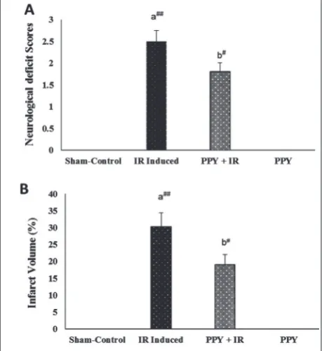
Evaluation of the neurological deficit score (NDS) is a major criterion for assessing the motor activity and the neuroprotective activity of any drug in an ischemic/reperfusion model. The results of the effica-cy of PPY on the neurological deficit score in experi-mental rats is presented in Fig. 1A. The neurological deficit score increased substantially (p<0.01) in IRI-induced rats as compared to sham-operated control rats owing to the MCAO/reperfusion. The above re-sults are in agreement with those of Gong et al. [18], who also reported that during an ischemic condition, overproduction of free radicals elicits neuronal
dam-age and results in impaired motor function, eventu-ally causing a neurological deficit. Supplementation of PPY for 7 days prior to IRI induction significantly diminished (29%; p<0.05) the neurological deficit score compared to MCAO/reperfusion-induced rats, implying that animals pretreated with PPY had a bet-ter functional outcome afbet-ter IRI. We hypothesized that PPY improved motor activity by attenuating ex-cessive free radical generation. Several studies have also indicated that PPY could exert antioxidant and free radical scavenging activities [19,20].
Cerebral infarct volume/area in IRI and PPY+IRI rats
The cerebral infarct volume/area in control and ex-perimental rats were examined after TTC staining (Fig. 1B). The MCAO/reperfusion-induced rats dis-played a concomitant elevation (p<0.01) in the in-farct volume/area. However, pretreatment with 100 mg/kg of PPY considerably lowered (41%; p<0.05) the infarct volume/area as compared to the IRI group. The above data indicated that PPY might
Fig. 1. Effects of PPY on the neurological deficit score (A) and cerebral infarct volume/area (B) in experimental rats. Data were expressed as the mean±standard deviation. Statistical signifi-cance: #p<0.05, ##p<0.01 (a) vs. control group, (b) vs. IR-induced
improve the antioxidant status and thus lower the neural injury, as well as the infarct volume/area. This improved outcome is consistent with the results of Mohibbullah et al. [21], who also demonstrated that
P. yezoensis exhibits a neuroprotective activity via
in-hibition of free radical generation and apoptosis due to the presence of polysaccharides with free radical scavenging activities. Moreover, numerous polysac-charides from different plant sources have also been shown to significantly reduce the neurological defi-cit score and infarct volume by inhibiting oxida-tive stress and inflammatory cytokines [13,14]. No evidence of neural deficits or infarct area in sham-operated control rats were observed as they were not occluded. The obtained results showed that the neuroprotective action of PPY was accomplished by lowering the neurological damage, which led to im-proved motor function.
The effect of PPY on cerebral antioxidants
Several researchers have pointed out that inflamma-tion and oxidative stress are the major pathophysi-ological events that contribute to ischemic stroke or brain injury. Inflammation and oxidative stress are interlinked with each other in most neurological dis-orders [1,4]. During the early phase of IRI induction, excessive free radicals are generated via activated mi-croglial cells [5]. Table 1 shows the effect of PPY on the activities of cerebral antioxidant components (SOD, CAT, GSH) in experimental rats. A considerable de-crease in the enzymatic activities of cerebral antioxi-dant enzymes (SOD by 37% and CAT by 22%; p<0.01) and the concentration of GSH (by 28%; p<0.01) were noted in IRI-induced rats, whereas pretreatment with PPY substantially improved these values (SOD 1.4-, CAT 1.2- and GSH 1.3-fold; p<0.05). Our results are in agreement with the data of Guo et al. [11].
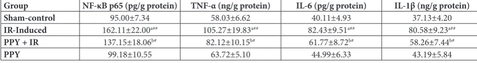
The effect of PPY on cerebral inflammatory markers in IRI rats
The inflammatory response is stimulated during the early phase of IR via excessive production of free radicals by activated microglial cells and neutrophil infiltration. The production of proinflammatory cy-tokines (IL-1β, Il-6, TNF-α) is upregulated, resulting in damage to the cerebrovascular endothelial cells as a result of the activation of the apoptotic cascade [5,6]. Table 2 shows the effect of PPY on the levels of cerebral cytosolic inflammatory markers, IL-1β, IL-6, TNF-α, as well of nuclear factor NF-κB p65 subunit. These markers were significantly increased (p<0.01) in MCAO/reperfusion-induced rats. Pretreatment with PPY for 7 days considerably suppressed the lev-els of these inflammatory markers (TNF-α by 22%, IL-6 by 26%, Il-1β by 27% and NF-κB p65 by 17%; p<0.05) as compared with the IRI-induced group and thereby exerted a protective effect on brain tissue. These results are in agreement with the results of Ryu et al. [22], who demonstrated that the polysaccharides (porphyrans) of P. yezoensis inhibit the secretion and
Table 1. Effect of PPY on the activities of cerebral antioxidants in experimental rats
Group (U/mg SOD
protein)
CAT
(U/mg protein)
GSH
(µg/mg protein)
Sham-control 3.56±0.33 62.60±7.92 8.83±0.99
IR-Induced 2.21±0.28a## 49.12±6.01a## 6.34±0.87a##
PPY+IR 3.14±0.18b# 59.05±8.18b# 8.28±1.07b#
PPY 3.45±0.25 63.32±9.48 8.49±1.16
Data are expressed as the mean±standard deviation (SD). Statistical significance: #p<0.05, ##p<0.01 (a) vs. control group, (b) vs. IR-induced
group. One unit (U) of SOD activity is defined as the amount of enzyme required to inhibit 50% of superoxide radicals at 550 nm. One unit (U) of CAT activity is defined as the amount consumed to inhibit H2O2 radical
at 405 nm. IR – Ischemic reperfusion insult, PPY – polysaccharides of
Porphyra yezoensis.
Table 2. Effect of PPY on the levels of cerebral inflammatory markers in experimental rats.
Group NF-κB p65 (pg/g protein) TNF-α (ng/g protein) IL-6 (pg/g protein) IL-1β (ng/g protein)
Sham-control 95.00±7.34 58.03±6.62 40.11±4.93 37.13±4.20
IR-Induced 162.11±22.00a## 105.27±19.83a## 82.43±9.51a## 80.58±9.23a##
PPY + IR 137.15±18.06b# 82.12±10.15b# 61.77±8.72b# 58.26±7.44b#
PPY 99.18±10.55 63.72±5.10 44.99±6.33 43.19±5.84
Data were expressed as the mean ± standard deviation (SD). Statistical significance: #p<0.05, ##p<0.01 (a) vs. control group, (b) vs.
expression of Tα and IL-1β by suppressing NF-κB. Moreover, pretreatment or preconditioning with
Ginkgo biloba polysaccharides notably abolished the
activation of microglial cells by inhibiting NF-κB and ultimately downregulated the proinflammatory cy-tokines IL-1β, IL-6 and TNF-α [23].
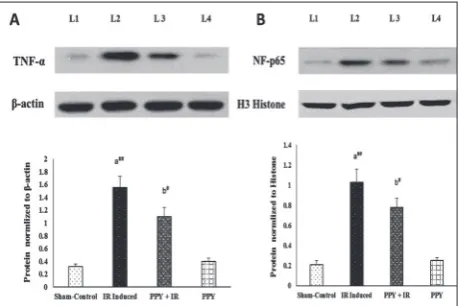
Protein expressions of TNF-α and NF-κB p65 in IRI and PPY+IRI rats
NF-κB is a crucial inflammatory factor that con-trols the expression of various proinflammatory cytokines. To confirm the antiinflammatory ef-fect of PPY, we quantified the protein expression of the nuclear fraction of the NF-κB p65 subunit and TNF-α. Fig. 2. shows the effects of PPY on cerebral protein expressions of cytosolic TNF-α (Fig. 2A) and nuclear NF-κB p65 (Fig. 2B) in experimental rats. The protein levels of cytosolic TNF-α and nuclear NF-κB p65 were significantly upregulated (p<0.01) in the MCAO/reperfusion-induced rats. However, pretreatment with PPY significantly downregulated (p<0.05) the protein expression of cytosolic TNF-α (Fig. 2A) and nuclear NF-κB p65 (Fig. 2B) by 26% and 28%, respectively, in comparison to the MCAO/ reperfusion-induced group. Similarly, Jiang et al. [24] pointed out that pretreatment with polysaccha-rides of P. yezoensis can inhibit the translocation of
NF-kB p65 from the cytosol to the nuclei, resulting in the downregulation of the production of various proinflammatory cytokines, including TNF-α, in the LPS-treated RAW 264 cell line model.
The presented results show that PPY exerts neu-roprotective activity by attenuating the inflamma-tory response. This study has some limitations, such as a lack of standard drugs for comparison, and that we did not evaluate the neutrophil infiltration levels (by myeloperoxidase, MPO) and apoptotic markers to confirm the protective effect of PPY. However, in future studies we aim to overcome these limitations.
CONCLUSION
The results of the present study indicated that PPY could moderately improve neurological motor func-tion by lowering the infarct volume through abolish-ing neuronal damage, oxidative stress and inflam-matory response. This work points to the use of PPY with standard neurological drugs for combating IR-related disorders. Further in-depth studies are needed to explore the mechanism underpinning the neuroprotective activity of PPY.
Acknowledgments: Financial support was provided by the
Nan-tong Health Youth Fund Projects (WQ2014027)
Author contributions: CS and JG conceived and designed the
study. FW and CS were involved in collection of data. JG, CS and DC conducted the experiments. FW and DC assisted in the statistical analysis of the data. JG, CS and FW helped in drafting this manuscript.
Conflict of interest disclosure: There are no competing interests to disclose
REFERENCES
1. Ai J, Wan H, Shu M, Zhou H, Zhao T, Fu W, He Y. Guhong injection protects against focal cerebral ischemia-reperfu-sion injury via anti-inflammatory effects in rats. Arch Pharm Res. 2017;40(5):610-22.
2. Yu X, Zhou C, Yang H, Huang X, Ma H, Qin X Hu J. Effect of ultrasonic treatment on the degradation and inhibition cancer cell lines of polysaccharides from Porphyra yezoensis. CarbohydrPolym. 2015;117:650-6.
3. Khatri R, McKinney AM, Swenson B, Janardhan V. Blood-brain barrier, reperfusion injury, and hemorrhagic transfor-mation in acute ischemic stroke. Neurol. 2012;79(13):S52-S57.
Fig. 2. Effects of PPY on cerebral protein expression of TNF-α
(A) and the NF-κB p65 subunit (B). Data are expressed as the mean±standard deviation. Statistical significance: #p<0.05, ##p<0.01 (a) vs. control group, (b) vs. IR-induced group. L1
4. Radenović L, Selaković V, Anđus PR. Neuroprotectionby MK-801 following cerebral ischemia in Mongolian gerbils. Arch Biol Sci. 2008;60(3):341-6.
5. Chen Y, Li Y, Xu H, Li G, Ma Y, Pang YJ. Morin mitigates oxidative stress, apoptosis and inflammation in cere-bral ischemic rats. Afr J Tradit Complement Altern Med. 2017;14(2):348.
6. Yan J, Zheng M, Zhang D. Chrysophanol liposome precondi-tioning protects against cerebral ischemia-reperfusion injury by inhibiting oxidative stress and apoptosis in mice. Int J Pharmacol. 2014;10(1):55-68.
7. Niwa K, Iida S, Kato A, Kawai H, Kikuchi N, Kobiyama A, Aruga Y. Genetic diversity and introgression in two culti-vated species (Porphyra yezoensis and Porphyra tenera) and closely related wild species of Porphyra (Bangiales, Rho-dophyta). J Phycol. 2009;45(2):493-502.
8. Lee HA, Kim IH, Nam TJ. Bioactive peptide from Pyropia yezoensis and its anti-inflammatory activities. Int J Mol Med. 2015;36(6):1701-6.
9. Zhang LX, Cai CE, Guo TT, Gu JW, Xu HL, Zhou Y, Wang Y, Liu CC,He PM. Anti-cancer effects of polysaccharide and phycocyanin from Porphyra yezoensis. J Mar Sci Tech. 2011;19(4):377-82.
10. Zhou C, Yu X, Zhang Y, He R, Ma H. Ultrasonic degrada-tion, purification and analysis of structure and antioxidant activity of polysaccharide from Porphyra yezoensis udea. Carbohydr. Polym. 2012;87(3):2046-51.
11. Guo TT, Xu HI, Zhang LX, Zhang JP, Guo YF, Gu JW, He PM. In vivo protective effect of Porphyra yezoensis polysac-charide against carbon tetrachloride induced hepatotoxicity in mice. Regul Toxicol Pharmacol. 2007;49(2):101-6. 12. Qian L, Zhou Y, Ma JX. Hypolipidemic effect of the
poly-saccharides from Porphyra yezoensis. Int J Biol Macromol. 2014;68:48-49.
13. Xie Y, Zhang B, Zhang Y. Protective effects of Acanthopanax
polysaccharides on cerebral ischemia–reperfusion injury and its mechanisms. Int J Biol Macromol. 2015;72:946-50. 14. Wang T, Li Y, Wang Y, Zhou R, Ma L, Hao Y, Jin S, Du J,
Zhao C, Sun T. Lycium barbarum polysaccharide prevents focal cerebral ischemic injury by inhibiting neuronal apop-tosis in mice. PloS One. 2014;9(3):e90780.
15. Longa EZ, Weinstein PR, Carlson S, Cummins R. Revers-ible middle cerebral artery occlusion without craniectomy in rats. Stroke. 1989;20(1):84-91.
16. Türeyen K, Vemuganti R, Sailor KA, Dempsey RJ. Infarct volume quantification in mouse focal cerebral ischemia: A comparison of triphenyltetrazolium chloride and cresyl vio-let staining techniques. J Neurosci Met. 2004;139(2):203-7. 17. Wu S, Hao J, Gao W, Zhou B, Man S, Huang L. Neuroprotec-tive effect and chemical composition of traditional Chinese medicines, zha-chong-shi-san-wei pill, against ischemic stroke on middle cerebral ischemia occlusion in rats. Lat Am J Pharm. 2013;32(7):1020-6.
18. Gong G, Xiang L, Yuan L, Hu L, Wu W, Cai L Yin L, Dong H. Protective effect of glycyrrhizin, a direct hmgb1 inhibi-tor, on focal cerebral ischemia/reperfusion-induced inflam-mation, oxidative stress, and apoptosis in rats. PLoS One. 2014;9(3):e89450.
19. Zhou C, Wang Y, Ma H, He R. Effect of ultrasonic degrada-tion on in vitro antioxidant activity of polysaccharides from
Porphyra yezoensis (rhodophyta). Revista De Agaroquimicay Tecnologia De Alimentos. 2008;14(6):479-86.
20. Liu F, Liu J, Gu J, Zhang L, Shen W, Guo T, Liu C, He P. Ex vivo anti-oxidation activity of polysaccharides from red alga
Porphyra yezoensis. Res J Pharmacol. 2007;1(3):61-6. 21. Mohibbullah M, Bhuiyan MMH, Hannan MA, Getachew P,
Hong YK, Choi JS, Choi TS, Moon IS. The edible red alga
Porphyra yezoensis. Cell Mol Neurobiol. 2016;36(5):669-82. 22. Ryu J, Kwon MJ, Nam TJ. Nrf2 and nf-κb signaling pathways
contribute to porphyra-334-mediated inhibition of uva-induced inflammation in skin fibroblasts. Marine Drugs. 2015;13(8):4721-32.
23. YangY, Liu P, Chen L, Liu Z, Zhang H, Wang J, Sun X, Zhong W, Wang N, Tian K. Therapeutic effect of Ginkgo biloba
polysaccharide in rats with focal cerebral ischemia/reperfu-sion (i/r) injury. Carbohydr Polym. 2013;98(2):1383-8. 24. Jiang Z, Hama Y, Yamaguchi K, Oda T. Inhibitory effect of