Research Article
a
August
2017
Computer Science and Software Engineering
ISSN: 2277-128X (Volume-7, Issue-8)
An Automated Brain Tumor Detection in MRI using Firefly
Optimized Segmentation
Prabhjot Kaur, Amardeep Kaur
Punjabi University Regional Centre, Mohali, Punjab, India
DOI: 10.23956/ijarcsse/V7I8/0164
Abstract— In the medical field brain tumor detection is an important application. The existing techniques of segmentation has various limitations .Existing techniques ignored the medical images which have poor quality or low brightness. Segmentation becomes the challenging issue as the image contains non-uniform object texture, cluttered objects, different image content and image noise. New technique of segmentation is proposed by research to detect tumor from MR images using firefly algorithm, then tumor is segmented and its features are extracted from MR image. The main goal of Research to design an algorithm for MRI based brain tumor segmentation using firefly algorithm and to improve the accuracy of the tumor detection. Fireflies produce a reaction in their body which produce light , this chemical reaction is called bioluminescent. By using firefly technique it is possible to detect and localize tumor accurately. For comparative analysis, various parameters are used to demonstrate the superiority of proposed method over the conventional ones.
Keywords— Image segmentation, Brain Tumor, MRI, Enhanced Darwinian Particle Swarm Optimization (EDPSO), Firefly.
I. INTRODUCTION
Medical Image processing is a successful field to doctors which helps them for the analysis of complex disease such as cancer, brain tumor, heart attack, kidney stones, and so on.
A Brain tumor is a complex set of abnormal cells in brain. Brain tumor can be of two types: cancerous and non-cancerous. This is lead to brain damage. The detection of brain tumor is a very challenging task, in which special care is taken while using medical image processing techniques. Various scanning applications for medical image analysis and techniques are available such as MRI, CT scan, and X-rays. Brain tumor is a big cause of death worldwide and related abnormalities constitute for major changes in life. A great research has been done in the last decade for brain tumor in the region of cerebral cancer diagnosis [1][3]. Varieties of image processing techniques are available to be applied on various imaging modalities for tumor detection that will detect certain features of the tumors such as the shape, border and texture [4]. These features will make the detection processes more accurate and easier as there are some standard characteristics of each feature for a specific tumor. Brain tumor segmentation requires the efficient knowledge of pathology and understanding the intensity and shape of MRI image [5]
The paper is classified into different sections: part 1 is the introduction section, part 2 includes motivation and then part 3 is about proposed work then part 4 contains results and discussions and at last, part 5 is conclusion and future enhancement.
II. MOTIVATION
Brain tumor is a collection of abnormal cells in the brain. The unwanted growth of the tumor cells affects the normal functions of the brain, which causes the tumor and that increases chances of death among human beings. The brain tumor diagnosis is a very challenging task because invalid diagnosis will leads to severe trouble. Each brain cell of human body is surrounded together in a complex way and the structure of the brain is internally highly complicated. In most of the research developed countries shows that the number of people passes away in 2010 due to brain tumor, survey says [1]. Early prediction of the brain tumor is very important and motivated task which is usually done by MRI and CT Scans.
Computed tomography (CT) and Magnetic Resonance Imaging (MRI) have revolutionized the study of the brain by allowing researchers to look at the brain non-invasively [2]. These diagnostic imaging techniques have allowed for the first time the noninvasive evaluation of brain structure, allowing researchers to infer causes of abnormal function due to different diseases. Between the two techniques MRI is taken as input due to following reasons:
MRI does not use ionizing radiation, and is thus preferred over CT in children and patients requiring multiple imaging examinations.
MRI has a much greater range of available soft tissue contrast, depicts anatomy in greater detail, and is more sensitive and specific for abnormalities within the brain itself.
MRI scanning can be performed in any imaging plane without having to physically move the patient. MRI contrast agents have a considerably smaller risk of causing potentially dangerous allergic reaction MRI allows the evaluation of structures that may be obscured by artifacts from bone in CT images.
ISSN(E): 2277-128X, ISSN(P): 2277-6451, DOI: 10.23956/ijarcsse/V7I8/0164, pp. 290-295
Due to the low attraction towards the destination the Swarms in EDPSO does-not provide better results. So to overcome this drawback the firefly algorithm is used because of the following reasons:
1) Everywhere fireflies are unisex so that match up firefly will be attracted to be in succession fireflies; 2) The outburst of a firefly is affected or determined by the landscape of the objective function.
Figure1. Steps for image processing
III. PROPOSED WORK
Fireflies are characterized by their flashing light produced by biochemical process bioluminescence. Such flashing light may serve as the primary courtship signals for mating. Besides attracting mating partners, the flashing light may also serve to warn off potential predators. Note that in some firefly species some adults are incapable of bioluminescence. These species attract their mates due to pheromone, similarly to ants.
In fireflies, bioluminescent reactions take place from light producing organs called lanterns. The most bioluminescent organisms provide only slowly modulated flashes (also glows). In contrast, adults in many firefly species are able to control their bioluminescence in order to emit high and discrete flashes. The lanterns light-production is initialized by signals originating within the central nervous system of firefly.
Most firefly species rely on bioluminescent courtship signals. Typically, the first signalers are flying males, who try to attract flightless females on the ground. In response to these signals, the females emit continuous or flashing lights. Both mating partners produce distinct flash signal patterns that are precisely timed in order to encode information like species identity and sex. Females are attracted according to behavioral differences in the courtship signal. Typically, females prefer brighter male flashes. It is well known that the flash intensity varies with the distance from the source. Fortunately, in some firefly species females cannot discriminate between more distant flashes produced by stronger light source and closer flashes produced by weaker light sources.
Firefly flash signals are highly conspicuous and may therefore deter a wide variety of potential predators. In the sense of natural selection, where only the strongest individual can survive, flash signals evolve as defense mechanisms that serve to warn potential predators.
Two features are characteristics for swarm intelligence: self-organization and decentralized decision making. Here, autonomous individuals live together in a common place as, for example, bees in hives, ants in anthills, etc. In order to live in harmony, some interaction or communication is needed among group members who live together (sociality). In fact, individuals within a group cannot behave as if they are solitary, but must adapt to the overall goals within the groups. The social life of fireflies is not just dedicated to foraging, but more to reproduction. These collective decisions are closely connected with the flashing light behavior that served as the main biological foundation for developing the firefly algorithm.
Firefly is an insect that mostly produces short and rhythmic flashes that produces by a process of bioluminescence. The function of flashing light is to attract partners or attract potential prey.
Everywhere fireflies are unisex so that match up firefly will be attracted to be in succession fireflies.
The attractiveness of the fireflies is correlative with the brightness of the fireflies, thus the less attractive firefly will move forward to the more attractive firefly.
ISSN(E): 2277-128X, ISSN(P): 2277-6451, DOI: 10.23956/ijarcsse/V7I8/0164, pp. 290-295
Fig.2 Flowchart of Proposed Methodology
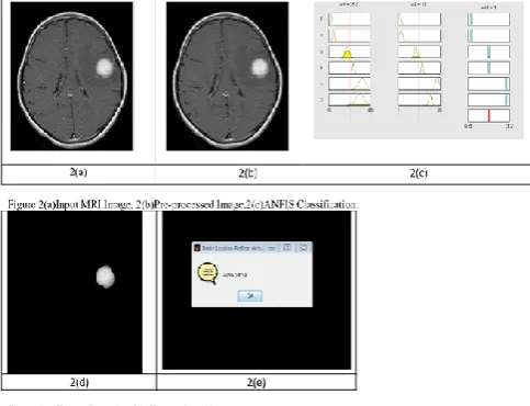
1. Pre-processing: This stage is used to remove the noise from MR image to enhance the image quality. Median filter is employed to remove the noise. The researcher of image processing asserts that median filter is better than linear filter [1].Image background does not contain any valuable information but it increases the processing time. To save computational time desired region extracted from MR image. So, skull is removed from noise free MR image using brain surface extraction algorithm. The output of this stage is noise free MR image which contains only human brain.
2. Extraction: The de noising MR image fed to KIFCM technique. The cluster centers calculated by
Mu=(1:k)*m/(k+1)
Where k, is the number of clusters and m is defined as:
m=max(MRIimage+1)
Then, distance between points and cluster centers helps to assign each point to the nearest cluster center based upon minimum distance then re-compute the new cluster centers. On the other side, some points are away from any cluster centers. Therefore, new cluster centers and the clustered points can enter to the loop that calculates the new distances and clustering the points due to membership values. This looping step takes less number of iterations than the random selection because the initial centers of the clusters were not randomly chosen which saves time and effort. The output of this stage is the clustering image.
After clustering stage, tumor is obtained from the background using thresholding technique. Lighter part in an image is the tumor area.
3. Implementation: Improved Sobel edge detection is implemented on the MR image with fuzzy. The proposed technique decreases the thickness of boundary lines of regions and enhances the accuracy of the obtained image. Then the final tumor is obtained which filtered again with median filter to remove any unwanted noise. At last, the results compare the proposed technique with existing technique based upon some parameters.
IV. RESULT AND DISCUSSION
A dataset signifies an instance of real world information object. DICOM dataset is used to evaluate the performance of the proposed firefly technique to detect the cancer. DICOM is free online dataset for biomedical images. These datasets are easily available for research and teaching. In the proposed approach, dataset contain 150 images from DICOM out of which 100 images are considered for testing purpose that are used for classification as normal and abnormal. To classify images into normal and abnormal 50 images are used as training set. Images used for brain tumor detection are of size 256*256 pixel.
Start
Input MR Image
De-Noising MR Image
Apply Edge Detection and segmentation
Extract brain tumor and localize
using EDPSO Filtration using Gaussian Filter
Results Comparison
End
Extract brain tumor and localize using
Firefly Filtration using
ISSN(E): 2277-128X, ISSN(P): 2277-6451, DOI: 10.23956/ijarcsse/V7I8/0164, pp. 290-295
The parameters used in the study are defined below. TP = No. of resulted images having brain tumor Total number of images
TN = No. of images that have not tumor Total number of images
FP = No.ofimages that have not tumor and detected positive Total number of images
FN= No. of images have tumor and not detected Total number of images
PERFORMANCE EVALUATION
1) Accuracy: Accuracy is defined as the degree of something which is true or exact value. Accuracy= No. of TP+ No. of TN … (equ.1)
No. of TP+TN+FP+FN
2) Sensitivity: (also called true positive rate) measures the ability of positives that are correctly recognized. Sensitivity= TP . ………(equ.2)
(TP+FN)
3) Specificity: (also called the true negative rate) measures the ability of negatives that are correctly recognized. Specificity= TN . ……… (equ.3)
(TN+FP)
Where,
TP = True Positive TN = True Negative FP = False Positive FN= False negative
Confusion matrix using EDPSO
Table 1: Confusion matrix using EDPSO n=100 Abnormal Normal Abnormal TP = 88 FN = 3 Normal FP = 2 TN = 7
Confusion matrix using Firefly
ISSN(E): 2277-128X, ISSN(P): 2277-6451, DOI: 10.23956/ijarcsse/V7I8/0164, pp. 290-295
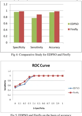
Comparison of EDPSO and Firefly
Table 3: Comparative Study of EDPSO and Firefly Technique
Techniques Metrics
EDPSO Firefly
Specificity 0.96 0.98 Sensitivity 0.77 0.88 Accuracy 95% 98%
In the table 3 a comparative study for existing and proposed technique is described. From the table it is clear that the image parameters are better in case of proposed system as it is more robust than the existing one.
Fig 4: Comparative Study for EDPSO and Firefly
Fig 5: EDPSO and Firefly on the basis of accuracy
V. CONCLUSION AND FUTURE ENHANCEMENT
In this paper, Firefly algorithm is comparatively studied against EDPSO algorithm. The proposed technique consists of MRI image, Image Enhancement, Edge Detection, Segmentation and final classification. The DICOM dataset that have been used in research. This image dataset contain 150 brain MRI images, including 90 brain images with tumor and the other 8 brain images without tumor. The detection of brain tumor is a very challenging task, in which special care is taken while using medical image processing techniques. In this research the accuracy of the brain tumor detection is improved according to the area and the acceptance rate.
In Future Enhancement, the tumor may be extracted using the unsupervised learning using SVM or ANN. The accuracy and average time consumption of the tumor extraction may be improved.
ACKNOWLEDGEMENT
This Research paper would not have been possible without the guidance and the help of Ms. Amardeep Kaur, Assistant Professor, and Punjabi University Regional Centre for IT & Management, Mohali. I also extend my profound thanks to all my professors, who have been a constant source of inspiration for me throughout this work. I would like to thanks Dr. Devinderpal Singh Head, Punjabi University Regional Centre for IT & Management, Mohali for his support. I am immensely grateful to my parents for everything they have always been doing for me and supporting me for my decision.
0 0.2 0.4 0.6 0.8 1 1.2
Specificity Sensitivity Accuracy
EDPSO
ISSN(E): 2277-128X, ISSN(P): 2277-6451, DOI: 10.23956/ijarcsse/V7I8/0164, pp. 290-295
REFERENCES
[1] Srikanta Patnaik, "Automated Brain Tumor Segmentation and Detection in MRI using Enhanced Darwinian Particle Swarm Optimization(EDPSO)", 2nd International Conference on Intelligent Computing, Communication & Convergence, vol: 92, 2016, pp: 475-480
[2] P. Dvorak, K. Bartusek, W. G. Kropatsch, Z. Smekal, "Automated Multi-Contrast Brain Pathological Area Extraction from 2D MR Images", Journal of Applied Research and Technology, vol: 13, 2015, pp: 58-69 [3] Sushmit Ghosh, Soham Kundu, Sushovan Chowdhury, AurpanMajumder, "Optimal Statistical Structure
Validation of Brain Tumors Using Refractive Index", 3rd International Conference on Recent Trends in Computing, vol: 57, 2015, pp: 168-177
[4] Asra Aslam, Ekram Khan, M.M. Sufyan Beg, "Improved Edge Detection Algorithm for Brain Tumor Segmentation", Second International Symposium on Computer Vision and the Internet, vol: 58, 2015, pp: 430-437
[5] J. Mehena, M. C. Adhikary, "Brain Tumor Segmentation and Extraction of MR Images Based on Improved Watershed Transform", IOSR Journal of Computer Engineering, e-ISSN: 2278-0661,p-ISSN: 2278-8727, vol: 17, issue 1, Feb 2015, pp: 1-5
[6] Sahil J Prajapati, Kalpesh R Jadhav, "Brain Tumor Detection By Various Image Segmentation Techniques With Introducation To Non Negative Matrix Factorization", International Journal of Advanced Research in Computer and Communication Engineering, ISSN (Online) 2278-1021, ISSN (Print) 2319-5940, vol. 4, issue 3, March 2015, pp: 599-603
[7] Yash Sharma, MeghaChhabra, "An Improved Automatic Brain Tumor Detection System", International Journal of Advanced Research in Computer Science and Software Engineering, ISSN: 2277 128X, vol: 5, issue 4, 2015, pp: 11-15
[8] Simran Arora, Gurjit Singh, "A Study of Brain Tumor Detection Techniques", International Journal of Advanced Research in Computer Science and Software Engineering, ISSN: 2277 128X, vol: 5, issue 5, May 2015, pp: 1272-1278
[9] Brundha B, Nagendra Kumar M, "MR Image Segmentation of brain to detect brain tumor and its area calculation using K-Means clustering and Fuzzy C-Means algorithm", International Journal For Technological Research In Engineering, ISSN (Online): 2347 - 4718, vol: 2, issue 9, May-2015, pp: 1781-1785
[10] Eman Abdel-Maksoud, Mohammed Elmogy, Rashid Al-Awadi, "Brain tumor segmentation based on a hybrid clustering technique", Egyptian Informatics Journal, ISSN: 1110-8665, Vol: 16, 2015, pp: 71-81