An Echocardiographic Comparison of
Sutureless and Conventional Aortic Valve
Replacement: A Matched Case-Control Study
Aaron R Casha
1,2, Alexander Manché
1, Liberato Camilleri
3, Kentaro Yamagata
4,
Stephanie Santucci
5, Marilyn Gauci
5, Joseph Galea
1.
1. Department of Cardiothoracic Surgery, Mater Dei Hospital, Malta 2. Faculty of Medicine and Surgery, University of Malta, Malta
3. Department of Statistics and Operations Research, Faculty of Science, University of Malta 4. Department of Cardiology, Mater Dei Hospital, Malta
5. Department of Anaesthesia, Mater Dei Hospital, Malta
Corresponding author: Prof Aaron R Casha,
Faculty of Medicine & Surgery, University of Malta Medical School, Mater Dei Hospital, Msida MSD 2090, Malta. E-mail: [email protected]
Introduction
Calcific aortic stenosis is a common disorder in the western world and is related to aging [1-3]. It is becoming increasingly more common with a prevalence of 3% in those aged 75 years and over [4]. Aortic valve stenosis causes a left ventricular pressure overload and a compensatory myocardial response of wall thickening and concentric hypertrophy resulting from myocardial muscle fibre thickening and sarcomerogenesis [5].
Sarcomeres thicken in response to continued systolic stress by adding filaments in parallel but do not increase in length [6]. The increase in left ventricular mass in isolated aortic stenosis is predictive of heart failure due to systolic dysfunction regardless of the severity of the valvular obstruction [7]. In other words the compensatory response of ventricular hypertrophy may in itself lead to progression of the pathology [7].
* Corresponding author. E-mail: [email protected]
Abstract
Background:
Patients at a high operative risk for conventional aortic valve replacement (AVR) may be offered sutureless valve implantation. Sutureless valves resemble conventional valves but incorporate an anchoring mechanism without using annular sutures.
Methods
Pre-operative and six month post-operative echocardiography data from our first year, single centre experience of sutureless valves was compared to conventional aortic valve replacements in patients matched for operative risk. Left ventricular ejection fraction, mean and peak AV gradients and inter-ventricular septal thickness, effective orifice area (EOA) and indexed effective orifice area (iEOA) were measured.
Results
The drops in mean and peak pre- to post-operative gradients were greater in the sutureless group, p=0.039 and p=0.001 respectively. Post-operative EOA was 1.69 cm2 and 1.26 cm2 (p=0.001) in the sutureless and conventional groups. Similarly iEOA was 0.93 cm2 and
0.74 cm2 (p=0.001) in the sutureless and conventional groups. There was also a reduction in patient prosthesis mismatch (PPM) in the
sutureless group as compared to the conventional group (Chi square test p=0.026). Post-operative inter-ventricular septal thickness was 1.13 cm2 in the sutureless group and 1.35 cm2 in the conventional group (p=0.011).
Conclusions
Use of sutureless valves with a stent framework resulted in larger EOA and iEOA and a diminution in PPM; and lead to a statistically significant faster regression in inter-ventricular septal thickness that is a measure of left ventricular mass. The rate and extent of regression in left ventricular hypertrophy after AVR is important since it determines long-term survival including mortality, heart failure and decreased admission rates.
Keywords: Sutureless valve; rapid deployment aortic valve; left ventricular hypertrophy; regression
Citation: Casha AR, Manché A, Camilleri L, Yamagata K, Santucci S, Gauci M, Galea J. An Echocardiographic Comparison of Sutureless and Conventional Aortic Valve Replacement: A Matched Case-Control Study: International
Cardiovascular Forum Journal. 2018;14:16-20. DOI: 10.17987/icfj.v14i0.536
Aortic valve replacement relieves the high aortic valve gradient, permits regression of the left ventricular wall hypertrophy and returns life expectancy back to normal [8,9]. It also leads to better quality of life, better NYHA (New York Heart Association) status and decreases hospitalisation rates [10]. Surgery was denied in one third of elderly patients with severe, symptomatic AS with older age and left ventricular dysfunction being the most striking characteristics of those denied surgery [11]. The greater morbidities and higher-risk profile of the current patient cohort has lead to the development of transcatheter aortic valve implantation (TAVI) and rapid deployment valves.
The aim of this study was to assess the difference in the early rate of regression in left ventricular hypertrophy between rapid deployment valves and conventional xenograft aortic valves, since the rate and extent of regression in left ventricular
hypertrophy after aortic valve replacement is an important
determinant of long-term survival [12].
Methodology
Data of our first year twenty-two consecutive Perceval sutureless valve implantations performed in a single centre was compared with a similar series of twenty-two conventional Mitroflow pericardial aortic valve replacements. The two cohorts of patients we studied were matched for per-operative risk using age and current risk assessment scores including additive and logistic Euroscore risk scoring for predicted operative mortality. Data collected included mortality and stroke rate, risk score, angiographic left venticulography data, patient demographic data, as well as crossclamp and bypass times. Patients referred for a combined aortic valve replacement and coronary artery revascularization were excluded from the study. All patients underwent procedures with identical blood cardioplegia and cardiopulmonary management protocols, and the roller-pump heart lung machines, oxygenators and tubing used were sourced from the same manufacturers throughout the study. A telephone interview was performed in order to assess changes in patients’ New York Heart Association (NYHA) status.
Echocardiography
Echocardiography examination and measurements were performed pre- and post-operatively within 6 months of the operation following published guidelines from the American Society of Echocardiography and other societies [13] in order to assess the initial rate of left ventricular regression. Left ventricular ejection fraction, mean and peak AV gradients and inter-ventricular septal thickness and effective orifice area (EOA) were measured. Indexed effective orifice area (iEOA) was calculated in order to characterize the degree of patient prosthesis mismatch (PPM). An iEOA ≤ 0.85 cm2/m2 was used to designate the threshold for aortic valve PPM, values between 0.65–0.85 cm2/m2 categorized moderate PPM and < 0.65 cm2/m2 severe PPM following Pibarot’s criteria [14].
Statistics
Independent samples t-test was used to compare parameters from the sutureless series and conventional controls, using the t-test for parametric data and the Mann-Whitney test for non-parametric data as determined by the Kolgomarov-Smirnoff test in SPSS statistics software (SPSS 23, IBM Inc., Armonk, New York, USA). Results were presented as mean and standard
deviation (SD) to indicate the dispersion of the values in each dataset. A binomial logistic regression model used to identify statistically significant predictors between valve type and pre-operative and post-pre-operative variables. The chi-squared test was used to analyze 2 × 2 contingency tables comparing the two valve groups and the presence or absence of patient- prosthesis mismatch (PPM). The criterion of a p-value < 0.05 was required for statistical significance.
Informed consent was obtained from each patient in this study. The study protocol conforms to the ethical guidelines of the 1975 Declaration of Helsinki as approved by the local institutional review committee.
Results
Pre-operative and operative parameters
These parameters are shown in Table 1. The Mitroflow patients were smaller and lighter, most likely due to the fact that there were more women in this group compared to the Perceval group. This resulted in a higher mass p = 0.021, higher body surface area (BSA) p = 0.045 and higher Body Mass Index (BMI) p = 0.027 in the Perceval rapid deployment group. There was also an advantageous lower cross-clamp (p = 0.001) time but not bypass time (p = 0.054) in the rapid deployment group. A binomial logistic regression model,
Table 1
Parameter MeanPerceval n=22 Mitroflow n=22SD Mean SD P-value
Pre-operative
Height (cm) 155.7 7.4 154.24 8.65 0.472
Weight (kg) 78.8 14.1 69.41 13.083 0.021
BSA (m2) 1.78 0.17 1.68 0.183 0.045
BMI (kg/m2) 32.5 5.7 28.99 4.87 0.027
Age (years) 76.9 4.9 78.29 0.507 0.416
Gender
(M:F ratio) 1.75 - 0.5 - 0.543§
Parsonnet
score 21.5 6.5 22.7 6.3 0.287
Additive
Euroscore 7.14 1.32 7.06 1.47 0.425
Logistic
Euroscore 7.51 3.07 7.69 3.65 0.434
NYHA class 2.04 0.69 2.28 0.70 0.240
Op
Cross-clamp
time (min) 42.7 8.2 59.59 10.387 0.000
Bypass time
(min) 60.8 14.1 71.24 15.193 0.054
Post-operative
Mortality
(number) 1 - 0 - 0.328
Stroke
(number) 0 - 0 - 1.000
Bleeding (ml) 456 134 404 162 0.277
Transfusion
(units) 1.36 0.77 0.89 1.02 0.119
NYHA class 1.46 0.50 1.31 0.45 0.251
Length of stay
(days) 11.2 5.5 10.3 6.7 0.665
between valve types and predictors showed that cross clamp time was an independent statistically significant variable (p = 0.003). There was no statistical difference between the two groups in the improvement in NYHA status after operation. There was one death in the Perceval group in an 82-year-old male that occurred in the fourth post-op week due to acute respiratory distress syndrome (ARDS) and multi organ failure.
Echocardiography
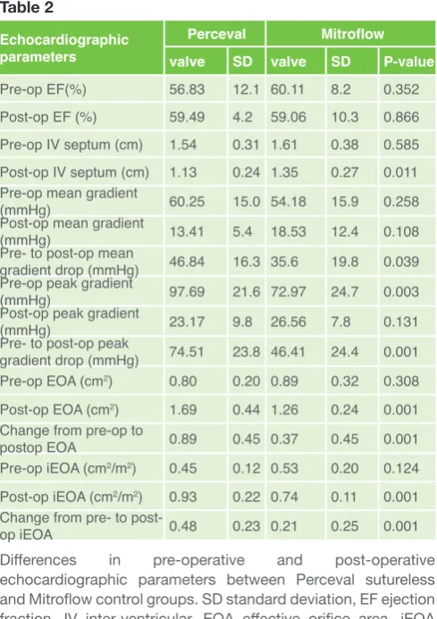
Results of pre-operative and post-operative echocardiographic parameters are shown in Table 2. Mean and peak pre- to post-operative gradients decreased more in the sutureless group, p=0.039 and p=0.001 respectively. Post-operative EOA was 1.69 cm2 and 1.26 cm2 (p=0.001) in the sutureless and conventional groups. Similarly iEOA was 0.93 cm2 and 0.74 cm2 (p=0.001) in the sutureless and conventional groups. Post-operative inter-ventricular septal thickness was 1.13 cm2 in the sutureless and 1.35 cm2 in the conventional groups (p=0.011). There was also a reduction in patient-prosthesis mismatch (PPM) in the sutureless group as compared to the conventional group (p<0.001) using Pibarot’s criteria [14]. In the rapid deployment group, there were 82% with no mismatch and 9% each with moderate and severe mismatch. In the conventional group 54% had no mismatch, 32% had mild mismatch and 14% severe mismatch. The presence or absence of RDW between the two patient groups was statistically significant, Chi-square test p=0.026.
Discussion
An ideal prosthetic aortic valve should be quick to insert and
present the lowest possible gradient. The first attribute should make aortic valve replacement (AVR) surgery safer, improving short-term results and permitting marginal, high-risk patients to be operated. The second attribute leads to a lower resistance to flow and enhances left ventricular regression and therefore improves long-term results.
Category of valves used
The Livanova Perceval S bioprosthetic valve is a surgical sutureless self-expanding valve without a sewing ring. It has a basic construction similar to the Pericarbon Freedom Stentless valve, which however requires suturing of the inflow and outflow sides. Instead the Perceval valve has an additional supra-annular aortic stent made of a super-elastic alloy. Three temporary and removable anchoring sutures allow accurate positioning of the prosthesis [15] to the nadir of the sinuses of Valsalva in the aortic root. A further important adjunct to the Perceval valve is the sub-annular curtain, which acts to diminish the risk of paravalvular leak as this is an important haemodynamic complication. In a population of patients implanted with TAVI valves, paravalvular leak was associated with a significant difference in mortality (25 versus 0%; p = 0.04) [16]. Its major advantage is in shortening cross clamp time [17].
The Sorin Mitroflow valve was introduced in 1982 and is an established valve with good clinical results [18,19]. In this pericardial valve, the leaflet hinge point is located on the outside of the strut. This is important as it leads to a larger orifice area while at the same time diminishes stresses at the hinge point as it is supported by the strut during valve closure when the pressure gradient across the valve is the highest.
Shrestha reported a cumulative life expectancy survival with Perceval valves that was worse than conventional biological valves from 2.5 years post-operatively. However in this study the sutureless group had a statistically significant higher operative risk with a logistic EuroSCORE of 20.4 ± 10.7 as compared to the conventional group at 16.7 ± 10.4 (p = 0.05) [20]. Therefore patient groups in this study were matched for operative risk order to eliminate this confounding factor.
Echocardiography
Echocardiography showed a doubling in post-operative in iEOA with Perceval sutureless valves, compared to a 33% increase in the Mitroflow conventional group. The difference in post-operative effective orifice area was probably due to the absence of a sewing ring in the Perceval valve and also because the stent exerts a radial distending force that may stretch the annulus. Though both the peak and mean gradients were lower, this did not reach statistical significance. However the differences in mean and peak pre- to post-operative gradients were greater in the sutureless group, p=0.039 and p=0.001 respectively. Our results contrast with those reported by Sepehripour that indicated that sutureless valves suffer from a higher mean valve gradient than conventional AVs [21]; however, this paper compared sutureless valves from six papers to the conventional arm of the PARTNER Trial (transcatheter versus surgical aortic-valve replacement in high-risk patients).
The post-operative EOA was 0.43cm2 lower and the iEOA 0.19cm2 lower in the Perceval sutureless group compared to the Mitroflow Table 2
Echocardiographic parameters
Perceval Mitroflow
valve SD valve SD P-value
Pre-op EF(%) 56.83 12.1 60.11 8.2 0.352
Post-op EF (%) 59.49 4.2 59.06 10.3 0.866
Pre-op IV septum (cm) 1.54 0.31 1.61 0.38 0.585
Post-op IV septum (cm) 1.13 0.24 1.35 0.27 0.011
Pre-op mean gradient
(mmHg) 60.25 15.0 54.18 15.9 0.258
Post-op mean gradient
(mmHg) 13.41 5.4 18.53 12.4 0.108
Pre- to post-op mean
gradient drop (mmHg) 46.84 16.3 35.6 19.8 0.039
Pre-op peak gradient
(mmHg) 97.69 21.6 72.97 24.7 0.003
Post-op peak gradient
(mmHg) 23.17 9.8 26.56 7.8 0.131
Pre- to post-op peak
gradient drop (mmHg) 74.51 23.8 46.41 24.4 0.001
Pre-op EOA (cm2) 0.80 0.20 0.89 0.32 0.308
Post-op EOA (cm2) 1.69 0.44 1.26 0.24 0.001
Change from pre-op to
postop EOA 0.89 0.45 0.37 0.45 0.001
Pre-op iEOA (cm2/m2) 0.45 0.12 0.53 0.20 0.124
Post-op iEOA (cm2/m2) 0.93 0.22 0.74 0.11 0.001
Change from pre- to
post-op iEOA 0.48 0.23 0.21 0.25 0.001
conventional group. This improvement in indexed effective orifice area in the Perceval group resulted in a statistically significant reduction in PPM in the Perceval group (p<0.001), even though the patients in the Perceval group were heavier (p=0.021). This is important since PPM is associated with poorer long-term outcome due to worse haemodynamics and less regression of ventricular hypertrophy [16]. No aortic root enlargement operations are performed routinely in our centre as although aortic root enlargement may reduce trans-valvular gradients and therefore the incidence of prosthesis- patient mismatch, this has not been shown to result in an improvement in long-term clinical outcomes [22]. The advantageous increase in EOA and iEOA in the stentless Perceval group is demonstrated in the early regression in left ventricular hypertrophy by a statistically significant difference of 0.22cm in post-operative inter-ventricular septal thickness between the two groups p=0.011.
Our results compare favourably with those from other series; for example mean effective orifice area (EOA) in our series was 1.69 ± 0.44 cm2 compared to 1.4 ± 0.4 cm2 and 1.6 ± 0.4 cm2 in other series. Similarly mean gradient was 13.4 ± 5.4 mmHg whilst results in published series ranged from 10 ± 5 to 14 ± 6 mmHg and peak gradient was 23.2 ± 9.9 mmHg compared to a range from 19 ± 8 to 27 ± 11 mmHg [23].
Advantages of rapid deployment valves
Rapid deployment AVR can be considered as a substitute to conventional AVR, especially for elderly patients with intermediate to high operative risk [24]. Another use is as a replacement in stentless bioprosthesis failure [25]. Rapid deployment valves reduce complexity of conventional valve operations [26] yet permit precise reproducible positioning [15].
Whilst the design of rapid deployment valves is similar in concept to Trans Aortic Valve Implantation (TAVI) prosthesis [27], the surgical approach differs in that it provides direct visualization and permits excision of the aortic valve and adjacent calcifications instead of lateral compression by the TAVI stent. The decalcification of the valve is important in that it may reduce the possibility of embolic showers particularly to the brain although this remains unproven [28]. The major degree of crimping required with TAVI valves may reduce prosthesis durability [29].
The reduction in implantation time with rapid deployment valves results in lower cross-clamp times should contribute to a reduction in postoperative complications [30,31]. The absence of a conventional sewing ring should lead to improved haemodynamics with greater EOA and lower post-operative aortic valve gradients, particularly important in older patients with small aortic roots [20]. Sutureless valves result in a higher rate of heart block compared to conventional AVR, with approximately 10% requiring a permanent pacemaker [24]. The mortality, neurological deficit, renal failure and post-operative bleeding rates of sutureless and conventional valves are similar, but there is an increased incidence of endocarditis and paravalvular leaks [21].
Left ventricular regression
Left ventricular hypertrophy is a dangerous consequence of the pressure load on the left ventricle resulting from aortic stenosis [32]. There is an independent relationship between indexed-EOA and the extent of regression in left ventricular mass after AVR
[33]. There is lesser regression of left ventricular hypertrophy after AVR with PPM, with a smaller decrease in chamber internal dimension in patients with PPM compared to those without a mismatch [34]. In our series sutureless valve implantation resulted in a larger iEOA and a marked decrease in PPM and this would be expected to result in more complete LV regression. An enhanced LV mass regression after AVR is associated with better long-term survival [35]. We have shown that there is a statistically significant difference in the rate of early (before 6 months post-op) LV regression when a sutureless prosthesis was used, suggestive of better long-term outcomes including mortality, heart failure and decreased admission rates [10,36].
Limitations
This case-matched study was performed with a small number of patients. This may add biases caused by not assigning patients randomly to a treatment arm, resulting in an augmented risk of introducing confounding variables.
Conclusion
Use of sutureless valves resulted in larger EOA and iEOA, decreased PPM and lead to a statistically significant faster regression in early inter-ventricular septal thickness, indicating early left ventricular mass reduction and modelling. The rate and extent of regression in left ventricular hypertrophy after
aortic valve replacement is an important determinant of
long-term survival including mortality, heart failure and decreased admission rates.
Declarations of interest
The authors declare no conflict of interest.
Acknowledgements
The authors state that they abide by the authors’ responsibilities and ethical publishing guidelines of the International Cardiovascular Forum Journal [37].
References
1. Lindroos M, Kupari M, Heikkilä J, Tilvis R. Prevalence of aortic valve abnormalities in the elderly: an echocardiographic study of a random population sample. J Am Coll Cardiol. 1993;21(5):1220-5.
2. Stewart BF, Siscovick D, Lind BK, Gardin JM, Gottdiener JS, Smith VE, Kitzman DW, Otto CM. Clinical factors associated with calcific aortic valve disease. Cardiovascular Health Study. J Am Coll Cardiol. 1997;29(3):630-4. 3. Nkomo VT, Gardin JM, Skelton TN, Gottdiener JS, Scott CG, Enriquez-
Sarano M. Burden of valvular heart diseases: a population-based study. Lancet 2006;368:1005–1011. DOI: 10.1016/S0140-6736(06)69208-8 4. Vahanian A, Otto CM. Risk stratification of patients with aortic stenosis. Eur
Heart J. 2010;31:416–23. DOI: 10.1161/CIRCIMAGING.115.003421 5. Grossman W, Jones D, McLaurin LP. Wall stress and patterns of hypertrophy
in the human left ventricle. J Clin Invest. 1975; 56: 56-64.
6. Russell B, Curtis MW, Koshman YE, Samarel AM. Mechanical Stress-Induced Sarcomere Assembly for Cardiac Muscle Growth in Length and Width. Journal of molecular and cellular cardiology. 2010;48(5):817-823. DOI: 10.1016/j.yjmcc.2010.02.016
7. Kupari M, Turto H, Lommi J: Left ventricular hypertrophy in aortic valve stenosis: preventive or promotive of systolic dysfunction and heart failure? Eur Heart J. 2005, 26: 1790-1796. DOI: 10.1093/eurheartj/ehi290 8. Manché A., Camilleri L., & Gauci D. (2016). Does aortic valve replacement
restore normal life expectancy? A twenty-year relative survival study. International Cardiovascular Forum Journal, 6, 46-53. DOI: https://doi. org/10.17987/icfj.v6i0.138
9. Lindblom D, Lindblom U, Qvist J, Lundstrom H: Long-term relative survival rates after heart valve replacement. J Am Coll Cardiol. 1990, 15: 566-573. 10. Lindman BR, Stewart WJ, Pibarot P, Hahn RT, Otto CM, Xu K et al. Early
11. Iung B, Cachier A, Baron G, Messika-Zeitoun D, Delahaye F, Tornos P et al. Decision-making in elderly patients with severe aortic stenosis: why are so many denied surgery? Eur Heart J 2005;26(24):2714–2720. DOI: 10.1093/ eurheartj/ehi471
12. De Paulis R, Sommariva L, Colagrande L, De Matteis GM, Fratini S, Tomai F, Bassano C, Penta de Peppo A, Chiariello L. Regression of left ventricular hypertrophy after aortic valve replacement for aortic stenosis with different valve substitutes. J Thorac Cardiovasc Surg. 1998;116(4):590-8.
13. Zoghbi WA, Chambers JB, Dumesnil JG, Foster E, Gottdiener JS, Grayburn PA et al. Recommendations for evaluation of prosthetic valves with echocardiography and Doppler ultrasound. J Am Soc Echocardiogr. 2009;22(9):975–1014. DOI: 10.1016/j.echo.2009.07.013
14. Pibarot P, Dumesnil JG. Prosthesis-patient mismatch: definition, clinical impact, and prevention. Heart. 2006;92(8):1022–9. DOI: 10.1136/ hrt.2005.067363
15. Gilmanov D, Miceli A, Bevilacqua S, Farneti P, Solinas M, Ferrarini M et al. Sutureless implantation of the Perceval S aortic valve prosthesis through right anterior minithoracotomy. Ann Thorac Surg. 2013;96(6):2101–8. DOI: 10.1016/j.athoracsur.2013.07.007
16. Santarpino G, Fischlein T. Use of sutureless prosthetic aortic valves in cardiac surgery. G Ital Cardiol (Rome) 2014;15(3):170–6. DOI: 10.1714/1463.16167
17. Igor B, Stefano M, Mariachiara C, Davide S, Ottavio A. Early predictors of permanent pacemaker implantation in sutureless aortic valve replacement with Perceval bioprosthesis: a conduction disorders analysis. J Heart Health Cir 2016;1:1.
18. Lorusso R, Gelsomino S, De Cicco G, Vizzardi E, Faggiano P, Carella R et al. The Italian study on the Mitroflow postoperative results (ISTHMUS): a 20-year, multicentre evaluation of Mitroflow pericardial bioprosthesis. Eur J Cardiothorac Surg 2011;39(1):18–26. DOI: 10.1016/j.ejcts.2010.03.069 19. Jamieson WR, Koerfer R, Yankah CA, Zittermann A, Hayden RI, Ling H
et al. Mitroflow aortic pericardial bioprosthesis–clinical performance. Eur J Cardiothorac Surg 2009;36(5):818–824. DOI: 10.1016/j.ejcts.2009.05.020 20. Shrestha M, Maeding I, Höffler K, Koigeldiyev N, Marsch G, Siemeni
T et al. Aortic valve replacement in geriatric patients with small aortic roots: are sutureless valves the future? Interact Cardiovasc Thorac Surg 2013;17(5):778–782. DOI: 10.1093/icvts/ivt291
21. Sepehripour AH, Harling L, Athanasiou T. What are the current results of sutureless valves in high-risk aortic valve disease patients? Interact Cardiovasc Thorac Surg 2012;14(5):615–621. DOI: 10.1093/icvts/ivs011 22. Kulik A, Al-Saigh M, Chan V, Masters RG, Bédard P, Lam BK et al.
Enlargement of the small aortic root during aortic valve replacement: is there a benefit? Ann Thorac Surg 2008;85(1):94–100. DOI: 10.1016/j. athoracsur.2007.07.058
23. Powell R, Pelletier MP, Chu MW, Bouchard D, Melvin KN, Adams C. The Perceval sutureless aortic valve: review of outcomes, complications, and future direction. Innovations (Phila) 2017;12(3):155-73. DOI: 110.1097/ IMI.0000000000000372
24. Vogt F, Pfeiffer S, Dell’Aquila AM, Fischlein T, Santarpino G. Sutureless aortic valve replacement with Perceval bioprosthesis: are there predicting factors for postoperative pacemaker implantation? Interact Cardiovasc Thorac Surg 2016;22(3):253–8. DOI: 10.1093/icvts/ivv330
25. Kim JH, Kim JB, Choi SY. Sutureless aortic valve replacement in stentless bioprosthesis failure. Interact Cardiovasc Thorac Surg 2015; 21(6):801– 802. DOI: 10.1093/icvts/ivv255
26. Santarpino G, Pfeiffer S, Concistrè G, Grossmann I, Hinzmann M, Fischlein T. The Perceval S aortic valve has the potential of shortening surgical time: does it also result in improved outcome? Ann Thorac Surg 2013;96(1):77– 81. DOI: 10.1016/j.athoracsur.2013.03.083
27. Phan K, Tsai YC, Niranjan N, Bouchard D, Carrel TP, Dapunt OE et al. Sutureless aortic valve replacement: a systematic review and meta-analysis. Ann Cardiothorac Surg 2015;4(2):100–11. DOI: 10.3978/j. issn.2225-319X.2014.06.01
28. Santarpino G, Pfeiffer S, Jessl J, Dell’Aquila AM, Pollari F, Pauschinger M et al. Sutureless replacement versus transcatheter valve implantation in aortic valve stenosis: a propensity-matched analysis of 2 strategies in high-risk patients. J Thorac Cardiovasc Surg 2014;147(2):561–7. DOI: 10.1016/j.jtcvs.2013.10.025
29. Khoffi F, Heim F, Chakfe N, Lee JT. Transcatheter fiber heart valve: Effect of crimping on material performances. J Biomed Mater Res B Appl Biomater 2015;103(7):1488–97. DOI: 10.1002/jbm.b.33330
30. Laborde F, Fischlein T, Hakim-Meibodi K, Misfeld M, Carrel T, Zembala M, et al. Clinical and haemodynamic outcomes in 658 patients receiving the Perceval sutureless aortic valve: early results from a prospective European multicentre study (the Cavalier trial). Eur J Cardiothorac Surg 2016;49(3):978-86. DOI: 10.1093/ejcts/ezv257
31. Flameng W, Herregods MC, Hermans H, Van der Mieren G, Vercalsteren M, Poortmans G et al. Effect of sutureless implantation of the Perceval S aortic valve bioprosthesis on intraoperative and early postoperative outcomes. J Thorac Cardiovasc Surg 2011;142(6):1453-7. DOI: 10.1016/j. jtcvs.2011.02.021
32. Haider AW, Larson MG, Benjamin EJ, Levy D. Increased left ventricular mass and hypertrophy are associated with increased risk for sudden death. J Am Coll Cardiol. 1998, 32: 1454-1459.
33. Villa E, Troise G, Cirillo M, Brunelli F, Tomba MD, Mhagna Z et al. Factors affecting left ventricular remodeling after valve replacement for aortic stenosis. An overview. Cardiovasc Ultrasound 2006;4:25. DOI: 10.1186/1476-7120-4-25
34. Tasca G, Brunelli F, Cirillo M, Dalla Tomba M, Mhagna Z, Troise G et al. Impact of valve prosthesis-patient mismatch on left ventricular mass regression following aortic valve replacement. Ann Thorac Surg 2005;79(2):505-510. DOI: 10.1016/j.athoracsur.2004.04.042
35. Ali A, Patel A, Ali Z, Abu-Omar Y, Saeed A, Athanasiou T et al. Enhanced left ventricular mass regression after aortic valve replacement in patients with aortic stenosis is associated with improved long-term survival. J Thorac Cardiovasc Surg 2011;142(2):285-91. DOI: 10.1016/j.jtcvs.2010.08.084 36. Une D, Mesana L, Chan V, Maklin M, Chan R, Masters RG et al. Clinical
impact of changes in left ventricular function after aortic valve replacement: analysis from 3112 patients. Circulation 2015;132(8):741-7. DOI: 10.1161/ CIRCULATIONAHA.115.015371