Address for correspondence
Dr. Yousuf Abd Mallick, MBBS, FCPS Consultant Dermatologist, Dermatology Unit The Indus Hospital, Main Campus-Korangi, R-386, Sector 16 A, Buffer Zone (Gulshan-e-Waseem), North Karachi, Karachi, Pakistan Ph: 03432687716, +92-21-36950203 Email: [email protected]
Original Article
Frequency of chronic venous insufficiency in patients
of Schamberg’s disease
Introduction
Schamberg’s disease (SD) is also known as Purpura Pigmentosa Progressiva.1 World-wide Schamberg’s disease is the most common type of pigmented purpuric dermatoses (PPD).2 PPD represents a rare group of diseases which clinically manifest as various forms of purpura
and pigmentation on the skin. Despite its rarity, PPD have been reported in all races and ethnicities across the globe. Domenico Majocchi (1849–1929), an Italian dermatologist, described the first ever PPD in 1896 as “purpura annularis telangiectaticum” which later on was given the name- Majocchi’s disease.3 After that many other types of PPD were reported and described in the literature. The current list now includes Schamberg’s disease, Majocchi’s disease, lichen aureus, pigmented purpuric lichenoid dermatitis of Gougerot and Blum, itching purpura of Lowenthal and eczematid-like purpura of Doucas and Kapetanakis.4,5 Unilateral variant of SD, linear pigmented purpura and Yousuf Abd Mallick, Sidra Akhlaq Hussain*
Department of Dermatology, The Indus Hospital, Main Campus-Korangi, Karachi * Department of Dermatology, Abbasi Shaheed Hospital, Karachi
Abstract Objective To determine the frequency of chronic venous insufficiency in patients of Schamberg’s
disease.
Methodology The study was cross-sectional & conducted in Dermatology outpatient department at Saifee Hospital Trust, Karachi, Pakistan. The duration of study was eleven months i.e. from April 2017 to March 2018. Clinically diagnosed, 60 cases of Schamberg’s disease were included via non probability consecutive sampling technique. Age of patients was 18 years and above. Clinical examination for varicose veins and Doppler ultrasonography of the lower extremities were performed and findings were recorded.
Results In our study, the mean age was 44.43±13.83 years. Out of 60 patients, 27 (45%) were male and 33 (55%) were female. Only 13 (21.67%) patients had a positive family history of Schamberg’s disease. In our study, 18 (30%) patients had clinically detected varicose veins of lower limbs while variable degrees of venous insufficiency were detected in 28 (46.67%) patients on Doppler ultrasound of the lower extremities.
Conclusion The number of patients with chronic venous insufficiency in Schamberg’s disease is
significantly higher and it might be one of the main predisposing factors for this disease. However, further case-control studies are required to calculate its prevalence and to confirm its aetiopathogenic association.
Key words
granulomatous variant of pigmented purpura are amongst the very rare types.4
All these disorders appear similar on histopathology and share common features which include narrowing of the lumen and endothelial swelling of superficial cutaneous blood vessels, extravasation of erythrocytes, perivascular T lymphocyte infiltration and marked haemosiderin deposition in dermis and dermal macrophages.5
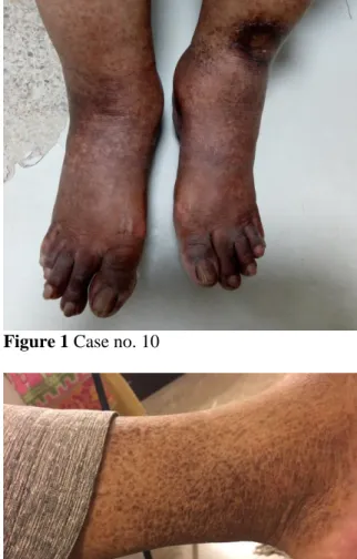
The hallmark of Schamberg’s disease is progressive, asymptomatic and non-palpable pigmentary changes.6 Clinically, SD manifests as orange‐red flat macules and patches of variable sizes with diagnostic “cayenne pepper” spots on the borders. Old lesions transform to yellow-brown colour. These lesions are oval or irregular in outline and pinpoint petechiae usually found inside the patches.5 SD has a very chronic course, sometimes it is of life-long duration. The disease continues to appear in successive crops over many years mostly at same places. The site of predilection is lower extremities but it can occur on trunk, thighs, buttocks; even arms and forearms can be affected.1,5 SD is completely asymptomatic, mostly occurs in adult population but it can also affect children7 and males are more often affected than females.6 (Figure 1 & 2)
There is no definitive treatment, neither cure is available for SD. Topical and oral corticosteroids, phototherapy, PUVA, photodynamic therapy,8 intense pulsed light (IPL),9 griseofulvin, pentoxifylline,7 cyclosporine, oral rutoside with ascorbic acid,4 colchicine7,10 and fractional non-ablative 1540nm erbium:YAG laser11 have been used with variable success. Sometimes, the pigmentation is resistant to all known treatments and besides psychological support, nothing can be offered by physicians and dermatologists.
Figure 1 Case no. 10
Figure 2 Case no. 14
The etiology of this disorder is still unknown. Prolonged standing in day-to-day work leading to gravitational effects and increased venous pressure is an important predisposing factor in many cases.12 The systemic diseases which have been correlated are diabetes mellitus, hypertension, rheumatoid arthritis, chronic hepatitis C virus infection and hyperlipidemia.13-16 Drug induced SD has also been reported on few occasions.16,17 Recently a study from Turkey reported variable degrees of venous insufficiency in 75% of the patients of PPD on Doppler ultrasonography of the lower extremities.18
was designed to determine the frequency of chronic venous insufficiency (CVI) in patients of Schamberg’s disease.
Patients and Methods
This study was conducted in OPD of Dermatology department, Saifee Hospital Trust, Karachi. The data collection was done for 11 months from April 2017 to March 2018. The cases were selected via non probability consecutive sampling technique. We took a sample of 60 adult subjects meeting the selection criteria for this study after written informed consent.
Clinically diagnosed, untreated cases of Schamberg’s disease for at least 6 months, of either sex group, age 18 years and above, with or without family history of SD were selected for the study. Patients with lower limb injuries, congenital and skeletal abnormalities, vascular malformations, malignancies, pregnant & lactating mothers and patients under treatment for SD were excluded.
SD is diagnosed clinically and then detailed clinical examination for varicose veins was performed in the OPD in lying down and standing positions. Trendelenburg and Tourniquet tests were performed. The Doppler ultrasound (DUS) of venous systems of both lower limbs was performed in ultrasound department by using 7.5 MHz Doppler ultrasound probe. The room temperature was maintained between 18°C and 22°C throughout the procedure. The standard protocol was followed.19 This consists of three steps:
1. DUS was first performed in orthostatic position.
2. Then DUS examination of the superficial veins of the lower limbs was performed in standing position. The patient was standing
upright on a stool and the limb under examination was rotated outward.
3. Then DUS was performed in supine position. The patient was lying on a padded bed.
All steps were followed in all patients. All findings were recorded on a specified form by the researcher. Permission from the institutional ethical review committee was taken prior to the conduction of the study.
The data feeding and analysis was performed on computer package SPSS (statistical package of social sciences) version 21.0. Clinical characteristics were summarized in terms of frequencies and percentages for qualitative variables (gender, family history, clinically varicose veins and Doppler findings). Mean+S.D for quantitative variable (age) was done. Stratification was done with regard to gender, family history of SD and clinically varicose condition to see the effect of outcome and post-stratification Fisher Exact test applied.
p <0.05 was considered as significant.
Results
Out of 60 patients, 27 (45%) were male and 33 (55%) were female. The mean age was 44.43±13.83 years. Only 13 (21.67%) patients had a positive family history of Schamberg’s disease. In our study, 18 (30%) patients had clinically detectable varicose veins of both lower limbs. Doppler ultrasound (DUS) of the lower extremities showed variable degrees of venous insufficiency in 28 (46.67%) patients while 32 (53.33%) patients showed normal DUS findings.
p-value was <0.001.
of the patients showed isolated insufficient deep venous system. All those 18 (30%) patients who have clinically detected varicose veins of lower limbs showed signs of CVI on DUS. So, we divided patients into 3 groups based on DUS findings; first with insufficient superficial venous system (superficial group), second with insufficient both deep and superficial venous systems (both group) and third with normal DUS findings group.
Stratification for gender with respect to DUS findings showed that 10 males (43.5%) and 13 females (56.5%) had insufficient superficial venous system; 3 males (60%) and 2 females (40%) had insufficient both deep and superficial venous systems; and 14 males (43.8%) and 18 females (56.3%) showed unaffected or normal venous systems. p-value was 0.854.
Stratification for family history with respect to DUS findings showed that 7 (30.4%) patients had positive while 16 (69.6%) patients had negative family history of SD in the superficial group. 1 (20%) patient had positive while 4 (80%) patients had negative family history of SD in both groups. 5 (15.6%) patients had positive while 27 (84.4%) patients had negative family history of SD in the normal DUS findings group. p-value was 0.365.
Stratification for clinically varicose condition with respect to DUS findings showed that 14 (60.9%) patients had and 9 (39.1%) patients did not have varicose veins on examination in the superficial group. 4 (80%) patients had and 1 (20%) patient did not have varicose veins on examination in both group. In the normal DUS findings group, all 32 (100%) patients had normal examination and none of the patients showed clinically detectable varicose veins. p -value was <0.001.
Discussion
In 1901, Schamberg’s Disease (Purpura Pigmentosa Progressiva) was first described17 as a disorder which causes discoloration of the skin, predominantly of the lower extremities but it can also involve buttocks, trunk and even upper extremities. The exact cause of SD still remains unknown.17 Multiple cofactors are reported in the literature which are thought to be associated with SD. The most important cofactor is venous hypertension.20 Other important ones are orthostatic pressure, recurrent lower limb infections, regular strenuous exercises, capillary fragility, ingestion of some chemicals and various drugs.16,20 The pathological manifestations of chronic venous insufficiency (CVI) develop as a consequence of venous hypertension.21 First of all tissue edema develops, followed by red cell extravasation and perivascular fibrin deposition leading to impaired arterial flow along with some other local immune disturbances. There is also concurrent damage to the lymphatic system which leads to impaired clearance of cellular metabolites.21 Hence hemosiderin deposition becomes a predominant feature of CVI.11
The earliest clinical manifestation of venous hypertension is lower limb edema,21 followed by venous stasis, venous eczema, trophic skin lesions, pigmentation, lower limb pain and variable degrees of varicosities.19 Other serious and mostly irreversible cutaneous complications which develop subsequently in patients of CVI are atrophie blanche, lipodermatosclerosis, venous ulcers, livedoid vasculopathy, PPD and
Pseudo-Kaposi’s sarcoma
(Acroangiodermatitis).21 Most of the venous ulcers are caused by incompetence of the superficial venous system of the lower limb.22
includes diabetes mellitus, thyroid disorders, hereditary spherocytosis, rheumatoid arthritis, systemic lupus erythematosus (SLE), osteomyelitis, hepatic diseases & porphyria, hematological disorders and various malignancies.18 But their exact relation and contribution towards disease development is yet to be identified. Furthermore, psychological issues such as anxiety and depression are also common in patients suffering from CVI and its complications.20 The recurrent nature and resistant to treatment course of the disease upsets the mental well-being of sufferers and make them vulnerable to various psychological diseases.
There is generally a male predominance in Schamberg’s disease but our study showed a subtle female dominance i.e. 55%. Similar results were also reported by Kim et al.16 and Cho et al.23 from Korea. We reported a positive family history of SD in 21.67% which no other study has documented yet. It indicates that some genetic predisposition to this disease also exists. Our study reported varicose dilatation in 30% of patients. Gönül et al.18 reported this in 20% of patients. We reported venous insufficiency in 46.67% of patients. Gönül et al. also reported this in 75% of patients.18
To the best of our knowledge, this is the first study in Pakistan which reports chronic venous insufficiency by Doppler ultrasound in patients of SD. Detection of varicose veins clinically, and early detection of CVI on DUS necessitates management of venous hypertension and referral to the vascular surgeon. Early referral, multidisciplinary team care and prompt treatment may save patients of SD not only from serious and irreversible cutaneous complications but also from psychological diseases like anxiety and depression.
Conclusion
Schamberg’s disease is reported as a consequence of venous disease for decades but little is known about its aetiology and pathogenesis. We have reported a high frequency (46.67%) of chronic venous insufficiency among patients of Schamberg’s disease. It might be the main predisposing factor for this disorder. Further multi-centre, large scale studies are required to build its aetiopathogenic role in development of Schamberg’s disease.
References
1. Haddad CJ, Lacle JH, Haddad CM. An uncommon cause of skin discoloration: Purpura Pigmentosa Progressiva. Int J Pathol Clin Res 2015; 1: 11-2.
2. Torrelo A, Requena C, Mediero IG, Zambrano A. Schamberg'spurpura in children: a review of 13 cases. J Am Acad Dermatol 2003; 48: 31-3.
3. Tristani-Firouzi P, Meadows KP, Vanderhooft S. Pigmented purpuric eruptions of childhood: a series of cases and review of literature. Pediatr Dermatol 2001; 18: 299–304.
4. Sardana K, Sarkar R, Sehgal VN. Pigmented purpuric dermatoses: an overview. Int J Dermatol 2004; 43: 482–8.
5. Leslie AT. Purpura. In: Griffiths CEM, Barker J, Bleiker T, Chalmers R, Creamer D, editors. Rook’s textbook of dermatology. Vol 4. 9th ed Oxford: Wiley-Blackwell; 2016 p. 101.1-101.27.
6. Schetz D, Kocić I. A new adverse drug reaction: Schamberg’s disease caused by amlodipine administration – a case report. Br J Clin Pharmacol 2015; 80(6): 1477-8. 7. Cavalcante MLLL, Masuda PY, Brito FF,
Pinto ACVD, Itimura G, Nunes AJF. Schamberg’s disease: case report with therapeutic success by using colchicine. An Bras Dermatol 2017; 92(2): 246-8.
8. Kim SK, Kim EH, Kim YC. Treatment of pigmented purpuricdermatosis with topical photodynamic therapy. Dermatology 2009; 219: 184–6.
disease with advanced fluorescence technology. J Drugs Dermatol 2012; 11(4): 528–9.
10. Geller M. Benefit of colchicine in the treatment of Schamberg’s disease. Ann Allergy Asthma Immunol 2000; 85(3): 246. 11. Hilerowicz Y, Sprecher E, Gat A, Artzi O.
Successful treatment of Schamberg’s disease with fractional non-ablative 1540 nm erbium:glass laser. J Cosmet Laser Ther 2018; 16: 1-4.
12. Sharma L, Gupta S. Clinicoepidemiological study of pigmented purpuricdermatoses. Indian Dermatol Online J 2012; 3: Shimizu H. Pigmented purpuricdermatosis in mixed cryoglobulinaemia associated with rheumatoid arthritis and hepatitis C infection. Acta Derm Venereol 2005; 85(5): 460-1.
13. Raslan HM, Ezzat WM, Abd El Hamid MF, Emam H, Amre KS. Skin manifestations of chronic hepatitis C virus infection in Cairo, Egypt. East Mediterr Health J 2009; 15(3): 692-70.
14. Tato BP, Marinero Escobedo S, Pérez González YC, Sánchez Albisua B, PolimónOlabarrieta I, Encabo Mayoral B. Granulomatous variant of pigmented purpuricdermatosis. Am J Dermatopathol 2012; 34(7): 746-8.
15. Kim DH, Seo SH, Ahn HH, Kye YC, Choi JE. Characteristics and clinical
manifestations of pigmented purpuric dermatoses. Ann Dermatol 2015; 27(4): 404-10.
16. Chatterjee S. Schamberg disease: uncommon reaction to a common drug. CMAJ 2009; 181(12): E275.
17. Gönül M, Çakmak SK, Özcan N, Oğuz ID, Gül U, Bıyıklı Z. Clinical and laboratory findings of pigmented purpuricdermatoses. Ann Dermatol 2014; 26(5): 610-4.
18. Galeandro AI, Quistelli G, Scicchitano P, Gesualdo M, Zito A, Caputo P, et al. Doppler ultrasound venous mapping of the lower limbs. Vasc Health Risk Manag 2012; 8: 59-64.
19. Barron GS, Jacob SE, Kirsner RS. Dermatologic complications of chronic venous disease: medical management and beyond. Ann Vasc Surg 2007; 21(5): 652-62.
20. Parsi K. Dermatological manifestations of venous disease: part 1. Aust N Z J Phlebology 2007; 10(1): 7-15.
21. Simon DA, Dix FP, McCollum CN. Management of venous ulcers. BMJ 2004; 328: 1358-62.