R E S E A R C H
Open Access
Association between cytokine profiles and
lung injury in COVID-19 pneumonia
Li-Da Chen
1†, Zhen-Yu Zhang
2†, Xiao-Jie Wei
3†, Yu-Qing Cai
4, Weng-Zhen Yao
4, Ming-Hui Wang
4,
Qiu-Fen Huang
4and Xiao-Bin Zhang
4*Abstract
Background:Coronavirus disease 2019 (COVID-19) is a new respiratory and systemic disease caused by severe acute respiratory syndrome coronavirus 2 (SARS-CoV-2) infection. The purpose of the present study was to investigate the association between cytokine profiles and lung injury in COVID-19 pneumonia.
Methods:This retrospective study was conducted in COVID-19 patients. Demographic characteristics, symptoms, signs, underlying diseases, and laboratory data were collected. The patients were divided into COVID-19 with pneumonia and without pneumonia. CT severity score and PaO2/FiO2ratio were used to assess lung injury.
Results:106 patients with 12 COVID-19 without pneumonia and 94 COVID-19 with pneumonia were included. Compared with COVID-19 without pneumonia, COVID-19 with pneumonia had significantly higher serum interleukin (IL)-2R, IL-6, and tumor necrosis factor (TNF)-α. Correlation analysis showed that CT severity score and PaO2/FiO2were significantly correlated with age, presence of any coexisting disorder, lymphocyte count,
procalcitonin, IL-2R, and IL-6. In multivariate analysis, log IL6 was the only independent explanatory variables for CT severity score (β= 0.397,p< 0.001) and PaO2/FiO2(β=−0.434,p= 0.003).
Conclusions:Elevation of circulating cytokines was significantly associated with presence of pneumonia in COVID-19 and the severity of lung injury in COVID-COVID-19 pneumonia. Circulating IL-6 independently predicted the severity of lung injury in COVID-19 pneumonia.
Keywords:Coronavirus disease 2019, Severe acute respiratory syndrome coronavirus 2, Cytokine, Lung injury, Pneumonia
Backgrounds
In December, 2019, a cluster of patients with “unknown
viral pneumonia” were reported in Wuhan, Hubei
prov-ince, China. Then it was confirmed that the disease was caused by a novel coronavirus which was named severe acute respiratory syndrome coronavirus 2
(SARS-CoV-2). In March, the World Health Organization (WHO) declared that the outbreak of coronavirus disease 2019 (COVID-19) has become a global pandemic. Up to May 6, COVID-19 has spread to more than 200 countries with over 3600, 000 laboratory confirmed cases around the world and over 250, 000 death cases. The rapid spread of this disease around the world poses a severe threat to global health.
COVID-19 is a new respiratory and systemic illness with multiple organ damage, among which the lung is the main target organ. Post-mortem lung tissue of COVID-19 patients revealed extensive alveolar oedema, proteinaceous exudate, fibrin deposition, and immune
© The Author(s). 2020Open AccessThis article is licensed under a Creative Commons Attribution 4.0 International License, which permits use, sharing, adaptation, distribution and reproduction in any medium or format, as long as you give appropriate credit to the original author(s) and the source, provide a link to the Creative Commons licence, and indicate if changes were made. The images or other third party material in this article are included in the article's Creative Commons licence, unless indicated otherwise in a credit line to the material. If material is not included in the article's Creative Commons licence and your intended use is not permitted by statutory regulation or exceeds the permitted use, you will need to obtain permission directly from the copyright holder. To view a copy of this licence, visithttp://creativecommons.org/licenses/by/4.0/. The Creative Commons Public Domain Dedication waiver (http://creativecommons.org/publicdomain/zero/1.0/) applies to the data made available in this article, unless otherwise stated in a credit line to the data.
* Correspondence:zhangxiaobincn@xmu.edu.cn
†Li-Da Chen, Zhen-Yu Zhang and Xiao-Jie Wei contributed equally to this
work.
4
Department of Pulmonary and Critical Care Medicine, Zhongshan Hospital, Xiamen University; Teaching Hospital of Fujian Medical University, No. 201, Hubin Nan Road, Siming District, Xiamen, Fujian Province 361004, People’s Republic of China
cell infiltration [1]. Similar to other viral infection dis-ease such as severe acute respiratory syndrome (SARS) and middle east respiratory syndrome (MERS), the cyto-kine storm was believed to be one of the major mecha-nisms which contribute to acute lung injury (ALI) and disease development [2,3]. A previous study found that intensive care unit (ICU) patients with COVID-19 had higher plasma levels of interleukin (IL)-10, IL-2, IL-7,
tumor necrosis factor (TNF)-α, IP-10, monocyte
chemo-attractant protein 1, Macrophage inflammatory protein
1A than those in non-ICU patients with COVID-19 [4].
Another study including 21 COVID-19 cases reported that severe cases had increased IL-2R, IL-6, IL-10, and
TNF-αwhen compared to moderate cases [5]. However,
there is no data evaluating the relationship between cytokine status and lung injury in COVID-19 pneumonia patients.
In the present retrospective study, we focused on the relationship between cytokine profiles and lung injury in COVID-19 pneumonia patients. First, we aimed to com-pare cytokine profiles between COVID-19 patients with pneumonia and without pneumonia. Second, we aimed to evaluate the relationship between cytokine profiles and lung injury assessed by computed tomographic (CT) findings and PaO2/FiO2ratio in COVID-19 patients with pneumonia.
Methods
Patients
This was a retrospective observational study carried out
in Optics Valley Branch ofTongji Hospital. Consecutive
discharged patients in Optics Valley Branch of Tongji
Hospital treated by Fujian Medical Team aiding Hubei province were enrolled in the study between January 28, 2020 and March 30, 2020. Inclusion criteria were as fol-lows: 1. COVID-19 patients who had clinical symptoms were confirmed by positive SARS-CoV-2 real-time RT-PCR results. 2. Patients had completed laboratory data of cytokines. Patients with age less than 18 years old were excluded. This retrospective study was approved by the Ethics Committee of Zhongshan Hospital, Xiamen University. Informed consent was obtained from patients involved before data were collected retrospectively.
Data collection
The medical records of all COVID-19 patients with positive SARS-CoV-2 real-time RT-PCR results were reviewed. The demographic data, comorbidities, clin-ical symptoms, signs, first time of laboratory findings during hospitalization, chest CT findings were col-lected. All data were checked by a team of trained physicians.
Grouping criteria
COVID-19 patients were classified as mild cases, moder-ate cases, severe cases, and critical ill cases according to the guidelines for diagnosis and management of COVID-19 (7th edition, in Chinese) released by National Health Commission of China. Mild cases: the clinical symptoms are mild and no pneumonia manifestation can be found in imaging. Moderate cases: patients have symptoms such as fever and respiratory tract symptoms, etc., and pneumonia manifestation can be seen in im-aging. Severe cases: adults who meet any of the following criteria: respiratory rate≥30 breaths/min; SpO2≤93% at rest; PaO2/FiO2≤300. Patients with greater than 50% le-sion progresle-sion wihin 24 to 48 hours in pulmonary im-aging were also defined as severe cases. Critically ill cases: patients who meet any of the following criteria: occurrence of respiratory failure requiring mechanical ventilation; presence of shock; other organ failure that requires monitoring and treatment in the ICU. We fur-ther group mild cases as COVID-19 without pneumonia and moderate cases, severe cases, critical ill cases as COVID-19 with pneumonia.
CT severity score
CT images were reviewed and scored independently by two respiratory and critical care physicians who were blinded to the clinical information in a consistent man-ner. CT severity score was evaluated based on the cri-teria as previously described [6, 7]. Briefly, each of the five lung lobes was assessed for percentage of the area involved. It was defined as none (0%), minimal (1–25%),
mild (26–50%), moderate (51–75%), or severe (76–
100%), with corresponded lobe score of 0, 1, 2, 3, 4, re-spectively. A CT severity score was calculated by sum-ming the five lobe scores. The total score ranges from 0 to 20.
Cytokine measurement
Blood samples were collected from the patients on ad-mission or the second day after adad-mission. Serum cyto-kines including IL-1β, IL-2R, IL-6, IL-8, IL-10, and
TNF-α were measured using chemiluminescent immunoassay
(CLIA) by Siemens Immulite 1000 analyzer according to the manufacturer’s instructions.
Statistical analysis
Data analyses were performed using SPSS v 22.0 (SPSS Inc., Chicago, IL). Normally distributed, skewed, and cat-egorical data were described using mean ± SD, median (interquartile range), and number (percentage), respect-ively. Student’s t test was conducted for two group com-parison when variables were normally distributed;
otherwise, the Mann–Whitney test was used. One-way
comparison when variables were normally distributed; otherwise, the Kruskal–Wallis H(K) test was used. Chi-square test or Fisher exact test were used to compare categorical variables. Spearman rank test was performed to test correlations between variables. In order to deter-mine the independent predictors of lung injury, stepwise multiple linear regression analysis was performed. All descriptive data not in normal distribution were log-transformed before multivariate analysis. Statistical sig-nificance was determined asp< 0.05.
Results
Demographic data and clinical signs and symptoms
A total of 106 COVID-19 patients with 12 mild cases, 69 moderate cases, and 25 severe cases were included. They were further divided into two groups: COVID-19
with-out pneumonia (n= 12) and COVID-19 with pneumonia
(n = 94). The baseline demographic and clinical data in
different groups are presented in Table1. COVID-19 pa-tients with pneumonia were older and had a higher
re-spiratory rate than COVID-19 patients without
pneumonia. The presence of any coexisting disorder and
symptom of fever were more common in COVID-19 pa-tients with pneumonia than in those without pneumo-nia. The comparison of the data among the three groups was also performed. Age, the rate of any coexisting dis-order, respiratory rate, and temperature increased, while SpO2decreased significantly with the aggravation of the COVID-19 severity.
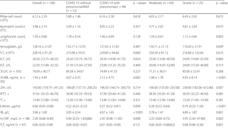
Laboratory data
The laboratory data in different groups are summarized in Table2. D-dimer, fibrinogen, and high sensitive C re-action protein (hs-CRP) were higher in COVID-19
pa-tients with pneumonia than in those without
pneumonia. Laboratory data including blood routine, liver injury index, cardiac injury index, and other coagu-lation index were similar in both groups. The compari-son of the data among the three groups showed a positive association between creatine kinase-MB, lactic
dehydrogenase, D-dimer, fibrinogen, procalcitonin
(PCT), hs-CRP and COVID-19 severity. A negative asso-ciation between lymphocyte count, hemoglobin and COVID-19 severity was observed. There was a trend
Table 1The baseline demographic and clinical data in different groups
Overall (n= 106) COVID-19 without pneumonia/Mild (n= 12)
COVID-19 with pneumonia(n= 94)
p–values Moderate (n= 69) Severe (n= 25) p–values
Age, years 52.75 ± 16.09 43.92 ± 13.73 53.87 ± 16.08 0.043 51.41 ± 15.77 60.68 ± 15.23 0.005 Male sex, n(%) 53 (50.0) 4 (33.3) 49 (52.1) 0.220 34 (49.3) 15 (60.0) 0.309 Coexisting disorder
Any, n(%) 25 (23.6) 0 (0.0) 25 (26.6) 0.041 14 (20.3) 11 (44.0) 0.007 Hypertension,
n(%)
17 (16.0) 0 (0.0) 17 (18.1) 0.108 9 (13.0) 8 (32.0) 0.028 Diabetes
mellitus, n(%)
13 (12.3) 0 (0.0) 13 (13.8) 0.169 7 (10.1) 6 (24.0) 0.103 CHD, n (%) 2 (1.9) 0 (0.0) 2 (2.1) 1.000 1 (1.4) 1 (4.0) 0.578 COPD, n (%) 1 (0.9) 0 (0.0) 1 (1.1) 1.000 1 (1.4) 0 (0.0) 1.000 Symptoms
Fever, n (%) 56 (52.8) 3 (25.0) 53 (56.4) 0.040 37 (53.6) 16 (64.0) 0.082 Cough, n (%) 52 (49.1) 5 (41.7) 47 (50.0) 0.587 34 (49.3) 13 (52.0) 0.839 Sputum
production, n (%)
14 (13.2) 1 (8.3) 13 (13.8) 0.596 7 (10.1) 6 (24.0) 0.179
Fatigue, n (%) 20 (18.9) 0 (0.0) 20 (21.3) 0.076 13 (18.8) 7 (28.0) 0.124 Dyspnea, n (%) 7 (6.6) 0 (0.0) 7 (7.4) 0.328 3 (4.3) 4 (16.0) 0.097 Vital signs
Temperature(°C)
36.50 (36.30–36.80) 36.50 (36.33–36.60) 36.50 (36.30–36.83) 0.270 36.50 (36.30–36.65) 36.90 (36.60–37.70) 0.001 Respiratory Rate 20.00 (20.00–20.00) 19.50 (19.00–20.00) 20.00 (20.00–20.25) 0.002 20.00 (20.00–20.00) 20.00 (20.00–22.00) 0.001 Heart Rate 89.89 ± 13.29 93.17 ± 14.48 89.47 ± 13.15 0.366 88.94 ± 13.94 90.92 ± 10.80 0.545 SpO2(%) 98.00 (97.00–98.00) 98.00 (97.25–99.00) 98.00 (97.00–98.00) 0.174 98.00 (97.00–99.00) 96.00 (93.50–98.00) < 0.001
toward increased neutrophil count and activated partial thromboplastin time with the aggravation of the disease severity, but did not reach statistical significance.
Cytokine profiles between COVID-19 without and with pneumonia
Compared with COVID-19 without pneumonia,
COVID-19 with pneumonia had significantly higher
serum IL-2R, IL-6, and TNF-α. Serum cytokines
includ-ing IL-2R, IL-6, IL-8, and TNF-α.were increased signifi-cantly with COVID-19 severity. The cytokine profiles in different groups are showed in the Table3.
Correlation analysis between lung injury and cytokine in COVID-19 with pneumonia
We used CT severity score and PaO2/FiO2 ratio to
as-sess lung injury in COVID-19 patients with pneumonia. Arterial blood gas analysis was not routinely performed in COVID-19 patients. Generally, it was more often per-formed in more severe COVID-19 patients. In total, 94 COVID-19 patients with pneumonia had the data of CT severity score, and 51 COVID-19 patients with
pneumo-nia had the data of PaO2/FiO2 ratio. In COVID-19
pa-tients with pneumonia, severe group had significantly
higher CT severity score and lower PaO2/FiO2 than
moderate group (Fig. 1). Correlation analysis between
Table 2The laboratory data in different groups
Overall (n= 106) COVID-19 without pneumonia/Mild (n= 12)
COVID-19 with pneumonia(n= 94)
p–values Moderate (n= 69) Severe (n= 25) p–values
White-cell count, ×109/L
6.12 ± 2.29 5.80 ± 1.46 6.16 ± 2.38 0.618 6.03 ± 2.17 6.49 ± 2.92 0.615
Neutrophil count,× 109/L
3.98 ± 2.14 3.44 ± 1.16 4.05 ± 2.23 0.357 3.77 ± 2.02 4.81 ± 2.64 0.075
Lymphocyte count,
×109/L 1.50 ± 0.68 1.78 ± 0.54 1.46 ± 0.69 0.128 1.59 ± 0.67 1.12 ± 0.66 0.003
Hemoglobin, g/L 128.16 ± 21.07 132.17 ± 13.70 127.65 ± 21.83 0.487 130.71 ± 21.13 119.20 ± 21.91 0.049
PLT, ×109/L 228.16 ± 91.20 215.08 ± 59.31 229.83 ± 94.60 0.600 234.59 ± 81.12 216.68 ± 125.64 0.615
ALT, U/L 24.50 (13.75–40.25) 24.50 (10.75–39.75) 24.50 (14.00–41.75) 0.654 25.00 (13.00–40.50) 24.00 (14.00–42.50) 0.883
AST, U/L 22.50 (15.00–32.25) 21.50 (15.50–27.50) 23.00 (15.25–35.25) 0.460 20.00 (14.50–52.00) 24.00 (15.50–46.00) 0.313
CK,U/L (n= 105) 76.09 ± 40.77 84.58 ± 34.67 74.99 ± 41.53 0.227 71.31 ± 36.51 85.00 ± 52.41 0.268
CK-MB, ng/mL (n = 105)
1.95 ± 4.49 0.67 ± 0.33 2.12 ± 4.75 0.083 1.08 ± 1.78 4.95 ± 8.14 < 0.001
LDH, U/L 193.00 (159.75–241.25) 180.00 (157.75–200.25) 196.50 (160.75–260.75) 0.214 190.00 (155.00–235.00) 239.00 (182.00–672.80) 0.007
APTT, s 37.65 (35.55–40.70) 36.90 (35.33–39.53) 37.90 (35.60–41.20) 0.440 38.30 (35.65–41.20) 36.50 (32.25–40.50) 0.085
PT, s 13.40 (12.80–13.93) 13.20 (12.90–13.68) 13.40 (12.83–14.00) 0.321 13.40 (12.90–14.00) 13.20 (11.85–14.30) 0.281
d-dimer,μg/mL 0.06 (0.05–0.08) 0.22 (0.22–0.22) 0.37 (0.22–0.81) 0.009 0.28 (0.22–0.64) 0.70 (0.32–1.50) < 0.001
FIB, g/L 3.96 ± 1.60 3.05 ± 0.54 4.07 ± 1.65 < 0.001 3.78 ± 1.47 4.89 ± 1.87 0.001
Hs-CRP, mg/L (n= 98) 2.30 (0.68–8.40) 0.90 (0.33–1.83)(86) 2.45 (0.98–11.85) 0.008 2.25 (0.60–8.75) 3.95 (2.30–47.80) 0.002
PCT, ng/ml (n= 97) 0.06 (0.05–0.08 0.06 (0.05–0.07) 0.07 (0.05–0.09) 0.121 0.06 (0.05–0.08)(62) 0.08 (0.06–0.30) 0.001
Abbreviation:ILinterleukin,TNFtumor necrosis factor,Hs-CRPhigh-sensitivity C-reactive protein,CTcomputed tomography,PCTprocalcitonin,COVID-19 coronavirus disease 2019,PLTplatelet,ALTalanine aminotransferase,ASTaspartate aminotransferase,CK creatine kinase, LDHlactic dehydrogenase,APTT activated partial thromboplastin time,PTprothrombin time,FIBfibrinogen
Table 3The cytokine profile in different groups
Overall (n= 106) COVID-19 without pneumonia/Mild (n= 12)
COVID-19 with pneumonia(n= 94)
p–values Moderate (n= 69) Severe (n= 25) p–values
IL-1β, pg/mL 5.00 (5.00–5.00) 5.00 (5.00–5.00) 5.00 (5.00–5.00) 0.726 5.00 (5.00–5.00) 5.00 (5.00–5.00) 0.829 IL-2R, U/mL 424.50 (268.75–692.75) 260.00 (229.25–358.50) 463.50 (281.50–716.75) 0.002 381.00 (266.00–631.00) 725.00 (471.00–968.50) < 0.001 IL-6, pg/mL 3.48 (1.63–11.39) 1.85 (1.50–2.74) 3.86 (1.79–13.60) 0.016 2.90 (1.59–7.29) 17.05 (3.95–120.00) < 0.001 IL-8, pg/mL 9.30 (6.25–12.48) 7.05 (6.15–11.18) 9.35 (6.45–12.95) 0.223 8.90 (5.80–12.05) 11.50 (9.05–18.00) 0.036 IL-10, pg/mL 5.00 (5.00–5.00) 5.00 (5.00–5.53) 5.00 (5.00–5.00) 0.849 5.00 (5.00–5.00) 5.00 (5.00–6.44) 0.198 TNF-α, pg/
mL
7.60 (6.10–9.70) 6.35 (5.43–7.15) 7.95 (6.28–9.95) 0.016 7.50 (6.10–9.70) 8.70 (7.40–11.85) 0.007
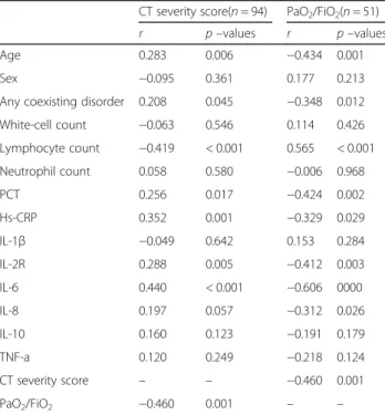
lung injury index and demographic data, laboratory data, as well as cytokines is presented in Table 4. CT severity score and PaO2/FiO2 were significantly correlated with age, presence of any coexisting disorder, lymphocyte count, PCT, IL-2R, and IL-6. IL-8 was significantly cor-related with PaO2/FiO2, but not with CT severity score. The correlations between cytokines and CT severity
score, PaO2/FiO2 are showed in Figs. 2 and 3,
respectively.
Predictors of lung injury in COVID with pneumonia
Stepwise multiple linear regression analyses were used to evaluate independent variables associated with CT se-verity score and PaO2/FiO2. The variables with statistical significance in Table4 were taken as candidates for fur-ther stepwise multiple linear regression analyses. Inde-pendent variables including age, presence of any coexisting disorder, lymphocyte count, log IL-6, log IL-8, log IL-2R, log PCT and log hs-CRP were entered into the regression model, and CT severity score was taken as dependent variable. The results showed that only log IL-6 was included in the final model (β= 0.397, adjusted r2= 0.147, p< 0.001). When PaO2/FiO2 was taken as dependent variable, the analysis identified the log IL-6 as
the only independent explanatory variables for PaO2/
FiO2(β=−0.434, adjusted r2= 0.169,p= 0.003).
Discussion
Main findings of this retrospective study are as follows: 1. COVID-19 patients with pneumonia had higher levels
of IL-2R, IL-6, and TNF-α than COVID-19 patients
without pneumonia. 2. Both IL-2R and IL-6 were statis-tically correlated with the severity of lung injury
accessed by CT severity score and PaO2/FiO2 in
COVID-19 patients with pneumonia.3. IL-6 was the in-dependent predictor of the severity of lung injury in COVID patients with pneumonia after controlling for confounders. The findings of this study highlight the role of cytokines in mediating lung injury in COVID-19 pneumonia.
Cytokine storm is characterized by excessive inflam-matory reaction in which proinflaminflam-matory cytokines are increasingly released in response to microbial infection. The process can result in tissue injury and an
unfavor-able prognosis in infectious disease [8]. This
phenomenon has been noted in COVID-19 [9] as well as
other coronavirus disease such as SARS [10] and MERS
[11]. ALI/acute respiratory distress syndrome (ARDS) is a common consequence of a cytokine storm in systemic circulation and the lung alveolar environment [11]. As early as 2004, Wong et al. [12] found that SARS patients had marked elevation of Th1 cytokine interferon (IFN)-gamma, inflammatory cytokines IL-1, IL-6 and IL-12 for at least 2 weeks after disease onset. Another study focus-ing on the cytokine profiles in MERS patients found that the severe group had significantly higher serum levels of IL-6 and CXCL-10 than the mild group, which suggested that IL-6 and CXCL-10 were elevated in MERS patients who developed severe diseases [13].
Some researchers have noticed the cytokine responses associated with SARS-CoV-2 infection and investigated the cytokine profiles in COVID-19 patients. Huang et al.
[4] found that ICU COVID-19 patients had higher
plasma levels of cytokine profiles than those in non-ICU
Fig. 1Comparisons of the CT severity score and PaO2/FiO2between moderate group and severe group (a. CT severity score;
b. PaO2/FiO2)
Table 4Spearman rank correlation coefficients between lung injury (CT severity score and PaO2/FiO2) and demographic data,
laboratory data, and cytokines
CT severity score(n= 94) PaO2/FiO2(n= 51) r p–values r p–values Age 0.283 0.006 −0.434 0.001 Sex −0.095 0.361 0.177 0.213 Any coexisting disorder 0.208 0.045 −0.348 0.012 White-cell count −0.063 0.546 0.114 0.426 Lymphocyte count −0.419 < 0.001 0.565 < 0.001 Neutrophil count 0.058 0.580 −0.006 0.968 PCT 0.256 0.017 −0.424 0.002 Hs-CRP 0.352 0.001 −0.329 0.029 IL-1β −0.049 0.642 0.153 0.284 IL-2R 0.288 0.005 −0.412 0.003 IL-6 0.440 < 0.001 −0.606 0000 IL-8 0.197 0.057 −0.312 0.026 IL-10 0.160 0.123 −0.191 0.179 TNF-a 0.120 0.249 −0.218 0.124 CT severity score – – −0.460 0.001 PaO2/FiO2 −0.460 0.001 – –
COVID-19 patients. Hou et al. [14] showed that lympho-cytes were significantly decreased while cytokines
in-cluding IL-8, TNF-α, IL-2R, IL-10 and IL-6 were
significantly increased with increased severity of
COVID-19. SARS-CoV-2 infection could result in injury to multiple organs leading to multiorgan failure. Previ-ous studies mainly focused on the relationship between cytokines profiles and the severity of COVID-19, which
was characterized by a respiratory and systemic
infec-tious disease. Lung was the main targeted organ during SARS-CoV-2 infection and ARDS was the most import-ant cause of COVID-19 death. Therefore, we aimed to evaluate the role of cytokines in lung injury in COVID-19 patients and attempted to find out a potential thera-peutic target for the management lung injury in COVID-19 pneumonia.
Fig. 2Correlations between cytokines and CT severity score (a. IL-1β;b. IL-2R;c. IL-6;d. IL-8;e. IL-10;f. TNF-α)
In this study, CT severity score and PaO2/FiO2 were used to evaluate extent of lung lesions and hypoxemia respectively. These two indexes were chosen based on the murray score [15], which is used to characterize the severity of lung injury. We revealed that COVID-19 pa-tients with pneumonia had significantly higher levels of
serum IL-2R, IL-6, and TNF-αthan COVID-19 patients
without pneumonia. This result indicated that the eleva-tion of cytokines was significantly associated with pres-ence of COVID-19 pneumonia. Then we further analyzed data of patients with COVID-19 pneumonia and found that both serum IL-2R and IL-6 were statisti-cally correlated with the severity of lung injury. The findings suggested that cytokines play an important role in the lung injury in COVID-19 pneumonia. We noted that lymphocyte count was positively correlated with
PaO2/FiO2in COVID-19 patients with pneumonia. This
was supported by a previous study, which showed that the lower lymphocyte count was commonly seen in COVID-19 patients and was significantly correlated with disease severity. The results of flow cytometry showed that the lower lymphocyte count in COVID-19 was largely attributed to the decrease in number of T
lym-phocytes including CD4+ T cells and CD8+ T cells [5].
Our results indicated that the COVID-19 pneumonia pa-tients with significantly lower lymphocyte count should be closely monitored due to higher risk of respiratory failure and ARDS.
The results of the present study have some clinical im-plications. We found that IL-6 was the independent pre-dictor of the severity of lung injury in COVID patients with pneumonia after controlling for confounders. The findings directly provide the evidence supporting the fa-vorable outcomes of IL-6R blockers tocilizumab
treat-ment in COVID-19 patients. Tocilizumab can
specifically block IL-6 from binding to the soluble and membrane-bound IL-6R and inhibit signal transduction of inflammatory process [16]. It has been suggested as a treatment of COVID-19 [17]. A study included 25 severe COVID-19 patients who received tocilizumab therapy reported that tocilizumab was associated with dramatic decline in inflammatory markers, radiological improve-ment and reduced ventilatory support requireimprove-ments [18]. Another study retrospectively analyzing the outcomes of tocilizumab treatment in 21 severe and critical COVID-19 patients showed that tocilizumab was associated with immediate improvement of the symptoms, hypoxygen-mia, and CT opacity changes in most of the patients [19]. Our results, together with other study findings, suggest that IL-6 could serve as a useful marker to guide tocilizumab therapy in COVID-19. It also can be used to predict the efficacy of tocilizumab therapy in COVID-19. Targeted therapy based on IL-6 level may be helpful to alleviate lung injury in COVID-19 pneumonia and
decrease mortality. SARS-CoV-2 infection can lead to injury of multiple organs, it will be interesting to explore whether IL-6 in systemic circulation mediates other organ or tissue injury in COVID-19. Future studies are needed to clarify this issue.
Several limitations of this study should be considered when interpreting results. Firstly, the sample size of the present study was relatively small, especially the number of patients in the group of COVID-19 without pneumo-nia was limited; thus statistical non-significance may occur because of insufficient power. Secondly, the study was a retrospective design, which might result in some biases (eg, unclear records, incomplete data). Thirdly, there was not any critical ill case in the present study, which might restrict the generalizability of the results to critical ill COVID-19 patients. Fourthly, only 6 cytokines which are assumed to play crucial roles in COVID-19 pneumonia were evaluated. Other cytokines, which may also play important roles in infectious diseases were not measured in the present study..
Conclusions
Our study showed that elevation of circulating cytokines was significantly associated with presence of pneumonia in 19 and the severity of lung injury in COVID-19 pneumonia. Circulating IL-6 independently predicted the severity of lung injury in COVID-19 pneumonia. The findings of our study could help to better under-stand the role of cytokines in COVID-19 associated lung injury and highlight a potential therapeutic target for the management lung injury in COVID-19 pneumonia.
Abbreviations
COVID-19:Coronavirus disease 2019; SARS-CoV-2: Severe acute respiratory syndrome coronavirus 2; CT: Computed tomography; IL: Interleukin; WHO: World Health Organization; SARS: Severe acute respiratory syndrome; MERS: Middle east respiratory syndrome; ICU: Intensive care unit; TNF: Tumor necrosis factor; hs-CRP: High-sensitivity C-reactive protein; PCT: Procalcitonin; ALI: Acute lung injury; ARDS: Acute respiratory distress syndrome;
IFN: Interferon
Acknowledgements
Not applicable.
Authors’contributions
LDC, ZYZ, XJW and XBZ conceived and designed the study, analyzed the data and drafted this manuscript. YQC, WZY, MHW and QFH contributed to analysis of the data, and revising of the manuscript. All authors have read and approved the final manuscript.
Funding
This work was supported by Youth Research Fund from Fujian Provincial Health Bureau (2018-2-65), and Natural Science Foundation of Fujian Province (2018 J01393). The sponsor had no role in study design, study conduction, data collection, data analysis, data interpretation, or writing of
thereport.
Availability of data and materials
Ethics approval and consent to participate
This retrospective study was approved by the Ethics Committee of Zhongshan Hospital, Xiamen University. All subjects gave signed informed consent.
Consent for publication
Not applicable.
Competing interests
The authors declare that they have no known competing financial interests or personal relationships that could have appeared to influence the work reported in this paper.
Author details
1Department of Respiratory and Critical Care Medicine, Zhangzhou Affiliated
Hospital of Fujian Medical University, Zhangzhou, Fujian Province, China.
2Department of Geriatrics, Zhongshan Hospital, Xiamen University; Teaching
Hospital of Fujian Medical University, Xiamen, Fujian, China.3Department of Pulmonary and Critical Care Medicine, Fujian Third People’s Hospital, Fuzhou, Fujian, China.4Department of Pulmonary and Critical Care Medicine,
Zhongshan Hospital, Xiamen University; Teaching Hospital of Fujian Medical University, No. 201, Hubin Nan Road, Siming District, Xiamen, Fujian Province 361004, People’s Republic of China.
Received: 23 May 2020 Accepted: 23 July 2020
References
1. Yao XH, Li TY, He ZC, Ping YF, Liu HW, Yu SC, Mou HM, Wang LH, Zhang HR, Fu WJ, et al. A pathological report of three COVID-19 cases by minimally invasive autopsies. Zhonghua Bing Li Xue Za Zhi. 2020;49(0):E009. 2. Shimabukuro-Vornhagen A, Godel P, Subklewe M, Stemmler HJ, Schlosser
HA, Schlaak M, Kochanek M, Boll B, von Bergwelt-Baildon MS. Cytokine release syndrome. J Immunother Cancer. 2018;6(1):56.
3. Moore JB, June CH. Cytokine release syndrome in severe COVID-19. Science. 2020;368(6490):473–4.
4. Huang C, Wang Y, Li X, Ren L, Zhao J, Hu Y, Zhang L, Fan G, Xu J, Gu X, et al. Clinical features of patients infected with 2019 novel coronavirus in Wuhan, China. Lancet. 2020;395(10223):497–506.
5. Chen G, Wu D, Guo W, Cao Y, Huang D, Wang H, Wang T, Zhang X, Chen H, Yu H, et al. Clinical and immunological features of severe and moderate coronavirus disease 2019. J Clin Invest. 2020;130(5):2620–9.
6. Chung M, Bernheim A, Mei X, Zhang N, Huang M, Zeng X, Cui J, Xu W, Yang Y, Fayad ZA, et al. CT imaging features of 2019 novel coronavirus (2019-nCoV). Radiology. 2020;295(1):202–7.
7. Li K, Fang Y, Li W, Pan C, Qin P, Zhong Y, Liu X, Huang M, Liao Y, Li S. CT image visual quantitative evaluation and clinical classification of coronavirus disease (COVID-19). Eur Radiol. 2020;30(8):4407–16.
8. Tisoncik JR, Korth MJ, Simmons CP, Farrar J, Martin TR, Katze MG. Into the eye of the cytokine storm. Microbiol Mol Biol Rev. 2012;76(1):16–32. 9. Sun X, Wang T, Cai D, Hu Z, Chen J, Liao H, Zhi L, Wei H, Zhang Z, Qiu Y,
et al. Cytokine storm intervention in the early stages of COVID-19 pneumonia. Cytokine Growth Factor Rev. 2020;53:38–42.
10. Huang KJ, Su IJ, Theron M, Wu YC, Lai SK, Liu CC, Lei HY. An interferon-gamma-related cytokine storm in SARS patients. J Med Virol. 2005;75(2):185– 94.
11. Channappanavar R, Perlman S. Pathogenic human coronavirus infections: causes and consequences of cytokine storm and immunopathology. Semin Immunopathol. 2017;39(5):529–39.
12. Wong CK, Lam CW, Wu AK, Ip WK, Lee NL, Chan IH, Lit LC, Hui DS, Chan MH, Chung SS, et al. Plasma inflammatory cytokines and chemokines in severe acute respiratory syndrome. Clin Exp Immunol. 2004;136(1):95–103. 13. Kim ES, Choe PG, Park WB, Oh HS, Kim EJ, Nam EY, Na SH, Kim M, Song KH,
Bang JH, et al. Clinical progression and cytokine profiles of Middle East respiratory syndrome coronavirus infection. J Korean Med Sci. 2016;31(11): 1717–25.
14. Hou H, Zhang B, Huang H, Luo Y, Wu S, Tang G, Liu W, Mao L, Mao L, Wang F, et al. Using IL-2R/lymphocytes for predicting the clinical progression of patients with COVID-19. Clin Exp Immunol. 2020;201(1):76–84.
15. Murray JF, Matthay MA, Luce JM, Flick MR. An expanded definition of the adult respiratory distress syndrome. Am Rev Respir Dis. 1988;138(3):720–3.
16. Jones SA, Scheller J, Rose-John S. Therapeutic strategies for the clinical blockade of IL-6/gp130 signaling. J Clin Invest. 2011;121(9):3375–83. 17. Fu B, Xu X, Wei H. Why tocilizumab could be an effective treatment for
severe COVID-19? J Transl Med. 2020;18(1):164.
18. Alattar R, Ibrahim TBH, Shaar SH, Abdalla S, Shukri K, Daghfal JN, Khatib MY, Aboukamar M, Abukhattab M, Alsoub HA, et al. Tocilizumab for the treatment of severe coronavirus disease 2019. J Med Virol. 2020.https://doi. org/10.1002/jmv.25964.
19. Xu X, Han M, Li T, Sun W, Wang D, Fu B, Zhou Y, Zheng X, Yang Y, Li X, et al. Effective treatment of severe COVID-19 patients with tocilizumab. Proc Natl Acad Sci U S A. 2020;117(20):10970–5.
Publisher’s Note