CHARACTERIZATION OF THE CASZ1-DEPENDENT MECHANISMS ESSENTIAL FOR CARDIOMYOCYTE DEVELOPMENT
Kerry M. Dorr
A dissertation submitted to the faculty of the University of North Carolina at Chapel Hill in partial fulfillment of the requirements for the degree of Doctor of Philosophy in
Genetics and Molecular Biology in the School of Medicine.
Chapel Hill 2015
Approved by:
Frank Conlon
Terry Magnuson
Li Qian
Joan Taylor
ii © 2015 Kerry M. Dorr
iii ABSTRACT
Kerry M. Dorr: Characterization of the Casz1-Dependent Mechanisms
Essential for Cardiomyocyte Development
(Under the direction of Frank L. Conlon)
The heart is one of the first structures to form during development and is required for embryonic growth and survival. The four-chambered mammalian heart arises from a series of complex processes during embryonic development that includes the
specification and differentiation of the different cardiac cell types within the heart,
proliferation, and morphological movements of the early heart fields. Early development of the heart is governed by hyperplastic growth in which cardiac cells undergo mitogen-dependent activation during the G1-phase of the cell cycle. Though numerous growth factor signals have been shown to be required for cardiomyocyte proliferation, genetic studies have only identified a limited number of transcription factors that act to regulate the entry of cardiomyocytes into S-phase. Casz1 is an evolutionarily conserved
transcription factor that is essential for heart development; however, there are vast deficiencies in our understanding of the mechanism by which Casz1regulates aspects of cardiac development.
Here we report that Casz1is expressed in, and gives rise to, cardiomyocytes in the first and second heart fields. We show through the generation of a cardiac
iv
of the cardiac chambers. We further demonstrate that Casz1 is essential for the proliferation of cardiomyocytes in both heart fields and that loss of Casz1 leads to severe cardiac hypoplasia, ventricular septal defects, and a decrease in cardiomyocyte cell number. Additionally, we report that the loss of Casz1 leads to a prolonged or
v
vi
ACKNOWLEDGEMENTS
There are many people that have been critical to the success of my graduate studies. I would first like to thank current and past members of the Conlon Lab for helpful discussions, advice, support, and technical know-how: Kathleen Christine, Chris Showell, Panna Tandon, Erin Kaltenbrun, Lauren Kuchenbrod, Lauren Waldron, Nirav Amin, Stephen Sojka, Kerry Dorr, Marta Charpentier, Chris Slagle, Michelle Villasmil, Leslie Kennedy, and Carrie Wilczewski. Special thanks to Chris Showell for being an excellent mentor and friend my first few years in the lab, and to Erin Kaltenbrun for being my lab BFF. This experience would not have been the same without her sarcastic and charming wit.
vii
I would also like to thank my committee members Terry Magnuson, Li Qian, Joan Taylor, and Andy Wessels. I am so grateful for the mentorship and support that I have received from each of them. One of the things that drew me to UNC was the faculty commitment to good training, and my committee has demonstrated this commitment throughout my graduate experience. I am extremely appreciative for their insights and helpful discussions on my work.
viii
TABLE OF CONTENTS
LIST OF FIGURES ... xii
LIST OF TABLES ... xv
LIST OF ABBREVIATIONS ... xvi
CHAPTER 1: INTRODUCTION The cardiomyocyte cell cycle ... 3
Cardiomyocyte cell cycle control and growth ... 5
Proliferation ... 6
Binucleation ... 9
Hypertrophy ... 12
Growth factors and signaling pathways that regulate proliferation... 15
Hippo ... 15
Wnt ... 17
Insulin-like growth factor ... 20
Neuregulin ... 21
Periostin ... 22
Fibroblast Growth Factor ... 23
MicroRNAs regulate cardiomyocyte proliferation ... 26
Transcription factors that regulate cardiomyocyte proliferation ... 28
ix
Jumonji ... 29
Hopx ... 30
Meis1 ... 31
Tbx1 ... 32
Tbx5 ... 32
Tbx20 ... 33
Casz1-Dependent Mechanisms ... 34
Dissertation Goals ... 35
References ... 42
CHAPTER 2: CASZ1 IS REQUIRED FOR CARDIOMYOCYTE G1-TO-S PROGRESSION DURING MAMMALIAN CARDIAC DEVELOPMENT INTRODUCTION ... 77
MATERIALS AND METHODS ... 79
RESULTS ... 88
Casz1 is expressed in the developing myocardium ... 88
Casz1-expressing cells give rise to first and second heart field derivatives ... 90
Casz1 is essential for cardiac development ... 91
Casz1 is required for growth of the cardiac chambers ... 93
Casz1 embryonic nulls phenocopy Casz1f/f;Nkx2.5Cre/+ embryos ... 93
Casz1 is essential in the second heart field ... 94
x
Casz1 is required for cardiomyocyte G1-toS-phase transition ... 96
DISCUSSION ... 97
Casz1 and the G1-to-S phase transition ... 98
REFERENCES ... 123
CHAPTER 3: DISCUSSION AND FUTURE DIRECTIONS Discussion ... 129
Casz1 is an essential cardiac transcription factor required for development ... 130
Casz1 regulates G1-to-S-phase cardiomyocyte cell cycle progression ... 132
Future Directions ... 135
REFERENCES ... 145
APPENDIX 1: CASZ1 PROMOTES VASCULAR ASSEMBLY AND MORPHOGENESIS THROUGH THE DIRECT REGULATION OF AN EGFL7/RHOA-MEDIATED PATHWAY INTRODUCTION ... 157
MATERIALS AND METHODS ... 159
RESULTS ... 163
EC expression of CASZ1 is evolutionarily conserved ... 163
CASZ1 is required for vascular patterning and lumen morphogenesis ... 164
CASZ1 regulates EC behavior ... 166
Isolation of the Egfl7/miR-126 locus by CASZ1 chromatin immunoprecipitation ... 168
xi
EGFL7 functions downstream of CASZ1 to control vascular
morphogenesis ... 170
EGFL7 and miR-126 function downstream of CASZ1 to regulate cell adhesion and shape ... 172
CASZ1 and EGFL7 regulate RhoA ... 173
DISCUSSION ... 174
CASZ1 regulates EC behavior via EGFL7/RhoA to promote assembly of a well-branched vascular network ... 175
CASZ1 and RhoA ... 175
Transcriptional regulation of Egfl7 and miR-126 ... 177
Conclusions ... 178
REFERENCES ... 209
APPENDIX 2: TRANSCRIPTIONAL REGULATION OF BLOOD VESSEL FORMATION: THE ROLE OF THE CASZ1/EGFL7/RHOA PATHWAY INTRODUCTION ... 215
REFERENCES ... 220
APPENDIX3: THE CARDIAC TBX5 INTERACTOME REVEALS A CHROMATIN REMODELING NETWORK ESSENTIAL FOR ATRIAL SEPTATION INTRODUCTION ... 221
MATERIALS AND METHODS ... 222
RESULTS ... 229
DISCUSSION ... 234
xii
LIST OF FIGURES
Figure 1.1. The cardiomyocyte cell cycle ... 36
Figure 1.2. Control of cardiomyocyte growth ... 37
Figure 1.3. Cardiomyocyte structure ... 38
Figure 1.4. Growth factors that regulate cardiomyocyte proliferation ... 39
Figure 1.5. miRNAs that regulate cardiomyocyte proliferation ... 40
Figure 2.1. Casz1 is expressed in the developing heart ... 100
Figure 2.2. Lineage tracing with Casz1CreERT2-neolabels cardiomyocytes in first and second heart field derivatives ... 102
Figure 2.3. Casz1 is required for cardiac development ... 103
Figure 2.4. Casz1 null embryos phenocopy Casz1 cardiac null embryos ... 105
Figure 2.5. Casz1 is required in the second heart field ... 107
Figure 2.6. Casz1 regulates cardiomyocyte proliferation ... 109
Figure 2.7. Casz1 is essential for cardiac G1 to S cell cycle transition ... 111
Figure S2.1. CASZ1 is expressed in PML bodies ... 112
Figure S2.2. Generation of a Casz1CreERT2-neolineage tracing allele... 113
Figure S2.3. Generation of a conditional Casz1 allele ... 115
Figure S2.4. Nkx2.5Cre Lineage analysis ... 117
Figure S2.5. Casz1 cardiac null embryos exhibit severe cardiac defects ... 118
Figure S2.6. Casz1 is required for myocardial development ... 119
Figure S2.7 RNA-Seq Analysis ... 120
Figure S2.8 Loss of Casz1 does not lead to programmed cell death ... 121
xiii
Figure A1.2. CASZ1 is Required for Vascular Development and Lumen
Formation ... 181
Figure A1.3. CASZ1 Regulates EC Behavior ... 183
Figure A1.4. CASZ1 Directly Activates Egfl7 Transcription ... 185
Figure A1.5. EGFL7-Depletion in Embryos and HUVECs Phenocopies CASZ1-Depletion ... 187
Figure A1.6. EGFL7 and miR-126 Play Distinct Roles Downstream of CASZ1 ... 190
Figure A1.7. A model describing CASZ1 function in endothelial cells ... 192
Figure SA1.1. CASZ1 is Required for Vascular Development ... 193
Figure SA1.2. CASZ1 is Required for EC Proliferation ... 195
Figure SA1.3. Sequence Alignment of miR-126 and Expression of miR-126 in CASZ1-Depleted Embryos ... 197
Figure SA1.4. EGFL7-Depletion Phenocopies CASZ1-Depletion During Vascular Development ... 199
Figure SA1.5. miR-126 Depletion Does Not Phenocopy CASZ1-Depletion During Vascular Development ... 201
Figure SA1.6. Efficacy of Ad-Egfl7 and Ad-miR-126 ... 203
Movie SA1.1. Time-lapse imaging of control and shCasz1 HUVECs ... 205
Movie SA1.2. EGFL7-depletion phenocopies CASZ1-depletion in HUVECs ... 205
Movie SA1.3. Restoration of Egfl7 or miR-126 levels rescues the adhesion defects of CASZ1-depletion ... 205
Movie SA1.4. Time-lapse imaging of sprouting control and shCasz1 HUVECs ... 206
Movie SA1.5. EGFL7-depletion phenocopies CASZ1-depletion in the sprouting angiogenesis assay ... 206
Figure A2.1. Proper assembly of vascular networks ... 219
Figure A3.1. TBX5 interacts with the NuRD complex ... 236
xiv
Figure A3.3. TBX5 functions to represses neural and cancer
genes in cardiac tissue ... 239 Figure A3.4. Congenital heart disease associated mutations of TBX5
disrupt TBX5-NuRD complex activity ... 241 Figure SA3.1 Generation of the Tbx5Aviknock-in mouse ... 243 Figure SA3.2 Transcription interaction network assembled by
STRING analysis ... 244 Figure SA3.3 TBX5 and the NuRD complex genetically interact ... 245 Figure SA3.4 Transcriptional targets not repressed by TBX5 ... 246 Figure SA3.5 The TBX5NID is necessary and sufficient for interaction
of TBX5 with CHD4 ... 247 Figure SA3.6 Sequence alignment of the TBX5NID from 48 TBX5
Ortholgues ... 248 Figure SA3.7 The TBX5/NuRD complex binds to neural and cancer
genes in vivo ... 249 Figure SA3.8 Targets repressed by TBX5 in a NuRD independent manner ... 250
xv
LIST OF TABLES
Table 1.1. The major cyclins and cyclin-dependent kinases
involved in the cardiomyocyte cell cycle ... 41
Table S2.1. Casz1 primer sequences ... 122
Table SA1.1 Putative direct targets of CASZ1... 207
Table SA1.2 Oligonucleotides used for quantitative Real-Time PCR ... 208
Table SA3.1. Gene-ontology analysis of candidate interactions from TBX5 complexes... 251
Table SA3.2. Genes differentially expressed in E9.5 Tbx5 null hearts by RNA-Seq Analysis ... 251
Table SA3.3. Genes potentially repressed by TBX5 ... 251
Table SA3.4. Genotyping primers for Tbx5Avi/Avi; Rosa26BirA/BirAmice ... 251
Table SA3.5. TBX5 target gene primers for transcriptional assays ... 252
Table SA3.6. Site directed mutagenesis primer pairs. Amino acid changes are represented in bold ... 253
xvi
LIST OF ABBREVIATIONS
ACTC Cardiac Actin
AD Activation Domain
α/β-MHC Alpha/Beta-Myosin Heavy Chain
AMPK AMP-activated protein kinase
APC Adenomatosis Polyposis Coli
AS Atrial Septum
ASD Atrial Septal Defects
Avi Biotin acceptor peptide
AVSD Atrioventricular Septal Defects
BAC Bacterial Artificial Chromosome bHLH Basic Helix Loop Helix
BIO 6-bromoindirubin-3′-oxime
BMP Bone Morphogenetic Protein
BrdU 5’-bromo-2’-deoxyuridine
CAC Common Atrial Chamber
CC Cardiac Crescent
CDKs Cyclin-dependent kinases
CHD Congenital Heart Disease
ChIP chromatin immunoprecipitation
CL Compact Layer
cTNT Cardiac troponin T
xvii DAPI 4',6-diamidino-2-phenylindole
DE Differential Expression
E Embryonic day or Eye
EC Endothelial Cells
ECM Extracellular Matrix
EdU 5-ethynyl-2’deoxyuridine
EGFL7 Epidermal Growth Factor-Like Domain 7
EGFP Enhanced green fluorescent protein
EN Endocardium or Endothelial Cells
ESC Embryonic Stem Cell
EPI Epicardium
ERK Extracellular-signal-related kinase
EXE Extra-embryonic tissue
FACS Fluorescence activated cell sorting FG Foregut
FGF Fibroblast growth factor
FGFR Fibroblast growth factor receptor tyrosine kinase
FHF First heart field
FILA FilaminA
GSK3 Serine-threonine kinase glycogen synthase kinase 3
GMT GATA4, MEF2C and TBX5
GO Gene Ontology
xviii HB Hindbrain
HCM Hypertrophic Cardiomyopathy
HDAC Histone Deacetylase
HOS Holt-Oram Syndrome
HEK Human Embryonic Kidney
HT Heart Tube
HUVECs Human umbilical vein endothelial cells IGF Insulin-like growth factor
iPSCs induced pluri-potent stem cells
IVS Interventricular Septum or of intersomitic vessels JNK c-Jun N-terminal kinase
LA Left atria
LB Limb Bud
LH Looped Heart
LSC Laser scanning cytometry
LV Left ventricle
LEF Lymphoid Enhancer Binding Factor miRNA MicroRNA
MI Myocardial infarction
MIMS Multi-isotope Imaging Mass Spectrometry
MTA Metastasis-associated Protein
MBD3 Methyl-CpG Binding Domain Protein 3
MO Morpholino
xix MEF2 Myocyte Enhancer Factor 2 MOI multiplicity of infection
MP Myocardial Progenitor Cells
MS Mass Spectrometry
MV Mitral valve
MY Myocardium MYBPC3 Cardiac myosin-binding protein C MYH7 Cardiac β-myosin heavy chain MYL2 Regulatory myosin light chain MYL3 Essential myosin light chain
N Neural Tube
15N Stable Isotope of Nitrogen
NB Neuroblastoma
NID NURD interacting domain
NLS Nuclear Localization Signal
NP Nasal Placode
NRG1 Neuregulin1
NT Neural Tube
NuRD Nucleosome Remodeling and Deacetylase
OFT Outflow Tract
P Postnatal day
xx
PECAM Platelet endothelial cell adhesion molecule PFA Paraformaldehyde pHH3 phosphorylated Histone H3
PME Pharyngeal Mesoderm
PML Promyelocytic Leukemia
R Restriction Point
RA Right atria
RAF Rapidly accelerated fibrosarcoma kinase
RAS Rat sarcoma kinase
RASIP1 Ras interacting protein 1
RB Retinoblastoma protein
RLM-RACE RNA ligase-mediated rapid amplification of cDNA ends
ROCK Rho kinase
RTKs Trans-membrane receptor tyrosine kinases RT-PCR Reverse transcriptase polymerase chain reaction
RV Right ventricle
SEM Standard Error of Measure or Scanning Electron Microscopy SHF Second heart field
shRNA short hairpin RNA
S Somites or Sarcomere
SOS Son of Sevenless
SRF Serum Response Factor
xxi
T Telencephalon
Tbx T-box Containing Protein
TGFβ Transforming Growth Factor Beta TMY Tropomyosin
TNNI3 Cardiac Troponin I
TNNT2 Cardiac Troponin T
TPM1 α-Tropomyosin
TR Trabeculae
TV Tricuspid Valve
UTR Untranslated Region
VEGF Vascular Endothelial Growth Factor VSD Ventricular Septal Defect
VVN Vitelline Vein Network WT Wild-type
1
Chapter 1: Introduction
The heart is one of the first structures to form during development, and heart
precursor cells are among the first cells of the epiblast to undergo gastrulation (Lawson
et al., 1991; DeRuiter et al., 1992; Parameswaran and Tam, 1995; Tam et al., 1997;
Saga et al., 1999; Saga et al., 2000; Buckingham et al., 2005; Devine et al., 2014;
Lescroart et al., 2014). In murine development, following ingression in the early to
mid-primitive streak stages, mesodermal heart precursor cells begin to migrate
anterolaterally as a sheet, and by the late primitive streak stages, they are localized as
identifiable bilateral fields that lie on either side of the ventral midline forming a structure
known as the cardiac crescent (Parameswaran and Tam, 1995; Tam et al., 1997; Saga
et al., 2000; Shenje et al., 2014). Heart precursor cells that form the cardiac crescent
are derived from two mesodermal populations: termed a first heart field (FHF) and a
second heart field (SHF). Cardiac precursors of the first heart field undergo
differentiation and assemble into a primitive heart tube by embryonic day 8.5 (E8.5)
(Tam et al., 1997; Saga et al., 1999; Meilhac et al., 2004; Buckingham et al., 2005;
Tzouanacou et al., 2009; Devine et al., 2014; Lescroart et al., 2014). This tube is
lengthened by the addition of newly differentiated myocardium derived from the SHF,
giving rise to the arterial and venous poles (Kelly et al., 2001; Mjaatvedt et al., 2001;
Waldo et al., 2001; Cai et al., 2003; Meilhac et al., 2003; Meilhac et al., 2004; Zaffran et
2
During the early stages of heart development, cardiomyocytes of the FHF and
SHF are highly proliferative, resulting in substantial growth of the embryonic heart (Tam
et al., 1997; Buckingham et al., 2005; Tzouanacou et al., 2009; van den Berg et al.,
2009; Dominguez et al., 2012; Meilhac et al., 2014). In both rodents and humans, the
overall rate of cardiomyocyte proliferation gradually declines concomitant with the onset
of terminal cardiomyocyte differentiation (Soonpaa et al., 1996; Christoffels et al., 2000;
Pasumarthi and Field, 2002; Sedmera et al., 2003; Toyoda et al., 2003; Walsh et al.,
2010; Ikenishi et al., 2012). After this period, the mammalian heart continues to grow,
largely through hypertrophy and recruitment of cells from extra-cardiac sources
including the neural crest and epicardium (Viragh and Challice, 1981; Gorza et al.,
1988; Kirby, 1990; Kirby and Waldo, 1995; Li et al., 1996; Creazzo et al., 1998; Manner
et al., 2001; Brown and Baldwin, 2006; Ahuja et al., 2007; Winter and Gittenberger-de
Groot, 2007; Zhou and Pu, 2008; van Wijk et al., 2009; Kelly, 2012; Maillet et al., 2013).
All animal life is dependent upon the sustained function of the heart. It is
therefore not surprising that genetic and acquired cardiac diseases can result in
devastating consequences with congenital heart defects representing the number one
type of human birth defect, and cardiovascular disease accounting for the most
common cause of adult death worldwide (Hoffman, 1995; Hoffman and Kaplan, 2002;
Lopez et al., 2006; Heron et al., 2009; Dolk et al., 2010a; Dolk et al., 2010b; van der
Linde et al., 2011; Fahed et al., 2013; Pediatric Cardiac Genomics et al., 2013). The
mammalian heart has limited regenerative potential, which is in stark contrast to the fish
or amphibian heart, which retains robust capacities for regeneration via the activation of
3
et al., 2006; Jopling et al., 2010; Kikuchi et al., 2010; Jopling et al., 2012; Piatkowski et
al., 2013). Cardiomyocyte loss is the causal etiology of many forms of heart disease;
therefore, strategies aimed at repopulating the damaged heart with replacement
cardiomyocytes could potentially restore cardiac structure and more importantly, cardiac
function.
The cardiomyocyte cell cycle
The eukaryotic cell cycle consists of four coordinated processes that include cell
growth, DNA replication, distribution of duplicated chromosomes into daughter cells,
and cell division. The cycle of most eukaryotic cells is composed of four distinct phases:
S-phase, where DNA synthesis occurs; M-phase, where karyokinesis and cytokinesis
occur; and G1-phase and G2-phases, where cells continue to grow and prepare for entry
into their respective, subsequent phase. The central core of the cell cycle machinery
comprises the cyclin-dependent kinases (Cdks) and cyclins. In most eukaryotes, there
are different Cdks that control different stages of the cell cycle. Cdks are activated via
complex formation with phase-specific cyclins, leading to changes in the
phosphorylation of intracellular proteins that both regulate cell cycle events and
advance the cell cycle through the stage that the particular Cdk controls (Mitchison,
1971; Baserga, 1985; Murray and Hunt, 1993; Sherr, 1993; Morgan, 1997; Pavletich,
1999; Nurse, 2000; Hochegger et al., 2008; Lim and Kaldis, 2013; Alberts, 2015). Cdk
activity is terminated by either cyclin protein degradation or by Cdk inhibitors, such as
INK family members (p15, p16, p18 and p19) and Cip/Kip family members (p21, p27
4
al., 1994; Roberts et al., 1994; Chan et al., 1995; Hirai et al., 1995; Russo et al., 1996;
Brotherton et al., 1998; Russo et al., 1998; Pavletich, 1999; Sherr and Roberts, 1999;
Lim and Kaldis, 2013). Cyclin protein synthesis and degradation determine the timing of
the activation of the cyclin’s partner Cdk. The Cdks are critical for cell cycle progression
because their inactivation prevents mitosis (Figure 1.1) (Devault et al., 1991; van den
Heuvel and Harlow, 1993; Morgan, 1997; Nurse, 2000; Lim and Kaldis, 2013; Alberts,
2015). There are four classes of cyclins that are defined by the cell cycle stage during
which they complex with Cdks: G1-cyclins, which promote passage through the start of
the cell cycle; G1/S-cyclins, which commit the cell to DNA replication; S-cyclins, which
initiate DNA replication; and M-cyclins, which promote mitosis (Sherr, 1993; Morgan,
1997; Hochegger et al., 2008; Alberts, 2015).
Cell cycle activity is an essential component of cardiac differentiation and
morphogenesis. Detailed analyses of cyclins and Cdks have revealed dramatic changes
in cell cycle patterns during embryonic and postnatal stages of cardiac development.
Positive cell cycle regulators are highly expressed in the embryonic heart and
downregulated in the adult heart while cell cycle inhibitors show the opposite expression
pattern (Soonpaa and Field, 1998; Pasumarthi and Field, 2002; Goetz et al., 2006;
Ahuja et al., 2007; Walsh et al., 2010; Ikenishi et al., 2012). Protein expression
analyses by western blot for cyclin E, cyclin A, cyclin D1, and cyclin B1 in heart
ventricles demonstrate a synchronous change in expression during embryonic and
postnatal stages. Cyclin levels are high during early embryonic stages (E12) and
gradually decrease as embryogenesis proceeds with minimal levels detected at birth.
5
decline, and cyclins are undetectable in adult ventricles. Concomitant with these data,
western blot analyses of the cyclin E-Cdk2, cyclin A-Cdk1/2, and cyclin B1-Cdk1
complexes also demonstrated a similar synchronous pattern with high levels present in
early embryogenesis (E12), followed by a gradual decline, a surge at P5, and a rapid
decline to undetectable levels in adult ventricles (Ikenishi et al., 2012). Terminally
differentiated cardiomyocytes show repression of cell cycle activators such as the
Cyclin/Cdk complexes (described above), Myc, and E2F transcription factors.
Conversely, negative cell cycle regulators such as INK4, p21, p27, retinoblastoma
protein (Rb), and cyclin-dependent kinase inhibitors are increased (Table 1.1) (Kim et
al., 1994; Flink et al., 1998; Li et al., 1998; Pasumarthi and Field, 2002).
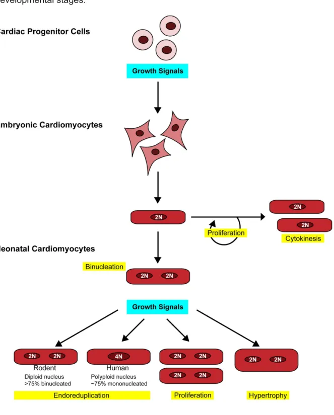
Cardiomyocyte cell cycle control and growth
Mammalian cardiomyocytes exhibit three developmentally determined forms of
cell cycle control and growth: proliferation, binucleation and hypertrophy. During
embryonic development, the heart primarily grows via hyperplasia or cell proliferation,
which is defined as an increase in cell number by cell division. Shortly after birth,
proliferation ceases and cardiomyocytes undergo cell-cycle withdrawal, following an
additional round of DNA synthesis and mitosis without cytokinesis, which leads to the
formation of binucleated cardiomyocytes. The postnatal heart primarily grows via
hypertrophy, or an increase in cell size (Figure 1.2) (Erokhnia, 1968a; Erokhnia, 1968b;
Dowell and McManus, 1978; Clubb and Bishop, 1984; Oparil et al., 1984; Goldspink et
al., 1986; Li et al., 1996; Soonpaa et al., 1996; Bergmann et al., 2009; Walsh et al.,
6
required for normal heart morphogenesis and determines the appropriate heart size
necessary for pumping a large volume of blood.
Proliferation
Tritiated thymidine analyses (Erokhnia, 1968a; Erokhnia, 1968b) that measure
DNA synthesis demonstrate that cardiomyocyte proliferation is high during early murine
embryogenesis at E8, with labeling indices of approximately 70% of precardiac
mesodermal cells. These levels start to decrease around E10-12, with labeling indices
plunging to 45%. This decrease is concomitant with the onset of cardiomyocyte
differentiation (Erokhnia, 1968a; Erokhnia, 1968b). As embryogenesis proceeds,
cardiomyocyte proliferation gradually declines (Erokhnia, 1968a; Erokhnia, 1968b;
Soonpaa et al., 1996; Toyoda et al., 2003; Walsh et al., 2010; Zeng et al., 2014).
Fluorescence-activated cell sorting (FACS) analyses of purified populations of E14
cardiomyocytes treated with 5’-bromo-2’-deoxyuridine (BrdU) revealed cardiomyocyte
DNA synthesis levels of 22.95% (Walsh et al., 2010). This work is further supported by
5-ethynyl-2’deoxyuridine (EdU) analyses, an alternative thymidine analog, of sectioned
cardiac tissue at E10, E17, P7, P21 and in the adult, demonstrating a decrease in
cardiomyocyte proliferation as embryogenesis proceeds, with no EdU incorporation
observed after P21 (Zeng et al., 2014).
The proliferative capacity of adult cardiomyocytes is quite limited; the frequency
and source of new cardiac cells is somewhat controversial, as different labs have
reported dissimilar results, most likely due to differences in experimental techniques and
7
Field, 1998; Bergmann et al., 2009; Walsh et al., 2010; Mollova et al., 2013; Senyo et
al., 2013; Ali et al., 2014; Naqvi et al., 2014). Tritiated thymidine analyses of α-cardiac
myosin heavy chain-positive ventricular myocardium revealed DNA synthesis levels of
0.0006% in wild-type 12 week-old adult murine cardiomyocytes (Soonpaa and Field,
1997). Additionally, DNA synthesis levels increased to 0.0083% in 12 week old mice
subjected to a myocardial injury, in which the myocardium was cauterized midway
between the apex and base of the heart (Soonpaa and Field, 1997). FACS analysis of cardiomyocyte populations treated with BrdU revealed DNA synthesis levels of 9.6% for
P3 neonates, which dropped to 0.94% by P7, 0.02% at P14, and was undetectable by 3
weeks of age (Walsh et al., 2010). More recently, using genetic fate-mapping with
stable isotope labeling and multi-isotope imaging mass spectrometry (MIMS), it was
shown that cardiomyocytes proliferate at a low rate by the division of pre-existing
cardiomyocytes during normal aging. This process increases 4-fold in areas adjacent to
myocardial injury (Senyo et al., 2013). MIMS is a technique that utilizes a rare stable
isotope of nitrogen (15N) and allows for high-resolution quantitation (Steinhauser et al.,
2012). Treatment with 15N-thymidine for 8 weeks at three different timepoints beginning
at P4 (neonates), and in 10 week (young adult) and 22 month-old (old adult) adults
followed by MIMS analysis revealed dramatic changes in DNA synthesis throughout
aging (Senyo et al., 2013). The percentage of 15N-labeled cardiomyocytes per day in the
neonatal heart was 1.00%; percentages in the young adult heart were 0.015% and
0.007% in the old adult heart (Senyo et al., 2013). In line with these observations,
recent mouse studies revealed that mammals possess the ability to regenerate heart
8
Interestingly, a recent mouse study confirmed the previously identified postnatal time
window during which cardiomyocyte cell numbers increased during P1-4, and
suggested the existence of a second tight, transient, proliferative time window at P15,
leading to a 40% increase in predominantly binucleated cardiomyocytes (Naqvi et al.,
2014).
In humans, cardiomyocyte proliferation can be observed until 20 years of age,
which is much longer than anticipated, based on the data detailed above for mice and
rats. Of particular significance and in contrast with mice, myocardial growth in humans
occurs through the simultaneous cycles of cardiomyocyte proliferation and cell
enlargement. However, similar to mice, cardiomyocyte cycling and division decreases
with age (Li et al., 1996; Soonpaa and Field, 1997; Soonpaa and Field, 1998; Bergmann
et al., 2009; Mollova et al., 2013). A novel 14C-based, birth-dating technique, which can
establish the age of cardiomyocytes based on the integration of 14C into human DNA,
demonstrated that cardiomyocytes renew with an annual turnover rate of 1.9% for a 19
year-old individual, decreasing to 1% between the ages of 21 and 40 years, with levels
decreasing to 0.45% by 75 years of age (Bergmann et al., 2009). A more recent study
supporting these results utilized a combined approach that first determined the
percentage of cardiomyocytes in M-phase by laser-scanning cytometry (LSC) of
myocardial sections that stained positive for phosphorylated histone H3 (pHH3), an
M-phase marker, followed by a stereological quantification technique to quantify the
number of cardiomyocytes present in the myocardium. LSC is a highly sensitive,
microscopy-based cytofluorometry method that measures fluorescence emitted from
9
dissection method is a stereological technique that obtains quantitative information
about three-dimensional tissues from two-dimensional sections (Muhlfeld et al., 2010).
This study identified 0.8–1.1 ͯ 109 newly generated cardiomyocytes during the first year
of life; 0.9 ͯ 109 new cardiomyocytes during years 1–10; and 0.6 ͯ 109 new
cardiomyocytes during years 10–20 (Mollova et al., 2013). Consistent with these data,
morphometric analyses revealed that during physiological hypertrophy, the
cardiomyocyte nuclear DNA content remains constant. However, as the heart grows in
size and when the weight of the myocardium reaches approximately 210 grams, there
appears to be a linear increase in the percentage of cardiomyocyte nuclear DNA
content as cardiac mass increases. Because human hearts do not change nuclear
number with hypertrophy, this observation indicates that new cardiomyocytes are
generated during this period of growth (Adler, 1975; Adler and Costabel, 1975; Adler
and Friedburg, 1986; Herget et al., 1997; Laflamme and Murry, 2011).
Binucleation
The cell cycle ends in the division of the cytoplasm by cytokinesis. In a typical
cell, cytokinesis accompanies every round of mitosis. However, some cells undergo
mitosis without cytokinesis, resulting in a multinucleated cell (Rappaport, 1996; Glotzer,
1997; Field et al., 1999). This type of endoreduplication generally occurs in tissues with
high metabolic activity and in cells that are terminally differentiated (MacAuley et al.,
1998; Larkins et al., 2001; Gonzalez et al., 2006; Zielke et al., 2013). In the heart, the
transition from hyperplastic to hypertrophic growth is associated with the formation of
binucleated cardiomyocytes (Oparil et al., 1984), resulting from incomplete cell division
10
embryonic burst of proliferation (E10–12), karyokinesis and cytokinesis are matched,
resulting in cardiomyocyte proliferation. In contrast, during early neonatal stages (P4–6),
karyokinesis occurs in the absence of cytokinesis, resulting in binucleation of
cardiomyocytes (Li et al., 1996; Soonpaa et al., 1996; Li, F. et al., 1997b; Walsh et al.,
2010). In mice and rats, binucleation begins around P4; by 3 weeks of age, 85–90% of
cardiomyocytes are binucleated with normal diploid DNA content in each nucleus
(Clubb and Bishop, 1984; Li et al., 1996; Soonpaa et al., 1996; Porrello et al., 2011). In
humans, these percentages range from 25–57% (Schmid and Pfitzer, 1985; Olivetti et
al., 1996). Notably, humans do not display a transition to predominantly binucleated
cardiomyocytes, maintaining relatively high levels (~64%) of mononucleated
cardiomyocytes up to 20 years of life. However, cardiomyocyte cytokinesis ceases after
this time point, resulting in mononucleated cells with tetraploid (4n) or higher DNA
content (Adler and Costabel, 1975; Olivetti et al., 1996; Mollova et al., 2013).
The mechanism by which binucleation occurs remains unclear. However, the
actin-myosin contractile ring, the structure that generates the force necessary to
separate the daughter cells after mitosis, forms during the binucleation process
indicating that the molecules involved in later stages of cytokinesis are responsible for
binucleation (Li, F. et al., 1997b; Miller, 2011). One possible explanation is that the
presence and incomplete disassembly of myofibrils in the equator region where
cleavage furrows normally form could physically impede cytokinesis (Li, F. et al.,
1997a). In muscle, for example, contractile myofibrils are organized into sarcomeres.
Recent evidence suggests that during karyokinesis, the sarcomeric Z-disks and
11
sarcomeric structure is absent, indicating that cardiomyocyte division involves
sarcomeric disassembly (Figure 1.3) (Bersell et al., 2009). Interestingly, recent studies
suggest that multinucleation could result from defects in the recruitment of the
contractile ring protein, anillin (Straight et al., 2005; Smith et al., 2013). Anillin binds to myosin II heavy chain and helps to focus myosin at the contractile ring (Straight and Field, 2000). Depletion of anillin leads to disorganized contractility and cytokinesis failure (Field and Alberts, 1995; Straight et al., 2005; Smith et al., 2013).
The physiological significance of possessing binucleated cells also remains to be
elucidated. There are few examples of multinucleated cells in higher-level vertebrates;
however, most examples of multinucleation occur in highly metabolically active cells
such as osteoclasts, hepatocytes and muscle cells (Holtzer et al., 1957; Okazaki and
Holtzer, 1966; Kahn and Simmons, 1975; Hall and Ralston, 1989; Manolagas and Jilka,
1995; Rossi et al., 2000; Miyaoka et al., 2012). Multinucleated cells often occur in
pathological conditions due to aberrant cell cycle control (Gillespie and Stevens, 1972;
Most et al., 1997; Sagona and Stenmark, 2010; Weihua et al., 2011). It has been
hypothesized that multinucleation is advantageous, generating twice the amount of RNA
for greater protein synthesis. Indeed, transcriptional activity that influences specialized
cellular functions has been implicated in other multinucleated cell types (Boissy et al.,
2002). Another advantage specific to muscle cells could be adaptation to the
physiological process of contraction, which requires myofibrils to maintain their
organization along their entire lengths. An increase in cross-sectional area could lead to
12
Hypertrophy
The size of an organ depends both on the number and size of the cells it
contains. In mammals, nutritional status, growth factors, and hormones play important
roles in the control of organ size (Conlon and Raff, 1999). The heart is an essential
pump and its size is tightly regulated across species to ensure proper function. The
heart must be large enough to generate a physiological cardiac output, but not so large
as to block cardiac output, which is common in cardiomyopathies (Seidman, 2000;
Seidman and Seidman, 2001; Heallen et al., 2011). During the postnatal period, the
cardiomyocytes of both rodents and humans undergo a period of physiological growth,
increasing in size by 30–40-fold (Adler and Costabel, 1975; Li et al., 1996).
Between preadolescence (P10) and puberty (P35), mouse body weight (in
grams) increases by 3.8-fold, whereas mouse heart weight (in milligrams) increases by
3.5-fold. At the cellular level, cardiomyocyte length increases by 46 µm with little change
in diameter. During this period of growth, the left ventricle increases in weight 3.47-fold,
and cardiomyocyte volume increases 1.7-fold (Adler, 1975; Adler and Costabel, 1975;
Laflamme and Murry, 2011; Naqvi et al., 2014). This interesting discrepancy, suggests
that an increase in cardiomyocyte number also occurs during this time period (Naqvi et
al., 2014). A similar trend is observed in humans, where the cellular volume of
cardiomyocytes increases by 8.6-fold between birth and 20 years of age; this increase
is concomitant with an increase in the number of cardiomyocytes (Mollova et al., 2013)
as well as an increase in the number of non-cardiomyocytes including fibroblasts and
endothelial cells (Adler and Costabel, 1975). Physiological hypertrophy is initiated by
13
and receptor (VEGFR1), insulin and insulin-like growth factor 1 (IGF1). These trigger intracellular signalling pathways that regulate protein synthesis, metabolism and energy production (Reiss et al., 1996; Bellomo et al., 2000; Karpanen et al., 2008; Bry et al., 2010). Growth signals are controlled by ERK1/2, PI3K, AKT and mTOR complex 1 (mTORC1), whereas AMP-activated protein kinase (AMPK) governs metabolic adaptive reprogramming (Maillet et al., 2013) (Bueno et al., 2000; Lips et al., 2004; Purcell et al., 2007; Kehat and Molkentin, 2010; Kehat et al., 2011; Horman et al., 2012).
One very important distinction with regard to hypertrophy is to highlight the difference between normal hypertrophy during heart growth and pathological
hypertrophy. Cardiomyocytes increase in size in response to biomechanical stress, a process known as pathological hypertrophy (Seidman and Seidman, 2001).
Pathological hypertrophy is functionally, mechanistically, and histologically distinguished from normal embryonic and postnatal myocardial growth by characteristic changes in cardiac myocyte shape and volume (Bishop, 1990; Dorn, 2007). In humans,
14
level, the major features of HCM are cardiomyocyte and myofibrillar disarray,
hypertrophy, and myocardial fibrosis. Genetic causes of HCM, which are autosomal dominant, are now estimated to account for at least 50% of sporadic childhood onset cases (Seidman, 2000; Seidman and Seidman, 2001; Morita et al., 2008).
Consequently, the need for genetic screens for HCM could greatly impact human health.
Adult onset pathological hypertrophy is also induced by acquired cardiac deficiencies such as hypertension, valvular disease, myocardial infarction (MI), and endocrine disorders. In these types of cardiac pathologies, chronic pressure overload results in concentric hypertrophy with a small, thick-walled left ventricle that is
associated with significantly increased risk of heart failure and malignant arrhythmia (Grossman et al., 1975; Levy et al., 1990b; Levy et al., 1990a; Koren et al., 1991; Morita et al., 1996; Dorn et al., 2003; Frey et al., 2004; Heineke and Molkentin, 2006; Dorn, 2007; van Berlo et al., 2013). Genes upregulated in adult onset pathological
hypertrophy include brain natriuretic peptide, angiotensin-converting enzyme, the endothelin receptor, and the β-adrenergic receptor kinase (Iemitsu et al., 2001; Dorn and Force, 2005). In addition, studies have implicated the calcium–calcineurin–NFAT and protein kinase A pathways in regulating the pathological growth of the heart. Other examples of mediators of pathological cardiac hypertrophy are the p38 and c-Jun N-terminal kinase (JNK) branches of the MAPK cascade. Sustained p38 or JNK activation in the heart leads to cardiomyopathy and heart failure (Heineke and Molkentin, 2006; Maillet et al., 2013; van Berlo et al., 2013).
15
Growth factors that regulate cardiomyocyte proliferation
Cardiomyocytes from both FHF and SHF undergo hyperplastic growth in
response to input from a large network of growth factor signaling pathways; such
pathways integrate a suite of transcription factors and microRNAs (miRNAs), which are
proposed to directly regulate the cardiac cell cycle (Lavine et al., 2005; Ahuja et al.,
2007; Bersell et al., 2009; Heallen et al., 2011; Porrello et al., 2011; Xin et al., 2011;
Eulalio et al., 2012; von Gise et al., 2012; Wadugu and Kuhn, 2012; Xin et al., 2013).
Many of these growth factors are secreted by the epicardium, an epithelial sheet of cells
that forms over the heart, and several lines of evidence suggest that cardiomyocyte
proliferation in the developing myocardium is influenced by these secreted factors
(Rumyantsev, 1977; Chen et al., 2002; Sengbusch et al., 2002; Kang and Sucov, 2005;
Sun et al., 2007; Sucov et al., 2009; Shen et al., 2015). Additionally, in cell culture,
many mitogens have been shown to stimulate the proliferation of fetal cardiomyocytes
(Figure 1.4) (Armstrong et al., 2000).
Hippo pathway
16
include the Ste-20-like protein kinases Mst1 and Mst2, which form a complex with the WW-repeat scaffolding protein Salv that phosphorylates Lats1 and Lats2. These Lats kinases complex with Mob, which phosphorylates and inactivates Yap and Taz. Yap and Taz partner with transcription factors, such as Tead, to regulate gene expression (Chan et al., 2005; Callus et al., 2006; Dong et al., 2007; Zhao, B. et al., 2007; Lei et al., 2008; Praskova et al., 2008; Pan, 2010).
The conditional depletion of the Salv gene using Nkx2.5-Cre (Moses et al., 2001), an early cardiac marker, leads to perinatal lethality due to heart enlargement (cardiomegaly) (Heallen et al., 2011). Furthermore, phenotypic analysis at E14.5 revealed that cardiac expression of a Salv mutant caused expansion of trabecular and subcompact ventricular myocardial layers, thickened ventricular walls, and enlarged ventricular chambers, but showed no change in myocardial cell size (Heallen et al., 2011). In addition, conditional mutants of Lats2 and Mst1/2 showed similar myocardial expansion phenotypes in the heart (Heallen et al., 2011).
Yap1 is a major target of the Hippo signaling pathway and is inactivated by Hippo kinase activity (Dong et al., 2007; Zhao, B. et al., 2007). Conditional depletion of the Yap1 gene in the embryonic heart, utilizing either the Tnnt2-Cre (Jiao et al., 2003) or the Nkx2.5-Cre driver (Moses et al., 2001), results in cardiac abnormalities and
embryonic lethality by E16.5 due to myocardial hypoplasia (Xin et al., 2011; von Gise et al., 2012). Yap1-null mutants showed a significant reduction in the cardiomyocyte mitotic index by pHH3 staining as well as reduced DNA synthesis by EdU analysis, indicating that Yap1 is required for cardiomyocyte proliferation (Xin et al., 2011).
17
vitro and in vivo. Treatment of embryos at E8.5 with a doxocycline-inducible Yap1
transgene results in embryonic lethality by E15.5, with embryos exhibiting cardiomegaly (von Gise et al., 2012). Histological examination revealed dramatic myocardial
overgrowth with moderate thickening of the compact myocardium and marked
expansion of the trabecular myocardium. Furthermore, Yap1 gain-of-function mutants showed enhanced cardiomyocyte proliferation (von Gise et al., 2012).
The above-mentioned studies, demonstrate that Hippo signaling inhibits
cardiomyocyte proliferation. A recent study found that depletion of Hippo signaling in the adult myocardium is an endogenous repressor of cardiomyocyte renewal and
regeneration. Inactivating either the Salv or Lats1/2 genes with a tamoxifen-inducible Cre, the Myh6CreERT2 transgene (Sohal et al., 2001), in the adult heart leads to cell cycle re-entry and a significant induction of DNA synthesis, measured by EdU and Ki67 analyses (Heallen et al., 2011). Moreover, Hippo deficiency promoted efficient heart regeneration in both postnatal cardiac apex resection and adult myocardial infarction models, revealing a crucial, inhibitory role for Hippo signaling in cardiomyocyte renewal and regeneration (Heallen et al., 2013).
Wnt pathway
β-catenin-18
independent pathway (Komiya and Habas, 2008). The canonical Wnt signaling pathway begins with Wnt proteins binding to a coreceptor complex composed of the frizzled (Fzd) family, seven-pass transmembrane proteins and lipoprotein receptor-related 5/6 (Lrp5/6) proteins. Wnt receptor binding activates the intracellular effector protein dishevelled, which in turn inactivates a protein complex that includes the constitutively active serine-threonine kinase glycogen synthase kinase 3 (Gsk3), as well as the
scaffolding proteins axin and adenomatosis polyposis coli (APC). This complex normally phosphorylates β-catenin and targets it for degradation (He et al., 2004; Gordon and Nusse, 2006; Komiya and Habas, 2008; MacDonald et al., 2009; Rao and Kuhl, 2010). The hallmark of the canonical Wnt pathway is the accumulation and translocation of the adherens junction associated-protein β-catenin into the nucleus (He, 2004; Gordon and Nusse, 2006). In contrast, the noncanonical pathway is further divided into two distinct branches: the planar cell polarity pathway, known for its role in epithelial cell orientation, and the Wnt/Ca2+ pathway, known for its role in cell movement during gastrulation (Strutt, 2002; Klein and Mlodzik, 2005; Barrow, 2006; Green et al., 2008; Komiya and Habas, 2008; De, 2011) .
Wnt signaling is essential for cardiogenesis and regulates many aspects of cardiac morphogenesis as well as the self-renewal and differentiation of cardiac
19
cardiomyocytes with the small molecule 6-bromoindirubin-3′-oxime (BIO), a specific inhibitor of Gsk3 promoted proliferation in vitro (Meijer et al., 2003). Stimulation of both neonatal and adult rat cardiomyocytes with BIO induced S-phase entry, upregulated positive cell cycle regulators, and increased the cardiomyocyte mitotic index. In addition, BIO treatment elevated β-catenin activity in cardiomyocytes, suggesting that the
increase in proliferative ability may be due, in part, to the activation of the canonical Wnt pathway (Tseng et al., 2006).
Interestingly, a recent study (Heallen et al., 2011) revealed that Hippo signaling inhibits Wnt/β-catenin to regulate heart size. Gene expression profiling in Salv-cardiac null mice revealed a dramatic upregulation of canonical Wnt target genes, including
Sox2, Snai2/Slug, and Birc5/Survivin in the heart (Heallen et al., 2011). Immunostaining using β-catenin-specific antibodies revealed a 4-fold increase in nuclear β-catenin, which is normally localized to the cytosol. Genetic interaction studies with Salv-null mice combined with a conditional β-catenin-null allele (Hippo-deficient embryos with reduced β-catenin dosage) resulted in suppression of the Hippo cardiomyocyte overgrowth phenotype and returned gene expression profiles to wild-type levels(Heallen et al., 2011). These findings indicate that Wnt signaling is required for the upregulation of cardiomyocyte proliferation and cardiomegaly in Hippo mutants. Furthermore, the authors demonstrated by chromatin immunoprecipitation (ChIP) studies that Yap and β-catenin are contained within a common regulatory complex on Sox2 and Snai2.
20
control of cardiomyocyte growth by Hippo and canonical Wnt signaling (Heallen et al., 2011).
Insulin-like Growth Factor (IGF) pathway
Insulin-like growth factors 1 and 2 (IGF1 and 2) are secreted ligands that are important for embryonic growth (Le Roith, 1997). IGF2 signaling is mediated by the type-1 IGF receptor (IGF1R), whereas receptor-mediated endocytosis of type-2 IGF receptor (IGF2R) causes the degradation of excess IGF2 (Ludwig et al., 1995). In cell culture, both IGFs stimulate the proliferation of primary fetal ventricular cardiomyocytes (Liu et al., 1996; Hertig et al., 1999), and both are expressed in the epicardium during myocardial compact zone expansion (Fraidenraich et al., 2004; Li et al., 2011). Notably, combined treatment of cultured whole mouse embryos with NRG1 and IGF1 results in a substantial increase in DNA synthesis, as well as the expansion of the ventricular
compact zone leading to chamber growth and maturation (Hertig et al., 1999).
Interestingly, injection of exogenous IGF1 leads to a partial rescue of cardiac defects observed in Id-null embryos (Fraidenraich et al., 2004). Id proteins inhibit cell lineage-specific gene expression and differentiation of several different bHLH transcription factors required for development (Norton and Atherton, 1998; Norton et al., 1998; Sikder et al., 2003). Id-null mice exhibit severe cardiac abnormalities leading to embryonic lethality. Treatment of Id-null mice with IGF1 results in enhanced myocardial
proliferation and a rescue of embryonic lethality (Fraidenraich et al., 2004). The role of IGF1 in cardiomyocyte proliferation is further enhanced by the transgenic
21
promoter (α-MHC), leading to a significant increase in cardiomyocyte cell number and cardiomegaly (Reiss et al., 1996).
IGF2 is encoded by the imprinted gene Igf2 and is expressed only from the paternal allele in most tissues (DeChiara et al., 1991). Genomic imprinting affects
several mammalian genes and results in the expression of those genes from only one of the two parental chromosomes (Reik and Walter, 2001). Depletion of IGF2R, also
encoded by an imprinted gene, in mice leads to excess IGF2, causing severe hyperplasia, with heart size 2–5 times larger than that of a wild-type littermate. In addition to heart failure, these mice exhibit respiratory abnormalities and die at birth (Lau et al., 1994; Wang et al., 1994; Eggenschwiler et al., 1997). Igf2-null embryos exhibit a substantial decrease in ventricular wall cardiomyocyte proliferation leading to severe hypoplasia. Furthermore, the conditional depletion of both IGFRs in embryonic hearts also leads to a significant decrease in myocardial compact zone proliferation at E12.5 (Li et al., 2011).
Neuregulin/ErbB pathway
Neuregulin1 (NRG1) is a cell-cell signaling protein that is a ligand for receptor tyrosine kinases (RTKs) of the epidermal growth factor receptor family (ErbB1 – 4) (Falls, 2003). Binding of NRG1 to ErbB4 leads to heterodimerization with ErbB2 or homodimerization with ErbB4 (Fuller et al., 2008). Deletion of NRG1, ErbB2, or ErbB4
22
cardiomyocytes to proliferate. Following NRG1 stimulation, cardiomyocytes re-enter the cell cycle from S-phase and disassemble sarcomeres, enabling cytokinesis.
Furthermore, the conditional deletion of ErbB4 in the adult myocardium leads to a complete ablation of DNA synthesis as measured by EdU incorporation and a 20% reduction in the number of cardiomyocyte nuclei. Conversely, overexpression of ErbB4 in the adult heart leads to a 3-fold increase in EdU incorporation. Together these data demonstrate that NRG1 induces proliferation of differentiated cardiomyocytes in vivo. Most strikingly, NRG1-treated animals show significant improvements in cardiac function and structure following myocardial infarction (Bersell et al., 2009). However, a recent study contradicted these findings, demonstrating that nine consecutive daily injections of NRG1β1 did not result in an increase in DNA synthesis in either the normal or infarcted adult mouse heart (Reuter et al., 2014). Therefore, the potential for NRG1 to be used as a therapeutic regimen for cardiac regeneration remains unclear. Notably, during embryogenesis NRG1 does not play a role in proliferation, but participates in signaling between the endocardium and myocardium that is required for differentiation and maturation (Grego-Bessa et al., 2007).
Periostin
23
mesenchyme myocardium interface (Norris et al., 2004; Litvin et al., 2005; Butcher et al., 2007). It is re-expressed in adult life following cardiac injury (Stanton et al., 2000; Wang et al., 2003). Postn-null mice are viable and show very mild phenotypes, none of which are cardiovascular (Rios et al., 2005). However, following myocardial infarction,
Postn-null mice show reduced scar formation, but no change in rates of cardiomyocyte proliferation (Shimazaki et al., 2008; Lorts et al., 2009). Overexpression of full-length periostin via the α-MHC promoter in differentiated cardiomyocytes also did not increase cardiomyocyte cell cycle activity (Lorts et al., 2009). Conversely, the local delivery of a periostin peptide in rats immediately after myocardial infarction increased
cardiomyocyte proliferation, reduced scar formation, and improved both ventricular remodeling and myocardial function. In addition, periostin stimulates mononucleated cardiomyocytes to enter a full mitotic cell cycle (Kuhn et al., 2007). The discrepancy between these findings has yet to be explained but could be due to either the molecular mechanisms involved (peptide vs. gene) or the timing of delivery (acute vs. chronic overexpression (Senyo et al., 2014).
Fibroblast Growth Factor (FGF) pathway
Fibroblast growth factors (FGFs) encoded by a large family of genes are
polypeptide signaling proteins with many important functions during development. FGFs activate cell surface FGF receptors (FGFRs) which are trans-membrane RTKs. During embryonic development, FGFs play diverse roles in regulating cell proliferation,
24
variants (b-splice forms) and mesenchymal variants (c-splice forms) (Miki et al., 1992; Chellaiah et al., 1994; Naski and Ornitz, 1998; Ornitz and Itoh, 2001; Pownall and Isaacs, 2010). Binding of FGF ligands leads to the autophosphorylation of the FGFR, allowing interaction of the FRS2 docking protein, followed by activation of the
GRB2/SOS complex. Activated SOS then activates RAS and triggers a phosphorylation cascade leading to the activation of RAF, MEK, and mitogen-activated protein kinase (ERK) (Kouhara et al., 1997; Rochais et al., 2009; Pownall and Isaacs, 2010). FGFR signals are also transduced by the phospholipase C-γ pathway and the
phosphoinositide-3 kinase (PI3K) pathway (Bottcher and Niehrs, 2005; Thisse and Thisse, 2005; Beenken and Mohammadi, 2009; Knights and Cook, 2010; Pownall and Isaacs, 2010).
A role for FGF signaling in cardiomyocyte proliferation has been demonstrated from flies to humans (Beiman et al., 1996; Ornitz and Itoh, 2001; Moon, 2006; Lavine and Ornitz, 2008; Moon, 2008). Several studies have demonstrated that FGFs are mitogenic for cardiomyocytes in culture (Speir et al., 1992; Engelmann et al., 1993; Pasumarthi et al., 1996). In addition, overexpression of FGFR1 in rat cardiomyocytes leads to increased proliferation (Sheikh et al., 1999). Fgf8 depletion in cardiac
mesoderm leads to embryonic lethality due to proliferative defects in the SHF and a failure to properly elongate the developing heart tube (Ilagan et al., 2006; Park et al., 2006). Fgf10 was the first identified marker of the SHF (Kelly et al., 2001). However,
embryos lacking Fgf10 exhibit abnormal ventricular morphogenesis and malpositioning
of the heart in the thoracic cavity, but no outflow tract (OFT) alignment defects
25
Islet1-Cre) (Srinivas et al., 2001) results in a substantial reduction in proliferation (Park et al., 2008). FGFs can be classified within subfamilies, which share similar receptor-binding properties and overlapping patterns of expression (Ornitz and Itoh, 2001). Fgf9,
Fgf16 and Fgf20 compose one subfamily and are all expressed in the epicardium and endocardium of the developing heart (Colvin et al., 1999; Lavine et al., 2005). Fgf9-null mice die at birth due to lung hypoplasia and an enlarged dilated heart (Colvin et al., 2001). However, during embryonic development, Fgf9-null hearts are hypoplastic and exhibit decreased cardiomyocyte proliferation (Colvin et al., 2001; Lavine et al., 2005). Interestingly, double-knockout mice for Fgfr1 and Fgfr2 also display cardiac hypoplasia and decreased proliferation. Further work has demonstrated that FGF9 signals to the myocardium from the epicardium through Fgfr1c and Fgfr2c (Lavine et al., 2005). In line with these studies, mice lacking Fgf16 also show decreased cardiomycoyte proliferation (Hotta et al., 2008).
FGF signaling has also been shown to play a role in adult cardiomyocyte proliferation. Mammalian cardiomyocytes control the cell cycle via p38 MAP kinase, which regulates the expression of genes required for mitosis, including cyclin A and cyclin B. p38 activity is inversely correlated with cardiac growth during development, and its overexpression blocks fetal cardiomyocyte proliferation. Cardiac-specific p38α knockout mice show enhanced cardiomyocyte proliferation and pronounced cytokinesis (Engel et al., 2005; Engel et al., 2006). In contrast, genetic activation of p38 in a
26
proliferation and improved cardiac function. The addition of FGF1 alone or in
combination with the p38 inhibitor also improved angiogenesis around the scar area, possibly contributing to the survival of newly generated cardiomyocytes (Engel et al., 2006). Furthermore, the apoptosis observed with ischemia-reperfusion injury is attenuated by FGF-1 in the adult myocardium (Cuevas et al., 1997).
MicroRNAs regulate cardiomyocyte proliferation
The regulatory pathways that govern heart development and growth are modulated by several miRNAs. miRNAs, 21–25 nucleotides in length, are noncoding RNAs that play important regulatory roles in biology by targeting mRNAs for cleavage or repressing translation (Bartel, 2004; Liu and Olson, 2010; Small and Olson, 2011; Xin et al., 2013). miRNAs can be classified into three categories: intergenic, intronic, and exonic. Intergenic miRNAs are derived from their own transcriptional units in the intergenic regions of the genome. Intronic and exonic miRNAs are located within the introns and exons of host genes (protein-coding or nonprotein-coding genes) and are usually cotranscribed and coexpressed with their host genes (Bartel, 2004). Nucleotides 2–8 of the miRNA, termed the “seed” sequence located at the 5’ end, are essential for target recognition and binding. Based on the seed sequence, miRNAs can suppress collections of mRNAs that encode proteins participating in similar biological networks (Bartel, 2009).
27
development. By contrast, they have opposing roles later in the cardiac lineage when miR-1 promotes and miR-133 inhibits cardiomyocyte differentiation (Zhao, Y. et al., 2007; Ivey et al., 2008; Small and Olson, 2011). However, only a few have been
identified that specifically regulate cardiomyocyte proliferation (Liu and Olson, 2010; Xin et al., 2013). To identify pathways involved in cardiomyocyte proliferation, a
high-throughput miRNA screen was conducted, and 40 miRNAs were identified that induced DNA synthesis and increased cytokinesis in neonatal rat cardiomyocytes (Eulalio et al., 2012). Of these, miR-590-3p and miR-199a-3p were subsequently implicated in
cardiomyocyte proliferation during injury repair (Shin et al., 2002; Kook and Epstein, 2003; Eulalio et al., 2012). Interestingly, these miRNAs contain different seed
sequences, but have several of the same predicted targets. Both were found to target the Homer1 gene, which modulates Ca2+ signaling in the heart through its interaction with the Ca2+ release channel ryanodine receptor (RyR). Also, both target the Hopx gene, which encodes a homeodomain protein that inhibits cardiomyocyte proliferation (Kook and Epstein, 2003; Trivedi et al., 2010; Grubb et al., 2012). In vivo,
overexpression of miR-590-3p and miR-199a-3p in neonatal mice results in increased cardiomyocyte proliferation. Of particular note, the presence of these two miRNAs in cardiomyocytes reduces fibrotic scar size and improves cardiac function following myocardial infarction, suggesting a potential therapeutic use. Interestingly, miR-590-3p but not miR-199a-3p, was found to target Clic5, a cell proliferation inhibitor,
28
proliferation in neonatal mice and rats, what role, if any, they play in embryonic cardiomyocyte proliferation, remains to be established.
In addition, miR-15 and miR-195 (members of the miR-15 family) have been shown to regulate the cardiomyocyte cell cycle. miR-195 is highly upregulated in neonatal mouse ventricles at P10 after cardiomyocytes have exited the cell cycle (Porrello et al., 2011; Porrello et al., 2013). Overexpression of miR-195 in the developing heart leads to severe hypoplasia and cell cycle arrest during G2-phase. Further work showed that miR-195 targets Chek1, a cell cycle checkpoint kinase that promotes G2-to-M-phase transition and thus mitotic progression (Porrello et al., 2011). Conversely, inhibition of miR-15 leads to an increase in cardiomyocyte proliferation in neonatal and adult mice, and improved cardiac function following injury (Hullinger et al., 2012; Porrello et al., 2013).
Transcription factors that regulate cardiomyocyte proliferation
Cardiomyocyte proliferation is an intrinsic component of normal cardiac
morphogenesis and is dependent upon cardiac-specific transcriptional regulators as
well as exogenous signals, as described above (Dunwoodie, 2007; Smith and Bader,
2007; Lavine and Ornitz, 2008; Ikenishi et al., 2012).
Foxp1
29
al., 2004). Foxp1 is expressed in cardiomyocytes, in the endocardium, and in cells underlying the cushion mesenchyme (Lu et al., 2002; Wang et al., 2004). Global loss of the Foxp1 gene results in a complex, and slightly confusing, cardiac phenotype
characterized by defects in outflow tract septation, increased myocardial proliferation, and thinning of ventricular myocardium (Wang et al., 2004). The observations of increased cell proliferation and loss of ventricular mass in Foxp1 mutants suggest that Foxp1 may regulate cardiomyocyte proliferation in a complex manner. Loss of Foxp1 in endothelial cells using Tie2-Cre (Kisanuki et al., 2001) results in embryonic lethality that correlates with decreased ventricular mass and reduced cardiomyocyte proliferation. This finding contrasts with the increased proliferation observed in global loss-of-function mice (Zhang et al., 2010), which is the result of decreased Fgf3/16/17/20 expression in the endocardium. Loss of Foxp1 specifically in the myocardium using the Nkx2.5-Cre
(Moses et al., 2001) leads to increased proliferation (particularly in the trabecular zone) increased ventricular wall thickness, and ventricular septation defects. Interestingly, Foxp1 directly represses Nkx2.5 in the myocardium, and loss of Foxp1 leads to
increased Nkx2.5 expression (Zhang et al., 2010). Moreover, transgenic overexpression of Nkx2.5 results in increased cardiomyocyte proliferation and ventricular wall thickness, similar to that in myocardial-deficient Foxp1 mutants. Together, these data indicate that Foxp1 regulates the balance of myocardial growth in development, resulting in the controlled exit of cardiomyocytes from the cell cycle (Zhang et al., 2010).
Jumonji
30
embryonic development (Gregory et al., 1996; Kortschak et al., 2000; Jung et al., 2005a). Jumonji mutant mice exhibit hyperproliferation and overexpression of cyclin D1 (Takeuchi et al., 1995; Takeuchi et al., 1999; Toyoda et al., 2003; Jung et al., 2005b; Jung et al., 2005a). Interestingly, jumonji downregulates growth via an interaction with the cell cycle regulator, retinoblastoma protein (Rb) (Jung et al., 2005b). Further studies, revealed that enhanced expression of cyclin D1 in mutant mice decreased GATA4 protein levels and inhibited the differentiation of cardiomyocytes in a Cdk4/6-dependent manner (Nakajima et al., 2011).
Hopx
Hopx is a transcriptional corepressor that is essential for the normal development of the mammalian heart. It is expressed in the embryonic and postnatal myocardium, and functions downstream of the early cardiac-specific transcription factor, Nkx2-5. Hopxmodifies the expression of cardiac-specific genes and thereby finely regulates heart development (Kook and Epstein, 2003; Kook et al., 2006). Hopx inhibits the transcriptional activity of serum response factor (SRF) in cardiomyocytes, and, by doing so, modulates cardiac growth and proliferation. Hopx inhibits SRF-dependent
31
embryos, GATA4 hyperacetylation is associated with a substantial increase in cardiomyocyte proliferation, upregulation of GATA4 target genes, and subsequent lethality. These data suggest that HDAC2, Hopx and GATA4 coordinately regulate cardiomyocyte proliferation during embryonic development (Trivedi et al., 2010).
Meis1
Meis1 is a homeodomain transcription factor that is required for normal cardiac
development and hematopoiesis (Imamura et al., 2002; Hisa et al., 2004; Azcoitia et al., 2005). siRNA-mediated knockdown of Meis1 in neonatal rat cardiomyocytes results in a
3-fold increase in cardiomyocyte proliferation. Cardiac-specific deletion of Meis1
induces cardiomyocyte proliferation and most notably, extends the postnatal
proliferative time window without any deleterious effects on cardiac function.
Conversely, the overexpression of Meis1 in cardiomyocytes decreases proliferation.
Furthermore, Meis1is required for the transcriptional activation of the Cdk inhibitors p15ink4b, p16ink4a, and p21Cip1, suggesting that Meis1 is a critical regulator of the cardiomyocyte cell cycle, prompting cell cycle exit (Mahmoud et al., 2013).
T-box genes
32
2005; Singh et al., 2005; Stennard et al., 2005; Stennard and Harvey, 2005). They are also of great clinical relevance as mutations in Tbx genes cause a number of human disorders, such as DiGeorge syndrome (Tbx1) and Holt-Oram syndrome (Tbx5) (Basson et al., 1994; Li, Q. Y. et al., 1997; Bruneau et al., 2001; Jerome and
Papaioannou, 2001; Lindsay et al., 2001; Packham and Brook, 2003; Naiche et al., 2005; Baban et al., 2014).
The pharyngeal mesodermal progenitor cells of the SHF are situated medially to
the FHF, that gives rise to the linear heart tube and are distinguished by the expression
of genes encoding the transcription factors Isl1 and Tbx1 (Kelly et al., 2001; Mjaatvedt
et al., 2001; Waldo et al., 2001; Cai et al., 2003; Xu et al., 2004). Conditional depletion
of Tbx1 results in a shortened and narrow outflow tract and severe hypoplasia of the
SHF due to the requirement for Tbx1 in the pharyngeal mesoderm during cardiac
looping. Furthermore, Tbx1 positively regulates proliferation in the SHF through the
transcriptional regulation of Fgf8, Fgf10, and Fgfr1 (Garg et al., 2001; Hu et al., 2004;
Xu et al., 2004; Zhang et al., 2006; Liao et al., 2008).
Tbx5 is expressed very early in heart development in the cardiac crescent and
the early heart tube. As the tube develops, Tbx5 expression becomes restricted to the
cardiac regions that will form the sinus venosus, atria, and left ventricle (Bruneau et al.,
1999; Liberatore et al., 2000). Tbx5 is required for normal heart development and Tbx5-
null mice arrest around E10.5 due to severe cardiac defects (Bruneau et al., 2001).
Furthermore, mice heterozygous for the Tbx5 gene display many of the same cardiac
33
malformations (Basson et al., 1997; Li, Q. Y. et al., 1997; Bruneau et al., 2001). In
Xenopus embryos, depletion of the Tbx5 gene results in a dramatic decrease in cardiomyocyte number due to defects in cell proliferation. Furthermore, Tbx5 gene depletion leads to G1/S-phase arrest and a decrease in the embryonic cardiac mitotic index. In contrast, overexpression of Tbx5, which we show also leads to heart-specific defects, results in an increase in the cardiac mitotic index and has an inverse effect on the timing of the cardiac differentiation program. Collectively, these studies demonstrate that Tbx5 is both necessary and sufficient to determine the length of cardiac G1/S-phase and the timing of the cardiac differentiation program (Brown et al., 2005; Goetz et al., 2006).