Humanized
H19/Igf2
locus reveals diverged imprinting
mechanism between mouse and human and reflects
Silver
–
Russell syndrome phenotypes
Stella K. Hura, Andrea Freschib, Folami Ideraabdullahc, Joanne L. Thorvaldsena, Lacey J. Luensea, Angela H. Wellera, Shelley L. Bergera, Flavia Cerratob,1, Andrea Ricciob,d,1, and Marisa S. Bartolomeia,1
aDepartment of Cell and Developmental Biology, University of Pennsylvania Perelman School of Medicine, Philadelphia, PA 19104;bDepartment of
Environmental, Biological, and Pharmaceutical Sciences and Technologies, Second University of Naples, 81100 Caserta, Italy;cDepartment of Genetics,
University of North Carolina at Chapel Hill, Chapel Hill, NC 27599; anddInstitute of Genetics and Biophysics A. Buzzati-Traverso, 80131 Naples, Italy
Edited by Shirley Tilghman, Princeton University, Princeton, NJ, and approved July 14, 2016 (received for review February 24, 2016)
Genomic imprinting affects a subset of genes in mammals, such that they are expressed in a monoallelic, parent-of-origin–specific man-ner. These genes are regulated by imprinting control regions (ICRs), cis-regulatory elements that exhibit allele-specific differential DNA methylation. Although genomic imprinting is conserved in mammals, ICRs are genetically divergent across species. This raises the funda-mental question of whether the ICR plays a species-specific role in regulating imprinting at a given locus. We addressed this question at theH19/insulin-like growth factor 2(Igf2) imprinted locus, the misregulation of which is associated with the human imprinting disorders Beckwith–Wiedemann syndrome (BWS) and Silver–Russell syndrome (SRS). We generated a knock-in mouse in which the en-dogenousH19/Igf2ICR (mIC1) is replaced by the orthologous human ICR (hIC1) sequence, designated H19hIC1
. We show that hIC1 can functionally replace mIC1 on the maternal allele. In contrast, pater-nally transmitted hIC1 leads to growth restriction, abnormal hIC1 methylation, and loss ofH19andIgf2imprinted expression. Imprint establishment at hIC1 is impaired in the male germ line, which is associated with an abnormal composition of histone posttransla-tional modifications compared with mIC1. Overall, this study reveals evolutionarily divergent paternal imprinting at IC1 between mice and humans. The conserved maternal imprinting mechanism and function at IC1 demonstrates the possibility of modeling maternal transmission of hIC1 mutations associated with BWS in mice. In addition, we propose that further analyses in the paternal knock-inH19+/hIC1
mice will elucidate the molecular mechanisms that may underlie SRS.
genomic imprinting
|
DNA methylation|
H19|
IGF2|
Silver–Russell syndromeG
enomic imprinting is a conserved, epigenetic process in mam-mals that regulates the expression of a small number of genes in a monoallelic, parent-of-origin–specific manner. Typically clus-tered within domains, the parental-specific expression of imprinted genes is controlled by acis-regulatory element, the imprinting control region (ICR). During gametogenesis, ICRs acquire dif-ferential DNA methylation patterns according to the sex of the germ cells. This DNA methylation is maintained in somatic cells after fertilization but is erased in primordial germ cells, allowing the establishment of sex-specific imprints in mature gametes. The proper establishment, maintenance, and erasure of imprints are crucial for the correct expression of imprinted genes. Misregulation of imprinted genes is associated with human imprinting disorders, including Beckwith–Wiedemann syndrome (BWS), an overgrowth disorder, and Silver–Russell syndrome (SRS), an undergrowth disorder (1–3).Mouse models have been valuable to the study of imprinting at theH19/insulin-like growth factor 2(Igf2) locus, serving as a proxy for the orthologous human locus. On distal mouse chromosome 7, reciprocal imprinting of the paternally expressed fetal growth factor gene,Igf2,and the maternally expressed noncoding RNA,
H19,is regulated by the ICR located betweenH19andIgf2, herein termed imprinting center 1 (IC1) (4). IC1 is hypomethylated in female germ cells and forms a CCCTC-binding factor (CTCF)-dependent insulator in somatic cells, preventing the interaction of the Igf2promoters with downstream enhancers that are shared betweenH19andIgf2. CTCF binding at the maternal IC1 is crit-ical for maintaining its methylation-free status and silencingIgf2 expression (5). In contrast, IC1 DNA methylation is acquired dur-ing spermatogenesis. Methylation at IC1 blocks CTCF binddur-ing and allowsIgf2expression.
Comparative genome studies have revealed extensive con-servation ofH19/Igf2in therians (6). Consistently, key features of imprinting, as well as spatial organization of the mouse and human loci, are shared, including DNA methylation patterns, CTCF-binding sites (CTSs), and cis-regulatory elements. No-tably, however, the length of IC1 and the number of CTSs within IC1 have diverged; the ∼5-kb human IC1 has seven CTSs, whereas the corresponding mouse sequence is ∼2 kb and has four CTSs. In addition, with the exception of the CTCF-binding motifs, IC1 exhibits low sequence homology between mice and humans (7, 8). This raises the question of whether the human IC1 sequence could successfully regulate H19/Igf2 imprinting in the mouse. If it can, then an in vivo model for imprinting disorders associated with mutations at human IC1 could be generated (9).
Significance
Genomic imprinting is essential for mammalian development. Curiously, elements that regulate genomic imprinting, the im-printing control regions (ICRs), often diverge across species. To understand whether the diverged ICR sequence plays a
species-specific role at theH19/insulin-like growth factor2 (Igf2)
imprin-ted locus, we generaimprin-ted a mouse in which the human ICR (hIC1) sequence replaced the endogenous mouse ICR. We show that the imprinting mechanism has partially diverged between mouse and human, depending on the parental origin of the hIC1 in mouse. We also suggest that our mouse model is optimal for studying
the imprinting disorders Beckwith–Wiedemann syndrome when
hIC1 is maternally transmitted, and Silver–Russell syndrome when
hIC1 is paternally transmitted.
Author contributions: S.K.H., A.F., F.I., J.L.T., F.C., A.R., and M.S.B. designed research; S.K.H., A.F., F.I., J.L.T., L.J.L., and A.H.W. performed research; S.L.B. contributed new reagents/ana-lytic tools; S.K.H., J.L.T., and M.S.B. analyzed data; and S.K.H., F.C., A.R., and M.S.B. wrote the paper.
The authors declare no conflict of interest.
This article is a PNAS Direct Submission.
1To whom correspondence may be addressed. Email: [email protected], andrea.
Although interspecies compatibility of human IC1 was previ-ously investigated in a transgenic mouse model (8), the human transgene failed to exhibit the expected imprinting pattern. The transgene acquired DNA methylation in male germ cells in a copy number-dependent manner, but the imprint was not stably maintained in somatic cells. In addition, the humanH19transgene was abnormally expressed on paternal transmission. Interpretation of these observations is complicated by transgene copy number variation, however. Furthermore, because long-range chromatin looping plays an essential role inH19/Igf2imprinting (10, 11), the transgene insertion site may influence the phenotype. Thus, it is imperative to test the functionality of the human IC1 element at the orthologous locus.
Here we generated a knock-in mouse model in which the en-dogenous mouse IC1 (mIC1) was replaced by human IC1 (hIC1). Our goal was to investigate the extent to which hIC1 can functionally replace mIC1. We found that hIC1 properly reca-pitulates mIC1 function on the maternal allele, whereas hIC1 fails to properly regulate theH19/Igf2locus on the paternal allele. hIC1 is incompletely methylated in the male germ cells of knock-in mice, which is associated with increased enrichment of dimethylation of histone H3 at lysine 4 (H3K4me2) on hIC1. Overall, this study reveals interspecies incompatibility of hIC1 in the mouse male germ line. Importantly, we show that abnormal histone modification composition at hIC1 may affect the proper establishment of DNA methylation at hIC1 during mouse germ cell development.
Results
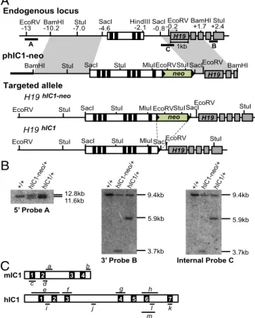
Generation of theH19hIC1Allele.To determine whether hIC1 could functionally substitute for the orthologous mouse sequence, we replaced the endogenous mIC1 with hIC1 by gene targeting in em-bryonic stem (ES) cells (Fig. 1A). Even though we obtained highly chimeric mice after blastocyst injection of the targeted ES cells, germ-line transmission of the targeted allele was inefficient. Only one female pup with theH19hIC1allele was live-born out of>250 agouti pups; all other agouti pups were wild-type, suggesting that the pups inheriting theH19hIC1allele might be dying prenatally. The single live-born female knock-in pup was of noticeably smaller size com-pared with its wild-type siblings and remained small.
The neomycin resistance cassette (NeoR) was excised by cross-ing the female to EIIA-Cre male on a C57BL/6J (B6) background (Jackson Laboratories). Germ-line transmission of the targeted allele and excision of NeoR were confirmed by Southern blot analysis (Fig. 1B). When bred to a B6 male, the female knock-in mouse was fertile, and wild-type and knock-in progeny were born in the expected Mendelian ratios with no sex bias. Embryonic lethality on paternal transmission was again observed after NeoR excision. The use of knock-in males in a B6/CF1 mixed strain for paternal transmission did not resolve the embryonic lethality. These results rule out NeoR and pure B6 background as being solely re-sponsible for the failure to obtain mutant pups. TheH19hIC1allele was maintained through maternal transmission in a B6 background.
The Maternally TransmittedH19hIC1
Allele Can Functionally Substitute for mIC1.To investigateH19andIgf2imprinting when the targeted allele was maternally transmitted, we bred femaleH19hIC1/+mice to B6 (CAST7) mice, which have aMus musculus castaneus chromo-some 7 on a B6 background (12). This cross allows the parental origin ofH19andIgf2expression to be distinguished in F1 progeny. Heterozygous H19hIC1/+ mice were compared with their wild-type littermates (H19+/+). TheH19hIC1/+andH19+/+mice were born in Mendelian ratios with no sex bias and no difference in neonatal weight (Fig. 2A). We assayed expression and IC1 methylation in neonatal livers, whereH19 and Igf2 are highly expressed, and detected monoallelic expression in all cases (Fig. 2B). Consistently, total expression levels of H19andIgf2 were statistically equivalent inH19+/+and H19hIC1/+livers, as mea-sured by quantitative real-time PCR (qRT-PCR) (Fig. 2C).
DNA methylation at hIC1 on the maternal allele and endogenous mIC1 on the paternal allele was measured by bisulfite mutagenesis of genomic DNA, followed by pyrosequencing. The maternal hICI was hypomethylated, as expected (Fig. 2D). Methylation at several other ICRs inH19hIC1/+livers was normal, suggesting that the general imprinting machinery is functioning normally (Fig. S1A).
Finally, hIC1 was properly hypomethylated in the oocytes of H19hIC1/+females (Fig. 2E). We repeated these analyses in two sequential generations of theH19hIC1allele maternal transmission offspring and obtained the same results. Overall, these data illustrate that hIC1 can functionally replace mIC1 on the maternal allele.
The Paternally TransmittedH19hIC1Allele Leads to Abnormal Insulation at theH19/Igf2Locus.To investigateH19andIgf2imprinting on the paternal allele, we bred maleH19hIC1/+mice to B6 (CAST7) mice.
12.8kb
11.6kb 9.4kb
5.9kb
3.7kb 9.4kb
5.9kb
3.7kb
3’ Probe B Internal Probe C
+/+ hIC1-neo/+hIC1/+ +/+ hIC1-neo/+hIC1/+
5’ Probe A
+/+ hIC1-neo/+hIC1/+
mIC1
hIC1
a
d
b
e f g h
j l
m c
i k
B
A
C
+2.4
B
1kb EcoRV
-13 BamHI-10.2 -7.0StuI SacI-4.6 HindIII-2.1 SacI-0.8 EcoRV
-0.2 BamHI+1.7 StuI
A
H19
C
EcoRV
StuI StuI StuISacIEcoRV
BamHI SacI
neo BamHI
MluI
H19 Endogenous locus
phIC1-neo
H19hIC1 H19hIC1-neo Targeted allele
EcoRV SacI StuI EcoRV StuI
EcoRV SacI StuI
H19 neo
MluI StuI
StuI SacIEcoRV StuI
EcoRV SacI StuI
H19 MluI
1 2 3 4
1 2 3 4 5 6 7
Fig. 1. Targeting strategy to generate theH19hIC1allele. (A) Schematics of the
endogenous locus, targeting vector (phIC1-neo), correctly targeted allele (H19hIC1-neo), and targeted allele after excision of the neoR cassette (H19hIC1).
Depicted are the IC1 (white rectangle) with CTCF-binding sites (black blocks within the IC1),H19exons (gray rectangles), pBluescriptIIKS sequence (bold line), neoR cassettes (green rectangles), loxP sites (black arrowheads), and en-dogenous mouse DNA (thin line). Restriction sites and their relative positions (in kb) to theH19transcription start sites are indicated above the endogenous locus. Probes (A, B, and C) used for Southern blot analyses are shown as thick lines below the endogenous locus. (B) Southern blot analysis to confirm correct targeting of the alleles. Genomic DNA from wild-type (+/+), hIC1-neo/+, and hIC1/+mice was either digested with EcoRV-MluI and hybridized to external 5′
probe A or digested with StuI and hybridized to external 3′probe B or to in-ternal probe C. (C) Depiction of mIC1 (Top) and hIC1 (Bottom) highlighting regions analyzed by qRT-PCR (a–m). IC1s are illustrated in the orientation shown inA, with each CTS numbered.a, bisulfite treatment followed by se-quencing for mIC1;bandc, ChIP–qRT-PCR for mIC1;d, pyrosequencing for mIC1;e–h, bisulfite treatment followed by sequencing for hIC1;i–k, ChIP– qRT-PCR for hIC1;l, pyrosequencing for hIC1;m, bisulfite treatment followed by sequencing for hIC1 (used for oocytes). Details are provided inMaterials and Methods.
GENET
All live-born neonates were wild-type, similar to what was ob-served when breeding for germ-line transmission in chimeric mice, suggesting that paternal transmission of theH19hIC1allele is em-bryonic lethal. To investigate this possibility, we isolated embry-onic day (E)15.5 conceptuses. Although theH19+/hIC1conceptuses were viable, the H19+/hIC1 embryos and placentas were smaller and weighed significantly less compared with those ofH19+/+(Fig. 3A). Anecdotally, such a size difference was not apparent at E10.5, and a trend toward a smaller size was observed at E12.5. The fetal/ placental weight ratio was not different between E15.5H19+/+and H19+/hIC1, demonstrating thatH19+/hIC1tissues were proportion-ately smaller (Fig. S2A).
Allele-specific RNA analysis revealed biallelic H19 in E15.5 H19+/hIC1livers and placentas, with equal expression derived from the two parental alleles (Fig. 3BandFig. S2D), suggesting
com-plete derepression of paternalH19. In contrast, paternalIgf2 ex-pression was barely detectable, indicating complete reex-pression ofIgf2(Fig. 3BandFig. S2D). Consistently, qRT-PCR analyses
revealed approximately 3.4-fold and 1.5-fold increases ofH19in liver and placenta, respectively, and undetectableIgf2inH19+/hIC1 compared with H19+/+embryos in both tissues (Fig. 3C and
Fig. S2E). Similar results were observed in E9.5 whole embryos
(Fig. S2BandC).
We next examined the extent to which methylation at hIC1 cor-related with abnormal expression in heterozygous livers and pla-centas. hIC1 was completely unmethylated on the paternal allele, resembling the endogenous mIC1 on the maternal allele (Fig. 3D andFig. S2F). This unusual methylation pattern was not due to a
gross defect in the methylation machinery, because methylation at other ICRs was normal inH19+/hIC1embryos (Fig. S1B).
Finally, to determine whether the hypomethylated state of hIC1 is associated with ectopic binding of CTCF on the paternal allele, we performed allele-specific chromatin immunoprecipitation (ChIP) followed by quantitative real-time PCR (ChIP–qRT-PCR) for CTCF in E12.5 mouse embryonic fibroblasts (MEFs). As expected, CTCF bound only to the unmethylated hIC1 on the maternal allele and did not bind to the methylated mIC1 on the paternal allele in H19hIC1/+MEFs. In contrast, CTCF bound to both the unmethy-lated mIC1 on the maternal allele and the unmethyunmethy-lated hIC1 on the paternal allele inH19+/hIC1MEFs (Fig. 3E). The results dem-onstrate that paternal hIC1 is unable to acquire or maintain the hypermethylated state of endogenous mIC1, fails to repress H19, and instead gains a CTCF-dependent insulator function.
Incomplete Establishment of Genomic Imprinting at hIC1 During Spermatogenesis.Because the paternal allele in E15.5 H19+/hIC1 embryos was hypomethylated, we assayed DNA methylation at earlier stages (Fig. S3). We did not detect any methylation at hIC1 as early as the blastocyst stage, suggesting that either methylation was not established during spermatogenesis or methylation was established but was lost during preimplantation development (Fig. S3). To distinguish between these possibilities, we examined DNA methylation at hIC1 in sperm of H19hIC1/+males, and observed partial methylation (Fig. 4 A and B). As positive controls, we analyzed methylation at endogenous hIC1 in human sperm sam-ples from two fertile men as well as at endogenous mIC1 in H19hIC1/+ sperm and found that all were hypermethylated, as expected (Fig. 4AandB).
To explore whether the methylation at hIC1 inH19hIC1/+sperm could be maintained after the first cleavage division, we assayed H19+/hIC1 two-cell embryos in which the zygote had undergone one round of mitosis. We found reduced methylation levels (close to one-half less) at hIC1 in two-cell embryos compared with ma-ture sperm (Fig. 4A). These results demonstrate that the DNA methylation is partially established at hIC1 during spermatogen-esis, but is not maintained in preimplantation development.
Increased Enrichment of H3K4me2 at hIC1 in Spermatogenic Cells.To investigate factors that may inhibit complete establishment of DNA methylation at hIC1 during spermatogenesis, we examined histone posttranslational modifications at hIC1. Parental allele-specific histone modifications have been described at mIC1 in both somatic and germ cells (14–18). Several studies have sug-gested an antagonistic relationship between “activating marks,” such as dimethylation and trimethylation of histone H3 at lysine 4 (H3K4me2 and H3K4me3, respectively), and DNA methylation (17–20). Other studies have shown a strong relationship between “repressive marks,”such as trimethylation of histone H3 at lysine 9 (H3K9me3), and DNA methylation (21). In fact, H3K4 meth-ylation is found preferentially on the hypomethylated maternal IC1, and H3K9me3 is found on the hypermethylated paternal IC1 in mouse and human somatic cells (11, 14). We hypothesized that depletion of H3K9me3, increased enrichment of H3K4me2, or both contribute to the inability to fully establish DNA methylation at hIC1.
Spermatogenic cells were fractionated by the STA-PUT method, and chromatin was isolated from a round spermatid-enriched fraction (22). ChIP–qRT-PCR analyses in round spermatids revealed that hIC1 had fivefold greater enrichment of H3K4me2 p
A
B
C
D
E
+/+ hIC1/+ m p
+/+ hIC1/+ m H19
Igf2
W
e
ight (g)
Relative Expression Relative Expression
% Methylation
Igf2 H19
mIC1 hIC1
CTS2 CTS6
+/+ hIC1/+
+/+ hIC1/+
+/+
hIC1/+
hIC1/+ (m)
hIC1/+ (p)
+/+
IC1
Fig. 2. Maternal transmission of theH19hIC1allele. (A) Neonatal (P0) weight
compared with mIC1 (Fig. 4C). Unlike previous studies that did not report significant enrichment of H3K9me3 above background at mIC1 in the male germ line (14, 17), we obtained a ChIP signal above background. This discrepancy could be attributed to the different antisera used for H3K9me3. Nevertheless, there was no difference in the enrichment of H3K9me3 between hIC1 and mIC1. Our finding that the difference in H3K4me2 between mIC1 and hIC1 is greater than the difference in H3K9me3 sug-gests that (i) H3K9me3 does not affect the acquisition of DNA methylation at hIC1 during spermatogenesis, and (ii) enrichment of activating histone marks at hIC1 contributes in part to the in-complete establishment of imprinting at hIC1 during spermato-genesis (Fig. 4C).
Abnormal Placental Morphology in H19+/hIC1
. Because H19+/hIC1 embryos display similar phenotypes to those of many patients with SRS who present with IC1 hypomethylation, including alteredH19 andIgf2expression and growth defects (23, 24), we hypothesized that these embryos can serve as a model for SRS. Placental growth defects are prevalent among individuals with SRS (24); thus, we further characterized H19+/hIC1 placentas to study potential mechanisms underlying SRS associated with IC1 hypomethylation. We performed histological analyses on E15.5H19+/hIC1placentas to explore whether abnormal placentation could contribute to embryonic growth restriction, given thatH19andIgf2play essential roles in placental development (25, 26). In addition to being smaller (∼74% of H19+/+), H19+/hIC1 placentas displayed an increased junctional/labyrinthine zone ratio, indicative of abnormal placenta morphology (Fig. S2GandH).
Discussion
Using a mouse model replacing endogenous mIC1 with hIC1, we have shown that the ability of hIC1 to functionally replace mIC1 depends on the parental origin of the hIC1 allele. Although the
main aim of this study was to investigate interspecies compatibility of hIC1 in the mouse system, we anticipate that findings from this study also will provide insight into modeling and further examining imprinting disorders such as BWS and SRS.
Several groups have reported that a subset of patients with BWS carry mutations at IC1, and that these mutations are largely as-sociated with IC1 hypermethylation, reduced H19expression, and biallelicIgf2expression. Notably, these IC1 mutations (i.e., microdeletions and point mutations) manifest BWS clinical phe-notypes when the mutant allele is maternal in origin (9, 27, 28). Determining the extent to which these mutations contribute to the molecular and clinical phenotypes of BWS is challenging, because IC1 hypermethylation is mosaic in the patients, suggesting that not all cells are aberrantly DNA-methylated. Moreover, clinical phe-notypes of the patients are highly variable, possibly as a conse-quence of either the mosaicism or the genetic background of the individuals (27). In this study, we have shown that maternal transmission of hIC1 can functionally replace mIC1 by properly regulating imprinted expression and hIC1 methylation. Thus, our findings raise the exciting possibility of modeling IC1 mutations associated with BWS in mice via maternal transmission.
In contrast, paternal transmission of hIC1 leads to loss ofH19 andIgf2imprinting;H19displays biallelic and increased expression, andIgf2is silenced. Offspring inheriting hIC1 paternally also exhibit severe growth restriction. Of note, previous studies have shown that Igf2null neonates are born smaller but viable (29, 30), suggesting that prenatal lethality ofH19+/hIC1is not due solely to the loss of Igf2. Mice from two independentH19knockout models exhibited overgrowth, suggesting a growth-suppressing role ofH19(31, 32). In addition, ectopic expression of H19 caused late-gestation le-thality (33). Thus, changes in bothH19andIgf2may synergistically contribute to the severe growth restriction and prenatal lethality ofH19+/hIC1.
A
C
B
D
E
Fig. 3. Paternal transmission of theH19hIC1allele. (A) E15.5 fetal and placental weight of wild-type (+/+) and+/hIC1 mutant offspring. (B) Allele-specific
expression ofH19andIgf2in E15.5 livers analyzed by RFLP. PCR cycle numbers varied between wild-type (+/+) and+/hIC1 forIgf2(Table S1; seeTable S2for primers). (C) Total expression ofH19andIgf2in E15.5 livers analyzed by qRT-PCR. (D) Percent methylation at IC1 in E15.5 livers measured by pyrosequencing. Assaydwas used for wild-type (+/+) and+/hIC1(m), and assaylwas used for+/hIC1(p) (Fig. 1C). (E) CTCF binding at mIC1 and hIC1 in heterozygous (hIC1/+and +/hIC1) E12.5 MEFs analyzed by ChIP–qRT-PCR. Assaysb,c,i, andkwere used (Fig. 1C); results from two biological replicates are shown separately. They-axis denotes percent input of CTCF IP normalized to nonspecific IgG (percent input of CTCF−percent input of IgG). **P<0.01; ***P<0.001; ****P<0.0001, two-tailed Student’sttest with equal variance (A) or unequal variance (C). InA–D: wild-type (+/+),n=8;+/hIC1,n=6 (from two litters). Bars represent the mean±SEM; error bars inDare too small to be seen on the graph.
GENET
Previous studies have shown that abnormal H19andIgf2 ex-pression is linked to both placental and embryonic growth defects. Deletion of a placenta-specificIgf2transcript was found to result in growth restriction of embryos in late gestation (34). In a mouse model in which H19 was deleted and Igf2 expression was in-creased, both the placenta and the fetus were overgrown at E19 (35, 36). In humans, placental growth defects are common in in-dividuals with BWS and SRS (24, 37). We observed thatH19+/hIC1 placentas not only are smaller, but also have abnormal placental morphology. Although the contribution of an abnormal placenta to fetal growth defects is unclear, it is noteworthy thatH19is more highly expressed in the labyrinthine zone compared with the junctional zone (38), and thatIgf2null mice display a dispropor-tionate reduction in the labyrinthine zone compared with the junctional zone (39). These observations suggest a potential major growth-suppressing effect of increasedH19and silencedIgf2 ex-pression in the labyrinthine zone.
We also have shown that hIC1 is partially methylated in the male germ line ofH19hIC1/+mice. This phenotype is in contrast with that of other mouse models that carry mIC1 mutations; in
those mice, methylation is properly established at nonmutated CpGs in the male germ line (5, 40). This finding suggests that interspecies communication between mouse and human is in-effective in establishing IC1 methylation. Based on our finding that hIC1 is abnormally enriched with activating H3K4me2 marks in the male germ cells, it is tempting to speculate that somatic histone modification marks carried from the maternal allele are not completely erased in the male germ cells. (Note that hIC1 was necessarily transmitted maternally to generate offspring.) Consequently, the establishment of DNA methylation at hIC1 is inhibited. This finding adds to the growing consensus that H3K4 methylation marks are inhibitory to de novo DNA methylation in the germ line, whereas repressive histone marks do not play major role in the establishment of methylation at ICRs (14, 16–18, 20). However, it is equally possible that the hypomethylated state of DNA attracts H3K4me2 at hIC1 by an unknown mechanism. More detailed time course analyses of DNA methylation and H3K4me2 enrichment in the primordial germ cells and early-stage male germ cells will provide insight into this hypothesis. Alternatively, there might be an inherent difference between mIC1 and hIC1 in terms of acquisition of methylation. A noncoding transcript has been detected at mIC1 during methylation acquisition in male germ cells (41), suggest-ing a potential role of transcription in the establishment of meth-ylation at mIC1. Whether the same holds true at hIC1 remains to be determined, however.
Finally, we have shown that the partially established methylation at hIC1 in the male germ line is not properly maintained during preimplantation development. We also have shown that CTCF ec-topically binds to hIC1 on the paternal allele in somatic cells. Similar results have been reported for an earlier mouse model in which CpGs within the CTCF-binding sites at the mIC1 were mutated to abrogate methylation, while keeping the CTCF-binding motifs intact (5). There, although methylation was properly established in the male germ cells at the mIC1, it was not maintained during pre-implantation development (5). These data suggest that CTCF binding inhibits the maintenance of DNA methylation in somatic cells, although the mechanism remains unknown. Alternatively, hIC1 might lack properties that allow the mouse imprint main-tenance machinery to properly recognize the sequence. It is also possible that hIC1 contains inhibitory signals that block accessi-bility and/or activity of the mouse imprint maintenance machinery. In conclusion, we have elucidated hitherto unreported princi-pals regarding the conservation of ICR function at theH19/Igf2 locus and molecular mechanisms associated with SRS. First, evi-dence of incomplete histone reprogramming at hIC1 suggests that the mechanism regulating histone reprogramming at IC1 in the germ line has diverged between mouse and human. In this regard, it would be interesting to explore whether an IC1 ortholog of a species more closely related to mouse could recapitulate the wild-type epigenetic pattern on paternal transmission. Second, despite the fact that IC1 hypomethylation is the most common epi-mutation found in individuals with SRS (24), the molecular mechanism underlying the phenotype remains elusive. Obstacles to addressing this question include mosaicism of the epimutation in patients and the lack of a suitable genetic model system. We suggest that the paternal transmission of hIC1 in mice can be used to study the molecular mechanisms underlying SRS associated with IC1 hypomethylation. Future experiments, such as breeding H19hIC1/+ males with H19null females, will help elucidate the extent to whichH19 contributes to the SRS-like phenotype. In addition, identifying pathways altered by IC1 hypomethylation may shed light on the physiology of SRS.
Materials and Methods
Targeting Vector.Detailed information on the hIC1 target vector is provided
inSI Materials and Methods.
A
B
C
Fig. 4. Incomplete establishment of imprinting at the hIC1 in knock-in male germ cells. (A) Percent methylation at IC1 measured by pyrosequencing; assaysdandlwere used (Fig. 1C). From left to right, results for endogenous hIC1 in mature sperm samples from fertile men (hSP1 and hSP2), the en-dogenous mIC1 and targeted hIC1 in knock-in sperm (KISP) from adult mice, and the targeted hIC1 in pools ofH19+/hIC1two-cell stage embryos (KI2 cells)
ES Cells and Mouse Generation, Breeding, and Genotyping.Details regarding ES cell targeting, Southern blot analyses, and mouse generation, breeding, and genotyping are provided inSI Materials and Methods.
Gene Expression Analysis.RNA isolation and cDNA synthesis was performed as
described previously (42). For qRT-PCR, total expression levels ofH19andIgf2 were measured relative to the geometric mean of expression levels ofArbp (acidic ribosomal phosphoprotein P0),Nono (non–POU domain-containing, octamer-binding protein), andRpl13a(ribosomal protein L13A). For they-axis on the graph, the mean value of wild-type is set arbitrarily as 1. Details are provided inSI Materials and Methods.
DNA Methylation Analysis.gDNA isolation from neonatal, embryonic, and germ
cell samples; bisulfite treatment; and methylation analyses are described in detail inSI Materials and Methods.
Histology.Histological analysis was performed as described previously (43).
Mouse Spermatogenic Cell Fractionation.Round spermatid fractions of mouse
spermatogenic cells were collected using STA-PUT in two independent replicates,
and the purity of each fraction was verified as described previously (22, 44). Each collection used both testes of 12 heterozygous (H19hIC1/+) male mice. The purity
was measured as 87% for pool 1 and 86% for pool 2.
Isolation of MEFs.MEFs were isolated from individual day 12.5 embryos in a B6
background as described previously (15).
ChIP–qRT-PCR Analysis.ChIP–qRT-PCR was carried out as described previously
(44); details are provided inSI Materials and Methods. Each ChIP signal was calculated as the percent input of each immunoprecipitation (IP) normalized to nonspecific IgG (percent input of IP−percent input of IgG). In Fig. 4C, the y-axis denotes the ChIP signal of each histone mark normalized to that of total H3 (e.g., percent input of H3K4me2/percent input of total H3).
ACKNOWLEDGMENTS.This work was supported by US Public Health Service Grants GM51279 (to M.S.B.) and HD068157 (to M.S.B. and S.L.B.); Advanced Imaging Research Center Grant 8700 (to F.C.); Telethon-Italy Grant GGP15131 (to A.R.); European Union Seventh Framework Programme, INGENIUM 290123 (to A.R.); and National Institutes of Health Training Grant T32 GM07229 (to S.K.H.).
1. Lee JT, Bartolomei MS (2013) X-inactivation, imprinting, and long noncoding RNAs in health and disease.Cell152(6):1308–1323.
2. Kalish JM, Jiang C, Bartolomei MS (2014) Epigenetics and imprinting in human dis-ease.Int J Dev Biol58(2-4):291–298.
3. Eggermann T, et al. (2015) Imprinting disorders: A group of congenital disorders with overlapping patterns of molecular changes affecting imprinted loci.Clin Epigenetics7:123. 4. Ideraabdullah FY, Vigneau S, Bartolomei MS (2008) Genomic imprinting mechanisms
in mammals.Mutat Res647(1-2):77–85.
5. Engel N, West AG, Felsenfeld G, Bartolomei MS (2004) Antagonism between DNA hypermethylation and enhancer-blocking activity at the H19 DMD is uncovered by CpG mutations.Nat Genet36(8):883–888.
6. Smits G, et al.; SAVOIR Consortium (2008) Conservation of the H19 noncoding RNA and H19-IGF2 imprinting mechanism in therians.Nat Genet40(8):971–976. 7. Jinno Y, et al. (1996) Mouse/human sequence divergence in a region with a
paternal-specific methylation imprint at the human H19 locus.Hum Mol Genet5(8):1155–1161. 8. Jones BK, Levorse J, Tilghman SM (2002) A human H19 transgene exhibits impaired pa-ternal-specific imprint acquisition and maintenance in mice.Hum Mol Genet11(4):411–418. 9. Demars J, Gicquel C (2012) Epigenetic and genetic disturbance of the imprinted 11p15 region in Beckwith–Wiedemann and Silver–Russell syndromes.Clin Genet81(4):350–361. 10. Engel N, Raval AK, Thorvaldsen JL, Bartolomei SM (2008) Three-dimensional confor-mation at the H19/Igf2 locus supports a model of enhancer tracking.Hum Mol Genet 17(19):3021–3029.
11. Nativio R, et al. (2011) Disruption of genomic neighbourhood at the imprinted IGF2-H19 locus in Beckwith–Wiedemann syndrome and Silver–Russell syndrome.Hum Mol Genet20(7):1363–1374.
12. Mann MR, et al. (2003) Disruption of imprinted gene methylation and expression in cloned preimplantation stage mouse embryos.Biol Reprod69(3):902–914. 13. Bell AC, Felsenfeld G (2000) Methylation of a CTCF-dependent boundary controls
imprinted expression of the Igf2 gene.Nature405(6785):482–485.
14. Delaval K, et al. (2007) Differential histone modifications mark mouse imprinting control regions during spermatogenesis.EMBO J26(3):720–729.
15. Verona RI, Thorvaldsen JL, Reese KJ, Bartolomei MS (2008) The transcriptional status, but not the imprinting control region, determines allele-specific histone modifica-tions at the imprinted H19 locus.Mol Cell Biol28(1):71–82.
16. Lee DH, et al. (2010) CTCF-dependent chromatin bias constitutes transient epigenetic memory of the mother at the H19-Igf2 imprinting control region in prosper-matogonia.PLoS Genet6(11):e1001224.
17. Singh P, et al. (2013) De novo DNA methylation in the male germ line occurs by de-fault but is excluded at sites of H3K4 methylation.Cell Reports4(1):205–219. 18. Stewart KR, et al. (2015) Dynamic changes in histone modifications precede de novo
DNA methylation in oocytes.Genes Dev29(23):2449–2462.
19. Ooi SK, et al. (2007) DNMT3L connects unmethylated lysine 4 of histone H3 to de novo methylation of DNA.Nature448(7154):714–717.
20. Ciccone DN, et al. (2009) KDM1B is a histone H3K4 demethylase required to establish maternal genomic imprints.Nature461(7262):415–418.
21. Rose NR, Klose RJ (2014) Understanding the relationship between DNA methylation and histone lysine methylation.Biochim Biophys Acta1839(12):1362–1372. 22. Bryant JM, Meyer-Ficca ML, Dang VM, Berger SL, Meyer RG (2013) Separation of
sper-matogenic cell types using STA-PUT velocity sedimentation.J Vis Exp(80):e50648. 23. Gicquel C, et al. (2005) Epimutation of the telomeric imprinting center region on
chromosome 11p15 in Silver–Russell syndrome.Nat Genet37(9):1003–1007. 24. Yamazawa K, et al. (2008) Molecular and clinical findings and their correlations in
Silver–Russell syndrome: Implications for a positive role of IGF2 in growth de-termination and differential imprinting regulation of the IGF2-H19 domain in bodies and placentas.J Mol Med (Berl)86(10):1171–1181.
25. Bartolomei MS, Ferguson-Smith AC (2011) Mammalian genomic imprinting.Cold Spring Harb Perspect Biol3(7):a002592.
26. Reik W, et al. (2003) Regulation of supply and demand for maternal nutrients in mammals by imprinted genes.J Physiol547(Pt 1):35–44.
27. Beygo J, et al. (2013) The molecular function and clinical phenotype of partial dele-tions of the IGF2/H19 imprinting control region depends on the spatial arrangement of the remaining CTCF-binding sites.Hum Mol Genet22(3):544–557.
28. Abi Habib W, et al. (2014) Extensive investigation of the IGF2/H19 imprinting control region reveals novel OCT4/SOX2 binding site defects associated with specific methyl-ation patterns in Beckwith–Wiedemann syndrome.Hum Mol Genet23(21):5763–5773. 29. DeChiara TM, Efstratiadis A, Robertson EJ (1990) A growth-deficiency phenotype in heterozygous mice carrying an insulin-like growth factor II gene disrupted by tar-geting.Nature345(6270):78–80.
30. Baker J, Liu J-P, Robertson EJ, Efstratiadis A (1993) Role of insulin-like growth factors in embryonic and postnatal growth.Cell75(1):73–82.
31. Schmidt JV, Levorse JM, Tilghman SM (1999) Enhancer competition between H19 and Igf2 does not mediate their imprinting.Proc Natl Acad Sci USA96(17):9733–9738. 32. Ripoche MA, Kress C, Poirier F, Dandolo L (1997) Deletion of the H19 transcription unit
reveals the existence of a putative imprinting control element.Genes Dev11(12):1596–1604. 33. Brunkow ME, Tilghman SM (1991) Ectopic expression of the H19 gene in mice causes
prenatal lethality.Genes Dev5(6):1092–1101.
34. Constância M, et al. (2002) Placental-specific IGF-II is a major modulator of placental and fetal growth.Nature417(6892):945–948.
35. Leighton PA, Ingram RS, Eggenschwiler J, Efstratiadis A, Tilghman SM (1995) Dis-ruption of imprinting caused by deletion of the H19 gene region in mice.Nature 375(6526):34–39.
36. Angiolini E, et al. (2011) Developmental adaptations to increased fetal nutrient demand in mouse genetic models of Igf2-mediated overgrowth.FASEB J25(5):1737–1745. 37. Armes JE, et al. (2012) The placenta in Beckwith–Wiedemann syndrome: Genotype–
phenotype associations, excessive extravillous trophoblast and placental mesenchymal dysplasia.Pathology44(6):519–527.
38. Keniry A, et al. (2012) The H19 lincRNA is a developmental reservoir of miR-675 that suppresses growth and Igf1r.Nat Cell Biol14(7):659–665.
39. Coan PM, et al. (2008) Disproportional effects of Igf2 knockout on placental mor-phology and diffusional exchange characteristics in the mouse.J Physiol586(20): 5023–5032.
40. Engel N, Thorvaldsen JL, Bartolomei MS (2006) CTCF-binding sites promote tran-scription initiation and prevent DNA methylation on the maternal allele at the im-printed H19/Igf2 locus.Hum Mol Genet15(19):2945–2954.
41. Henckel A, Chebli K, Kota SK, Arnaud P, Feil R (2012) Transcription and histone methylation changes correlate with imprint acquisition in male germ cells.EMBO J31(3):606–615. 42. de Waal E, et al. (2014) In vitro culture increases the frequency of stochastic
epige-netic errors at imprinted genes in placental tissues from mouse concepti produced through assisted reproductive technologies.Biol Reprod90(2):22.
43. de Waal E, et al. (2015) The cumulative effect of assisted reproduction procedures on placental development and epigenetic perturbations in a mouse model.Hum Mol Genet24(24):6975–6985.
44. Bryant JM, et al. (2015) Characterization of BRD4 during mammalian postmeiotic sperm development.Mol Cell Biol35(8):1433–1448.
45. Thorvaldsen JL, Mann MR, Nwoko O, Duran KL, Bartolomei MS (2002) Analysis of sequence upstream of the endogenous H19 gene reveals elements both essential and dispensable for imprinting.Mol Cell Biol22(8):2450–2462.
46. Stadnick MP, et al. (1999) Role of a 461-bp G-rich repetitive element in H19 transgene imprinting.Dev Genes Evol209(4):239–248.
47. Kühn R, Rajewsky K, Müller W (1991) Generation and analysis of interleukin-4–deficient mice.Science254(5032):707–710.
48. Plasschaert RN, et al. (2014) CTCF binding site sequence differences are associated with unique regulatory and functional trends during embryonic stem cell differen-tiation.Nucleic Acids Res42(2):774–789.
49. Weaver JR, et al. (2010) Domain-specific response of imprinted genes to reduced DNMT1.Mol Cell Biol30(16):3916–3928.
50. Takai D, Gonzales FA, Tsai YC, Thayer MJ, Jones PA (2001) Large-scale mapping of methylcytosines in CTCF-binding sites in the human H19 promoter and aberrant hy-pomethylation in human bladder cancer.Hum Mol Genet10(23):2619–2626.
GENET