DEVELOPMENT OF A CELLULAR ANALYSIS PLATFORM
FEATURING ARRAYS OF PATTERNED MICROWELLS
FABRICATED ATOP PERMEABLE SUPPORTS
Douglas M. Ornoff
A dissertation submitted to the faculty at the University of North Carolina at Chapel Hill in partial fulfillment of the requirements for the degree of Doctor of Philosophy in the
Department of Pharmacology in the School of Medicine.
Chapel Hill 2014
iii
ABSTRACT
Douglas M. Ornoff: Development of a cellular analysis platform featuring arrays of patterned microwells fabricated atop permeable supports
(Under the direction of Nancy L. Allbritton)
Many human pulmonary diseases lead to accumulation of fluid in the alveoli, the air sacs located in the distal lung and at which gas exchange occurs. The most serious example of alveolar fluid buildup is in Acute Respiratory Distress Syndrome (ARDS), in which an insult to the lung results in injury to the cells lining the alveolus, leading to compromise of the alveolar capillary barrier and impaired gas exchange. Current ARDS mortality rates lie at 30-40%. Worsening this problem is the lack of disease-specific therapies for treating ARDS: the cornerstone of treatment is merely supportive respiratory care via mechanical ventilation.
Further investigations into treating ARDS have been hampered by unresolved
questions about the normal physiology of alveolar fluid transport. Therefore, new insights are needed in order to develop more effective ARDS therapies. The alveolus is lined by two types of cells: squamous alveolar type 1 cells that cover 98% of the alveolar surface area and small cuboidal alveolar type 2 cells. While studies have examined AT2 cells, the ion
iv
This dissertation describes the development of microfabricated devices that feature an array of microwells patterned atop a porous or otherwise permeable support. First, a method for fabricating 1002F photoresist into a freestanding microwell array is explored. Next, a strategy to co-fabricate freestanding 1002F films with the hydrogel chitosan to form
v
This work, being the longest block of prose I’ve ever written, is fittingly dedicated to the memory of Ms. Martha Woolery.
First, may this work be free of gross grammatical errors.
vi
ACKNOWLEDGEMENTS
vii
I would also like to thank my committee members and collaborators. Rob Nicholas, Tasha Blatt, Ross Dubose, Sam Wolff, and Ken Harden, thank you for all the help and assistance you’ve given me over these years. I count myself lucky that I have had people so willing to share their expertise and experience with me. Brian Button, Scott Randell, Leslie Fulcher, and Peter Bove, I thank you all for the outstanding insight and discussions on the nuances of respiratory epithelia, which have helped inspire so many of my ideas and so much of my creativity. Ashley Henderson, you have been such a great clinical mentor. Your
patience and passion for teaching are a rare find, and I hope I get the chance to learn more from you in the future.
I would not be here today if it were not for several individuals who helped me take the plunge into the world of science. Foremost among these is Ric Boucher, who has been such an inspiration to me in my pursuits. If I end up being even half as successful as you, I will consider myself greatly fortunate. Your ability to inspire hard work and creativity is truly unparalleled, and your ability to see potential in a skinny 19-year-old and open doors for him is greatly appreciated. To Eduardo Lazarowski and Catja van Heusden, I owe you such thanks for giving me my first break in science and taking me under your wings. To Matt Redinbo and Eric Ortlund, who first took the time to teach me how to be independent and to think to the next steps, thank you for all the lessons you taught me.
I would also like to acknowledge the UNC Medical Scientist Training Program, both faculty and students. Training in the two disciplines of clinical medicine and research science is not an easy undertaking, but this Program has strived to excel, and to do so with a
viii
definition of “a gentleman and a scholar.” Your willingness to offer your time and guidance, even to someone such as I whose field is set far apart from your own, has been unmatched. Alison Regan and Carol Herion have been so incredibly fun and supportive, and I have taken such heart from your humor and your dedication to each of us in the Program. Dr. Ryan Phillips, you have been a true friend to me and a great guide as I navigate the Allbritton Lab, the Pharmacology Department, and the MSTP.
To all my friends outside of science and the lab, you’ve become my family in Chapel Hill, and without each of you I would have burned out a long time ago. You have truly kept me going all these years with your constant friendship and ever-present good humor. I have so enjoyed all our game nights, trips to the beach, and bar trivia gatherings. A great deal of my motivation to work in science and medicine comes from the desire to see that others might have the good times and the friendship that we share. If every person in the world had friends like you, no one would feel unwanted. Aaron, John, Will, Josh, Neal, Chris, Chris, Ben, and Andrew, I affirm the borrowed statement: I have never been in a fraternity, but I count you all as my brothers. Lauren, Helen, Brandy, Sarah, Sarah, Suzanne, and Ashley, you all mean the world to me. Shannon, I want you to know how much your support has meant as I navigated the past few years. Thank you so much for believing in me.
ix
TABLE OF CONTENTS
LIST OF TABLES . . . xv
LIST OF FIGURES . . . xvi
LIST OF ABBREVIATIONS AND SYMBOLS . . . xviii
Chapter 1: Introduction . . . 1
1.1 Airway and Alveolar Disease: States of Disordered Pulmonary Fluid Homeostasis . . . 1
1.1.1 Respiratory epithelia and luminal fluid films . . . 1
1.1.1.1 Hierarchical organization of respiratory epithelia . . . 1
1.1.1.2 Air-liquid interfaces and luminal fluid films in the pulmonary tract . . . 5
1.1.2 Alveolar diseases and disordered fluid homeostasis . . . 6
1.1.3 Alveolar diseases: therapeutic options and shortfalls . . . 8
1.2 Physiology and Pharmacology of Pulmonary Liquid Homeostasis . . . 10
1.2.1 Airway surface liquid in bronchial and bronchiolar epithelia . . . 10
1.2.2 Ion and fluid transport by alveolar type 2 (AT2) epithelia . . . 14
1.2.3 Ion transporters and purinergic receptors in alveolar type 1 (AT1) epithelia . . . 17
1.2.4 Current methods for studying pulmonary fluid homeostasis . . . 20
1.3 Microscale devices for analysis of discrete numbers of cells . . . 23
x
1.3.2 Advantages of microscale devices . . . 25
1.3.3 Survey of previously described microscale devices relevant to alveolar cell culture . . . 26
1.3.3.1 Microscale devices applied to respiratory biology . . . 26
1.3.3.2 Microscale devices utilizing a permeable support . . . 28
1.3.4 Design guidelines for a microscale device applicable to AT1 cells . . . 31
1.4 Scope of the Dissertation . . . 33
1.5 Figures and Tables . . . 35
1.6 References . . . 43
Chapter 2: Characterization of freestanding photoresist films for biological and MEMS applications . . . 62
2.1 Introduction . . . 62
2.1.1 General approaches . . . 62
2.1.2 Lab-on-a-foil devices . . . 63
2.1.3 Photoresists . . . 64
2.1.4 Photoresists as lab-on-a-foil devices . . . 65
2.1.5 Overview . . . 66
2.2 Experimental Design . . . 67
2.2.1 Materials . . . 67
2.2.2 Fabrication and release of single-layer films . . . 67
2.2.3 Measurement of film properties . . . 68
2.2.4 Characterization of photoresist thin film release kinetics . . . 69
2.2.5 Patterned deposition of metal and protein. . . .70
xi
2.2.7 Seeding of AT2 primary cells . . . 71
2.3 Results and Discussion . . . 72
2.3.1 Fabrication of 1002F films and release from glass substrates . . . 72
2.3.2 Mechanical strength of freestanding 1002F films . . . 73
2.3.3 Fabrication of micropatterned freestanding 1002F films . . . 74
2.3.4 Fabrication of multilayered, micropatterned freestanding 1002F films . . . 75
2.3.5 1002F films as re-usable stencils for substrate patterning . . . 77
2.3.6 1002F films to address a single cell within a monolayer of cells . . . 79
2.4 Conclusions . . . 80
2.5 Figures and Tables . . . 82
2.6 References . . . 97
Chapter 3: Co-fabrication of 1002F and chitosan for freestanding films of permeable-bottomed microwell arrays . . . 102
3.1 Introduction . . . 102
3.1.1 General considerations . . . 102
3.1.2 Analysis of cellular heterogeneity . . . 102
3.1.3 Microfabricated platforms for analyzing cellular heterogeneity . . . . 104
3.1.4 Chitosan . . . 105
3.1.5 Overview . . . 107
3.2 Experimental Design . . . 107
3.2.1 Materials . . . 107
xii
3.2.3 Imaging . . . 109
3.2.4 Multi-layer fabrication . . . 109
3.2.5 Measurement of chitosan dry film thickness. . . 110
3.2.6 Measurement of 1002F:chitosan film optical properties . . . 110
3.2.7 Diffusion testing . . . 111
3.2.8 Calculation of diffusivity constant for chitosan membranes in 1002F:chitosan hybrid films . . . 113
3.2.9 Spatially controlled protein conjugation . . . 118
3.2.10 Isolated clonal expansion and analysis of heterogeneity of cell proliferation in entrapped, nonadherent cells . . . 119
3.2.11 Growth of AT1-like cells . . . 120
3.2.12 Statistical testing . . . 120
3.3 Results and Discussion . . . 121
3.3.1 Co-fabrication of freestanding 1002F:chitosan films . . . 121
3.3.2 Multi-layer 1002:chitosan:1002F sandwich films . . . 122
3.3.3 Chitosan hydrogel thickness measurements . . . 123
3.3.4 Optical properties . . . 125
3.3.5 Diffusion testing of micropatterned 1002F: chitosan hybrid films . . . 126
3.3.6 Calculation of diffusion coefficients and MW cutoffs for chitosan membranes in 1002F:chitosan hybrid films . . . 129
3.3.7 Spatially controlled protein conjugation . . . . . . 131
3.3.8 Survival and proliferation of nonadherent cells entrapped in chitosan-bottomed microwells . . . 132
xiii
3.3.10 Analysis of cell division heterogeneity in
entrapped, nonadherent cells . . . 134
3.3.11 Growth of primary human AT2 cells as AT1 surrogates . . . 136
3.4 Conclusions . . . 136
3.5 Figures and Tables . . . 138
3.6 References . . . 154
Chapter 4: Co-fabrication of 1002F and chitosan for freestanding films of permeable-bottomed microwell arrays . . . 161
4.1 Introduction . . . 161
4.1.1 Cellular polarization in epithelial tissues . . . 161
4.1.2 Advantages of microfabricated devices for studies of fundamental cell processes . . . . . . 162
4.1.3 Microfabricated devices for study of polarized cells . . . 162
4.1.4 Design of a protocol for micropatterning a track-etch membrane . . . 164
4.1.5 Overview . . . 166
4.2 Experimental Design . . . 167
4.2.1 Materials . . . 167
4.2.2 Photoresist-based micropatterning of track-etch membranes . . . 168
4.2.3 Imaging of freestanding, micropatterned track-etch membranes . . . . 169
4.2.4 Assessing micropore patency by measuring solute diffusion . . . 169
4.2.5 Testing epithelial cell polarization on micropatterned track-etch membranes . . . 170
xiv
atop track-etch membranes . . . 171
4.3.2 Optimization of fabrication parameters . . . 173
4.3.3 Testing membrane micropore patency using solute diffusion . . . 175
4.3.4 Growth of polarized MDCK cells in microwells patterned atop track-etch membranes . . . 176
4.4 Conclusions . . . 178
4.5 Figures and Tables . . . 179
4.6 References . . . 184
Chapter 5: Conclusions and Future Directions . . . 188
5.1 Overall summary and conclusions . . . 188
5.1.1 Freestanding 1002F photoresist films . . . 188
5.1.2 Chitosan-bottomed microwell arrays . . . 189
5.1.3 Photoresist-based micropatterning of polyester track-etch membranes . . . 189
5.2 Anticipated future directions . . . 190
5.2.1 Freestanding 1002F photoresist films . . . 190
5.2.2 Chitosan-bottomed microwell arrays . . . 191
5.2.3 Track-etch membranes micropatterned with an overlying photoresist layer . . . 192
5.2.4 Adaptation of technology prototypes for specific study of primary AT1 cells . . . 193
5.3 Potential for future therapies for and insights into ARDS . . . 194
xv
LIST OF TABLES
Table 1.1: Comparison of overall ion and water transport properties
between AT1 and AT2 cells . . . 41
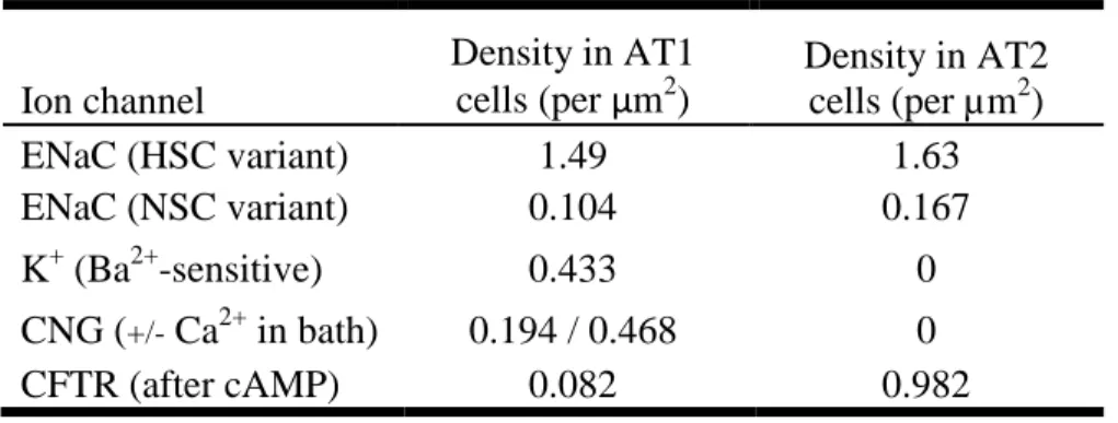
Table 1.2: Comparison of ion transporter density in AT1 and AT2 cells. . . 42
Table 2.1: Process parameters for 1002F photoresist films . . . 94
Table 2.2: Removal of 1002F films by immersion in water . . . 95
Table 2.3: Mechanical properties of 1002F films . . . 96
Table 3.1: Design overview of a microwell array platform for culture of single AT1 cells under an ALI . . . 152
Table 3.2: Comparison of diffusion constants to solute molecular weight for chitosan membranes in 1002F:chitosan hybrid films . . . 153
xvi
LIST OF FIGURES
Figure 1.1: Hierarchical organization of heterogeneous epithelial subtypes
in the respiratory tree . . . 35
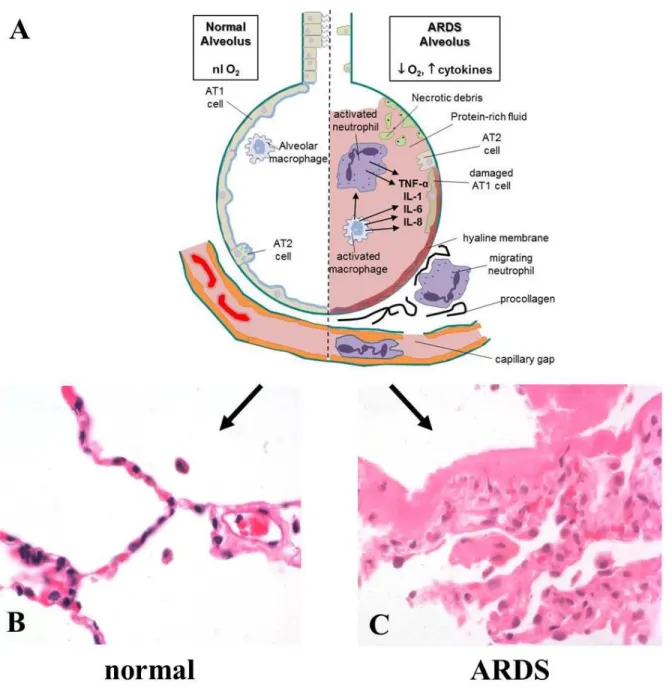
Figure 1.2: Alveolar pathology in ARDS . . . 36
Figure 1.3: Models of lung fluid homeostasis . . . 37
Figure 1.4: Real-time measurement of ASL and AvSL by confocal microscopy . . . 38
Figure 1.5: Insufficiency of standard culture models to study primary AT1 cells . . . 39
Figure 1.6: Design overview of a microwell array platform for culture of single AT1 cells under an ALI . . . 40
Figure 2.1: Fabrication and release of photoresist thin films . . . 82
Figure 2.2: Removal of 1002F films with various solvents . . . 83
Figure 2.3: Removal of 1002F films from fabrication substrate . . . 84
Figure 2.4: Tensile testing of freestanding, released 1002F films . . . 85
Figure 2.5: Flexibility of freestanding 1002F films . . . 86
Figure 2.6: Fabrication versatility of photoresist films . . . 87
Figure 2.7: Formation of high-aspect ratio through-holes in 1002F photoresist films . . . 88
Figure 2.8: Seeding of AT1-like cells into microstrainers . . . 89
Figure 2.9: Use of 1002F film as a stencil . . . 90
Figure 2.10: Application of photoresist thin films as inexpensive, re-usable stencils for metal deposition . . . 91
Figure 2.11: Protein surface micropatterning using freestanding microarray 1002F films as stencils . . . 92
Figure 2.12: 1002F films for loading exogenous molecules into single cells in a monolayer . . . 93
xvii
Figure 3.2: Imaging of released chitosan-bottomed microwell array films . . . 139
Figure 3.3: Fabrication of multi-layer 1002F:chitosan:1002F films . . . 140
Figure 3.4: Dependency of chitosan film thickness on spin coating speed . . . 141
Figure 3.5: Optical properties of chitosan films . . . 142
Figure 3.6: Permeability of micropatterned 1002F:chitosan films to small molecules . . . . 143
Figure 3.7: Diffusion of FITC-conjugated dextrans through chitosan membranes in micropatterned 1002F:chitosan films . . . 144
Figure 3.8: Visualization of FITC-dextran diffusion through chitosan membranes in individual microwells . . . 145
Figure 3.9: Determination of diffusion constants and molecular weight cut-off for chitosan membranes in micropatterned 1002F:chitosan films . . . 146
Figure 3.10: Selective and covalent modification of chitosan film surfaces . . . 147
Figure 3.11: Entrapment of non-adherent cells in culture medium micropockets . . . 148
Figure 3.12: Physical isolation of clonal cell populations in microwell medium pockets . . . 149
Figure 3.13: Physical isolation of clonal cell populations in microwell medium pockets . . . 150
Figure 3.14: Growth of primary AT1-like cells in laminin-coated 1002F:chitosan microwells . . . 151
Figure 4.1: Fabrication and release of photoresist-micropatterned track-etch membranes . . . 179
Figure 4.2: Imaging of track-etch membranes micropatterned with photoresist . . . 180
Figure 4.3: Verification of pore patency via solute diffusion . . . 181
xviii
LIST OF ABBREVIATIONS AND SYMBOLS
λ wavelength
Ω ohm
1002F EPON™ resin 1002F, an epoxy negative photoresist
2D two-dimensional
3D three-dimensional
A549 human alveolar adenocarcinoma cell line
Ab antibody
ADO adenosine
ALI air-liquid interface
APTES 3-aminopropyltriethoxysilane
Aqp aquaporin
ARDS Acute Respiratory Distess Syndrome ASL airway surface liquid
AT1 alveolar type 1
AT2 alveolar type 2
ATP adenosine triphosphate
Au gold
AvSL alveolar surface liquid
Ba/F3 non-adherent human leukemia cell line
BSA bovine serum albumin
xix Ca++ calcium cation
cAMP cyclic AMP
CF cystic fibrosis
CFTR cystic fibrosis transmembrane conductance regulator CHANL Chapel Hill Analytical and Nanofabrication Laboratory
Cl- chloride anion
cm centimeter
cm2 square centimeter
cm3 cubic centimeter
CO2 carbon dioxide
o
C degrees Celcius
D diffusion coefficient (alternatively, diffusivity)
Da daltons
ddH2O double-deionized water
DI deionized
DMEM Dulbecco's Modified Eagle Medium
DNA deoxyribonucleic acid
EtOH ethanol
ECM extracellular matrix
ECMO extracorporeal membrane oxidation
e.g. for example
EM electron microscopy
xx
FACS fluorescence-activated cell sorting
FBS fetal bovine serum
FITC fluorescein isothiocyanate
g gram
Gαq G-protein alpha-q subunit
Gαs G-protein alpha-s subunit
GBL γ-butyrolactone
GFP green fluorescent protein
GPA Granulomatosis with Polyangiitis
GPa gigapascals
GPCR G-protein coupled receptor
h hours
h thickness
H1299 human non-small cell lung adenocarcinoma cell line
H2O water
HCO3- bicarbonate anion
hr hours
HSC high-specificity cation
IPA isopropyl alcohol
i.e. meaning
ICU intensive care unit
Ig immunoglobulin
xxi
J joule
K0.5 half-maximal activity concentration
kDa kilodaltons
KGF keratinocyte growth factor
ln natural logarithm
log base-ten logarithm
m meter
M molar
% m/m percent mass per unit mass
m2 square meters
MAPK mitogen-activated protein kinase MDCK II Madin-Darby Canine Kidney MEMS microelectromechanical system(s)
mg milligram
min minute
mJ millijoule
mL milliliter
mm millimeter
mm2 square millimeters
mM millimolar
MPa megapascals
mRNA messenger ribonucleic acid
xxii
N number of moles
N2 nitrogen gas
Na+ sodium cation
NaCl sodium chloride
NaOH sodium hydroxide
NCI-H441 adherent human bronchiolar adenocarcinoma cell line
nL nanoliters
nm nanometers
nM nanomolar
NSC non-specific cation
O2 oxygen gas
P2X purine-gated cation channel
P2Y purine-activated GPCR
Pa pascals
PBS phosphate buffered saline
PCL periciliary layer
PCR polymerase chain reaction
PCT polycarbonate
PCTE polycarbonate track-etch
Pd palladium
PDMS polydimethylsiloxane
PEB post-exposure bake
xxiii PETE polyester track-etch
PGMEA propylene glycol methylether acetate
PKA protein kinase A
pL picoliters
pS picosiemens
PTFE polytetrafluoroethylene
RAGE Receptor for Advanced Glycation Endproducts
RNA ribonucleic acid
rpm rotations per minute
RPMI Rosalind Park Memorial Institute
RT-PCR reverse-transcriptase polymerase chain reaction
s seconds
S siemens
SEM scanning electron microscopy
sulfo-SMCC Sulfosuccinimidyl-4-(N-maleimidomethyl)cyclohexane-1-carboxylate SP-A surfactant protein A
SU-8 an epoxy negative photoresist
TC tissue culture
TEER transepithelial electrical resistance
Tg glass transition temperature
TNF-α tumor necrosis factor-alpha
µ micro
xxiv
µL microliters
µm micrometers or microns
µm2 square micrometers
µM micromolar
UTP uridine triphosphate
UV ultraviolet
V volume
1
Chapter 1: Introduction
1.1 Airway and Alveolar Disease: States of Disordered Pulmonary Fluid
Homeostasis
1.1.1 Respiratory epithelia and luminal fluid films
1.1.1.1 Hierarchical organization of respiratory epithelia
The human lung is a highly complex organ system whose function is to facilitate exchange of CO2, a by-product of energy metabolism, for the atmospheric O2 that drives oxidative chemical respiration, the reaction that nearly every human cell uses to produce energy.1
Inhaled air is conducted through the nasopharynx and down the trachea, where it enters the lungs via the left and right main pulmonary bronchi. Further movement through the respiratory tree – the secondary and tertiary bronchi, the terminal bronchioles, and the respiratory bronchioles – deposits the air in the alveoli, or air sacs, located at the most distal part of the lung and at which gas exchange actually occurs.1-3
2
membrane (Figure 1.1A).4 As will be explained below, recent research has discovered that these cells, despite their lack of participation in any gas exchange, play an important role in pulmonary fluid homeostasis and in the innate immune system of the lung.5-6 The epithelial layer here also has a number of goblet cells and exocrine secretory glands, both of which serve to secrete mucus onto the luminal surface. At the more distal level of the terminal bronchioles, the epithelial tissue begins to transition to a layer of simple cuboidal epithelium that replaces ciliated pseudostratified epithelium with ciliated cuboidal cells and goblet cells with “club” cells that are morphologically and functionally diverse (involved in secretion of surfactant-like glycosaminoglycans, IgA, and lysozyme, lysosome- and cytochrome P450-mediated metabolism of inhaled exogenous substances, and regeneration of the bronchiolar lining)(Figure 1.1B).4, 7-11 A transition again takes place as the respiratory tree further branches into the respiratory bronchioles, in which alveoli begin to appear in the walls. Alveoli are primarily located, however, at the most distal end of the respiratory bronchioles. Whether the alveoli lie in the walls of a respiratory bronchiole or at its distal end, a third transition of epithelial cell type occurs at the alveolar atrium, where cuboidal epithelium terminates and the epithelial layer becomes composed of two types of cells, termed alveolar type 1 (AT1) and alveolar type 2 (AT2) cells (Figure 1.1C).4
AT1 cells are squamous-type epithelia with large diameters – on the order of 100 µm – and extremely thin cytoplasms, save for a perinuclear region containing a number of
3
basement membrane.14 Together, AT1 cells are estimated to make up 40% of the alveolar cells and 97-98% of the total alveolar surface area.13
AT1 cells are thought to be terminally differentiated cells that arise from AT2 cells, cuboidal cells that make up the remaining 60% of alveolar cells but just 2-3% of the alveolar surface area.15 Classic experiments by Evans et al. and Adamson and Bowden, as well as more recent work, showed that AT2 cells function as progenitor cells that can divide and differentiate, eventually assuming an AT1 morphology in order to preserve the alveolar epithelial surface in the event an AT1 cell undergoes apoptosis or death.16-19 More advanced studies recently showed that in response to cellular injury (either AT2 cell-specific ablation or hyperoxic damage to AT1 cells), lineage-labeled AT2 cells clonally divide and self-renew in both mouse and human adult lungs, and that lipofibroblasts may help these cells form a specific pulmonary stem cell niche.20-21
AT2 cells have further importance as the source of pulmonary surfactant, a mixture of secreted proteins (surfactant proteins SP-A, -B, -C, and –D) and phospholipids (mainly dipalmitoylphosphatidylcholine).22 Not only does this surfactant mixture help to opsonize any stray bacteria that reach the alveolus, it also accomplishes the imperative task of maintaining alveolar and small airway patency by reducing the surface tension of the epithelial surface.23-24 In addition, AT2 cells synthesize and secrete immune effector molecules such as CXC chemokines and complement proteins.25-27
4
Gonzalez et al. analyzed freshly isolated AT1 and AT2 cells and cultured AT2 cells, finding over 600 genes differentially expressed in two-way comparisons between the three
populations.28 Treutlein et al. recently used single-cell RNA sequencing to examine distal lung cells from the developing lungs of embryonic mice, and noted the presence of a “bipotential progenitor” cell that had expressed both AT1 and AT2 markers, potentially identifying a progenitor state through which transdifferentiating AT2 cells pass as they begin to downregulate typical AT2 gene expression patterns (typically surfactant proteins SP A-D) and upregulate typical AT1 patterns (such as podoplanin, aquaporin-5 (Aqp5), caveolin-1, and receptor for advanced glycation end products (RAGE)).32 One possible interpretation of this data, which seem to argue against the classical model of AT2-to-AT1 transdifferentiation is that the “bipotential progenitor” represents a population of late stage, lung-specific, stem cells that give rise to a small group of differentiated, functional AT1 cells for the immediate needs of the lung and to a group of AT2 cells to replenish the AT1 population in the adult organism, once the pluripotent stem cell population has decreased. However, some have also called into question whether AT1 cells are truly terminally differentiated: a report by
Gonzalez et al. observes that AT1 cells can divide in vitro and stain for the proliferation marker OCT-4, and three other reports detail the use of keratinocyte growth factor (KGF) to attenuate or even reverse the AT2-to-AT1 transdifferentiation, as measured by expression of AT1 or AT2 markers Aqp5 and SP-A and activation of mitogen-activated protein kinase (MAPK) pathways.33-36
5
pathogens entering the alveolus in inhaled air and any cellular debris that results from turnover of the epithelial layer.4
1.1.1.2 Air-liquid interfaces and luminal fluid films in the pulmonary tract
In addition to a hierarchical organization of the cellular subtypes found in the epithelial layer, a second key feature of the respiratory tree is presence of a physiologic air-liquid interface (ALI) just above the epithelial tissue, due to the open-air lumen to which the epithelia that line the respiratory tract face. Flow of blood through capillary beds below the basement membrane on which the epithelia lie ensures delivery of nutrients and removal of metabolic by-products, as well as ensuring proper electrolyte balance across the cell
membrane and availability of fluid to maintain cellular hydration.4 While air in the lungs has relatively high humidity (achieved via humidification of inhaled air that takes place in the nasopharynx), respiratory epithelial tissue nonetheless greatly differs from other epithelial tissues that are bathed in large fluid volumes (large relative to the cell volume) on both sides of the basement membrane, basal and luminal.14 Importantly, the presence of an ALI appears to have a role in maintaining a differentiated and polarized phenotype for the epithelial layer and thus appears to be a key and necessary component of normal in vivo physiology, a concept that will be discussed further in section 1.2.4.37-40
However, rather than having an apical surface that is completely dry, epithelia at all levels of the respiratory tree (from the trachea to the alveolus) are known to have a thin sol-gel laer on the luminal surface. Thus, the true location of the respiratory ALI does not lie at the epithelial layer, but rather just above it. Similar to the hierarchical heterogeneity of epithelial subtype observed along the respiratory tree, the apical fluid layer varies in
6
airway surface liquid (ASL), to an estimated 0.2 µm in the alveoli, where it is known as the alveolar surface liquid (AvSL).41-42 Thinning of the fluid layer in the more distal parts of the lung reflects the need to minimize the diffusion distance and optimize gas exchange
efficiency.
The conservation of the apical fluid layer thickness within a given level of the respiratory tree suggests that the lung can homeostatically regulate the thickness (and hence the volume) of the fluid layer in both the airways and alveoli. Two opposing observations support this. First, because the water content of the humidified air in the lungs lacks the volume to match the estimated 20-30 mL that comprises the respiratory thin fluid layer over the surface area of the entire lung (~100 m2), let alone the estimated 700 mL of fluid thought to be lost in the proximal airways due to evaporation, the fluid layer must be generated from the respiratory tract itself (and not “raining out” from the air inhaled into it).41, 43 At the same time, the demonstration of rapid clearance of pulmonary fluid at birth, when the location of gas exchange moves from the placenta to the lung, and the observation that a small fluid bolus instilled into the lungs is cleared into the interstitium suggest that fluid clearance out of the respiratory lumen is a normal physiological process.44-46 However, as will be discussed below, this process of pulmonary fluid homeostasis can be disrupted by certain lung diseases.
1.1.2 Alveolar diseases and disordered fluid homeostasis
7
toxic inhalants, Goodpasture’s Syndrome, Granulomatosis with Polyangiitis (GPA), and connective tissue and autoimmune diseases (e.g. systemic lupus erythematosus).48-49
However, the most serious example of alveolar disease is Acute Respiratory Distress Syndrome (ARDS), in which an insult to the alveolar-capillary interface is enough to damage the alveolar epithelial cells, the capillary endothelial cells, and even the basement membrane separating the cell layers (Figure 1.2A).50 This compromise of the normal alveolar-capillary barrier (Figure 1.2B) increases its permeability, leading to the influx of a highly
proteinaceous fluid into the alveoli that results in severe and intractable alveolar flooding and a severe compromise in gas exchange. Numerous etiologies (most commonly septic
pneumonia or aspiration/inhalation injury) can cause ARDS, but the clinical pattern is the same: rapid onset of dyspnea and arterial hypoxemia that is refractory to O2 therapy,
followed by worsening respiratory failure as the alveoli further fill with fluid and the alveolar basement membrane becomes hyalinized with thick protein deposits (Figure 1.2C).50
8
macrophages, inflammation is reduced, and a slow repopulation of the alveolar and endothelial layers by resident progenitor cells occurs as those cells proliferate and spread over fresh extracellular matrix laid by down by fibroblasts activated by the inflammation.51
At its onset, ARDS is a medical emergency that usually necessitates admission to an intensive care unit (ICU), and epidemiologic data suggest that there are close to 200,000 cases of ARDS in the U.S. annually.50, 52 Up to 20% of patients in intensive care units on mechanical ventilation meet ARDS criteria, and current overall mortality rates of ARDS patients lie at 37-45%, and are as high as 50-60% for some patient groups, such as the elderly and those with pre-existing cardiovascular conditions.50, 53-54
The most serious complication shared by all of the diseases described above is the persistent presence of fluid in the alveolar space. This fluid can disrupt the gas exchange function of alveoli, owing to a great increase in the diffusion distance without a
corresponding increase in oxygen solubility in the fluid. Additionally, the fluid can also result in damage to the epithelial layer itself, further compromising gas exchange. The presence of intractable alveolar flooding in these disease states, but not in normal states, suggests a compromise of normal alveolar fluid homeostasis via an increased influx of fluid from the bloodstream or interstitium into the alveolus (increased rate of entry), a reduced clearance of fluid from the alveolar space (decreased exit rate), or both.55-56
1.1.3 Alveolar diseases: therapeutic options and shortfalls
Certain cases of alveolar flooding due to disease can be treated and resolved without complication. Pulmonary edema, for example, can be treated via a combination of diuretics (to promote alveolar fluid resorption) and appropriate management of the underlying
9
diseases, namely Goodpasture’s Disease, lupus, and GPA, can be managed with
immunosuppression, with fluid in the alveolus being gradually resorbed over time and the erythrocytes broken down by resident alveolar macrophages.49
ARDS, however, continues to present a treatment challenge, due to both the
persistence of alveolar fluid (owing to basement membrane disruption and hyalinization) and the damage to the epithelial and endothelial cells themselves. While the triggering etiology (e.g. sepsis) can sometimes be treated and resolved, the patient is left in a hypoxemic state with flooded, fibrotic alveoli, and hence respiratory support with a mechanical ventilator is the mainstay of ARDS treatment.56 Recent clinical trials advocating a switch from traditional to low-tidal volume ventilation have led to some improvement in relevant patient outcomes, namely hospital and overall mortality and number of ventilator-free days. 58-59 However, low-tidal volume ventilation is nonetheless only an improvement in supportive therapy, not a direct address of ARDS pathophysiology. Similarly, the FACTT Trial indicated that while patients who received conservative fluid management in the ICU in order to reduce alveolar flooding required less time on a mechanical ventilator, overall 60-day survival was not increased.60 Recently, clinical trials of both inhaled and infused β2-adrenergic receptor agonists that had demonstrated in vitro efficacy in potentiating fluid clearance by AT2 cell monolayers failed to show improvements in patient mortality.61-62 Other clinical trials have demonstrated the failure of inhaled nitric oxide, antioxidant compounds, and exogenous pulmonary surfactant in reducing morbidity or mortality.63 Even in those patients who survive ARDS and can be discharged from the ICU, functional morbidities remain. Herridge
10
Considering the prevalence and variety of etiologies that can result in ARDS (notably sepsis from either community or nosocomial sources) and the epidemiologic data on ARDS mortality, ARDS presents a significant source of morbidity and mortality to the public health. Given the central role alveolar flooding occupies in ARDS pathophysiology, a strategic approach to potentiating alveolar fluid removal, especially in the early stages of ARDS before basement membrane hyalinization occurs, is an important and pressing biomedical research goal.
1.2 Physiology and Pharmacology of Pulmonary Liquid Homeostasis
1.2.1 Airway surface liquid in bronchial and bronchiolar epithelia
In order to most efficiently develop a strategy, pharmacologic or otherwise, to potentiate alveolar fluid clearance, a full and complete understanding of alveolar fluid
homeostasis mechanisms and regulation is needed. A great deal of information exists on fluid homeostasis in the upper airways, and so provides a useful place to begin generating a model of fluid clearance in the more distal lung spaces.
11
luminal surface.5-6, 41, 67 Discoveries of reduced chloride anion (Cl-) secretion to, and
increased sodium cation (Na+) resorption from, the airway surface by epithelia of CF patients compared to healthy subjects were found in the 1980s via bioelectric measurements.68-72 Subsequent functional reconstitution of the gene product shown to be mutated in CF – termed the cystic fibrosis transmembrane conductance regulator (CFTR) - revealed it to be a chloride channel.73-76 As further research was undertaken to better understand how CFTR loss also resulted in abnormally high Na+ resorption through the epithelial sodium channel (ENaC), two studies by Matsui et al. suggested that these ion transport abnormalities resulted in depletion of the layer of fluid approximately 10 µm thick found on the luminal surface of ciliated airway epithelia.77-78 In normal airways, this apical fluid hydrates a layer the approximate thickness of the respiratory cilia (hence the term ‘periciliary layer,’ PCL), and the mucus sol-gel layer above it (together known as the airway surface liquid ASL). The presence of fluid in the PCL allows efficient and coordinated beating of the cilia atop the cells, allowing the movement of mucus in the overlying sol-gel layer up the respiratory tree and out of the lung in a process termed mucociliary clearance (MCC). The dehydration observed in CF airways leads to ciliary stasis and a reduction in mucociliary clearance. Based on data comparing healthy and CF subjects, airway fluid is thought to be maintained via a CFTR- and ENaC-dependent transport of chloride and sodium
12
This hypothesis also describes a regulatory mechanism for airway surface ion
transport: extracellular concentrations of the nucleotide adenosine triphosphate (ATP) and its metabolites. Knowledge that activation of protein kinase A (PKA) by cAMP stimulated chloride conductance in normal airway epithelia, but not in CF cells, suggested a role for cAMP-dependent PKA signaling in CFTR activation.79-81 Determination of CFTR’s primary structure identified several consensus phosphorylation sites for PKA, explaining the lack of response to cAMP or PKA in CF cells.73 Further research into the regulation of this ion transport system in normal airways revealed a role for constitutive release of ATP to the luminal surface, its subsequent metabolism by ecto-nucleotidases to adenosine, and activation by that adenosine of the A2b adenosine receptor, an apical membrane Gαs - associated GPCR.82-83 This activation, resulting in adenylate cyclase stimulation and increases in intracellular concentrations of cAMP, leads to PKA activation and
phosphorylation of CFTR. Subsequent increases in chloride secretion and ENaC inhibition by CFTR generates the osmotic gradient for water flow to the airway surface, which dilutes surface adenosine concentrations and reduces A2b receptor activation, thus providing negative feedback to the regulatory pathway.84
Interestingly, a second signaling pathway utilizing surface P2 purinergic receptors was coincidentally discovered that came to be a target for pharmacologically potentiating directed ion transport and correcting the disordered fluid homeostasis in CF.85 Early
13
ATP and uridine triphosphate (UTP) nucleotides to the extracellular surface of airway
epithelia.87-92 Later data demonstrated a connection between apical P2Y purinergic receptors, to which ATP and UTP are agonists, and Ca++-dependent increases in chloride
conductance.93-94 These P2Y purinoreceptors on the apical membranes of airway epithelia activate chloride secretion via a Gαq-mediated release of intracellular calcium that targets TMEM16A calcium-dependent chloride channels found in respiratory epithelia.95
In summary then, the leading model for fluid homeostasis in the airway involves active chloride and sodium ion secretion onto the airway surface, creating an osmotic pressure that draws water onto the apical membrane, where it hydrates the airway surface with a ~10 µm-thick fluid layer that facilitates proper cilia function and mucociliary
clearance (Figure 1.3A). Regulation of this CFTR- and ENaC-mediated chloride and sodium ion balance is achieved under normal conditions by the constitutive extracellular release and metabolism of ATP to adenosine, which fine-tunes airway surface hydration via A2b receptor binding and downstream cAMP-mediated activation of CFTR.6, 96 As will be discussed below, much of the same mechanistic and regulatory machinery of airway fluid homeostasis is also found in alveolar cells.
14
clinical success in improving lung function. Second, an understanding of how loss of CFTR-mediated ion transport results in defective fluid homeostasis led to the demonstration that inhaled doses of hypertonic saline could provide an osmotic draw that restores proper airway surface hydration to normal functional levels. By doing so, Donaldson et al. reported an increase in sustained mucus clearance and lung function in CF patients.102 Third, the identification of accessory purinergic signaling pathways led to the development of several drug candidates for potentiating chloride secretion, and thus airway surface hydration, through non-CFTR means. Synthetic analogues of ATP were found to potentiate in vitro
Ca++-dependent chloride secretion and were taken into clinical trials.103-104
1.2.2 Ion and fluid transport by alveolar type 2 (AT2) epithelia
In 1995 Bastacky et al. used scanning electron microscopy to show the presence of a continuous, 0.2 µm-thick apical fluid layer in alveoli.42 While this was the first direct measurement of the basal thickness of the alveolar surface liquid (AvSL), the first evidence that fluid homeostasis occurs in the alveolus had come a decade before, when a
demonstration by Matthay et al. of differential clearance rates of fluid and protein instilled into sheep alveoli was published at the same time that the first cultures of isolated primary AT2 cells were shown to participate in active ion transport.105-106 Since that time, alveolar ion transport has been studied extensively using a variety of models, including animal and human whole perfused lungs and an innovative lung slice preparation.107-108 Rat models have been widely used due to demonstrations that total lung fluid clearance rates in rats are similar to those of isolated, perfused human lungs ex vivo.107 Despite these clinically-relevant
15
and hence there remains a crucial role for basic in vitro studies of purified primary alveolar cells.
A number of published studies have specifically examined ion transport in AT2 cells. Early work showed that monolayers of cultured AT2 cells took up 22Na+ in an apical-to-basolateral direction and produced measurable transepithelial potential differences when cultured on permeable supports.106, 109 Further work by a number of groups has shown that rat AT2 cells, both in situ and cultured on permeable supports, contain the basolateral
Na/K/ATPase and ENaC. Bioelectric studies of rat AT2 cell monolayers and patch-clamp studies of individual rat AT2 cells have firmly established the functionality of these proteins.40, 46, 110-112 Studies by Fang et al. have demonstrated a role for CFTR in AT2 cells.113 ENaC has been a subject of particular investigation in the alveolar biology
community.111, 114 One general set of studies has focused on the balance and relative activity between two functional variants of ENaC. One variant, termed highly-selective (HSC) has a low, amiloride-sensitive conductance (approximately 6 pS, amiloride K0.5 = 37 nM) but a >40 fold selection of sodium over potassium cation, while the non-selective (NSC) variant is amiloride-insensitive (K0.5 = 2.2 µM) and cation nonselective, albeit with a higher
conductance (21 pS).114 Jain et al. noted that cultured AT2 monolayers preferentially expressed the HSC ENaC variant when cultured under an ALI, suggesting that a shift in ENaC function in AT2 cells can accompany a transition from a basal to a flooded
16
trying to reconcile failed clinical trials of β2-adrenergic agonists with in vitro studies suggesting the efficacy of this agonist class.115-125 An as-yet unresolved question concerns whether CFTR, or at least some kind of Cl- transport, is required for ENaC activation. Jiang
et al. showed that adrenergic stimulation of Na+ transport in AT2 cells involved the
activation of apical Cl- channels, while Mutlu et al. showed a similar effect but demonstrated a role for CFTR as the intermediate in ENaC activation.117, 119 Interestingly, CFTR-mediated Cl- secretion in confluent cultures of primary AT2 cells was shown to be calcium-dependent, a relationship not found in epithelia of the upper airways.126
Striking similarities in the regulation of ion transport and fluid homeostasis were also recently shown between bronchial epithelial and AT2 cells at the monolayer level. Bove et al. used confluent monolayers of AT2 cells to show that AT2 ion transport and fluid movement is regulated by nucleotides via purinergic signaling pathways similar to those of bronchial epithelia in the larger airways.126 Addition of ATP and UTP to AT2 cell
monolayers produced ion fluxes inhibited with ion transporter modulators. Similar addition of ATP and UTP to cell monolayers led to increases in apical fluid thickness, while addition of the nucleotide-scavenging enzyme apyrase produced the opposite effect. Moreover, AT2 cells grown in monolayers expressed the P2X4 and P2Y2 purinergic receptors.126 The significance of this report was twofold. First, its study of ion transport by monolayers of human AT2 cells validated similar data showing functional ion transporters in rat AT2 monolayers. Second, analysis of the role of nucleotide signaling in these monolayers
17
transport mechanism and regulation are made more interesting by the identification of such molecular machinery in AT1 cells as well.
1.2.3 Ion transporters and purinergic receptors in alveolar type 1 (AT1)
epithelia
While bronchial and AT2 cells have been studied extensively, the physiology of AT1 cells, which populate >98% of the alveolar surface area, remains poorly understood. For many years, this was due to technical difficulties in the isolation and purification of AT1 cells, which are larger, more fragile, and lower yielding in primary cell harvests than AT2 cells.127 One advance for the AT1 field came with the identification and use of antibodies to apical integral membrane proteins specific to AT1 cells: chiefly, RTI40 in rodents and HTI56 in humans.128-129 This advance enabled positive selection strategies that allowed for the isolation of AT1 populations at purities of >85%, with cellular viabilities of >95%.130-132 Typical isolation/purification protocols currently involve enzymatic digestion of the distal airspaces with elastase to liberate alveolar cells, labeling of the AT1, AT2, and macrophage cells with antibodies to cell-specific markers, and lastly either negative depletion by
magnetic bead immunoselection or positive enrichment by fluorescence-activated cell sorting (FACS).133
18
However, more recent advances in AT1 cell biology have called into question the idea that AT1 cells play only a passive role in alveolar fluid transport. This paradigm shift has occurred because of the identification of similar ion transport machinery in AT1 cells through radionuclide uptake, electrophysiology, and mRNA and protein-level expression studies. First to suggest a higher role were two independent studies by Borok and Johnson showing that isolated rat AT1 cells immunostain for ENaC and the Na/K/ATPase.127, 130 These proteins, both active in ion transport, were also shown in parallel rat alveolar tissue sections. Johnson also showed that isolated AT1 cells in suspension could actively take up 22
Na+ in an amiloride-inhibitable fashion.130 Cell-attached patch clamp studies of individual, nonconfluent AT1 cells showed function of HSC and NSC ENaC and CFTR, as well as a number of other channels such as potassium and cyclic-nucleotide gated (CNG) cation channels and the Cl-/HCO3- anion exchanger.131-132 Apart from showing electrophysiologic function of the ion channels previously identified by immunofluorescence, this work was significant in that measurements of channel density and area suggest that AT1 cells contain a significantly greater number of ion channels than AT2 cells. By applying the calculated channel density found in AT1 versus AT2 cells to the surface area of each cell type, AT1 cells were found to have a seven-fold greater osmotic permeability, a forty-fold higher number of Na+ channels, and six times as many CFTR channels.136 While a caveat central to these studies is that the measured cells were grown in suspension or unpolarized on glass, the identification of ion channels studied in vitro with positive in situ immunostaining of alveolar tissue sections for those same channels supports these data.
19
higher numbers of functional ion channels in AT1 versus AT2 cells, an alternative model theorizes that AT1 cells achieve the bulk of both ion and water transport (compared to the smaller AT2 cells) in the alveolus (Figure 1.3B). In this model, similar to that of the airway, Cl- and Na+ are moved across the apical membrane of both AT1 and AT2 cells by specific ion channels (presumably CFTR and ENaC, respectively), and water follows down its osmotic gradient through Aqp5.136-137 Curiously, patients with CF who thus lack functional CFTR do not exhibit signs of alveolar disease, suggesting the possibility of either an alternative primary anion transporter as the driver of alveolar fluid homeostasis, or the upregulation of an alternative, secondary mechanistic pathway in CF patients that could present a pharmacological target in all patients with alveolar disease.126
20
receptors. In the latter case, however, the lack of reliable P2Y or P2X receptor antibodies makes this difficult to achieve.
In summary, then, the expression of similar ion transport molecular machinery in bronchial, AT2, and AT1 cells, as well as apparent similarities in the pathways regulating ion transport in bronchial and AT2 cells, suggest conserved mechanisms and regulation of ion transport and fluid homeostasis in both the airway and the alveolus. Taken together, these similarities also provide compelling evidence that, if a detailed understanding of the physiology is available, successful pharmacological targets for potentiating alveolar fluid clearance can be identified for use in alveolar diseases.
Given the success of in vitro cultures of bronchial and AT2 cells in replicating the in vivo physiology, the logical direction for subsequent research, then, would be to determine whether in vitro cultures of purified AT1 cells directly participate in directed ion and fluid transport and to elucidate the mechanisms and regulatory elements of that process. However, testing this theory of ion and fluid transport by AT1 cells has proven difficult due to technical difficulties in AT1 culture, beyond the difficulties encountered in AT1 isolation: the
formation of intercellular gaps between AT1 cells in culture that render useless the gold standard methods for measuring ion and fluid transport in cultured cells.
1.2.4 Current methods for studying pulmonary fluid homeostasis
A great deal of the data used in formulating the current model of fluid homeostasis by bronchial epithelia, as well as a number of studies with AT2 cells, used monolayers of
21
commercially available and typically composed of either track-etched polyester or
microwoven fluoropolymer (for more details, see Chapter 4), feature pores that permit the exchange of fluid, ions, and other solutes across the support. By seeding cells onto the
support, supplying growth media to the compartment below the support, and evacuating fluid from the apical surface, the primary cells can be cultured across an ALI that ensures cells can obtain nutrients and expel metabolites from the culture medium below, while still replicating the ALI that characterizes their natural in vivo microenvironment.
The necessity for permeable supports is twofold: the maintenance of proper in vivo
22
two studies have nonetheless shown that ALI conditions influence bioelectric properties in AT2 monolayers as well.39-40
In addition to promoting a recapitulation of both the in vivo epithelial layer
architecture and the molecular physiology of the epithelial cells themselves, use of permeable supports also allows testing of tissue-scale epithelial function using bioelectric and confocal microscopic methods to study ion and fluid transport, respectively. In the first case, a setup known as an Ussing chamber is utilized to allow for real-time monitoring of voltage potential differences across an epithelial layer, with the capability to rapidly introduce interrogatory compounds such as ion transporter modulators to either the apical or basolateral faces of the cell.138 Supply of an inhibitor to a specific ion transporter present in the membranes of cells in the monolayer, for example, begets a reproducible and dose-dependent change in the transepithelial potential difference measured by the chamber apparatus. In this way, studies of ion channel expression can be used to guide the design of specific bioelectric experiments. In the second case, confocal microscopy is used to directly observe fluid movement across an epithelial layer. A typical protocol, pioneered by the Boucher Labs, involves labeling the apical surface fluid with a fluorophore-conjugated, high-molecular weight dextran and using a laser-scanning confocal microscope to scan through the apical fluid and epithelial cell layers. By measuring the thickness of the fluorescent layer above the cells, the thickness and volume of the surface liquid can be determined. Time course serial imaging of the fluid layer allows quantitation of water flux in response to perturbations of the mechanistic or regulatory molecular machinery (Figure 1.4).147
23
leakage of ions and fluid. Primary bronchial and AT2 cells readily form epithelial layers in culture that have transepithelial seals with resistances on the order of 500 Ω*cm2 at the minimum (Figure 1.5A).126, 138, 148 However, primary AT1 cells in culture spontaneously form intercellular gaps.132, 149 Comparison of these gaps, which reach sizes of up to 50 µm (Figure 1.5B), with in vivo alveolar ultrastructure led to the proposal that these gaps are the attempts of AT1 cells in culture to form pores of Kohn, an interalveolar gap involved in collateral ventilation of adjacent alveoli that prevent alveolar collapse. Regardless of their purpose, these gaps act to short-circuit bioelectric measurements and allow fluid leakage in microscopy studies, and so constitute a technical barrier that must be overcome for useful study of AT1 cells.132, 149
Given the imperative to directly assess ion and fluid transport by AT1 cells, and the problem posed by the intercellular gaps they form, I hypothesized that a novel cell culture platform fabricated with microscale features would enable the culture of small numbers of or even single primary AT1 cells while circumventing the problem posed by the AT1
intercellular gaps. Construction of such a platform lies in the field of microfabrication.
1.3 Microscale devices for analysis of discrete numbers of cells
1.3.1 Definition and use of microscale devices
24
or gradients of substrate stiffness. Combinations of these patterning schema open
applications such as directed or ordered cellular co-cultures, tailoring substrate stiffness for experiments on cellular mechanotransduction, or spatially-defined gradients of chemotactic factors.152-155
The most common methods in use in the microfabrication field include soft lithography, photolithography, and injection molding, with a number of others also available.156-159 Soft lithography most commonly uses the optically clear silicone polymer polydimethylsiloxane (PDMS) to create structures from a master mold. By casting uncured PDMS onto the mold, heat curing, and later removing the polymerized PDMS, a final structure of negative relief from the master mold is formed.158 Photolithography typically uses photoactive polymers known as photoresists to create its structures. By exposing the photoresist to UV light through a mask patterned with microscale 2D features, the photoresist can be selectively polymerized according to the mask pattern. Injection molding, typically done with cyclic olefin co-polymer, a thermoset, or a thermoplastic, involves the instillation of a polymer in its liquid form around a mold, after which the polymer is allowed to cool and set, forming a negative relief of the mold.
In addition to general classifications of device composition and construction, the majority of devices used in biomedical research endeavors and fabricated using these materials typically fall into one of several general design categories: valved or laminar flow microfluidics, droplet microfluidics, microwell arrays, micropedestal arrays, and
25
droplets composed of one phase that are driven through the device microchannels by flow of the other phase. Microwell arrays feature an ordered pattern of pits into which a cells or some other analyte of interest is deposited. Micropedestal arrays, the opposite relief pattern of microwell arrays, disperse the analyte, typically cells, onto the sides or top of small physical posts patterned onto a surface. While microwell and micropedestal arrays are both
patternings of physical features, micropatterned surfaces generally entail chemical patternings of some kind. Though a comprehensive review of the microfabrication field, apart from the focused survey described in sections below, is beyond the scope for this dissertation, there are several excellent reviews of this field, which is rapidly growing and becoming highly relevant for biomedical research.162-167 These surveys summarize recent advances not only in the materials and techniques of microfabricated devices, but also in the novel and innovative applications for which these devices are being used.
1.3.2 Advantages of microscale devices
A wealth of literature exists that describe in detail the advantages microscale devices bring to biomedical research. Chief among these advantages is the relative ease of adapting these devices for use with automated instrumentation and high throughput methods, which take advantage of the patterned nature of the devices via algorithms that not only enable rapid and precise analysis, but an orderly reference of assay results. Additionally, because of the small size of the features in these devices, a reduced consumption of reagents can be achieved. One unique advantage some, though not all, microscale devices have over
26
Most relevant to this dissertation is the compelling case that microscale devices make as tools for analyses of single or discretely small numbers of cells. Because the design of these devices allows for precise engineering of the shape, size, and surface chemistry of the area used for cell growth, these devices allow unparalleled control of the physical and chemical microenvironment to which a cell or group of cells is exposed as it grows. 150, 164, 170-171
By achieving such control, researchers gain the ability to create novel bio-inspired platforms that can precisely interrogate the mechanisms and regulation of fundamental cellular processes and to gather highly sensitive, cell-specific data platforms.165, 172-175 Be it due to the inherently small size of their features, or the frequent need for gathered data to meaningfully discern between groups of or even single cells, microscale devices are also uniquely suited to performing the highly sensitive measurements necessary for high throughput, single cell analysis.160-161, 176
Thus, given their advantages and utility as single-cell analytical platforms, a research goal of designing and building a microscale device for the study of primary AT1 cells was formed.
1.3.3 Survey of previously described microscale devices relevant to alveolar
cell culture
1.3.3.1 Microscale devices applied to respiratory biology
27
to studies of pulmonary surfactant dysfunction or depletion.177-179 Building on these past works, a study by Douville et al. created a microfluidic model of the alveolus in which sheets of primary murine alveolar cells or cells from the human immortalized alveolar epithelial A549 cell line were grown on a flexible membrane of PDMS subjected to a migrating ALI.180 This study demonstrated the role of migrating ALIs in worsening the cellular damage caused by cyclic stretch/compression as compared to cells that were entirely submerged or air-exposed when stretched, results relevant to research on ventilator-induced lung injury.
Additionally, a seminal study by the Ingber Group engineered a “lung-on-a-chip” device that featured a microporous membrane separating two microchannels.181 On one side of the membrane, a sheet of immortalized endothelial cells were exposed to a fluid filled microchannel. The opposite side of the membrane was covered with sheets of cells from either small airway NCI H-441 or alveolar A549 cell lines, creating an ALI across the membrane. A noteworthy feature of this device was the pair of microchannels that laterally bordered the membrane: reducing or normalizing air pressure to these side channels created stretch or relaxation forces that simulated the expansion of the pulmonary tissues that occurs during inspiration.
Several of the studies cited above are surveyed in a review of efforts to study
respiratory physiology on chip-based platforms.182 This review reiterates the need for an ALI in culture of pulmonary cells as a way to recapitulate true in vivo architecture. Several
28
membrane oxidation (ECMO), a direct therapeutic end goal not falling within the physiology- and pharmacology-oriented research goals of this dissertation.
While useful advances, none of these devices are suited for research with primary AT1 cells, for at least one of a variety of reasons: incompatibility with primary cell use, designs that do not feature permeable supports, or designs that do not appropriately replicate the physicochemical features of the alveolar architecture. For example, while the “lung-on-a-chip” represents an impressive engineering advance, its use of sheets of cells grown atop a membrane with large, 10 µm-diameter pores does not sufficiently replicate the alveolar microenvironment, which features smaller physical concavities approximately 250 µm in diameter whose basement membrane has submicron-scale porosity.1, 183 The microfluidic alveolar model developed by Douville et al. has similar shortfalls: sheets of cells 6 mm in diameter, grown on an impermeable membrane 100 µm thick, do not replicate the alveolar microenvironment seen by an AT1 cell. Given the shortcomings of these otherwise laudable devices as they relate to AT1 culture, other examples of microtechnology that utilize
permeable membranes were explored.
1.3.3.2 Microscale devices utilizing a permeable support
To date there has been some success in combining microscale technologies with porous or otherwise permeable materials, even if they have not been used in respiratory biology studies. Porous membranes are used most commonly in microfluidic setups
29
membranes and the PDMS layers are most commonly bonded either indirectly using a thin PDMS mortar layer or directly via the organosilane 3-aminopropyltriethoxysilane
(APTES).184-186 However, most of the studies cited above bond only the membrane edges, at the macroscopic scale, to the borders of PDMS layers. A literature review did not locate any studies that involve bonding membranes to PDMS films with a microscale pattern, likely due in one part to the relative difficulty in making through-hole arrays in such films in the first place and due in another part to difficulty in establishing uniform seals around each microwell that eliminate communicating gaps between them. Indeed, the smallest area of membrane exposed was still 4 mm2.187 Use of “microfluidic stickers” presents an alternative method that is chemically distinct from the sandwich methods described above, but nearly identical in general concept.188
Studies employing the PDMS-membrane sandwich method have used the membrane in a microfluidic setup in such varied applications as establishing standing chemical gradients to which cells can be exposed without the harsh shearing forces of high flow rates.188-190 In a general sense, seeding cells onto one side of the membrane allows for single-stream
perfusion at relatively slow rates – or even static culture – that do not introduce shearing stresses to the cell. On the other side of the membrane, flows from two entering fluid
30
and hemolysis-free filtration of plasma from whole blood.191-192 By using membranes with pores too small for red blood cells, whole blood infused into the system could be efficiently separated into two compartments, with one compartment retaining erythrocytes and
leukocytes and the other containing only plasma, with low shear stress and minimal erythrocyte hemolysis.
Other reports utilize the membrane to recapitulate the polarization seen in vivo of certain types of non-respiratory epithelial cells. For example, Jang and Suh created a
microfluidic “kidney-on-a-chip” using PDMS bonded to a porous membrane on which sheets of primary rat inner medullary collecting duct (IMCD) cells were cultured.193-194 The
McGuigan Group successfully patterned MDCK and retinal epithelial cells into
microcolonies atop commercially-available porous substrates.195 A device fabricated as an extension of “lung-on-a-chip” technology, described in the previous section, was used to grow cells from an immortalized colon adenocarcinoma-derived cell line, hence the term “gut-on-a-chip.”196
Alternatively, insertion of cylindrical electrodes on each side of the membrane allows measurement of the transepithelial electrical resistance (TEER) of a monolayer of cells cultured on the membrane.187 This concept was later extended in a microfluidic model of the blood-brain barrier that incorporated electrodes to measure TEER into the device.197
A few devices do attempt to introduce micropatterns onto porous membranes. The Takayama Group developed a method to microstamp PDMS that could pattern microwells 5 µm in height and as small as 50 µm in diameter atop a porous polyester membrane.198 Two reports – one of which used a combination of electrospun polyblend fibers and soft
31
combined microscale patterning with a unique permeable surface, but fabrication of these devices requires both large manpower and highly specialized equipment.199-200 A similar requirement for specialized equipment is one drawback to a recent report describing the ability to thermoform polyester or polycarbonate into films with 3D topographies.201 The fabricator must have ready access to not only a specialty thermoforming apparatus, but also to a heavy ion accelerator in order to track-etch pores into the final product.
Despite the many examples presented, these platforms have inherent limitations that prevent their immediate use or adaptation for AT1 cells. Chief among these limitations is an inability to pattern the membranes themselves, either directly or via the enclosures of a microfluidic device to the specifications needed by a device for primary AT1 culture. Indeed, the McGuigan Group’s cell colony micropatterns and the Takayama Group’s orthogonally-oriented microchannel array represent the works most relevant to culture of primary AT1 cells, but each of these two methods has detractions: the McGuigan Group’s micropatterns appear to be limited to features larger than approximately 250 µm (comparative AT1
diameter is 50-100 µm), and the Takayama Group’s PDMS microstamps were so thin (5 µm in height) that cells readily migrated out of the microwells and spread over the entire
surface.195, 198
1.3.4 Design guidelines for a microscale device applicable to AT1 cells
As discussed above, a simple adaptation of previously developed technology
32
small or discrete numbers of primary AT1 cells in a microenvironment mimicking that of the native alveolus (Figure 1.6A).
Such a platform should satisfy several design guidelines. First, its construction and assembly should be compatible with standard microfabrication techniques so as to minimize the need for designing entirely new fabrication methods. Second, the platform and its
prototypes should be relatively rapid and inexpensive to construct, so as to ensure an efficient use of manpower and materials. Third, the fabrication method should allow micron-level precision in patterning the platform. Figure 1.6B illustrates a number of the parameters that should be amenable to control, including the diameter of the microwell elements, the height of the microwell walls, and the interwell spacing. One special consideration will have to be the well diameter, as it would be desirable to pattern microwells the approximate diameter of AT1 cells as they exist in vivo. A second consideration will have to be given to the well depth. While it would be ideal that wells would be deep enough to prevent the migration or spreading of the AT1 cells out of the wells, wells must also be shallow enough that polarized, differentiated AT1 cells will have the transport capacity (energetically and kinetically) to be able to transport fluid in the well down to a volume that reflects the thin AvSL seen in vivo.