DAF-18/PTEN signals through AAK-1/AMPK to
inhibit MPK-1/MAPK in feedback control of
germline stem cell proliferation
Patrick Narbonne1¤*, Paul S. Maddox2, Jean-Claude Labbe´1
1 Department of Pathology and Cell Biology, Institut de Recherche en Immunologie et en Cance´rologie
(IRIC), Universite´ de Montre´al, Montre´al, Que´bec, Canada, 2 Department of Biology, University of North Carolina Chapel Hill, Chapel Hill, NC, United States of America
¤ Current address: De´partement de Biologie Me´dicale, Universite´ du Que´becàRivières, Trois-Rivières, Que´bec, Canada.
Abstract
Under replete growth conditions, abundant nutrient uptake leads to the systemic activation of insulin/IGF-1 signalling (IIS) and the promotion of stem cell growth/proliferation. Activated IIS can stimulate the ERK/MAPK pathway, the activation of which also supports optimal stem cell proliferation in various systems. Stem cell proliferation rates can further be locally refined to meet the resident tissue’s need for differentiated progeny. We have recently shown that the accumulation of mature oocytes in the C. elegans germ line, through DAF-18/PTEN, inhibits adult germline stem cell (GSC) proliferation, despite high systemic IIS activation. We show here that this feedback occurs through a novel cryptic signalling path-way that requires PAR-4/LKB1, AAK-1/AMPK and PAR-5/14-3-3 to inhibit the activity of MPK-1/MAPK, antagonize IIS, and inhibit both GSC proliferation and the production of addi-tional oocytes. Interestingly, our results imply that DAF-18/PTEN, through PAR-4/LKB1, can activate AAK-1/AMPK in the absence of apparent energy stress. As all components are conserved, similar signalling cascades may regulate stem cell activities in other organisms and be widely implicated in cancer.
Author summary
Adult stem cell proliferation rates respond to the needs for their differentiated progeny. For example, the lung stem cells of a smoker will divide more frequently than those of a non-smoker to constantly replenish smoke-damaged tissues. Molecularly, stem cell prolif-eration rates vary according to growth factor levels, some of which, such as insulin, are regulated systemically. What remains unclear is whether and how a more or less rapid turnover of differentiated cells interferes with growth factor signalling to ultimately set the stem cells’ proliferative rate. We show here that this is accomplished though a novel cryp-tic signalling pathway that locally antagonizes the effects of insulin through inactivating a more locally-acting growth factor pathway (ERK/MAPK signalling). Interestingly, a ubiquitous protein kinase (AMPK) that has primarily been implicated in energy stress
a1111111111 a1111111111 a1111111111 a1111111111 a1111111111 OPEN ACCESS
Citation: Narbonne P, Maddox PS, Labbe´ J-C
(2017) DAF-18/PTEN signals through AAK-1/AMPK to inhibit MPK-1/MAPK in feedback control of germline stem cell proliferation. PLoS Genet 13(4): e1006738.https://doi.org/10.1371/journal. pgen.1006738
Editor: Giovanni Bosco, Geisel School of Medicine
at Dartmouth, UNITED STATES
Received: August 31, 2016
Accepted: April 5, 2017
Published: April 14, 2017
Copyright:©2017 Narbonne et al. This is an open access article distributed under the terms of the Creative Commons Attribution License, which permits unrestricted use, distribution, and reproduction in any medium, provided the original author and source are credited.
Data Availability Statement: All relevant data are
within the paper and its Supporting Information files.
Funding: PN was supported by a postdoctoral
response constitutes an integral part of this feedback mechanism. This suggests that an abundance of differentiated cells is molecularly interpreted as being similar to energy stress at the level of the stem cells, likewise leading to the inhibition of their growth and proliferation.
Introduction
Stem cells can divide symmetrically to generate more copies of themselves or asymmetrically to produce one stem cell and a daughter cell destined for differentiation. Whether a stem cell will proliferate or differentiate is governed by niche signaling, which locally prevents differen-tiation [1]. Stem cell proliferation rates on the other hand, are largely controlled by growth fac-tors that act in parallel to niche signaling to stimulate stem cell activity independent of fate [2– 9]. Some of these growth factors are systemically controlled, such as insulin/IGF-1 signalling (IIS), which relays nutrient uptake information to cell growth/proliferation, largely through stimulating protein synthesis by activating the mTOR complex [10–12].
InC.elegans(Ce), activated IIS stimulates germline stem cell (GSC) proliferation through-out postembryonic life [2,3,9]. Genetic inactivation of IIS during early larval development leads to entry into a developmentally-arrested stage called dauer in which GSCs are fully quies-cent [2,13], while IIS inactivation at later stages of development or adulthood dramatically slows GSC proliferation rates [3,9]. Dauer entry is naturally promoted by food deprivation, which causes energy stress and leads to a down-regulation of IIS and activation the AMP-acti-vated protein kinase AAK (CeAMPK) [14], a master intracellular energy-stress sensor [15]. In this context, the proper establishment of GSC quiescence downstream of insulin receptor inac-tivation requires DAF-18 (CePTEN), PAR-4 (CeLKB1; an AMPK-activating kinase [15]), as well as germline AAK activity [2,14].
In addition to being subject to systemic growth factor regulation, the proliferation rates of certain stem cell populations are also affected by the demand for their differentiated progeny. This was first demonstrated inDrosophilaintestinal stem cells, the proliferation of which can be stimulated by cytokines released by damaged intestinal epithelial cells [5]. InDrosophila
hematopoietic progenitor cells, proliferation was found to be inhibited by differentiated hemo-cytes through an adenosine deaminase growth factor A (Adgf-A) signal [16]. In mammals, hair follicle stem cell proliferation is stimulated by their transit-amplifying progeny once these begin producing Sonic hedgehog (Shh) [17]. Interactions between stem cells and their differ-entiated progeny is thus a conserved phenomenon that is likely to be involved in the regulation of diverse types of stem cells.
Recently, we found that the proliferation rates of theC.elegansGSCs were negatively influ-enced by oocyte accumulation in sperm-less or sperm-depleted hermaphrodites [9].C.elegans
hermaphrodites produce a finite number of sperm during larval development, then switch to oocyte production during adult life, which continues until sperm stores are depleted. Sperm constitutively secrete major sperm proteins (MSPs) that activate cAMP signaling in the proxi-mal somatic gonad. cAMP leads to activation of OMA-1 and OMA-2 in the proxiproxi-mal-most oocyte, triggering oocyte maturation (i.e. the transition between diakinesis and metaphase of meiosis I, which is accompanied by nuclear envelope breakdown, rearrangement of the corti-cal cytoskeleton, and meiotic spindle assembly) and ovulation [18–21]. Thus, when sperm is absent or depleted, oocytes do not mature and begin to accumulate in the proximal gonad, eventually leading to the inhibition of further oocyte production, as well as the inhibition of GSC proliferation at the distal end [9,19,22]. Indeed, the GSCs of less or
sperm-Signalling for feedback control of adult stem cell proliferation
Canada Foundation for Innovation and the Fonds de Recherche du Que´bec – Sante´. The funders had no role in study design, data collection and analysis, decision to publish, or preparation of the manuscript.
Competing interests: The authors have declared
depleted hermaphrodites enter G2/M quiescence with stochastic transient bouts of prolifera-tion, leading to an overall drastically reduced mitotic index (MI) [9,23].
Feedback inhibition of GSC proliferation by oocyte accumulation requires DAF-18 activity, which acts to locally antagonize systemic IIS information and block GSC proliferation, specifi-cally in the gonad arm that is sperm-depleted and has accumulated oocytes. In this situation, DAF-18 does not block GSC proliferation by directly inhibiting IIS through antagonizing AGE-1 (CePI3K) activity because the transcription factor that AGE-1 eventually inhibits, DAF-16 (CeFOXO), remains inactivated in sperm-less animals and is dispensable for feedback GSC regulation [9]. Thus, we reasoned that DAF-18 was inhibiting GSC proliferation in paral-lel to IIS via another, yet unidentified, signalling mechanism. Our work here demonstrates how DAF-18 negatively regulates MPK-1 (CeMAPK) function through activating PAR-4 and AAK-1 (CeAMPKα1-catalytic subunit), in the absence of any apparent stress, to locally cou-ple GSC proliferation rates to the need for their differentiated progeny.
Results
PAR-4/LKB1 and AAK-1/AMPK couple GSC proliferation to oocyte
needs
We first sought to identify the regulators that, together with DAF-18 and independently of DAF-16, promote GSC quiescence in sperm depleted animals. Because PAR-4 and AMPK function with DAF-18 to promoteC.elegansGSC quiescence independently of DAF-16 during dauer development [2], we tested whether PAR-4 and/or AMPK were similarly required to promote GSC quiescence in sperm-depleted, adult day 4 (A4) hermaphrodites. We found that GSC quiescence was not induced in sperm-depleted A4 animals bearing either a strongpar-4
loss-of-function allele [24], or null mutations in both AMPKα-catalytic subunits (aak-1and
aak-2) (Fig 1A and 1B). These mutations similarly disrupted the promotion of GSC quiescence at A1 in sperm-less mutants, such that the GSC MI offog-1; par-4andfog-1; aak-1; aak-2 ani-mals was indistinguishable from that of wild-type aniani-mals orfog-1; daf-18double mutants (Fig 1A and 1C). We further noticed that bothfog-1; par-4double mutants andfog-1; aak-1; aak-2
triple mutants did not accumulate a large number of unfertilized oocytes in their proximal gonad, a feature offog-1single mutants. Accordingly, the proximal-most oocyte in these ani-mals spontaneously matured in the absence of sperm and was ovulated (Figs1DandS1), a phenotype also observed infog-1; daf-18mutants [9]. These results indicate that PAR-4 and AMPK are, like DAF-18, required to promote oocyte and GSC quiescence in adult hermaphro-dites following sperm depletion.
AAK-1/AMPK activity is required at multiple steps in feedback control of
GSC proliferation
All of the mutations uncoupling GSC proliferation from sperm availability also prevent oocyte quiescence, retention, and accumulation (Figs1C, 1DandS1; [9]). Although we had previ-ously suspected that oocyte accumulation triggers the inhibition of GSC proliferation, no con-dition had allowed us to directly ascertain this. We reasoned that OMA-1/2 were likely to be required for the ectopic oocyte maturation that we had observed in less or sperm-depletedaak-1mutants, thus potentially allowing us to uncouple oocyte accumulation and feedback signalling. In theseoma-1; oma-2double mutants, sperm is normally produced and activates MSP signaling in the proximal somatic gonad, but the oocytes fail to mature and they accumulate. As in sperm-lessfog-1orfog-2mutants (we did not observe any phenotypic differ-ences betweenfog-1andfog-2for any of the measured parameters [9], and thus these two
Fig 1. Sperm depletion requires PAR-4 and AAK-1 to promote GSC quiescence. (A) Representative distal germ lines
dissected from unmated A1 animals of the indicated genotypes, stained with DAPI (DNA; blue), anti-phospho[ser10] histone H3 (M-phase GSCs; green) and anti-HIM-3 (differentiated GSC progeny; red). Distal is to the left. Scale bar: 20μm. (B-C) The GSC MIs of (B) A4 sperm-depleted, or (C) A1 hermaphrodites of the indicated genotypes were determined. (B) Asterisks indicate statistical significance (P<0.05; One-way ANOVA with Holm pairwise comparisons vs N2). n, 22–26. (C) Asterisks indicate statistical significance (P<0.05; One-way ANOVA with Holm multiple comparisons). n, 15–32. (B-C) Dots mark averages, open boxes and brackets represent the whole sample, divided in quartiles. (D) Average number (±standard deviation) of diakinesis-stage oocytes per gonad arm in mated and unmated A1 hermaphrodites of the indicated genotypes. An asterisk indicates statistical significance vs N2, whereas two asterisks indicate difference vs both unmated N2 and unmated fog-1 (P<0.05; One-way ANOVA with Holm multiple comparisons). n, 24–30. (A-D) ns, not significant. Alleles, fog-1(q785); daf-18(nr2037); par-4(it57); aak-1(tm1944); aak-2 (ok524).
https://doi.org/10.1371/journal.pgen.1006738.g001
backgrounds were used interchangeably), GSC proliferation is also reduced inoma-1; oma-2
A1 double mutants (Fig 2A–2E; [9]).oma-1; oma-2double mutants thus phenotypically resemblefogmutants although they accumulate a smaller number of variably-sized oocytes that do not appear as compressed as those in the germ lines offogmutants [9,18] (also refer to Fig 3A–3C).
We thus generatedaak-1; oma-1; oma-2triple mutants to see if this would prevent oocyte maturation and induce oocyte accumulation in animals defective for feedback signalling to the GSCs. As expected, we found that oocytes did not undergo spontaneous maturation and accu-mulated inaak-1; oma-1; oma-2triple mutants. Notably, despite the presence of accumulated oocytes, the GSC MI remained elevated at A1 in these animals (Fig 2E), indicating that oocyte accumulation requires AAK-1 activity to promote GSC quiescence. Strikingly however, oocyte
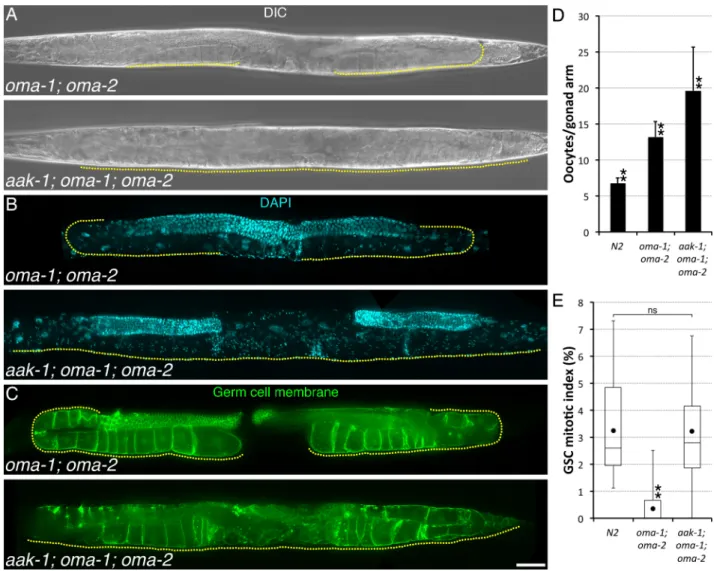
Fig 2. AAK-1 couples GSC proliferation to oocyte accumulation. (A-C) Representative (A-B) A1 or (C) A2 animals of the indicated
genotypes imaged using (A) Differential Interference Contrast (DIC), (B) DAPI (DNA; blue), or (C) mex-5p::mNeonGreen::PLCδ-PH (Germ cell membrane; green). A yellow dotted line delineates the area occupied by oocytes. Anterior is to the left and dorsal, up. Scale bar: 50μm. (D) Average number (±standard deviation) of diakinesis-stage oocytes per gonad arm in A1 hermaphrodites of the indicated genotypes. n, 24–28. (E) The GSC MIs of A1 hermaphrodites of the indicated genotypes were determined. n, 18–23. Dots mark averages, open boxes and brackets represent the whole sample, divided in quartiles. (D-E) Two asterisks indicate statistical significance (P<0.05; One-way ANOVA with Holm multiple comparisons) to all other samples. ns, not significant. (A-E) Alleles: aak-1(tm1944), oma-1 (zu405te33), oma-2(te51).
production did not slow down and these animals became filled with arrested, diakinesis-stage oocytes within 1–2 days (Fig 2A–2D). Diakenesis-arrested oocytes inaak-1; oma-1; oma-2 tri-ple mutants accumulated sometimes in multitri-ple, disorganized rows in the gonad arm, filled-up the uterus, and could eventually breach the gonad proper (Fig 2A–2C). Thus, we conclude that AAK-1 is required at three steps for feedback control of GSC proliferation: 1) to permit oocyte accumulation by preventing their spontaneous maturation and ovulation in the absence of sperm/MSP signal, 2) to slow-down oocyte production upon oocyte accumulation, and 3) to inhibit GSC proliferation.
AAK-1/AMPK blocks GSC proliferation in absence of stress
AMPK is activated by direct binding to 5’-AMP, the concentration of which can dramatically increase during energy stress, but also under many other stressful cellular circumstances that affect ATP usage/production. Significant AMPK activation however requires phosphorylation on a conserved activating residue, which is generally performed by LKB1, a constitutively active kinase [25]. Thus, AMPK has emerged as a master metabolic stress sensor that is turned ON in response to various kinds of stresses and acts to implement a modified energy homeo-stasis balance [15]. Well-fed sperm-less hermaphrodites (fogmutants) have normally activated IIS [9] and a wild-type lifespan [26], suggesting that they do not experience systemic energy stress. However, specific cells could still be subjected to stress during oocyte accumulation, leading to localized, cell-autonomous AMPK activation. Notably, the accumulating unfertil-ized oocytes infogmutants undergo visible compaction (Fig 3A and 3B; [19,27]) and could be experiencing mechanical stress. Consistent with this, large ribonucleoprotein (RNP) foci (also
Fig 3. AAK-1 is activated in the absence of oocyte stress. (A-D) Representative proximal germ lines dissected from unmated A1
animals of the indicated genotypes, stained with DAPI (DNA; blue) and anti-MEX-3 (a stress granule marker [27]; green). n, 5–7 germ lines were analysed per genotype. Proximal is to the left. Scale bar: 20μm. Alleles: aak-1(tm1944), oma-1(zu405te33), fog-2(oz40), oma-2 (te51).
https://doi.org/10.1371/journal.pgen.1006738.g003
known as P-bodies or stress granules) form in the compressed and arrested oocytes offog
mutants [27]. Similar RNP foci also form in the non-arrested (non-compressed) oocytes of sperm-bearing wild-type animals during heat shock, osmotic stress, or anoxia, suggesting that formation of these RNP foci is a general response of oocytes to stress [27]. Thus, AAK-1 activ-ity could be locally triggered in accumulated oocytes due to a localized stress response and act to block the production of further oocytes and GSC proliferation.
To test this hypothesis, we monitored the localization of MEX-3, a germline protein known to accumulate in RNP granules upon stress [27], infogmutants that were devoid of feedback signalling. We found that RNP foci did not form in the proximal oocytes offog-1; daf-18,fog-1; par-4orfog-1; aak-1mutants (S2A–S2D Fig). However we could not exclude that this was a consequence of the ectopic oocyte maturation phenotype that we observed in these mutants, which prevents oocyte accumulation (and compression). We therefore monitored MEX-3 localization in the arrested oocytes ofaak-1; oma-1; oma-2triple mutants, which accumulate but do not undergo oocyte maturation. We found that RNP foci were absent from arrested oocytes ofaak-1; oma-1; oma-2triple mutants but, to our surprise, they were also absent from the arrested oocytes ofoma-1; oma-2double mutants (Fig 3A–3D). This suggests that the arrested oocytes ofoma-1; oma-2double mutants are not experiencing any stress, unlike the seemingly compressed ones offogmutants. Thus, the formation of RNP foci in arrested oocytes is not required for feedback regulation of GSC proliferation. These results suggest that AAK-1 is functioning in this pathway as a signalling kinase rather than as an energy or stress sensor.
AAK-1/AMPK blocks GSC proliferation through inhibiting MPK-1/MAPK
In both pre-dauer and adult germ lines, DAF-18 is required to suppress GSC proliferation, but can do so independently of DAF-16, the ortholog of FOXO that functions as the main IIS effector inC.elegans[2,9,26,28]. DAF-18 was reported to impinge on the ERK/MAPK pathway independently of DAF-16 during vulva development and meiotic progression [29,30]. While theC.elegansortholog of ERK/MAPK, MPK-1, is present throughout the germ line, its dou-bly-phosphorylated active form (dpMPK-1) is detectable in two distinct regions of the adult hermaphrodite germ line: late pachytene-stage germ cells (termed “zone I”) and in growing and maturing oocytes (termed “zone II”) [30–32]. Accordingly,mpk-1is required for germ cell progression through the pachytene stage and is involved in oocyte growth and maturation [19,31]. Interestingly, it was also reported that nullmpk-1mutants, which can grow into sterile vulva-less adults, have a small germ line, which is compatible with reduced GSC proliferation [31].
We reasoned that if DAF-18 activity acts to down-regulate MPK-1 following sperm deple-tion, DAF-18 could potentially block GSC proliferation as well as germ cell progression through the pachytene stage. To test this hypothesis, we first verified thatmpk-1was indeed required for optimal GSC proliferation. As expected, we found that the GSC MI of A1 null
Fig 4. AAK-1 inhibits MPK-1 downstream of oocyte accumulation to promote GSC quiescence. (A-B) The GSC MIs of A1 hermaphrodites of the
indicated genotypes were determined. (A) Asterisks indicate statistical significance (P<0.05; Kruskal-Wallis with Mann-Whitney multiple comparisons) to all other samples. n, 18–30. (B) Asterisks indicate statistical significance (P<0.05; ANOVA with Holm multiple comparisons) to all other samples. n, 18–29. (A-B) Dots mark averages, open boxes and brackets represent the whole sample, divided in quartiles. (C) Average number (±standard deviation) of diakinesis-stage oocytes per gonad arm in mated and unmated A1 hermaphrodites of the indicated genotypes. Asterisks indicate statistical significance (P<0.05; ANOVA with Tukey HSD multiple comparisons) to all other samples within the same condition. Two asterisks indicate additional statistical significance (P<0.05; two-tailed t-test) vs mated animals of the same genotype. n, 24–32 (A-C). ns, not significant. (D-O) Representative germ lines extruded from unmated or continually mated animals of the indicated genotypes and ages, stained with DAPI (DNA; cyan) and a monoclonal mouse anti-MAPKYT that labels dpMPK-1 (green) are shown. White dotted lines underline zones I-II where elevated cytoplasmic dpMPK-1 is apparent. Zone I corresponds to late pachytene germ cells, and zone II, to diakinesis-stage oocytes [30]. Red dotted lines delineate areas of unspecific signal. Distal is to the left. Oocytes are labelled with negative values from the proximal end. n, 7–15 germ lines were analyzed for each condition; fluorescence intensity profiles across zone I are shown inS4 Figand other representative examples are shown inS5 Fig. dpMPK-1 signal were identical in A1 fog-1 and fog-2 germlines (S5 Fig). Scale bar, 50μm. (A-O) Alleles, fog-1(q785), aak-1(tm1944), mpk-1(ga117), daf-2(e1370), daf-18(ok480) (panel A), daf-18 (nr2037) (panels H-J), let-60(ga89)gf, par-4(it57), oma-1(zu405te33), fog-2(oz40), oma-2(te51), par-5(it55). par-5(it55) mutants are also homozygous for unc-22(e66).
https://doi.org/10.1371/journal.pgen.1006738.g004
mutants and A1 wild-type animals (Fig 4B). Consistent with a previously-reported phenotype oflet-60(gf)mutants [31],fog-1; let-60(gf)double mutants accumulated a greater number of smaller oocytes (Fig 4C). These results indicate that hyperactivation of Ras can alleviate GSC quiescence upon oocyte accumulation and suggest that inactivation of MPK-1 is required for oocyte accumulation to block GSC proliferation.
Ifdaf-18andmpk-1were acting as part of the same signalling pathway during feedback control of GSC proliferation, we would expect the effect of the two mutations not to add up, but that one (acting downstream) would cancel the effect of the other. We thus asked if a null mutation indaf-18modified the low GSC MI phenotype ofmpk-1null mutants. We found that A1mpk-1; daf-18double null mutants had a low GSC MI that was indistinguishable from that ofmpk-1single mutants (Fig 4A), indicating that MPK-1 acts downstream of (or in paral-lel to) 18 to maintain elevated GSC proliferative activity. To confirm that this DAF-18-to-MPK-1 axis of signalling that controls GSC proliferation is acting in parallel to IIS, we tested whether IIS inhibition would be additive to the loss of MPK-1 activity. Strikingly, we found that the GSCs of A1daf-2 mpk-1double mutants do not divide and have a null MI (Fig 4A), indicating that adult GSC proliferation absolutely requires at least one of these two growth factors and that they largely act in parallel.
To determine whether the stimulatory effect that the DAF-18-to-MPK-1 axis of signalling has on GSCs depends on IIS, we measured the GSC MI at A1 in bothdaf-2; par-4anddaf-2; let-60(gf)double mutants. We found that these two mutations, unlike a mutation indaf-18[9], did not significantly restore the GSC MI indaf-2mutants (S3 Fig), indicating that the DAF-18-to-MPK-1 axis of signalling does not directly inhibit IIS in wild-type adults, but specifically antagonizes activated IIS in sperm-less hermaphrodites. While loss of eitherdaf-18orpar-4
impairs the establishment of GSC quiescence during dauer development [2], we found that the ectopic activation of MPK-1 signalling inlet-60(gf)dauer animals had no significant effect on GSCs (S3 Fig). These results suggest that control of GSC proliferation during dauer formation does not require MPK-1 inhibition, and that MPK-1 inhibition is thus specifically required for feedback control in adults. Altogether, these results suggest that upon sperm depletion, DAF-18 inhibits MPK-1 activity through PAR-4 and AAK-1 to antagonize IIS independently of DAF-16, and block GSC proliferation in response to oocyte accumulation.
To further test this hypothesis, we evaluated the levels MPK-1 phosphorylation on activat-ing residues when oocytes accumulate in the absence of sperm. In wild-type animals, dpMPK-1 levels peak in the -dpMPK-1 oocyte, and the high level of proximal staining is dependent on MSP signalling [18,19]. In young adultoma-1; oma-2double mutants producing their first few oocytes, dpMPK-1 is transiently detectable in the first oocytes, indicating that they do receive the sperm MSP signal, however it is not maintained thereafter [18]. Thus, MPK-1 activation in zone II requires MSP signalling, but maintenance of MPK-1 activity is further dependent on oocyte maturation and/or the presence of OMA proteins. As previously reported, we found that dpMPK-1 levels in zones I-II decreased to background upon oocyte accumulation in A1
first in zone II and then in zone I, would then be required to block adult GSC proliferation, either directly or indirectly.
To gain insight into where MPK-1 inactivation may be required to inhibit GSC proliferation, we removedmpk-1activity fromgld-3 nos-3double mutant animals, in which GSC differentia-tion is completely blocked, preventing the formadifferentia-tion of zones I-II altogether, and leading to the formation of germline tumours [35,36]. We found that the lack ofmpk-1activity suppressed the growth of germline tumours ingld-3 nos-3animals (S5 Fig), indicating that during develop-ment, MPK-1 functions either cell autonomously in the GSCs and/or non-cell autonomously in the soma to promote GSC proliferation. Interestingly, we did not measure a significant differ-ence in the GSC MI betweengld-3 nos-3andgld-3 nos-3; mpk-1A1 animals (S6 Fig). This sug-gests that during adult life, MPK-1 activation in differentiated germ cells is required to promote GSC proliferation inmpk-1(+)animals. We conclude that upon sperm depletion, DAF-18, PAR-4 and AAK-1 are required to prevent OMA-1 and OMA-2 activation in the absence of MSP signalling and thus permit the accumulation of quiescent oocytes in the proximal gonad. Upon oocyte accumulation, the activation of DAF-18, PAR-4 and AAK-1 induces the inhibition of MPK-1 activity in zones I-II of the germline, which antagonizes IIS signals and induces GSC quiescence while also blocking meiotic progression and the production of further oocytes.
PAR-5/14-3-3 is required to inhibit MPK-1/MAPK and block GSC
proliferation in response to sperm depletion
The consensus recognition motif for AMPK is well defined and conserved [15,25], and there is an overlap between a subset of potential AMPK consensus phosphorylation sites and that of the signalling adaptor protein 14-3-3 [25,37,38]. The functional relevance of this motif overlap has been previously demonstrated. For example, phosphorylation of Raptor by AMPK in cul-tured mammalian cells has been demonstrated to induce 14-3-3 binding, which in turns leads to Raptor inactivation [37]. Interestingly, PAR-5 (theC.elegansgermline-expressed ortholog of 14-3-3 [39]) was found to be required for the maintenance of oocyte quiescence in an RNAi screen designed to isolate regulators of oocyte maturation [40]. This raised the possibility that following sperm depletion, binding between PAR-5 and AAK-1-phosphorylated MPK-1 regu-lators could lead to the inactivation of this pathway to couple both oocyte and GSC quiescence to sperm availability. To test this hypothesis, we first asked ifpar-5was required to couple GSC proliferation with oocyte needs inC.elegansadults. We found that RNAi inactivation ofpar-5
in eitherfog-1orfog-2mutants prevented feedback inhibition of GSC proliferation (Fig 5A). Similarly, GSC proliferation was not significantly inhibited in A1fog-1; par-5double mutants (Fig 5B). Thus, sperm depletion requires PAR-5 to inhibit GSC proliferation. As was the case fordaf-18,par-4andaak-1, inactivation ofpar-5caused spontaneous oocyte maturation and ovulation infog-1mutants, preventing their accumulation (Fig 5C) and stress granule forma-tion (S2 Fig). This suggests that all four genes act in the same signalling pathway to retroac-tively couple GSC proliferation to oocyte needs.
To determine if PAR-5 is required to inhibit MPK-1 activity in zone I following sperm deple-tion, we measured the levels of dpMPK-1 in A1fog-1; par-5loss-of-function mutants. As for fog-1; daf-18,fog-1; par-4andfog-1; aak-1mutants, we found that the germ line of A1fog-1; par-5
double mutants had increased levels of dpMPK-1 in zones I-II relative to the background, albeit levels in zone II were variable between gonads (Figs4O,S4andS5). Thus, PAR-5 is required to inactivate MPK-1 in late-pachytene stage germ cells following sperm depletion. Given these results and the functional overlap between the AMPK consensus site and that of 14-3-3, these results suggest that PAR-5 may act to mediate the effects of AMPK phosphorylation to ultimately inhibit MPK-1 activity in the pachytene area of the germ line to promote GSC quiescence.
Discussion
Our results uncover a novel signalling pathway that links, in two sequential steps, oocyte needs to the regulation of GSC proliferation inC.elegansadult hermaphrodites (Fig 6). We previ-ously demonstrated that DAF-18 was required to inhibit GSC proliferation downstream of sperm depletion, and that in the absence of sperm in these animals, unfertilized oocytes spon-taneously activated and were ovulated [9]. Here, we demonstrate that the lack of PAR-4, AAK-1 or PAR-5 causes a similar phenotype, disrupting oocyte quiescence and feedback regulation of GSC proliferation. Preventing oocyte activation in a feedback-defective mutant also revealed an additional coupling step, this time between oocyte maturation and oocyte production. In
aak-1; oma-1; oma-2triple mutants, loss of OMA-1 and OMA-2 prevents oocyte maturation, triggering their accumulation, while the loss of AAK-1 blocks feedback signalling to the rest of the germ line. In this case, neither GSC proliferation nor oocyte maturation is blocked and we observe hyper-accumulation of oocytes. Thus, in feedback-defective animals, the accumulation
Fig 5. Sperm depletion requires PAR-5 to promote GSC quiescence. (A-B) The GSC MIs of A1 hermaphrodites of the indicated
genotypes/conditions were determined. Dots mark averages, open boxes and brackets represent the whole sample, divided in quartiles. Ctrl, control RNAi vector L4440. ns, not significant. (A) Asterisks indicate statistical significance (P<0.05; Kruskal-Wallis with Mann-Whitney pairwise comparisons to respective RNAi controls). n, 22–28. (B) An asterisk indicates statistical significance (P<0.05; ANOVA with Tukey HSD multiple comparisons) to all other samples. n, 18–24. (C) Average number (±standard deviation) of diakinesis-stage oocytes per gonad arm in A1 hermaphrodites of the indicated genotypes. Asterisks indicate statistical significance (P<0.05; ANOVA with Tukey HSD multiple comparisons) to all other samples. n, 14–24. (A-C) Alleles, fog-1(q785), par-5(it55), fog-2(oz40). par-5(it55) mutants are also homozygous for unc-22(e66).
https://doi.org/10.1371/journal.pgen.1006738.g005
Fig 6. Model for feedback control of GSC proliferation. Arrows represent activation and bars represent inhibition.
The dotted lines represent the delay between the coupling of oocyte maturation/ovulation and the regulation of MPK-1 in zone I due to the time required for oocyte accumulation or its relief. Note that MPK-MPK-1 activity could regulate GSC proliferation indirectly through affecting pachytene progression (as represented), but also potentially through a parallel mechanism. Also note that we placed par-5 downstream of aak-1, but this is purely due to the overlap between the two proteins consensus target motif.
of diakinesis-arrested oocytes cannot block GSC proliferation or oocyte production. This rules out the possibility that oocyte accumulation is physically sufficient to promote GSC quies-cence, in which case the accumulated oocytes inaak-1; oma-1; oma-2triple mutants would have been expected to promote GSC quiescence.
We favor the possibility that oocyte accumulation indirectly promotes GSC quiescence through first causing an arrest in oocyte production, by blocking pachytene progression upstream in the germ line. Stalling of germ cell progression through meiosis would concomi-tantly inhibit GSC proliferation further upstream, in a chain of interdependent retrograde events, as we previously proposed [9]. In this scenario, AAK-1 would act as the gear that cou-ples oocyte accumulation to the arrest of oocyte production (and other feedback steps). The newly demonstrated involvement ofmpk-1in feedback GSC regulation fits well such a mode of operation. Indeed, our results (combined with those of others [9,19,30,40]) are consistent with a model in which sperm depletion blocks oocyte maturation and ovulation, causing their accumulation. After oocytes have accumulated up to a certain point, levels of dpMPK-1 in the pachytene zone of the germ line drop, blocking meiotic progression and the production of additional oocytes. Our results indicate that this inactivation of MPK-1 in pachytene-stage germ cells following oocyte accumulation requires DAF-18, PAR-4, AAK-1 and PAR-5 activi-ties, and that ectopic activation ofmpk-1signalling prevents oocyte accumulation from block-ing GSC proliferation. Lastly, we found that inactivation ofmpk-1suppressed the effects of a
daf-18null allele on GSC proliferation, indicating that MPK-1 functions downstream of, or in parallel to, DAF-18. Thus, we propose that following oocyte accumulation, DAF-18 activity leads to MPK-1 downregulation in pachytene-stage germ cells through activation of PAR-4 and AAK-1 (PAR-4 can directly phosphorylate and activate AAK-1/AMPK [14,15]), followed by AAK-1 phosphorylation of yet unidentified targets and their recognition by PAR-5, to inhibit MPK-1 signalling (Fig 6). How this inactivation of MPK-1 exactly inhibits GSC prolif-eration remains to be elucidated and is a focus for future investigation. However, our analysis of GSC proliferation in tumorous animals that lack differentiated germ cells imply thatmpk-1
activity may promote GSC proliferation partly in the soma and/or GSCs, as well as in differen-tiated germ cells in the adult, possibly in zone I. Despite their anticipated germline require-ment, the precise site(s) of PAR-4/LKB1 and AAK-1/AMPK action in feedback control of GSC proliferation remains to be determined.
Our results indicate that the regulation of larval and adult GSC proliferation is mechanisti-cally distinct. Feedback regulation of GSC proliferation by oocyte accumulation requires PAR-4/LKB1 and AAK-1/AMPK. These genes are also required for inhibition of GSC proliferation during dauer development but in this context they function downstream of insulin and/or TGF-βsignals [2]. Our work reveals that PAR-4 and AAK-1 control GSC proliferation upon oocyte accumulation in the absence of changes in IIS during adulthood. The molecular context of feedback regulation of GSC proliferation by oocyte accumulation is thus mechanistically distinct from inhibition of GSC proliferation during dauer development, despite some over-lap in gene requirements. Our results suggest that the inhibition of MPK-1 signalling in dif-ferentiated germ cells by AAK-1 may be an adult-specific mechanism through which GSC proliferation is controlled. While this mechanism must permit localized regulation of GSC proliferation, it appears to function only in animals that have activated IIS, perhaps because the inhibition of IIS also inactivates MPK-1 in germline zone I [30].
The involvement of AAK-1/AMPK in this feedback signal is particularly interesting because AMPK has been historically predominantly implicated in cellular stress response, typically inhibiting cell growth and proliferation in response to nutrient stress [2,15,25]. Hormones that increase intracellular Ca2+can however activate AMPK in the absence of stress via phosphory-lation by the calmodulin-dependent protein kinase CaMKKβ, but this is independent of
4/LKB1 [25,41–43]. To our knowledge, the feedback mechanism that we herein report is thus the first documented occurrence of AMPK activation in a PAR-4/LKB1-dependent context in the absence of any apparent stress, but in response to a specific signal. Although we did not provide molecular evidence of AAK-1/AMPK activation, we assume it is the case due to the concomitant requirement for the upstream AMPK activating kinase PAR-4/LKB1. As for the regulation of lipid reserves in dauer larvae [44], only one of the two catalytic subunit isoforms is sufficient to fulfill this function. Thus, an emerging role of AAK-1/AMPK is its function as a signalling molecule downstream of DAF-18/PTEN and PAR-4/LKB1 to inhibit stem cell pro-liferation through PAR-5/14-3-3-dependent MPK-1/MAPK inhibition.
Cancer is emerging as being a disease of the stem cells. For instance, cancer incidence was proposed to be related to the frequency of stem cell divisions [45], suggesting it is the stem cells themselves that can turn ill after going through a number of divisions and become tumor-igenic. Understanding how stem cell division rates are regulatedin vivois thus immediately relevant to cancer biology. In this regard, our results reveal that some tumour suppressor genes, such as PTEN and LKB1, may prevent cancer by limiting stem cell divisions when these divisions are not needed. Indeed, defective feedback inhibition of intestinal stem cells may well underlie the growth of the related benign hamartomas in Cowden’s and Peutz-Jegher’s syn-dromes, which are caused in humans by germline mutations in PTEN and LKB1, respectively [46–48]. As such, we predict that the mechanisms we have identified in feedback GSC regula-tion inC.elegansmay underlie cancer development in humans.
Materials and methods
C. elegans genetics
Animals were maintained at 15˚C on standard NGM plates and fedE.colibacteria (OP50) unless otherwise indicated [49]. The Bristol isolate (N2) was used as wild-type throughout. The following alleles, deficiencies and transgenes were used. LGI:fog-1(q785). LGII:gld-3 (q730),nos-3(q650),cpIs42[mex-5p::mNeonGreen::PLCδ-PH::tbb-2 3’UTR + unc-119(+)] (gen-erously provided by Drs Dan Dickinson and Bob Goldstein [50]). LGIII:daf-2(e1370),aak-1 (tm1944),mpk-1(ga117). LGIV:daf-18(nr2037,ok480),oma-1(zu405te33),par-5(it55),unc-22 (e66),let-60(ga89)gf. LGV:fog-2(oz40),par-4(it57),oma-2(te51),qIs56[lag-2p::GFP; unc-119 (+)]. LGX:aak-2(ok524). The following rearrangements were used.qC1[dpy-19(e1259) glp-1 (q339) qIs26[rol-6(su1006)gf; lag-2p::GFP]]III,hT2[bli-4(e937) let-?(q782) qIs48](I;III),mIn1 [mIs14 dpy-10(e128)] II,nT1[unc-?(n754) let-?] (IV;V). RNAi was induced by feeding from the L1 stage as previously described [51].
GSC mitotic index evaluation
Diakinesis oocytes counts
A1 animals were generated as above, fixed in Carnoy’s solution and stained with DAPI as pre-viously described [2]. The number of diakinesis-stage oocytes (having a least one condensed bivalent chromosome [52]) per gonad arm was then determined. In some backgrounds, the chromosomes in one to a few of the proximal-most oocytes sometimes appeared all condensed together and these were included in the analysis. Endomitotic oocytes were however excluded.
Staining
Whole animals were fixed in Carnoy’s solution and stained with DAPI [2]. For germline immunofluorescence, gonads were dissected out of the animal in a drop of PBS on a cover slip, which was then placed against a poly-L-lysine coated slide, submitted to a freeze-crack and stained as previously described [9,53]. Primary mouse monoclonal anti-phospho[ser10]-his-tone H3 (1:200, Cell Signaling #9706), mouse monoclonal anti-double-phospho[Thr202/ Tyr204]MAPK (anti-MAPKYT) (1:100, Cell Signaling #9106), rabbit polyclonal anti-HIM-3 (1:500, a generous gift from Dr Monique Zetka) [53], mouse monoclonal anti-MEX-3 (1:100, a generous gift from Dr Jim Priess) [27], and secondary A488-conjugated goat anti-mouse, or A546-conjugated goat anti-rabbit antibodies (both at 1:500, Invitrogen) were used. DAPI was used as a counterstain.
Image acquisition and processing
Images were acquired at a 0.75μm (Figs1and2B) or 1μm (Figs2C,3,4,S1,S2,S5andS6) intervals using a 20x (whole-worm) or a 60x (germ line) objective on either of two DeltaVision microscopes, deconvolved (whole-worm and distal germ lines), maximally-projected, stitched (whole-worm and whole germ lines), and thresholded using ImageJ. All images are maximal projections, except DIC images, which show a single focal plane. For display and ease of com-parison purposes, whole-worms and germ lines were computationally straightened using Ima-geJ, except forS5 Fig. Fluorescence intensity profiles were obtained and processed essentially as previously described [30] using ImageJ, except that they were obtained from stitched maxi-mal projections instead of single focal planes, and that the background from each stitched image was individually subtracted. Images were reconstructed by stitching multiple, overlap-ping acquisitions of the same sample using the “Grid/collection stitching with sequential images” plugin with default parameters in ImageJ [54]. Samples in Figs4andS4D–S4I, S4K and in S4J, S4L–S4Owere acquired on a different microscope/setting that gave rise to a lower signal-to-noise ratio. To partly (and conservatively) circumvent this issue, we defined the background germline signal as the highest signal intensity observed with the better signal-to-noise microscope/setting in animals without dpMPK-1 signal in zones I-II (e.g.fog-2, A1).
Supporting information
S1 Fig. Spontaneous oocyte maturation and ovulation indaf-18, par-4 and AMPK mutants.
(A-E) Representative A1 animals of the indicated genotypes were stained with DAPI. A white dotted line underlines the region occupied by arrested unfertilized oocytes. A red dotted line marks the uterine region, occupied by (A) embryos, (C-E) spontaneously matured endomi-totic oocytes, or (B) empty. Anterior is to the left and dorsal, is up. Alleles,fog-1(q185),daf-18 (nr2037),par-4(it57),aak-1(tm1944),aak-2(ok524). Scale bar, 100μM.
(TIF)
S2 Fig. Loss-of-function mutations indaf-18, par-4, aak-1 or par-5 block the formation of MEX-3 granules in the absence of sperm. (A-E) Representative A1 proximal gonads extruded
from animals of the indicated genotypes, stained with DAPI (cyan) and anti-MEX-3 (green), are shown. Diakinesis oocytes are negatively labeled from the proximal-most one. Positive numbers indicate ovulated oocytes (in utero) and asterisks mark activated oocytes (endomi-totic). Proximal is to the left. Alleles,fog-1(q185),daf-18(nr2037),par-4(it57),aak-1(tm1944),
par-5(it55). Scale bar, 20μM. (TIF)
S3 Fig. Effects ofpar-4(lf) and let-60(gf)on GSC proliferation in daf-2 mutants. (A) The
GSC MIs of A1 hermaphrodites of the indicated genotypes/conditions were determined. An asterisk indicates statistical significance (P<0.05; ANOVA with Tukey HSD multiple compari-sons) to all other samples. n, 18–34. (B) The number of GSC per animal at the dauer stage was evaluated as previously described [2] in animals of the indicated genotypes. P>0.05; two-tailed t-test. n, 25–37. (A-B) Dots mark averages, open boxes and brackets represent the whole sam-ple, divided in quartiles. ns, Not significant. Alleles,daf-2(e1370),par-4(it57),let-60(ga89)gf.
qIs56is present in thedaf-2; par-4strain. (TIF)
S4 Fig. dpMPK-1 signal intensity profiles related toFig 4. (D-O) The dpMPK-1 signal
inten-sity in arbitrary units (A.U.) of fluorescence was background subtracted and plotted as a func-tion of the distance across germline zone I (from early/mid-pachytene to oocytes). Asfog-2
mutants having accumulated oocytes are negative for dpMPK-1 [19,31], we used this condi-tion as reference and considered regions of more than 100 pixels wide having signal intensities above 50 A.U. as positive for dpMPK-1 (arrows delineate zone I, except in I where dpMPK-1 is found ectopically). Refer to the materials and methods for additional information.
(TIF)
S5 Fig. Additional images related toFig 4. (D’-P”‘) Additional representative germ lines
extruded from unmated or continually mated animals of the indicated genotypes and ages, stained with DAPI (DNA; cyan) and a monoclonal mouse anti-MAPKYT antibody labeling dpMPK-1 (green). (P’-P”‘) A1 fog-1 germ lines were indistinguishable from A1 fog-2 germ lines (E’-E”). Maximal projections in (J”) did not stitch properly.
(TIF)
S6 Fig. Differentiated germ cell-independent effects ofmpk-1 loss-of-function on GSC pro-liferation. (A-B) Three representative A1 animals of the indicated genotypes stained with
DAPI are shown. Germline tumours grow noticeably slower in the absence ofmpk-1. Anterior is to the left and dorsal, is up. Scale bar, 50μM. (C) The GSC MIs of A1 hermaphrodites of the indicated genotypes were determined. An area located around the bend of the germ line was randomly selected using the DAPI channel for MI analysis. ns, Not significant (P>0.05; two-tailed t-test). n, 25–30. (A-C) Alleles,gld-3(q730),nos-3(q650),mpk-1(ga117).
(TIF)
Acknowledgments
Author Contributions
Conceptualization: PN.
Data curation: PN.
Formal analysis: PN.
Funding acquisition: PN PSM JCL.
Investigation: PN.
Methodology: PN.
Project administration: PN.
Resources: PN PSM JCL.
Supervision: PSM JCL.
Validation: PN.
Visualization: PN.
Writing – original draft: PN.
Writing – review & editing: PN PSM JCL.
References
1. Morrison SJ, Kimble J: Asymmetric and symmetric stem-cell divisions in development and cancer. Nature 2006, 441:1068–1074.https://doi.org/10.1038/nature04956PMID:16810241
2. Narbonne P, Roy R: Inhibition of germline proliferation during C. elegans dauer development requires PTEN, LKB1 and AMPK signalling. Development 2006, 133:611–619.https://doi.org/10.1242/dev. 02232PMID:16407400
3. Michaelson D, Korta DZ, Capua Y, Hubbard EJA: Insulin signaling promotes germline proliferation in C. elegans. Development 2010, 137:671–680.https://doi.org/10.1242/dev.042523PMID:20110332
4. Shim J, Gururaja-Rao S, Banerjee U: Nutritional regulation of stem and progenitor cells in Drosophila. Development 2013, 140:4647–4656.https://doi.org/10.1242/dev.079087PMID:24255094
5. Jiang H, Patel PH, Kohlmaier A, Grenley MO, McEwen DG, Edgar BA: Cytokine/Jak/Stat Signaling Mediates Regeneration and Homeostasis in the Drosophila Midgut. Cell 2009, 137:1343–1355.https:// doi.org/10.1016/j.cell.2009.05.014PMID:19563763
6. Jiang H, Grenley MO, Bravo M-J, Blumhagen RZ, Edgar BA: EGFR/Ras/MAPK Signaling Mediates Adult Midgut Epithelial Homeostasis and Regeneration in Drosophila. Cell Stem Cell 2011, 8:84–95. https://doi.org/10.1016/j.stem.2010.11.026PMID:21167805
7. LaFever L, Drummond-Barbosa D: Direct Control of Germline Stem Cell Division and Cyst Growth by Neural Insulin in Drosophila. Science 2005, 309:1071–1073.https://doi.org/10.1126/science.1111410 PMID:16099985
8. Dalfo´ D, Michaelson D, Hubbard EJA: Sensory Regulation of the C. elegans Germline through TGF-β-Dependent Signaling in the Niche. Curr. Biol. 2012, 22:712–719.https://doi.org/10.1016/j.cub.2012.02. 064PMID:22483938
9. Narbonne P, Maddox PS, Labbe´ J-C: DAF-18/PTEN locally antagonizes insulin signalling to couple germline stem cell proliferation to oocyte needs in C. elegans. Development 2015, 142:4230–4241. https://doi.org/10.1242/dev.130252PMID:26552888
10. Narbonne P, Roy R: Regulation of germline stem cell proliferation downstream of nutrient sensing. Cell Div. 2006, 1:29.https://doi.org/10.1186/1747-1028-1-29PMID:17150096
11. Korta DZ, Tuck S, Hubbard EJA: S6K links cell fate, cell cycle and nutrient response in C. elegans germ-line stem/progenitor cells. Development 2012, 139:859–870.https://doi.org/10.1242/dev.074047 PMID:22278922
12. Dibble CC, Cantley LC: Regulation of mTORC1 by PI3K signaling. Trends Cell Biol. 2015, 25:545–555. https://doi.org/10.1016/j.tcb.2015.06.002PMID:26159692
13. Gems D, Sutton AJ, Sundermeyer ML, Albert PS, King KV, Edgley ML, Larsen PL, Riddle DL: Two Pleiotropic Classes of daf-2 Mutation Affect Larval Arrest, Adult Behavior, Reproduction and Longevity in Caenorhabditis elegans. Genetics 1998, 150:129–155. PMID:9725835
14. Narbonne P, Hyenne V, Li S, Labbe´ J-C, Roy R: Differential requirements for STRAD in LKB1-depen-dent functions in C. elegans. Development 2010, 137:661–670.https://doi.org/10.1242/dev.042044 PMID:20110331
15. Hardie DG, Ross FA, Hawley SA: AMPK: a nutrient and energy sensor that maintains energy homeo-stasis. Nat. Rev. Mol. Cell Biol. 2012, 13:251–262.https://doi.org/10.1038/nrm3311PMID:22436748
16. Mondal BC, Mukherjee T, Mandal L, Evans CJ, Sinenko SA, Martinez-Agosto JA, Banerjee U: Interac-tion between Differentiating Cell- and Niche-Derived Signals in Hematopoietic Progenitor Maintenance. Cell 2011, 147:1589–1600.https://doi.org/10.1016/j.cell.2011.11.041PMID:22196733
17. Hsu Y-C, Li L, Fuchs E: Transit-Amplifying Cells Orchestrate Stem Cell Activity and Tissue Regenera-tion. Cell 2014, 157:935–949.https://doi.org/10.1016/j.cell.2014.02.057PMID:24813615
18. Detwiler MR, Reuben M, Li X, Rogers E, Lin R: Two Zinc Finger Proteins, OMA-1 and OMA-2, Are Redundantly Required for Oocyte Maturation in C. elegans. Dev. Cell 2001, 1:187–199. PMID: 11702779
19. Miller MA, Nguyen VQ, Lee M-H, Kosinski M, Schedl T, Caprioli RM, Greenstein D: A Sperm Cytoskele-tal Protein That Signals Oocyte Meiotic Maturation and Ovulation. Science 2001, 291:2144–2147. https://doi.org/10.1126/science.1057586PMID:11251118
20. Govindan JA, Nadarajan S, Kim S, Starich TA, Greenstein D: Somatic cAMP signaling regulates MSP-dependent oocyte growth and meiotic maturation in C. elegans. Development 2009, 136:2211–2221. https://doi.org/10.1242/dev.034595PMID:19502483
21. Starich TA, Hall DH, Greenstein D: Two Classes of Gap Junction Channels Mediate Soma-Germline Interactions Essential for Germline Proliferation and Gametogenesis in Caenorhabditis elegans. Genet-ics 2014, 198:1127–1153.https://doi.org/10.1534/genetics.114.168815PMID:25195067
22. Morgan DE, Crittenden SL, Kimble J: The C. elegans adult male germline: Stem cells and sexual dimor-phism. Dev. Biol. 2010, 346:204–214.https://doi.org/10.1016/j.ydbio.2010.07.022PMID:20659446
23. Cinquin A, Chiang M, Paz A, Hallman S, Yuan O, Vysniauskaite I, Fowlkes CC, Cinquin O: Intermittent Stem Cell Cycling Balances Self-Renewal and Senescence of the C. elegans Germ Line. PLOS Genet 2016, 12:e1005985.
24. Watts JL, Morton DG, Bestman J, Kemphues KJ: The C. elegans par-4 gene encodes a putative serine-threonine kinase required for establishing embryonic asymmetry. Development 2000, 127:1467–1475. PMID:10704392
25. Hardie DG, Schaffer BE, Brunet A: AMPK: An Energy-Sensing Pathway with Multiple Inputs and Out-puts. Trends Cell Biol. 2016, 3:190–201.
26. Lin K, Hsin H, Libina N, Kenyon C: Regulation of the Caenorhabditis elegans longevity protein DAF-16 by insulin/IGF-1 and germline signaling. Nat. Genet. 2001, 28:139–145.https://doi.org/10.1038/88850 PMID:11381260
27. Jud MC, Czerwinski MJ, Wood MP, Young RA, Gallo CM, Bickel JS, Petty EL, Mason JM, Little BA, Padilla PA, et al.: Large P body-like RNPs form in C. elegans oocytes in response to arrested ovulation, heat shock, osmotic stress, and anoxia and are regulated by the major sperm protein pathway. Dev. Biol. 2008, 318:38–51.https://doi.org/10.1016/j.ydbio.2008.02.059PMID:18439994
28. Lee RY, Hench J, Ruvkun G: Regulation of C. elegans DAF-16 and its human ortholog FKHRL1 by the daf-2 insulin-like signaling pathway. Curr. Biol. CB 2001, 11:1950–1957. PMID:11747821
29. Nakdimon I, Walser M, Fro¨hli E, Hajnal A: PTEN Negatively Regulates MAPK Signaling during Caenor-habditis elegans Vulval Development. PLoS Genet. 2012, 8:e1002881.https://doi.org/10.1371/journal. pgen.1002881PMID:22916028
30. Lopez AL III, Chen J, Joo H-J, Drake M, Shidate M, Kseib C, Arur S: DAF-2 and ERK Couple Nutrient Availability to Meiotic Progression during Caenorhabditis elegans Oogenesis. Dev. Cell 2013, 27:227– 240.https://doi.org/10.1016/j.devcel.2013.09.008PMID:24120884
31. Lee M-H, Ohmachi M, Arur S, Nayak S, Francis R, Church D, Lambie E, Schedl T: Multiple Functions and Dynamic Activation of MPK-1 Extracellular Signal-Regulated Kinase Signaling in Caenorhabditis elegans Germline Development. Genetics 2007, 177:2039–2062.https://doi.org/10.1534/genetics. 107.081356PMID:18073423
32. Lackner MR, Kim SK: Genetic Analysis of the Caenorhabditis elegans MAP Kinase Gene mpk-1. Genetics 1998, 150:103–117. PMID:9725833
34. Beitel GJ, Clark SG, Horvitz HR: Caenorhabditis elegans ras gene let-60 acts as a switch in the pathway of vulval induction. Nature 1990, 348:503–509.https://doi.org/10.1038/348503a0PMID:2123303
35. Fox PM, Vought VE, Hanazawa M, Lee M-H, Maine EM, Schedl T: Cyclin E and CDK-2 regulate prolifer-ative cell fate and cell cycle progression in the C. elegans germline. Development 2011, 138:2223– 2234.https://doi.org/10.1242/dev.059535PMID:21558371
36. Hansen D, Schedl T: Stem cell proliferation versus meiotic fate decision in Caenorhabditis elegans. Adv. Exp. Med. Biol. 2013, 757:71–99.https://doi.org/10.1007/978-1-4614-4015-4_4PMID:22872475
37. Gwinn DM, Shackelford DB, Egan DF, Mihaylova MM, Mery A, Vasquez DS, Turk BE, Shaw RJ: AMPK phosphorylation of raptor mediates a metabolic checkpoint. Mol. Cell 2008, 30:214–226.https://doi. org/10.1016/j.molcel.2008.03.003PMID:18439900
38. Johnson C, Crowther S, Stafford MJ, Campbell DG, Toth R, MacKintosh C: Bioinformatic and experi-mental survey of 14-3-3-binding sites. Biochem. J. 2010, 427:69–78.https://doi.org/10.1042/ BJ20091834PMID:20141511
39. Morton DG, Shakes DC, Nugent S, Dichoso D, Wang W, Golden A, Kemphues KJ: The Caenorhabditis elegans par-5 Gene Encodes a 14-3-3 Protein Required for Cellular Asymmetry in the Early Embryo. Dev. Biol. 2002, 241:47–58.https://doi.org/10.1006/dbio.2001.0489PMID:11784094
40. Govindan JA, Cheng H, Harris JE, Greenstein D: Gαo/i and Gαs Signaling Function in Parallel with the MSP/Eph Receptor to Control Meiotic Diapause in C. elegans. Curr. Biol. 2006, 16:1257–1268.https:// doi.org/10.1016/j.cub.2006.05.020PMID:16824915
41. Hurley RL, Anderson KA, Franzone JM, Kemp BE, Means AR, Witters LA: The Ca2+/Calmodulin-dependent Protein Kinase Kinases Are AMP-activated Protein Kinase Kinases. J. Biol. Chem. 2005, 280:29060–29066.https://doi.org/10.1074/jbc.M503824200PMID:15980064
42. Woods A, Dickerson K, Heath R, Hong S-P, Momcilovic M, Johnstone SR, Carlson M, Carling D: Ca2 +/calmodulin-dependent protein kinase kinase-βacts upstream of AMP-activated protein kinase in mammalian cells. Cell Metab. 2005, 2:21–33.https://doi.org/10.1016/j.cmet.2005.06.005PMID: 16054096
43. Hawley SA, Pan DA, Mustard KJ, Ross L, Bain J, Edelman AM, Frenguelli BG, Hardie DG: Calmodulin-dependent protein kinase kinase-βis an alternative upstream kinase for AMP-activated protein kinase. Cell Metab. 2005, 2:9–19.https://doi.org/10.1016/j.cmet.2005.05.009PMID:16054095
44. Narbonne P, Roy R: Caenorhabditis elegans dauers need LKB1/AMPK to ration lipid reserves and ensure long-term survival. Nature 2009, 457:210–214.https://doi.org/10.1038/nature07536PMID: 19052547
45. Tomasetti C, Vogelstein B: Variation in cancer risk among tissues can be explained by the number of stem cell divisions. Science 2015, 347:78–81.https://doi.org/10.1126/science.1260825PMID: 25554788
46. Hemminki A, Markie D, Tomlinson I, Avizienyte E, Roth S, Loukola A, Bignell G, Warren W, Aminoff M, Ho¨glund P, et al.: A serine/threonine kinase gene defective in Peutz–Jeghers syndrome. Nature 1998, 391:184–187.https://doi.org/10.1038/34432PMID:9428765
47. Jenne DE, Reomann H, Nezu J, Friedel W, Loff S, Jeschke R, Mu¨ller O, Back W, Zimmer M: Peutz-Jeghers syndrome is caused by mutations in a novel serine threoninekinase. Nat. Genet. 1998, 18:38– 43.https://doi.org/10.1038/ng0198-38PMID:9425897
48. Liaw D, Marsh DJ, Li J, Dahia PLM, Wang SI, Zheng Z, Bose S, Call KM, Tsou HC, Peacoke M, et al.: Germline mutations of the PTEN gene in Cowden disease, an inherited breast and thyroid cancer syn-drome. Nat. Genet. 1997, 16:64–67.https://doi.org/10.1038/ng0597-64PMID:9140396
49. Brenner S: The Genetics of Caenorhabditis Elegans. Genetics 1974, 77:71–94. PMID:4366476
50. Shaner NC, Lambert GG, Chammas A, Ni Y, Cranfill PJ, Baird MA, Sell BR, Allen JR, Day RN, Israels-son M, et al.: A bright monomeric green fluorescent protein derived from Branchiostoma lanceolatum. Nat. Methods 2013, 10:407–409.https://doi.org/10.1038/nmeth.2413PMID:23524392
51. Fraser AG, Kamath RS, Zipperlen P, Martinez-Campos M, Sohrmann M, Ahringer J: Functional geno-mic analysis of C. elegans chromosome I by systematic RNA interference. Nature 2000, 408:325–330. https://doi.org/10.1038/35042517PMID:11099033
52. Labella S, Woglar A, Jantsch V, Zetka M: Polo Kinases Establish Links between Meiotic Chromosomes and Cytoskeletal Forces Essential for Homolog Pairing. Dev. Cell 2011, 21:948–958.https://doi.org/10. 1016/j.devcel.2011.07.011PMID:22018921
53. Couteau F, Nabeshima K, Villeneuve A, Zetka M: A component of C. elegans meiotic chromosome axes at the interface of homolog alignment, synapsis, nuclear reorganization, and recombination. Curr. Biol. CB 2004, 14:585–592.https://doi.org/10.1016/j.cub.2004.03.033PMID:15062099
54. Preibisch S, Saalfeld S, Tomancak P: Globally optimal stitching of tiled 3D microscopic image acquisi-tions. Bioinforma. Oxf. Engl. 2009, 25:1463–1465.
![Fig 1. Sperm depletion requires PAR-4 and AAK-1 to promote GSC quiescence. (A) Representative distal germ lines dissected from unmated A1 animals of the indicated genotypes, stained with DAPI (DNA; blue), anti-phospho[ser10] histone H3 (M-phase GSCs; gree](https://thumb-us.123doks.com/thumbv2/123dok_us/8272635.2191031/4.918.192.862.112.616/depletion-requires-quiescence-representative-dissected-animals-indicated-genotypes.webp)
![Fig 3. AAK-1 is activated in the absence of oocyte stress. (A-D) Representative proximal germ lines dissected from unmated A1 animals of the indicated genotypes, stained with DAPI (DNA; blue) and anti-MEX-3 (a stress granule marker [27]; green)](https://thumb-us.123doks.com/thumbv2/123dok_us/8272635.2191031/6.918.155.864.115.508/activated-absence-representative-proximal-dissected-animals-indicated-genotypes.webp)


