THE DESIGN AND APPLICATION OF PEPTIDES AND SYNTHETIC RECEPTORS TO CHEMICAL BIOLOGY
Effrat Libby Fayer
A dissertation submitted to the faculty of the University of North Carolina at Chapel Hill in partial fulfillment of the requirements for the degree of Doctor of Philosophy in the Department
of Chemistry (Organic) in the College of Arts & Sciences
Chapel Hill 2016
Approved by:
Marcey Waters
Nancy Allbritton
Michel Gagné
Jeffrey Johnson
ii
iii ABSTRACT
Effrat Libby Fayer: The Design and Application of Peptides and Synthetic Receptors to Chemical Biology
(Under the direction of Marcey Waters)
The need for chemical tools to probe biological process has become increasingly apparent
in the last decade. The work presented here focused on developing such tools, taking on two
different paths of development-one using supramolecular chemistry to aid in proteomics, a
highly popular focus of research, and the other making use of peptides to create reporters with
the ability to probe enzyme activity in cells.
Posttranslational Modifications (PTMs) of proteins are implicated in a wide range of
biological processes, including gene transcription, DNA replication and repair, mitosis, and
meiosis.1 Consequently, their dysregulation is linked to various diseases, including cancer,
asthma, and diabetes, among others, and can thus serve as valuable diagnostic indicators of
disease progression.1,2 Due to these biological ramifications, there is great interest in mapping
where, when, why, and how PTMs are installed and their subsequent downstream effects, though
this is often hampered by their presence in complex mixtures, consisting of mostly un-modified
proteins/peptides.
Using synthetic supramolecular receptors developed in our lab,3 an affinity
chromatography based method is described here that allows for the separation/enrichment of
posttranslationally methylated peptides from such mixtures. This takes advantage of the
iv
attached to a solid support, the receptors can be used to make a column, through which peptides
will travel at different rates based on the methylation states of lysine residues. This allows for the
separation of these peptides, drastically simplifying their detection and analysis.
Just as important as PTMs are the enzymes that install them. Dysregulation of various
enzymatic pathways is implicated in many diseases. The ability to monitor enzyme activity
within cells is becoming increasingly important, for promoting further discovery, as well as to
enable early detection and patient monitoring during treatment. The use of peptide substrates for
such assays is extremely advantageous, as they are the best mimics of the enzymes’ natural
substrates. Furthermore, the ease of synthesizing peptides allows them to be easily modified for
specialized function and detection, making them applicable to multiple types of assays.4 While
they work quite well for in vitro assays, they are incompatible with the cytosolic environment, as
they are rapidly destroyed by cytosolic peptidases.5
In this dissertation, a variety of approaches towards increasing the lifetime of peptides in
cytosolic environment were tested. Kinase substrates were selected as test peptides due to their
role in a diverse set of vital processes, and their importance in current drug development efforts.
For the most successful method, the rates of proteolytic degradation in cell lysates and in vitro
phosphorylation were measured and analyzed using capillary electrophoresis paired with laser
induced fluorescence (CE-LIF). Comparison to unmodified substrate peptides was used to assess
the effect of dimerization on protease resistance and substrate efficacy. Finally, a dimerized Abl
kinase substrate was used to monitor phosphorylation in living cells, demonstrating the utility of
this method for intracellular assays. We find that N-terminal dimerization provides comparable
v
accessibility, suggesting that this is a promising new method for developing peptide-based
vi
ACKNOWLEDGEMENTS
First and foremost, I would like to thank Marcey Waters for all of her guidance
throughout my time here. The environment you created in the lab and the relationship you
choose to have with your students served as major factors when I chose to join this lab, and I
never regretted it for a minute. I’d like to also thank my committee members, both for this
defense and oral examination: Nancy Allbritton, Mike Gagné, Jeff Johnson, Kevin Weeks, and
Dave Nicewicz. Your willingness to take the time to help me complete this is very much
appreciated.
I’d also like to thank the members of our lab. Nick and Brendan, for having endless
patience whenever I needed advice or had random questions, during their time here, and still
now. Kai, for spending her last weeks here teaching me how to carry her work on. Lauren for
letting me talk her ear off endlessly about anything and everything, and all other lab members
who helped create a warm, fun environment to work in. I would also like to thank members of
the Allbritton lab, especially Angie and Emilie, for teaching me whenever my research reached
points outside of my field of expertise, more biological and/or analytical than I ever expected,
and significantly expanding the scope of my knowledge.
I would especially like to thank Amber for being a great friend outside of lab and helping
to keep us both balanced during our time here. I would also like to thank Sarah, for doing the
vii
TABLE OF CONTENTS
LIST OF TABLES ...x
LIST OF FIGURES ... xi
LIST OF SCHEMES ... xv
LIST OF ABBREVIATIONS...xvi
Chapter I. The Use of Peptides as Reporters to Monitor Enzymatic Activity ...1
1. Introduction ...1
2. Peptides as substrates for the detection of enzymatic activity ...1
2.1. Approaches to reading fluorescent peptide-based probes ...3
2.2. Using peptides to monitor intracellular enzymatic activity ...5
3. Purpose of this work ... 10
Chapter II. Design, Synthesis, and Characterization of Protected Peptides (Protectides) for Kinases ... 11
1. Introduction ... 11
2. Analysis of an Unprotected Kinase Substrate ... 12
3. Beta-Hairpins as “Protectides” ... 13
4. Supramolecular “Protectide”... 17
viii
5.1. Synthesis ... 21
5.2. Determination of proteolytic stability ... 23
6. Dendrimers as “Protectides” ... 24
7. Dimerization as the Protection Strategy ... 26
7.1. Extension ... 29
7.2. Substrate Efficacy... 32
8. Conclusions ... 35
9. Experimental ... 36
Chapter III. Current Methods for Characterizing and Sensing Histone Posttranslational Modifications ... 46
1. Packaging of DNA ... 46
2. Posttranslational Modifications ... 47
3. Current Tools for Studying Histone PTMs ... 48
3.1. Enrichment of Modified Fragments ... 49
4. Purpose of This Work ... 53
Chapter IV. The Use of Small Molecule Receptors for Affinity Chromatography ... 54
1. Waters Group Mercaptophanes ... 54
1.1. Dynamic Combinatorial Chemistry (DCC) ... 54
1.2. Macrocyclic Receptor for Trimethyllysine ... 55
2. Modification of Receptors for Attachment to Resin ... 56
ix
2.2. Amine Linker ... 61
2.3. Biotinylated Receptors ... 64
3. Additional Binding Studies ... 65
3.1. Salt Study ... 67
4. Affinity chromatography ... 68
4.1. Direct Attachment ... 68
4.2. Attachment via Biotin ... 69
4.3. Initial Results ... 72
5. Conclusions and Ongoing Work ... 76
6. Experimental ... 77
x
LIST OF TABLES
Table 1. Half-lives of peptides 1-5 in HeLa cytosolic lysates.a ... 16 Table 2. Half-lives of peptides in HeLa cell lysates.a ... 32
Table 3. Extent of in vitro phosphorylation of peptides 1*, 14*, & 17*-20*
after 20 minutes. Ϯ Full separation could not be achieved, and
phosphorylation was not quantified.a... 33
Table 4. Half-lives of peptides in HeLa cytosolic lysates.a ... 35
Table 5. Dissociation constants for the binding of A2B to Ac-WGGG-
QTARKnSTG-NH2 (H3K9Men; n=0-3) as reported in the literature.152
The peptide sequence represents residues 5-12 of Histone 3, 3
glycines as spacers, and a tryptophan for concentration determination.
All peptides were acetal capped and amidated at the C-terminus. ... 56
Table 6. Dissociation constants for the binding of A2B-FLAG to Ac-WGGG- QTARKnSTG-NH2 (H3K9Men; n=0-3) as measured by ITC.a The
peptide sequence represents residues 5-12 of Histone 3, 3 glycines
as spacers, and a tryptophan for concentration determination. ... 59
Table 8. Dissociation constants for the binding of A2B and A2D to Ac-WGGG-
QTARKnSTG-NH2 (n=0 or 3) or Ac-YGGG-QTAXKSTG-NH2
(X=R or aR(Me2)) as measured by ITC.a The peptide sequences
represent residues 5-12 of Histone 3, 3 glycines as spacers, and a
tryptophan or tyrosine for concentration determination. ... 68
Table 9. Dissociation constants for the binding of prop-H3 3-8 peptides to
xi
LIST OF FIGURES
Figure 1. Quench-based kinase assay dependent on the recruitment of a reader protein ...3
Figure 2. Quench-based kinase assay. The fluorophore is quenched by
tyrosine, and upon phosphorylation, fluorescence is restored ...4
Figure 3. FRET-based protease assay.166 ...5
Figure 4. Intracellular phosphorylation assay analyzed by CE-LIF using a
fluorescent peptide substrate. ... 10
Figure 5. Control peptide 1. The substrate is in black, and the fluorophore is shown in green. ... 12 Figure 6. Electropherogram from a degradation assay of control peptide 1. ... 13 Figure 7. General design of peptide constructs. The PKB substrate is in black,
the spacer in red, the lysine conjugated fluorophore in green, and
the protecting group in blue. ... 13
Figure 8. An example peptide highlighting the noncovalent interactions of
the Andersen capping motif. ... 14
Figure 9. (A) Protected peptides 2-4. (B) Structure of peptide 2. The PKB substrate is in black, the PEG spacer in red, the lysine-conjugated
FAM in green, and the protecting β-hairpin in blue. ... 15
Figure 10.Degradation of peptides 1-5 in HeLa cytosolic Lysate. Error bars
represent the standard deviation from three trials. ... 16
Figure 11. Peptide 5. Protected by a β-hairpin at the N-terminus and two
glutamic acid residues at the C-terminus. ... 17
Figure 12. Binding of Urbach’s Cucurbit[7]uril to various phenylalanine derivatives ... 17
Figure 13. Schematic illustration of the inhibition of APN-mediated peptide
digestion at a Phe residue using CB[7] ... 18
Figure 14. Peptide 6. ... 18 Figure 15. Degradation of peptide 6, with and without CB[7], in HeLa
cytosolic lysates. Error bars represent the standard deviation of three runs. ... 20
Figure 16. Urbach’s rotaxane, stoppered by tetraphenylmethane groups ... 20
xii
Figure 18. RP-HPLC traces following the degradation of peptides 7 & 8 in
HeLa cytosolic lysates. ... 23
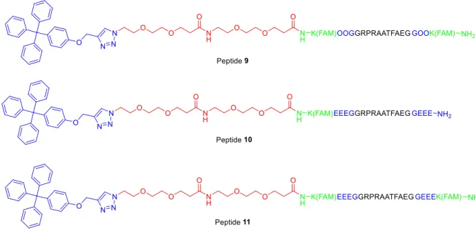
Figure 19. Peptides 9-11. ... 24
Figure 20. Peptide 14. ... 28
Figure 21. Degradation of peptides 1 & 14 as analyzed by CE-MS. *analysis done by Mac Gilliland in the Ramsey lab. Error bars represent the standard deviation of three runs. ... 29
Figure 22. Peptides 15 & 16. ... 30
Figure 23. Degradation of peptides 14-16 in HeLa cytosolic lysates. Error bars represent standard deviation of three runs. ... 30
Figure 24. (A) Peptide standards 17-18. (B) Peptide dimers 19-20. *Peptide 19 was synthesized with a PEG4 linker... 31
Figure 25. Degradation of control peptides 1, 17, & 18 and dimerized peptides 14, 19, & 20 in HeLa cytosolic lysate. Error bars represent the standard deviation of three runs. ... 31
Figure 26. Electropherograms following the in vitro phosphorylation of peptides 17* (A) and 19* (B). Time points were taken at t=0, 1, 3, 5, 10, 15, & 20 minutes. (C) & (D) are Electropherograms following the in vitro phosphorylation of peptide 20*. Complete separation was never achieved. ... 33
Figure 27. Electropherograms monitoring the phosphorylation of peptides 17* (A & B) and peptide 19* (C & D) in Baf/BCR-Abl cytosolic lysates. Time points were taken at (top to bottom) t=1, 1.5, & 2 hours. (A) & (C) are the negative controls in which no ATP was added. ... 34
Figure 28. Electropherograms measuring the amount of peptide 17* (B) and 19* (D) phosphorylated in Baf/BCR-Abl cells after 25 minutes of incubation. (A) and (C) show time points from in in vitro assays of peptide 17* and 19*, respectively, for comparison of migration times... 35
Figure 29. Representation of DNA packaging. ... 46
Figure 30. Representation of heterochromatin and euchromatin ... 47
Figure 31. PTMs found at different sites on histone tails ... 47
xiii
Figure 33. Examples of PTM enrichment methods for subsequent MS analysis. ... 50
Figure 34. β-elimination of O-GlcNAc and replacement with BAP via Michael addition. ... 50
Figure 35. Chemical derivatization and subsequent enzymatic cleavage for enrichment of glycoproteins or peptides ... 51
Figure 36. Affinity chromatography using a synthetic receptor for the separation of KMe3. ... 54
Figure 37. Cartoon representation of dynamic combinatorial chemistry (DCC). ... 55
Figure 38. Structure of A2B and monomers A & B. ... 56
Figure 39. LC/MS trace of a biased A2B-FLAG DCC library at 2 days. Run in a gradient from 5 to 80% B; solvent A=0.2% FA/H2O; Solvent B=0.2% FA/ACN. ... 58
Figure 40. HPLC traces of (A) Glycine•HCl buffer elutions (blue and black) and solution of A2B-FLAG in glycine•HCl buffer (green); (B) PBS buffer blank (light blue), TCEP in PBS buffer (green), and TCEP+monomer A in PBS buffer (dark blue), TCEP elution (black). All traces were run at 0100% B in 60 mins; A=0.1%TFA/H2O, B=0.1%TFA/ACN. ... 61
Figure 41. LC/MS trace of a biased A2B-NH2 library. Run in 1090%B in 25 mins; A/B=NH4OAc in H2O/ACN. ... 62
Figure 42. LC/MS trace of biased A2B-NH2-Fmoc library (3.3 mM B-NH2-Fmoc, 6.6 mM A, and 10.4 mM Me-Isoquinoline in 5.5 mL 5:3 THF:DMSO) after 7 days. Run in a gradient of 10-90%B in which A=0.2% FA/H2O and B=0.2% FA/ACN. ... 64
Figure 43. HPLC traces of (A) 1X PBS buffer (red), A2B-PEG3-Biotin/PBS buffer (black), incubation solution (green), post-incubation washes (light blue), and acid elutions (pink); (B) 1X PBS buffer (black), monomer A+TCEP in PBS buffer (blue), TCEP elution (green). ... 65
Figure 44. Dot-blot assay with A2B-PEG3-Biotin. ... 65
Figure 45. A2B and A2D. ... 67
Figure 46. Model peptides prop-H3K4prop and H3K4Me3. K4 is highlighted in red. ... 71
xiv
Figure 48. Elution patterns of H3K4Me3 and H3K4Ac in different buffers
analyzed by LC-MS/MS. ... 75
xv
LIST OF SCHEMES
Scheme 1. Synthesis of aryl cap and azido-PEG2-acid linker. ... 22
Scheme 2. Synthesis of azido-dendrimers. ... 25
Scheme 3. Synthesis of dendrimer protected peptides via click reaction. ... 26
Scheme 4. Synthesis of Trt-B. ... 57
Scheme 5. Synthesis of A2B-FLAG. ... 58
Scheme 6. Synthesis of B-NH2. ... 62
Scheme 7. Synthesis of B-NH2-Fmoc. ... 63
Scheme 8. Synthesis of A2B-PEG3-Biotin. ... 64
Scheme 9. Attachment of CX4 and A2B to amino-sepharose resin. ... 69
xvi
LIST OF ABBREVIATIONS
Ac Acetal
ACN, CH3CN Acetonitrile
ADP Adenosine Diphosphate
Alloc Allyloxycarbonyl
APN Aminopeptidase N
Arg,R Argenine
aRMe2 Asymmetric Dimethyl Argenine
sRMe2 Symmetric Dimethyl Argenine
ATP Adenosine Triphosphate
CB[x] Cucurbit[x]uril
CE Capillary Electrophoresis
CX4 Calix[4]arene
DBU 1,8-Diazabicyclo[5.4.0]undec-7-ene
DCC Dynamic Combinatorial Chemistry
DCC N,N'-Dicyclohexylcarbodiimide
DCM Dichloromethane
DIC N,N'-Diisopropylcarbodiimide
DIPEA N,N'-Diisopropylethylamine
DMEM Dulbecco’s Modified Eagle Medium
DMF Dimethylformamide
DMSO Dimethyl Sulfoxide
xvii
DPPA Diphenylphosphoryl Azide
EDTA Ethylenediaminetetraacetic acid
ELISA Enzyme-Linked Immunosorbent Assay
EtOAc Ethyl Acetate
FAM Carboxyfluorescein
FBS Fetal Bovine Serum
Fmoc Fluorenylmethyloxycarbonyl
FRET Förester Resonance Energy Transfer
HBTU N,N,N′,N′-Tetramethyl-O-(1H-benzotriazol-1-yl)uranium
hexafluorophosphate
HCTU N,N,N′,N′-Tetramethyl-O-(6-chloro-1H-benzotriazol-1-yl)uranium
hexafluorophosphate
HOBt Hydroxybenzotriazole
HP1 Heterochromatin Protein 1
HR ESI-MS High Resolution Electrospray Ionization Mass Spectrometry
HTS High Throughput Screening
IMAC Immobilized Metal Affinity Chromatography
ITC Isothermal Titration Calorimetry
ivDde 1-(4,4-dimethyl-2,6- dioxocyclohex-1-ylidene-3-methylbutyl
K(Me)1/2/3 Mono/Di/Trimethyl Lysine
Kd Dissociation Constant
LAP Leucine Aminopeptidase
LC Liquid Chromatography
xviii
LIF Laser Induced Fluorescence
Lys, K, KMe0 Lysine
MALDI Matrix-Assisted Laser Desorption Ionization
MeOH Methanol
MOAC Metal Oxide Affinity Chromatography
MPER Mammalian Protein Extraction Reagent
MS Mass Spectrometry
MSH Melanocyte-Stimulating Hormone
NH4OAc Ammonium Acetate
NHS N-hydroxysuccinamide
NMM N-Methylmorpholine
NMR Nuclear Magnetic Resonance
O-GlcNac O-linked β-N-acetylglucosamine
PBS Phosphate Buffered Saline
PEG Polyethylene Glycol
Phe Phenylalanine
PKB Protein Kinase B
PKC Protein Kinase C
PNGase F Peptide-N-Glycosidase F
POP Prolyl Oligopeptidase
PTK Protein Tyrosine Kinase
PTM Posttranslational Modification
xix
QD Quantum Dot
RP-HPLC Reversed-Phase High-Performance Liquide Chromatography
SAM S-adenosyl-L-methionine
SDS Sodium Dodecyl Sulfate
SP Substance P
TCEP Tris(2-carboxyethyl)phosphine
TFA Trifluoroacetic Acid
THF Tetrahydrofuran
TIPS Triisopropylsilane
TLC Thin Layer Chromatography
TOP Thimet Oligopeptidase
TPPII Tripeptidylpeptidase II
Trt Trityl
Chapter I. The Use of Peptides as Reporters to Monitor Enzymatic Activity
1. Introduction
The understanding of disease has evolved markedly over the last two centuries. With the
rapid advancements in analytics and the mapping of the human genome and proteome, scientists
have gone from merely treating external symptoms to the point of understanding them on the
molecular level. Dysregulated enzymes are now known to be notorious culprits in the causation
and progression of many diseases, and with this knowledge at hand, it has become increasingly
important to develop biosensors that allow us to monitor their activity. Such tools are extremely
useful for fundamental studies, allowing scientists to tackle questions concerning the function of
enzymes, as well as subsequent use of that knowledge to develop mimics or derivatives for the
purpose of binding and/or catalysis. Drug discovery also heavily relies on the toolbox of probes
for high throughput screening (HTS) of drug candidates and subsequent analysis of their
therapeutic efficacy.6 An expansion of the toolbox is foreseen to extend its use into biomedical
applications, including diagnostic assays and monitoring of disease progression and response to
therapeutics in patients. This area of research has recently sparked the interest of the scientific
community and the addition of new analytical techniques has advanced at a rapid pace. In this
chapter, examples of biosensors used to monitor enzyme activity will be discussed, focusing on
peptide-based probes.
2
Enzymatic activity is traditionally analyzed by radiolabeling, where incorporation of a
radioactive moiety into a substrate is quantified by scintillation counting, or by a numberof
antigenic approaches, such as Western blotting, enzyme-linked immunosorbent assay (ELISA),
and immunohistochemistry. Aside from radiolabeling, which has the obvious problem of having
to handle radioactive waste, these methods all rely on antibodies for recognition. This can be
seen as a disadvantage since they are expensive and difficult to produce, and batch to batch
variability can be the source of misleading results.7 The post reaction analysis required with
these methods is also quite time consuming, and while they can provide significant information,
they give only indirect measurement of catalytic activity, and are not able to provide real-time
kinetic data.8 For investigating cell lysates, all of the above methods suffer from the additional
disadvantage of requiring a large number of cells. In the context of biomedical applications, this
can become problematic, as obtaining large samples from patients can be challenging, and even
detrimental to the patient.
The use of peptide-based reporters for enzymatic assays has begun to overcome some of the
aforementioned limitations. Peptides are excellent mimics of the enzymes’ natural substrates,
making them particularly effective as probes for enzymatic activity. They can be designed with
high affinity and specificity to the enzyme of interest, or alternatively, as non-selective substrates
for an enzyme family, if so desired.4 Peptides further carry the advantage of being easy to
synthesize and modify, by hand or by automated synthesis, and cost effective. Finally, peptide
substrates can provide a direct readout, allowing one to monitor enzyme reactions in real-time.
Numerous approaches have been developed to pair such substrates with a detectable readout,
including the use of electrochemical,9–16 colorimetric,17 and element mass spectrometry18 output,
3
Instead, the focus here will be on using fluorescent peptide-based reporters. The addition of a
fluorophore to a peptide substrate allows for rapid sensitive detection that can be visualized and
quantified by various types of instrumentation, including fluorescence microscopy, liquid
chromatography with fluorescence detection, and capillary electrophoresis paired with laser
induced fluorescence (CE-LIF). Within that category alone, a number of approaches have
already been taken in translating enzyme activity into readable output. A few of the most popular
ones are described below, followed by the work being done to apply those to intracellular assays.
Approaches to reading fluorescent peptide-based probes
The use of solvatochromic fluorophores conjugated to peptide substrates has been extremely
popular in kinase assays. 21 Solvatochromic fluorophores are ones whose spectroscopic
properties change in response to changes in the polarity
of their surrounding environment. When placed in
proximity to a target residue on a peptide substrate,
modification of the peptide by an enzyme, or a
subsequent event, such as recruitment of a reader
protein (Figure 1), can lead to a change in fluorescence.
Examples of this can be seen in probes developed for
PKC,22 Src, 23 and cyclin-dependent kinase activity.24
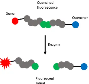
Another method used quite commonly is one based
on restoring the fluorescence of a quenched fluorophore. This approach has been seen in kinase
as well as protease assays. In the latter, a self-quenching fluorophore must be placed at several
residues on a peptide substrate in close proximity,25 or ones that come in close proximity due to
secondary structure.26 Upon enzymatic cleavage of the peptide substrate, the local concentration
4
of the fluorophore decreases, and fluorescence intensity increases in turn. In kinase assays,
quenching-based fluorescent assays can take multiple forms. Tyrosine and tryptophan, both
viable phosphorylation sites, are known to quench a variety of organic fluorophores through π-π
interactions.27,28 Phosphorylation can disrupt the stacking and result in a fluorescence
enhancement (Figure 2). Alternatively, the Lawrence lab developed the “deep-quench” method,
which they used to monitor PKA activity.29,30 In this construct, the fluorophore is quenched by a
molecule in solution. Upon phosphorylation, the corresponding phospho-peptide binds to a
phospho-recognition domain thereby effectively shielding the fluorophore from the quencher in
solution and restoring fluorescence. In a more recently reported form of this biosensor, a
positively charged fluorescent peptide was quenched by a negatively charged quencher. Upon
phosphorylation, the quencher was released due to electrostatic repulsion, leading to increased
fluorescence.31
Figure 2. Quench-based kinase assay. The fluorophore is quenched by tyrosine, and upon phosphorylation, fluorescence is restored. Reproduced from Chem. Soc. Rev. 2012, 41 (5), 1652 with permission of The Royal Society of Chemistry.
Förster resonance energy transfer (FRET) is perhaps one of the most common detection
methods among all biosensors. A donor-acceptor pair conjugated onto opposite ends of a peptide
substrate (within Förester radius), and upon enzymatic cleavage, the donor-acceptor pair are
separated, leading to a detectable change in fluorescence (Figure 3). This FRET-quenching
5
MMP-9,33 and HIV protease.34 Apart from
fluorescent dye molecules, much attraction has
been paid to utilizing semiconductor quantum dots
(QDs) to develop peptide-based FRET protease
sensors.35 Particle size and surface modification
control the absorbance and emission of QDs,
allowing one to adjust them to match various
fluorescent dyes. Several groups have successfully
used QD-conjugated peptides to monitor the
activity of various proteases, such as caspases,36 and Botulinum neurotoxin A.37 Impressively, QD-FRET methods are already being applied to microchip detection platforms, and are believed
to have great potential for commercialization. 38
All of the methods discussed above are based on monitoring changes in fluorescence.
Another option is to pair fluorescence detection with a separation method, thereby observing the
modified and un-modified peptide substrate simultaneously throughout an enzymatic reaction.
The Allbritton group, in collaboration with the Lawrence group, reported the detection of several
enzymes using capillary electrophoresis paired with laser induced fluorescence (CE-LIF). Using
this technique, modified and un-modified peptide substrate are separated due to the difference in
their electrophoretic mobility. They applied this method to monitoring the activity of several
enzymes, including Abl,39 PKB,40 PKT,41 and PTP.42 This method can also be applied to
monitoring protease activity, as the fragmentation products can be separated from the
un-degraded substrate, and even from each other.43,44
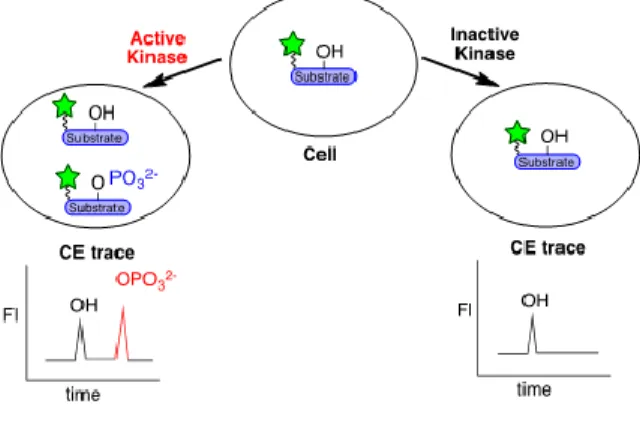
Using peptides to monitor intracellular enzymatic activity
6
While the number of peptide-based probes developed for in vitro detection of enzyme activity is vast, the examples of such probes used intracellularly are much fewer. Peptides are
generally incompatible with the cytosolic environment, as they are rapidly metabolized by
cytosolic peptidases.5 A limited number of aminopeptidases and endopeptidases are known to
exist free in the cytosol of cells, including tripeptidylpeptidase II (TPPII),45–47 thimet
oligopeptidase (TOP),48 prolyl oligopeptidase (POP),49 and leucine aminopeptidase (LAP).50,51 A
common feature of these peptidases is a narrow cleft that contains the catalytic site, and evidence
suggests that it is the N-terminus of peptides that initially enters the cleft.49,52–55 Several methods
in the literature have taken advantage of these features to create longer-living peptides, for use as
enzymatic reporters, as well as drug candidates.56 A few such approaches are discussed below.
Note that this is a limited overview, and other notable methods (commonly used in the
pharmaceutical industry), such as PEGylation57,58 and conjugation of macromolecules,59–61 will
not be covered here.
2.2.1. Peptidomimetics
The incorporation of non-canonical amino acids or amino acid analogues into peptide
regions important for enzymatic activity is expected to strongly affect their efficacy as substrates.
This holds true with proteases as well. β and γ-amino acids, N-methylated amino acids, and even
D-amino acids have been shown to increase a peptide’s resistance to proteolysis.62,63 For
example, Hamamoto et al. demonstrated this in their development of a small antimicrobial
peptide. Partial incorporation of D-amino acids made the peptide, though short and cationic,
more resistant to trypsin degradation than its L-amino acids counterpart.64 Tugyi et al. also
demonstrated that the stability of peptide immunogens was increased by incorporation of D-Thr
7
all D-amino acids was found to remain completely intact even after 24 hours of incubation with
pronase E, showing no degradation at all. However, an unstructured L-amino acid peptide
attached to the C-terminus of the D-beta-hairpin was degraded rapidly, suggesting that D-amino
acids do not prevent threading into the catalytic cleft of the enzymes.66 The use of β- and γ-
amino acids was also found to successfully increase resistance in peptides. Separate work done
by the Seebach and Gellman labs showed peptides made entirely of β- and γ- amino acids are
resistant to proteolysis by a number of different proteases.67–69 More significant modifications to
the peptide backbone to enhance resistance are also seen in the literature, including ester
linkages, peptoids, oligoureas, and azapeptides.57,70–72
In order to use such peptides as enzymatic substrates, they must maintain the ability to be
recognized by the target enzyme. One can infer that residues important for catalytic activity must
therefore remain unmodified. Optimal results can be achieved by careful analysis of cleavage
sites, followed by modifications at those sites alone. Proctor and co-workers used this approach
to develop protease resistant peptide probes for kinase activity. After incubation of a peptide
substrate in cytosolic lysate, the degradation products were analyzed using CE-LIF. Cleavage
sites were identified and the residues substituted with non-native amino acids. Through an
iterative design process, they were able to attain a 15-fold increase in the half-life of an Abl
substrate peptide,39 and a 4.6-fold increase in a PKB substrate peptide, which was then used as a
reporter to measure PKB activity in a single cell.40 In another example, Turner et al. showed that
placing 7- (S)-hydroxy-1,2,3,4-tetrahydroisoquinoline-3-carboxylic acid (L- Htc) in the place of
tyrosine in a PTK substrate peptide was enough to double the half-life in cell lysates, allowing it
to be used as an intracellular reporter.41 Though effective, this method requires multiple rounds
8
Additionally, each modification made to a peptide can also affect its efficacy as a substrate for
the enzyme of interest, and so a delicate balance must be maintained.
2.2.2. Secondary Structure
Research has shown that well-folded peptides have longer lifetimes in cytosolic
environments than those of their unstructured counterparts. This is believed to be due to the fact
that in their folded form, peptides are too large to enter the narrow catalytic cleft of cytosolic
proteases, thereby limiting their degradation. Work from our lab has shown evidence to that
effect. A series of β-hairpins composed of entirely natural amino acids were synthesized and the
rate of degradation in α-chemotrypsin, trypsin, and pronase E were measured. As was expected,
a direct correlation between the fraction folded and the stability of a peptide was seen, reaching
up to a 42-fold increase in half-life for the best folded β-hairpin.73 Taking advantage of this
correlation, along with the evidence that cytosolic proteases thread peptides in through the
N-terminus,49,52–55 our group further showed that the inclusion of a small β-turn at the N-terminus
of a linear peptide can extend its lifetime in cytosolic environment up to 10-fold.74 The protected
peptide, a known Abl substrate, maintained its biological activity, and was successfully
phosphorylated by the Abl kinase.74
Cyclic peptides have also exhibited substantial resistance to proteolysis.75–78 Such peptides
are bulky (preventing entrance to proteases’ catalytic cleft), lack an N-terminus (preventing
recognition by aminopeptidases), and unlike β-hairpins, which sample both a folded and
un-folded form in solution, they are permanently locked in that conformation. Examples of that can
be seen in nature; naturally occurring cyclic peptides such as Gramicidin and Polymyxin B are
metabolically stable, allowing them to be used as therapeutic agents.79 Inspired by some of these
9
extensively investigated in the literature. In a paper published in 1996, Kyb et al. synthesized
various cyclic analogues of Substance P (SP), a member of the tachykinin neuropeptide family,
and studied the effect of cyclization on its properties. Cyclized peptides, though with varying
biological activity, consistently showed increased metabolic stability, with up to 80% remaining
after 2 hour incubation in parotid gland slices (compared to 50% of the linear peptide within 6
minutes).80 In a more recent example, Hess et al. explored the effect of structural and
conformational modifications on the intestinal permeability and metabolic stability of
hydrophilic peptides. A library of 18 cyclic peptides was screened, and in all cases, cyclization
dramatically reduced proteolysis by brush border enzymes.81 They carried out the same tests on a
tetrapeptide derived from melanocyte-stimulating hormone (αMSH), a peptide with potential as a
therapeutic agent for treating obesity. As in the prior case, they found that cyclized analogues all
displayed improved metabolic stability.82
Although cyclization remains the most effective way to protect peptides from proteolytic
degradation, this method does not come without its challenges. Cyclization of peptides is not
always feasible, whether it’s due to a lack of suitable side chain functionalities, or because of the
importance of such functionality for bioactivity. Furthermore, the conformational constraints that
help maintain metabolic stability can also render a peptide inactive, locking it in a conformation
that is unfavorable for binding/catalysis.83 In collaboration with the Allbritton group, our group,
in an extension of the work described above, cyclized the β-turns at the N-terminus of an Abl
substrate peptide, thereby leaving the substrate region linear and the substrate efficacy
un-affected. This showed up to a 4-fold increase in lifetime over its non-cyclized counterpart
(40-fold increase over the linear peptide alone). This peptide was then used in an intracellular Abl
10
quickly.74 However, even in this best case scenario, the synthesis of cyclized peptides is no
trivial task, as is evident by the constant efforts to improve and develop new methods of
cyclization.84
3. Purpose of this work
As is evident by the above discussion, despite the great advantages of using peptides as
reporters, incorporating them into intracellular assays is challenging, as they get rapidly degraded
by proteases. While some methods exist to prevent that, none go without affecting the
efficacy/bioactivity of the peptide in question, and the most commonly used ones are also
synthetically and/or financially draining. To further the use of these probes, a generalizable and
synthetically simple method is needed to make them more stable in cytosolic environment.
Described below is the work done to develop a technique for increasing the protease resistance
of peptides, thereby allowing their effective use in intracellular enzymatic assays. A variety of
protecting strategies were tested for their ability to prevent or slow down proteolytic degradation
of a linear peptide. The most successful method was then applied to several different peptides,
and their efficacy as enzymatic substrates was evaluated. One of the protected peptides was then
carried forward to use as a substrate in an intracellular phosphorylation assay (Figure 4).
11
Chapter II. Design, Synthesis, and Characterization of Protected Peptides (Protectides) for Kinases
1. Introduction
To address the limitations in intracellular enzyme assays described in chapter one, we aimed
to develop protected peptide substrates, which we call “protectides”. Our design focused on the
three characteristics known to be common among cytosolic proteases: narrow catalytic cleft,
highly charged surface area, and threading of peptides in an N- to C-terminus manner.49,52–55 We
reasoned that entrance into the catalytic clefts can be prevented through sterics, by addition of
bulky groups, or electrostatic repulsion, by incorporating negative charge into peptides, at the
N-terminus. To begin our work on developing a protection method, a peptide sequence on which to
test protecting groups was needed. We envisioned ultimately using our protecting strategy on a
peptide substrate in an enzymatic assay. It was therefore ideal to test possible protecting groups
(termed “protectides”) directly on the substrate in mind. We decided to demonstrate our work on
kinase substrates. Kinases are a superfamily of enzymes that are responsible for the
phosphorylation of tyrosine, threonine, and serine. They play an important role in a large set of
vital processes, including cell differentiation, gene expression, and apoptosis.85 The involvement
of these enzymes in so many aspects of cell function makes cells extremely vulnerable to any
alterations in their function, be it due to mutations, or overexpression. Dysregulation of kinases
is in fact implicated in a wide range of diseases, including, but not limited to, diabetes, infectious
12
heavily investigated drugs,87 motivating the development of robust probes for intracellular kinase
assays.
2. Analysis of an Unprotected Kinase Substrate
Many peptide sequences are known to function as kinase substrates, and the choice of one for
these purposes was rather arbitrary. We initially decided to use the sequence
RKRDRLGTLGI-NH2, reported by Kunkel et al. to be a selective PKB substrate.88 The synthesis of this peptide,
however, turned out to be quite problematic due to aspartamide formation, and the desired
peptide was never isolated. This was quickly abandoned in favor of another PKB substrate
peptide, GRPRAATFAEG-NH2, previously used by our collaborators in the Allbritton lab.89
This peptide was cleanly synthesized by hand or by an automated synthesizer, and despite the
low overall charge, it did not present any solubility problems at the concentrations needed for
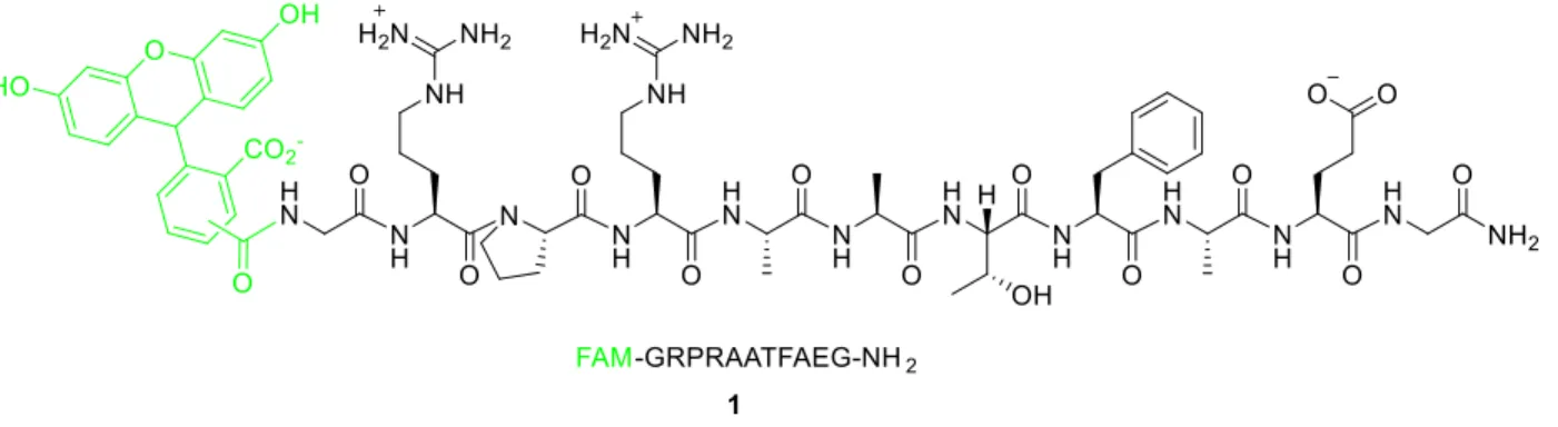
this work. The control peptide 1 was simply capped with (5,6)-carboxyfluorescein ((5,6)-FAM) at the N-terminus for visualization (Figure 5) and served as the standard for comparison for all of
the studies described below.
Figure 5. Control peptide 1. The substrate is in black, and the fluorophore is shown in green.
The stability of peptide 1 in the presence of cytosolic peptidases was tested by incubating in HeLa cell lysates at 37 °C. Aliquots were removed and quenched with equal amount of
13
capillary electrophoresis paired with laser induced fluorescence (CE-LIF) (Figure 6). Based on
peak integrations relative to an added internal standard ((5,6)-FAM), the half-life of the peptide,
the point at which 50% remained un-degraded, was found to be 15±2 minutes.
Figure 6. Electropherogram from a degradation assay of control peptide 1.
Throughout the rest of this chapter, various protecting methods, or “protectides”, are
evaluated. All protected peptides consist of the substrate sequence, shown in black, a PEG
spacer, shown in red, a fluorophore conjugated to a lysine residue, shown in green, and a
protecting moiety, shown in blue (Figure 7). All peptides, unless otherwise stated, were
synthesized via standard Fmoc solid phase chemistry, and all were amidated at the C-terminus.
Peptide characterization was done using HR ESI-MS or LC-MS.
Figure 7. General design of peptide constructs. The PKB substrate is in black, the spacer in red, the lysine conjugated fluorophore in green, and the protecting group in blue.
3. Beta-Hairpins as “Protectides”
Previous work in our lab has shown that appending small β-turn structures to the N-terminus
14
steric bulk preventing entrance into the narrow catalytic clefts of proteases. With this knowledge
at hand, we set out to examine the effect of attaching more highly folded and “click-cyclized”
β-hairpins to the N-terminus of a substrate peptide to see whether longer half-lives were achievable
with this method. Three peptide sequences known to fold well were adapted from the literature
and modified for our purposes: Ac-WIpOOWTGPS (ACAP1), Ac-WVWVpOOKIWTG
(ACAP2),90,91 and Ac-RK(N3)VKVpGOWIG(propargyl)Q (NHB).92 ACAP1 and ACAP2 were
designed by Andersen et al. with a unique capping motif that enhances folding and minimizes
terminal fraying of β-hairpins. This capping motif, “acyl-W-loop-WTG”, confers additional
stability through a face-to-edge Trp-Trp interaction, hydrogen
bonding of the Thr residue with the N-terminal acyl group and
the HN of Gly, and a CH-π interaction between the N-terminal
Trp and C-terminal Gly (Figure 8).91 Since the incorporation of
noncanonicalamino acids is known to increase protease
resistance,40,62 ornithines were incorporated in ACAP1 and
ACAP2 at positions that were shown to be unimportant for
folding. NHB, designed by Park and Waters, was shown to be amenable to cyclization via click
reaction, and in its cyclized form, showed no degradation after 48 hours of incubation with
Pronase E.92 We thus chose to investigate its effect, in both the cyclized and un-cyclized form,
on the PKB substrate. The PKB substrate was synthesized with each one of these hairpins
appended to its N-terminus (peptides 2-4, Figure 9) in a linear manner. The peptides’ half-lives in HeLa cytosolic lysate was then measured using CE-LIF for analysis.
15
Figure 9. (A) Protected peptides 2-4. (B) Structure of peptide 2. The PKB substrate is in black, the PEG spacer in red, the lysine-conjugated FAM in green, and the protecting β-hairpin in blue.
When appended to a β-hairpin, up to a 3-fold increase in half-life relative to peptide 1
was seen. Peptides 2 and 3 were within error of each other, with peptide 2 having a half-life of 26±3 minutes, and peptide 3 30±5 minutes. Peptide 4, in its unclicked form, showed the greatest improvement over the control peptide, with a half-life of 45±7 minutes (Figure 10& Table 1).
Since cyclized peptides are known to dramatically improve metabolic stability,75–78 it was
postulated that cyclization of peptide 4 would further enhance the lifetime of the protected substrate. Attempts at cyclizing peptide 4 (using the procedure outlined by Park & Waters92), however, proved futile, only further illustrating the impracticality of relying on peptide
16
Figure 10.Degradation of peptides 1-5 in HeLa cytosolic Lysate. Error bars represent the standard deviation from three trials.
Table 1. Half-lives of peptides 1-5 in HeLa cytosolic lysates.a
Peptide t1/2 (minutes)
Peptide 1 15±1
Peptide 2 26±3
Peptide 3 30±5
Peptide 4 45±7
Peptide 5 25±2
a Error was determined based on the standard deviation from three runs.
We chose to also test the effect of adding “protection” on the C-terminus of a peptide.
Since the addition of another β-hairpin on the C-terminus of the peptide would have been far
more synthetically challenging, we chose to take advantage of the negatively charged surface of
cytosolic proteases,5,48 and hoped that the addition of negative residues to the C-terminus of the
peptide would help by creating electrostatic repulsion between the peptide and the catalytic cleft
of peptidases. This hypothesis was tested on peptide 3. We initially attempted to synthesize it with four glutamic acid residues added to the C-terminus. However, this peptide was never
successfully isolated. Sequential reduction of the number of residues added was finally
successful, as the peptide was made and isolated with two glutamic acids added (peptide 5, Figure 11). This additional modification of the C-terminus, however, did not appear to have an
-20 0 20 40 60 80 100 120 140
0 20 40 60 80 100
17
effect on the peptide’s stability, as its half-life remained within error of peptide 3 (Figure 10& Table 1).
Figure 11. Peptide 5. Protected by a β-hairpin at the N-terminus and two glutamic acid residues at the C-terminus.
4. Supramolecular “Protectide”
In 2011, Urbach
demonstrated the ability of a
synthetic cucurbit[7]uril (CB[7])
to bind the 4-tert-butyl and 4-amino-methyl derivatives of
phenylalanine with
sub-micromolar dissociation constants (Kd). Binding was shown to be promoted by the positive
cooperativity between the N-terminal ammonium group and the side chain ammonium group in
the case of aminomethyl phenylalanine (Figure 12). Placement of these residues at the
N-terminus of tripeptides did not reduce the affinity, and in some cases even improved it due to
additional cooperativity by the peptide backbone.93 In 2013, Urbach et al. reported the use of
CB[7] to inhibit the degradation of a peptide by aminopeptidase N (APN), a non-specific
exopeptidase. In the presence of CB[7], digestion of a peptide stopped upon reaching a Phe or
(4-aminomethyl)Phe residue, and the remaining peptide was stable for at least 24 hours (Figure 13).
18
The extent of protection of an N-terminal residue was found to directly correlate to its binding
affinity to CB[7].94 This study, however was done in the presence of a single, purified peptidase.
We decided to see if this approach could be extended
to multiple peptidases, such as those in the cytosol of a
cell. Based on the above results, we speculated that
placement of a Phe or (4-aminomethyl)Phe residue at
the N-terminus of our peptide substrate could inhibit
degradation by aminopeptidases. Since
(4-aminomethyl)Phe is known to have a higher affinity
towards CB[7],93,94 we chose to test this hypothesis by
appending it to the N-terminus of our peptide.
Attempts were made to synthesize the peptide with a
free N-terminus to get maximal binding (Kd~1.88 nM),94 but the peptide was never successfully
isolated, or even detected in crude mixtures. We instead used the Ac-capped peptide (peptide 6, Figure 14), as CB[7] displays strong binding to peptides containing (4-aminomethyl)Phe as an
internal residue as well (Kd~510 nM).93
Figure 14. Peptide 6.
19
The stability of peptide 6 was assessed with and without CB[7] (commercially available) in the same manner described above, using CE-LIF for analysis. In the absence of CB[7], the
peptide was found to be 50% degraded within 5±0.4 minutes. In the presence of 2-fold excess of
CB[7] (20 µM), the half-life of the peptide was 20±1 minutes, representing a 4-fold increase
(Figure 15). It is possible that while the cucurbituril is large enough to block entrance into the
catalytic cleft of the peptidase APN, it is not large enough to do so for some other peptidases
found in the cytosol, as size differences do exist. Additionally, despite the strong binding
affinity, it is still a reversible process, just like the folding of β-hairpins, and the peptide could
thread into the proteolytic cleft while sampling the off state. While the concentrations used were
well above the reported Kd, it is important to note that these assays were carried out in PBS, and
not in ammonium phosphate buffer as was reported in the literature.94 The difference in buffer,
20
present in cell lysates), could have a profound effect on the binding properties of CB[7], though
this was not investigated further.
Figure 15. Degradation of peptide 6, with and without CB[7], in HeLa cytosolic lysates. Error bars represent the standard deviation of three runs.
5. Aryl Cap as “Protectide”
With the above results at hand, it was deemed best to move away from using reversible
processes for protection and focus on covalent attachment of protecting groups. In another article
published by Urbach and Ramalingam, they outlined the synthesis of 2 rotaxanes, each comprising a
viologen core threaded through a similar, this time cucurbit[8]uril macrocycle (CB[8]), and
stopperedby tetraphenylmethane groups (Figure 16).95 Since the tetraphenylmethane unit is large
enough to stopper the rotaxane, it was our hope that it would be large enough to act as a barrier 0 20 40 60 80 100 120 140
0 10 20 30 40 50 60 70
% P epti de R em ai ni ng Time (Minutes) Peptide 6 Peptide 6+CB[7]
21
to sterically restrict access of our substrates into the catalytic cleft of cytosolic peptidases,
thereby increasing the lifetime of the peptide substrates. Based on structural analysis of several
peptidases found in the cytosol, the tetraphenylmethane is larger than the entrance to the catalytic
site in some. Thus with its large size, unnatural structure, and unnatural linker to the peptide, we
hypothesized that is could hinder the recognition of the peptidic substrate by the peptidase,
thereby reducing threading into the catalytic cleft and extending its lifetime.
To test this hypothesis, a tetraphenylmethane unit was appended to the N-terminus of the
peptide substrate via a click reaction. Since FAM in itself is an unnatural component of this
peptide, its possible role was tested by synthesizing two versions of the protected substrate: one
in which the lysine conjugated FAM was on the N-terminal side (peptide 7), and one in which it was on the C-terminus (peptide 8). Since the peptide substrate has an overall charge of only +1, the addition of a large hydrophobic aryl cap was expected to cause solubility problems. To
circumvent that, ornithines were incorporated at the N- and C-terminus of the peptide substrate,
separated from it by a glycine residue (Figure 17).
Figure 17. Peptides 7 and 8.
22
The tetraphenylmethane stopper (aka the aryl cap) was synthesized in one step from
commercially available 4-tritylphenol by treatment with propargyl bromide and K2CO3 (Scheme
1) and characterized by 1H NMR.Attempts were made to synthesize a sulfonated protectide as
well, in hope of getting greater protection due to charge repulsion, but attempts at sulfonation of
the aryl cap led to inseparable product mixtures, the analysis of which (via 1H NMR and LC-MS)
was always inconclusive. This approach was therefore abandoned. The azido-PEG2-acid linker
was synthesized from cheap, commercially available diethylene glycol, starting with a
high-yielding desymmetrization using tBu-acrylate and sodium metal. The alcohol was then converted
into an azide using DPPA and DBU, and the tBu-ester was deprotected in 1:1 TFA:DCM to
reveal the carboxylic acid, which was then characterized by 1H NMR and ESI-MS (Scheme 1).
Scheme 1. Synthesis of aryl cap and azido-PEG2-acid linker.
Synthesis of peptides 7 and 8 was carried out entirely on resin, using standard solid phase peptide synthesis up until the final step, in which the aryl cap was attached via an overnight solid
23 Determination of proteolytic stability
The proteolytic stability of peptides 7 and 8 was tested in HeLa cell lysates, and the results were analyzed using analytical RP-HPLC (Figure 18). Surprisingly, peptides 7 and 8 both displayed half-lives shorter than that of the control peptide 1, with t1/2 of ~7 minutes and ~5
mins. For peptide 7 and peptide 8, respectively. This most likely reflects a more active batch of cell lysates, as peptide 1 was not run in parallel with this particular batch The placement of FAM did not appear to make a difference in their half-lives, though it is worth noting that peptide 8
was completely degraded after 45 minutes, while trace amountsof peptide 7 were still seen after an hour.
Figure 18. RP-HPLC traces following the degradation of peptides 7 & 8 in HeLa cytosolic lysates.
New versions of the protected substrate were synthesized (in the same way as above) with a
few changes in hopes of increasing resistance: FAM was incorporated on both the N and C
terminus (peptide 9), the ornithines on the ends were replaced with three glutamic acids on each side to induce electrostatic repulsion (peptide 10), or both (peptide 11). In all three cases, a linker that is 2 PEG units long was used (Figure 19). With all three, however, trouble was encountered
24
MALDI, and when run on the LC-MS, the only peaks seen were eluted off the column very
quickly.
Figure 19. Peptides 9-11.
6. Dendrimers as “Protectides”
As an extension of the steric blocking approach described above with the tetraphenylmethane
group, the use of dendrimers as a capping motif was investigated as a protecting method.
Dendrimers can be synthesized quite easily, making them practical to use. Their size can be
systematically increased, with every generation synthesized, until a sufficient size is reached to
effectively protect the peptides. Furthermore, their synthesis allows for the incorporation of
multiple negative charges at the periphery, which may further help with protection due to the
charge repulsion between them and the proteolytic cleft.49,54,55 This can also provide sites for the
conjugation of cell-penetrable ligands when applied intracellularly. To that end, benzyl
hydroxy-dendrimers were synthesized, beginning from commercially available methyl
4-methylbromobenzoate and 3,5-dihydroxybenzyl alcohol, following a standard literature
25
generation is reached), the benzylic alcohol at the focal point was converted to an azide to enable
conjugation to the peptide via a click reaction (Scheme 2). The synthesis was high yielding for
all the generations synthesized.
Scheme 2. Synthesis of azido-dendrimers.
Modeling suggested that the second generation ([G2]), third generation ([G3]), and fourth
generation ([G4]) dendrimers would be in the size range necessary to restrict access into the
clefts of cytosolic proteases (based on the crystal structure data available to date).49,52–55,97
Dendrimers (HO2C)4-[G2]-N3 and (HO2C)8-[G3]-N3 (see Scheme 2 for naming) were
synthesized. (HO2C)4-[G2]-N3 and (HO2C)8-[G3]-N3 were then clicked onto a purified propargyl
26
peptides 12 and 13, respectively. The reaction was done in 10 mM phosphate buffer (pH 8) using a synthesized tris-tri(methylazolyl)amine ligand (Scheme 3).
Scheme 3. Synthesis of dendrimer protected peptides via click reaction.
When tested for protease stability, peptide 12 showed a half-life of 35±7 minutes, an improvement over the unprotected peptide 1. It was expected that peptide 13 would show an even greater increase in half-life as the size of the dendrimer increases. However, attempts at
analyzing the results of the degradation assay using both analytical RP-HPLC and CE-LIF
proved unsuccessful, as almost no signal was seen. This quench in fluorescence is most likely
due to the high local concentration of acid present in the periphery of the later generation
dendrimers. Later generation dendrimers were thus not pursued as protectides.
7. Dimerization as the Protection Strategy
All of the approaches taken thus far relied on adding bulk to the N-terminus of a peptide in
order to block its entrance to the catalytic cleft of cytosolic peptidases. We decided to take a step
back and focus merely on masking the N-terminus to hinder recognition without relying on
27
hypothesized that N-terminal dimerization of a peptide could mask its N-terminus in the same
way that cyclization would, but without the synthetic challenges or added conformational
constraints that can often decrease substrate efficacy. Unlike N-terminal bulk, it will not add
un-necessary cargo that could bring about unforeseen changes in the peptide’s properties and
activity. The idea of peptide dimers was brought about by recent work reported by Kier and
Andersen. In a comprehensive study on the effect of various capping motifs on the folding
properties of β-hairpins, Kier et al. introduced dicarboxylate capping motifs, among others,
leading to the capping of two β-hairpins with one shared cap, thereby forming dimeric adducts.
This was synthetically very simple, requiring just 1 equivalent of a dicarboxylic acid coupled
during solid-phase peptide synthesis like any other amino acid.90 We adapted this method to
create a dimer of the PKB substrate using succinic acid as the linker (peptide 14). This followed the same general structure of all other protected peptides, with the addition of a glycine spacer
28 Figure 20. Peptide 14.
The synthesis of the peptide dimer was successful on the automated synthesizer, on the
microwave synthesizer, and by hand. Optimal results were achieved when the substrate sequence
was synthesized on the automated synthesizer and all proceeding steps were done by hand.
Dimerization proceeded efficiently, and the dimer was the major product of the reaction,
according to LC/MS peak integration. Some succinic acid coupled to only one peptide was seen
as well, and no peptide was seen that remained uncoupled. Despite the reported purity of the
purchased carboxy-PEG3-amine (reported to be 99.5% pure), significant amounts of dimers with
differing (longer) lengths of PEG chains were observed by ESI-MS, even after multiple rounds
of purification by RP-HPLC. We hypothesized that a longer PEG chain may make dimerization
more favorable, but since the PEG3 dimer remained the major product and the 1-2 unit change in
the length of the PEG chain was not expected to have a significant effect on stability, we
29
degradation assay revealed a half-life of 420±24 minutes, a 28-fold increase over peptide 1. Such an improvement in stability has not been seen before outside the realm of cyclized peptides. To
confirm that these results do not reflect co-elution of the parent peptide and fragments, CE-MS
was used to analyze the samples (done by Mac Gilliland; Ramsey lab). Based on integration of
the peaks relative to an added internal standard, the half-life of peptide 1 was 5 minutes, while the half-life of peptide 14 was 2 hours (Figure 21), representing the same fold increase seen by CE-LIF.
.
Figure 21. Degradation of peptides 1 & 14 as analyzed by CE-MS. *analysis done by Mac Gilliland in the Ramsey lab. Error bars represent the standard deviation of three runs.
Extension
7.1.1. Linker effect
To test the role of the linker, two additional versions of peptide 14 were synthesized, one using isophthalic acid (peptide 15) and one using terephthalic acid (peptide 16) as the linker (Figure 22). We were particularly interested to see if the rigidity of the linker will have any effect
on the stability of the peptide dimer. When tested against peptide 14, however, peptides 15 & 16
30
not affect the degradation resistance. This can be seen as advantageous, potentially allowing for
the incorporation of dyes or conjugation sites in the linker, for example.
Figure 22. Peptides 15 & 16.
Figure 23. Degradation of peptides 14-16 in HeLa cytosolic lysates. Error bars represent standard deviation of three runs.
7.1.2. Substrate scope
To evaluate the generality of this approach, we applied it to other kinase substrates. Dimers
of a known PKC substrate (QKRPSQRSKYL-NH2)98 and Abl substrate
(EAIYAAPFAKKK-NH2)99 were synthesized, using succinic acid as the linker. The Abl dimer (peptide 19) was
synthesized with a PEG4 linker. As we predicted, no dimers other than the desired one were
0 20 40 60 80 100 120
0 2 4 6 8 10
P
epti
de
inta
ct
(%)
Time (hours)
Peptide 14
Peptide 15
31
observed. The PKC dimer (peptide 20) was already synthesized with PEG3, so the mixture was
used. A standard for comparison in which the N-terminus of the substrate sequence was simply
capped with FAM was also synthesized for each substrate (Peptides 17 & 18, Figure 24).
Figure 24. (A) Peptide standards 17-18. (B) Peptide dimers 19-20. *Peptide 19 was synthesized with a PEG4 linker.
Analysis of peptide degradation showed an improvement with all peptides upon dimerization,
with a 24-fold and 7.5-fold increase for the Abl and PKC substrates, respectively (Figure 25&
Table 2).
Figure 25. Degradation of control peptides 1, 17, & 18 and dimerized peptides 14, 19, & 20 in HeLa cytosolic lysate. Error bars represent the standard deviation of three runs.
0 20 40 60 80 100 120
-100 0 100 200 300 400 500 600 700 800
P
epti
de
inta
ct
(%)
Time (minutes)
Peptide 17 Peptide 1
Peptide 18 Peptide 19
32 Table 2. Half-lives of peptides in HeLa cell lysates.a
Peptide Standards t1/2 (mins) Peptide Dimer t1/2 (mins) Fold Increase
Peptide 1 15±1 Peptide 14 420±24 28
Peptide 17 5±1 Peptide 19 120±5 24
Peptide 18 2±0.2 Peptide 20 15±3 7.5
a Errors are from the standard deviation of three runs.
Substrate Efficacy
7.2.1. In vitro Phosphorylation
To assess the ability of the dimerized peptides to act as substrates for their respective
enzymes, all peptides were synthesized using only one isomer of FAM ((5)-FAM) and a PEG4
chain (herein indicated with * by the peptide number) in order to simplify analysis, since dimers with differing lengths of PEG chains (and even the 5- and 6- isomers of FAM) can be separated
by CE, complicating the analysis. The peptides were incubated with purified kinase at 30 °C and
phosphorylation was monitored over time using CE-LIF (Figure 26A&B). The identification of
phosphorylated peaks was confirmed by HR ESI-MS. All dimer peptides were phosphorylated
by their respective kinase, on one or both of the phosphorylation sites, indicating that the
dimerization did not hinderthe peptide’s ability to act as a substrate. Dimers were
phosphorylated to a similar extent as their un-dimerized counterpart, suggesting the rate of
phosphorylation was not significantly perturbed (Table 3). Separation could not be achieved with
peptide 20*, so phosphorylation could not be quantified. However, new peaks were clearly seen growing in (compared to the negative control) (Figure 26C&D) and HR ESI-MS confirms
33
Figure 26. Electropherograms following the in vitro phosphorylation of peptides 17* (A) and 19* (B). Time points were taken at t=0, 1, 3, 5, 10, 15, & 20 minutes. (C) & (D) are Electropherograms following the in vitro phosphorylation of peptide 20*. Complete separation was never achieved.
Table 3. Extent of in vitro phosphorylation of peptides 1*, 14*, & 17*-20* after 20 minutes. Ϯ Full separation could not be achieved, and phosphorylation was not quantified.a
Peptide Standard % Phosphorylated Peptide Dimer % Phosphorylated
Peptide 1* 27±2 Peptide 14* 30±2
Peptide 17* 17±2 Peptide 19* 16±1
Peptide 18* 42±6 Peptide 20* ---
a Error represents the standard deviation of three runs
7.2.2. Phosphorylation in Cell Lysates
Since previous work in our lab has been done on increasing the lifetime of peptide 17, we chose to move forward with peptides 17* & 19* for comparison. The phosphorylation of peptide
19* in a cytosolic lysate was tested. Peptides 17* & 19* were incubated at 30°C in Baf/BCR-Abl cytosolic lysate containing ATP and a cocktail of phosphatase and protease inhibitors, and
phosphorylation was monitored using CE-LIF (Figure 27). Both peptides 17* & 19* were phosphorylated over time at comparable rates: peptide 17* was 87±2% phosphorylated after 2 hours, and peptide 19* was 86±0.1% phosphorylated relative to non-degraded peptide (due to the overlay, peaks for peptide 19* were de-convoluted as described previously44 using Origin 9.0.
34
Figure 27. Electropherograms monitoring the phosphorylation of peptides 17* (A & B) and peptide 19* (C & D) in Baf/BCR-Abl cytosolic lysates. Time points were taken at (top to bottom) t=1, 1.5, & 2 hours. (A) & (C) are the negative controls in which no ATP was added.
7.2.3. Phosphorylation in Living Cells
The performance of peptide 19* intracellularly was then assessed. Peptide 17* or 19* were pinocytosed, along with sodium pervanadate as phosphatase inhibitor, into live Baf/BCR-Abl
cells and incubated at 37°C for 25 minutes. It is worth noting that pinocytosis puts a significant
amount of stress on cells, making it an even more hostile environment for peptides. The cells
were then lysed and the cellular contents analyzed by CE-LIF (Figure 28). Peptide 17* seemed to have completely degraded, as no intact phosphorylated or un-phosphorylated peptide was seen.
Peptide 19*, however, did not exhibit complete degradation, and both un-phosphorylated and di-phosphorylated peptide were seen. The peptide dimer was 54±8% di-phosphorylated relative to
intact peptide remaining, and together, those constituted 19±2% of all species present.
Interestingly, unlike with the in vitro assays, no mono-phosphorylated peptide was detected. This could be due to the more crowded environment within a cell, making a second phosphorylation
event on the same peptide more favorable than the mono-phosphorylation of another substrate


![Figure 13. Schematic illustration of the inhibition of APN-mediated peptide digestion at a Phe residue using CB[7]](https://thumb-us.123doks.com/thumbv2/123dok_us/8271276.2190773/37.918.109.811.789.983/figure-schematic-illustration-inhibition-mediated-peptide-digestion-residue.webp)