Received 22 Jan 2016|Accepted 4 May 2016|Published 6 Jun 2016
PolyMetformin combines carrier and anticancer
activities for
in vivo
siRNA delivery
Yi Zhao
1,2, Wei Wang
1,2,3, Shutao Guo
1,2, Yuhua Wang
1,2, Lei Miao
1,2, Yang Xiong
1,2,4& Leaf Huang
1,2Metformin, a widely implemented anti-diabetic drug, exhibits potent anticancer efficacies. Herein a polymeric construction of Metformin, PolyMetformin (PolyMet) is successfully synthesized through conjugation of linear polyethylenimine (PEI) with dicyandiamide. The delocalization of cationic charges in the biguanide groups of PolyMet reduces the toxicity of PEI both in vitroandin vivo. Furthermore, the polycationic properties of PolyMet permits capture of siRNA into a core-membrane structured lipid-polycation-hyaluronic acid (LPH) nanoparticle for systemic gene delivery. Advances herein permit LPH-PolyMet nanoparticles to facilitate VEGF siRNA delivery for VEGF knockdown in a human lung cancer xenograft, leading to enhanced tumour suppressive efficacy. Even in the absence of RNAi, LPH-PolyMet nanoparticles act similarly to Metformin and induce antitumour efficacy through activation of the AMPK and inhibition of the mTOR. In essence, PolyMet successfully combines the intrinsic anticancer efficacy of Metformin with the capacity to carry siRNA to enhance the therapeutic activity of an anticancer gene therapy.
DOI: 10.1038/ncomms11822 OPEN
1Division of Molecular Pharmaceutics, Eshelman School of Pharmacy, University of North Carolina at Chapel Hill, Chapel Hill, NC 27599, USA.2Center for Nanotechnology in Drug Delivery, Eshelman School of Pharmacy, University of North Carolina at Chapel Hill, Chapel Hill, North Carolina 27599, USA.3State Key Laboratory of Natural Medicines, Department of Pharmaceutics, China Pharmaceutical University, Nanjing 210009, China.4Department of
T
he discovery of RNA interference (RNAi) has since provided a conserved cellular pathway to modulate or inhibit endogenous gene expression through shortinterfering RNA (siRNA) therapeutics1,2. However, naked
siRNA is susceptible to degradation by nucleases in blood serum and unable to cross the cell membrane due to its anionic
charge3,4. Thus, systemic delivery of RNAi therapeutics to
cancerous tissues remains a major challenge. To date, in vivo
siRNA delivery has been achieved by vector-based delivery systems with cationic or ionizable characteristics, including polycation5,6, lipid7–9 and inorganic nanoparticles10,11, and
RNA modifications/conjugations12,13. Among them, delivery
using cationic polymers composed of different cationic
compositions has become one of the most widely accepted
strategies for delivery of siRNA14,15. Yet, the functional
mechanisms of a polycation’s molecular composition on siRNA
delivery remain unclear16. Recently, materials have been
developed incorporating guanidine groups to increase cellular uptake and transfection17,18. Herein a polycationic bi-guanidine composed of the anticancer therapeutic, Metformin
(dimethyl-bi-guanide), has been developed for in vivo siRNA delivery.
Metformin, one of the most effective drugs against diabetes19, is also known as a therapeutic agent against cancers including lung cancer20,21, pancreatic cancer22, breast cancer23, gastric cancer24,25and so on. The anticancer efficacy of Metformin can be primarily attributed to the activation of AMP-activated protein
kinase (AMPK)26,27 and inhibition of the mammalian target of
rapamycin (mTOR)28,29. This cationic small molecular drug
shows excellent tolerability and can be dosed at up to 2 g per day due to low toxicity. Moreover, the cationic biguanide composition of Metformin makes its polymeric form a desirable carrier for siRNA delivery. Therefore, it is expected that polymeric Metformin (PolyMet) would serves dual purposes as both a gene carrier and an antitumour therapeutic to achieve combinational therapeutic efficacies against cancer. Human non-small-cell lung cancer (NSCLC) is a well-known, aggressive and metastatic refractory tumour that responds to treatment by Metformin30,31. Thus, a NSCLC tumour cell H460 was used to evaluate the efficacy of siRNA delivery and antitumour abilities of PolyMet in this study.
Our data show a possibility to facilitate vascular endothelial
growth factor (VEGF) siRNA in vivo delivery by PolyMet
nanoparticles and enhanced tumour growth. In the absence of RNAi, LPH-PolyMet nanoparticles, like metformin, activated the AMPK, inhibited the mTOR pathway and induced tumour autophagy and apoptosis. Thus, PolyMet successfully combines the intrinsic anticancer efficacy of Metformin with the capacity to carry siRNA to enhance the therapeutic activity of an anticancer gene therapy.
Results
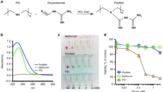
Synthesis and characterization of PolyMet polymer. Inspired by the fact that Metformin can be synthesized through a one-step reaction of dimethylamine hydrochloride and 2-cyanoguanidine (dicyandiamide), with heating (Supplementary Fig. 1), the Metformin polymer was designed using a similar method. To yield PolyMet, linear polyethylenimine (PEI) and dicyandia-mide were reacted under heat in acidic conditions (Fig. 1a). Linear PEI hydrochloride (0.2 g) and dicyandiamide (2 g) were mixed in 10 ml 2 M HCl solution. The reaction mixture was reacted at 100°C for 24 h, purified through an ultrafiltration tube to remove excess dicyandiamide, washed with deionized water for two times and lyophilized. The formation of PolyMet was verified
by using proton nuclear magnetic resonance (1H-NMR)
(Supplementary Fig. 2A) and matrix assisted laser desorption/
ionization time-of-flight (MALDI-TOF) (Supplementary
Fig. 2B,C) analyses. 1H-NMR (400 MHz, d6-dimethylsulfoxide)
spectrum exhibits all characteristic proton resonance peaks cor-responding to the present PolyMet molecules. The appearance of
the proton resonance at d2.86–3.75 p.p.m. in the product along
with the characteristic guanidium proton at d 5.80–6.40 p.p.m.
confirmed the formation of biguanide polymer. The spectrum also indicated near 95% substitution ratio of PolyMet by com-paring the area ratios of PEI (2.53–2.70 p.p.m.) and PolyMet (2.86–3.75 p.p.m.). MALDI-TOF of the PolyMet and free PEI was performed to determine the synthesis of PolyMet
(Supplementary Fig. 2B,C). The distribution centre
for PEI (ca. 570 Da) was completely red-shifted compared with PolyMet (ca. 1600 Da), which is consistent with the
1H-NMR result, indicating successful conjugation of PEI with
2-cyanoguanidine (Supplementary Fig. 2B). The average
molecular weight of PEI shown by MALDI-TOF analysis is smaller than the molecular weight we used for synthesis (ca. molecular weight is 4,300 Da), which might be due to the existence of numerous differently charged species of PEI,
since MALDI-TOF only detects singly charged species32.
Moreover, the extended MALDI-TOF mass spectra
(Supplementary Fig. 2C) show several series of oligomer ions
43 and 129m/z apart from each other, in agreement with the
PEI (43m/z for C2H5N unimer) and PolyMet (129m/z for
C4H11N5 unimer) repeat units, respectively. As described in
authoritative literatures33,34, Metformin can be characterized by (1) using ultraviolet absorption at the wavelength of maximum absorbance at about 233 nm or (2) exhibiting a red colour in the solution of nitroprusside/potassium hexacyanoferrate(III)/ sodium hydroxide. Both PolyMet and Metformin exhibited a maximum absorbance around 233 nm (Fig. 1b) and showed red colour in the colour test in a dose-dependent manner (Fig. 1c). These results suggest that after the reaction with dicyandiamide, the secondary amines in PEI had been completely replaced with biguanides.
Notably, PolyMet showed a significantly lower cytotoxicity (Fig. 1d) and threefold higher maximum tolerable dose compared with PEI (Supplementary Table 1), indicating PolyMet containing guanidine is more tolerable than its secondary amine-containing counterpart. But the lower maximum tolerable dose of PolyMet than Metformin (Supplementary Table 1) is perhaps attributed to the interaction of the cationic polymer with the serum proteins in the blood. In summary, PolyMet as a cationic polymer may be used to condense and deliver negatively charged siRNA with similar pharmacological properties and activity to Metformin due to the biguanide structure.
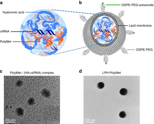
Preparation and characterization of LPH nanoparticles. It is well documented that siRNA forms a loose and unstable complex when mixed with only polycationic polymers due to its low
molecular weight and low charge density35,36. Therefore,
liposomes were used to coat complexes to protect siRNA from dissociation or clearance before it reaches the tumour in our study. First of all, hyaluronic acid (HA), a polyanionic and multivalently charged polysaccharide, was used to facilitate siRNA delivery and enhance the particle condensation. Then cationic 1,2-dioleoyl-3-trimethylammonium-propane chloride salt (DOTAP) liposome was applied to coat complex to prove the stability of complex. These core/shell-type nanoparticles,
referred to the lipid-polycation-hyaluronic acid (LPH)
nanoparticle were established for systemic delivery of siRNAs to
the tumour site37,38. Specifically, HA was mixed
with siRNA at 1:1 mole ratio, and modulated the N/P ratios of
an N/P ratio around 0.9, where a neutral complex was formed. In this case, 0.6 was chosen as the optimal ratio, as the complex
remained negatively charged (B 20 mV) with a relatively small
size (B100 nm). At this ratio the PEI/(HAþsiRNA) complex
had a similar charge and size with PolyMet/(HAþsiRNA)
complex (Supplementary Fig. 3). Next, DOTAP/cholesterol (1:1 mol/mol) cationic liposomes were added to the complex to form the lipid coating through charge–charge interactions. Studies have shown that the NSCLC cell H460 overexpresses sigma receptors17,39,40, thus a targeting ligand for the sigma
receptors, DSPE-PEG-anisamide, was used to deliver
nanoparticles to the tumour by the post-insertion method to form the outer leaflet of the liposome membrane. The size,
distribution, morphology and the characteristics of the
nanoparticles are summarized in Fig. 2 and Supplementary Table 2. The lipid coating reduced the complex’s size due to electrostatic interactions. The final nanoparticles were 70–80 nm with a positive charge of about 20 mV and 4.38% loading efficiency (Supplementary Table 2).
LPH-PolyMet combines siRNA carrier and anticancer activities. Previous studies have shown that VEGF siRNA suppresses angiogenesis and may be a potential therapeutic agent for
NSCLC41. Human NSCLC H460 tumour-bearing mice received
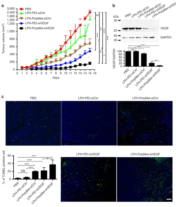
intravenous (i.v.) injections of different formulations with VEGF siRNA or control siRNA every other day. On successive i.v. injections into tumour-bearing mice, the greatest enhancement of antitumour efficacy was observed when LPH-PolyMet-siVEGF was administered (Fig. 3a).
Notably, the LPH-PolyMet-siCtrl group that contained control siRNA also showed a partial tumour suppression activity, indicating that poly-biguanide structure of PolyMet has the antitumour activity. Meanwhile, minimal suppression of tumour growth was found in the LPH-PEI-siCtrl group. One day after the
second injection, mice were killed and tumour lysates were prepared for western blot. As shown in Fig. 3b, LPH-PolyMet-siVEGF group showed robust knockdown of VEGF with about 95% of silencing compared with the phosphate-buffered saline (PBS) group. LPH-PEI-siVEGF had only a partial effect on the VEGF expression level with about 70% of knockdown in comparison with control group. LPH-PolyMet-siCtrl showed a slight knockdown effect. LPH-PEI-siCtrl had no measurable effect on the protein expression level of VEGF.
Terminal deoxynucleotidyl transferase dUTP nick end labelling (TUNEL) assay further confirmed the induction of apoptotic cells in tumours (Fig. 3c). The number of TUNEL-positive apoptotic cells after LPH-PolyMet-siVEGF treatment was about 40%, which was the highest among all groups. This indicates that elevated apoptosis occurred after downregulation of VEGF expression, which is consistent with studies reported elsewhere42. Treatment with LPH-PEI-siVEGF could only induced apoptosis in about 20% of cells. Importantly, we also observed that LPH-PolyMet-siCtrl exhibited significant higher TUNEL-positive population compared with PBS or LPH-PEI-siCtrl, indicating the apoptotic capability of PolyMet gene carrier itself.
PolyMet LPH was then investigated to determine its ability to carry other therapeutic siRNA. As an anti-apoptotic protein BCL-2 promotes cell survival, and inhibition of BCL-2 can
suppress tumour growth43,44. Herein siRNA used against BCL2
inhibited tumour growth, especially in the LPH-PolyMet-siBCL2 treatment group, which provided persistent suppression of tumour growth (Supplementary Fig. 4A). The level of BCL-2 after treatment was detected by western blot analysis, LPH-PolyMet-siBCL2 induced a significant downregulation of BCL-2 level in comparison with all other groups (Supplementary Fig. 4B) and the TUNEL assay further confirmed the induction of apoptotic cells in tumours. The number of TUNEL-positive apoptotic cells after LPH-PolyMet-siBCL2 treatment was about 70%, which was significantly higher than all other groups PEI
NH n +
N NH
NH
NH2 HCI, heat
NH2
N n
NH
NH NH
Dicyandiamide PolyMet
1.2
1.0
0.8
0.6
Absorbency
Viability
, % of control
0.4
0.2
0.0
–0.2
220 240 260
nm PolyMet
PolyMet
Metformin
Metformin
PEI
PolyMet
Metformin PEI PEI
60 20 6 2 0 (mM)
280 300
120
100
80
60
40
20
0
0.01 0.1
Drugs, mM 1 a
b c d
(Supplementary Fig. 4C). This indicates that elevated apoptosis occurred after downregulation of BCL2 anti-apoptotic protein expression in the LPH-PolyMet-siBCL2 group.
PolyMet was then tested for gene delivery and tumour inhibition in a melanoma model. As shown in Supplementary Fig. 5, the most effective treatment, LPH-PolyMet-siVEGF
resulted in a knockdown of VEGF level by B70% and a
suppression of tumour growth by about 80% in comparison with the PBS group in a 1205Lu melanoma model. Meanwhile, LPH-PEI-siVEGF also suppressed tumour growth effectively, but not as persistently as LPH-PolyMet-siVEGF.
Moreover, the antitumour activity of PolyMet was compared with other two commonly used polycations, poly-L-lysine (PLL) and protamine. PLL and Protamine were formulated in the LPH systems containing the same amount of VEGF siRNA as PolyMet. The antitumour efficacy and VEGF knockdown were presented in Supplementary Fig. 6. All LPH formulations had significantly suppressed tumour growth in comparison with PBS. However, there was no significant difference among PEI, PLL and protamine. The greatest enhancement of antitumour effect was observed with PolyMet. The expression of VEGF was also evaluated after day 16, as shown in Supplementary Fig. 6B. The VEGF level in the tumour was significantly lower than other groups, as well as the PBS control. Thus, PolyMet showed the greatest antitumour activity among all four polycations tested.
Mechanisms of PolyMet on the inhibition of tumour growth. Both clinical and preclinical studies have shown that Metformin
improved survival and/or antitumour efficacy25,45. Results
shown in Fig. 3 and Supplementary Figs 4, 5 and 6B indicate the tumour growth inhibition of LPH-PolyMet-siCtrl. It was thus hypothesized that in the absence of RNAi, PolyMet, like its unimer Metformin, may be responsible for some part of
anticancer activity. The effect of LPH-PolyMet and different nanoparticles on an H460 xenograft was studied. As shown in Fig. 4, treatment with LPH-PolyMet led to significant inhibition of cancer growth in comparison with PBS. A noticeable difference in tumour growth inhibition was observed between LPH-PEI and LPH-PolyMet (Fig. 4a), suggesting that PolyMet played an important role in enhancing antitumour activity. At the end of the experiment, the tumours of the animals treated with
LPH-PolyMet were o1% of the total body weight, which was
significantly smaller than the LPH-PEI (4%) and PBS (6%) groups (Fig. 4b). Moreover, no toxicity was detected in blood haematology serum chemistry or the histology of the major tissues (Supplementary Fig. 7 and Supplementary Table 3).
Furthermore, we compared the effect of LPH-PolyMet siRNA
nanoparticles with Metformin in an H460 xenograft
(Supplementary Fig. 8). Free Metformin (1.25 mg kg1)
equivalent to the unimer dose of PolyMet (1.25 mg kg1) that
was required to deliver 0.625 mg kg1of VEGF siRNA was used
in the study. It was confirmed that there is no significant difference between the PolyMet carrier (LPH-PolyMet-siCtrl) and Metformin, indicating that biguanide in the PolyMet copolymers are associated with the anticancer activities of Metformin. Both of the groups exhibit significantly higher antitumour growth activity compared with LPH-PEI-siCtrl or PBS while LPH-PolyMet-siVEGF has the best antitumour efficacy. In this case tumour growth was suppressed seven- to eightfold compared with the PBS group, and about twofold compared with the PolyMet carrier or the Metformin group, which suggests again that PolyMet successfully combine the intrinsic anticancer efficacy of Metformin with the tumour suppressive effects of VEGF siRNA to enhance the therapeutic activity of an anticancer gene therapy. It has been reported that Metformin inhibits cancer cells by
activating the AMPK and inhibiting the mTOR pathways28,46.
We checked the activity of PolyMet in these pathways and Hyaluronic acid
siRNA
PolyMet
DSPE-PEG-anisamide
Lipid membrane
DSPE-PEG
LPH-PolyMet PolyMet / (HA+siRNA) complex
100 nm 100 nm
a b
c d
compared it with that of Metformin. AMPK acts as a metabolic tumour suppressor that governs glucose and lipid metabolism47. In many cases, a low level of phosphorylation of AMPK is
correlated with poor prognosis after cancer treatment48.
We observed that following treatment with Metformin or
LPH-PolyMet, phosphorylation levels of AMPKa in the H460
tumour were significantly enhanced compared with the PBS
group (Fig. 5a). The activation of AMPKa did not occur in
the LPH-PEI group, suggesting PolyMet is the key factor in the
nanoparticle that can activate AMPKa pathway. mTOR is a
downstream effector of AMPK49. AMPK activation inhibits
mTOR and its downstream effector kinases50. Phosphorylation of
mTOR plays a pivotal role in the proliferation and survival of
cancer cells51. Therefore, we then evaluated the effects of
treatments on the activity of mTOR (Fig. 5a). Metformin and LPH-PolyMet treatments led to a significant inhibition of mTOR activity indicated by stronger reduction in p-mTOR/mTOR levels compared with the PBS group. The activity was not shared by LPH-PEI.
Autophagy is recognized as a potentially toxic mechanism for Metformin that results in the inhibition of cancer growth52,53. We evaluated whether autophagy can also be observed after treatment of LPH-PolyMet. Microtubule-associated protein light chain 3 b (LC3b) is a specific marker for autophagy initiation54. As shown 3,000
2,400 1,800 1,200 1,200
1,000
800
600
T
umour v
olume (mm
3)
% of
TUNEL-positiv
e cell
400
200
0
0 1 2 3 4 5 6 7 8 Days PBS
PBS LPH-PEI-siCtrl
LPH-PEI-siCtrl LPH-PolyMet-siCtrl
LPH-PolyMet-siCtrl LPH-P
olyMet-siCtr l
LPH-PEI-siVEGF
LPH-PEI-siVEGF
LPH-PEI-siVEGF
LPH-PolyMet-siVEGF
LPH-PolyMet-siVEGF LPH-P
olyMet-siVEGF
9 10 11 12 13 14 15 16
NS
***
***
***
**
**
*
20
40 30 30
kDa PBS LPH-PEI-siCtr l
LPH-P olyMet-siCtr
l
LPH-PEI-siVEGF LPH-P
olyMet-siVEGF PBS
LPH-PEI-siCtr l
LPH-P olyMet-siCtr
l
LPH-PEI-siVEGF LPH-P
olyMet-siVEGF PBS
LPH-PEI-siCtr l
VEGF
GAPDH
VEGF/GAPDH
100 80 60 40 20 0
*** *** *
**
*** *** ***
***
*
NS
NS
60
40
20
0
a b
c
Figure 3 | LPH nanoparticles composed of PolyMet can systemically deliver VEGF siRNA to the tumour site and inhibit tumour growth.(a) H460 tumour-bearing mice were injected i.v. every other day and tumour volumes were measured every day. (b) H460 tumour VEGF protein levels after two injections were measured by western blot analysis. Bar chart inbrepresent quantitative analysis of relative normalized VEGF band intensity (Image J). (c) TUNEL staining (green) in H460 tumour cells after treatment with siRNA in different formulationsin vivo. Nucleic acid was stained with 4,6-diamidino-2-phenylindole (blue). Bar chart incis a quantitative analysis of % of TUNEL-positive cells. Five randomly selected microscopic fields were quantitatively analysed on Image J. Data are mean±s.e.m. (n¼5 per group) analysed by two-way analysis of variance with Tukey’s correction. Data are representative of
in Fig. 5b, Metformin- and LPH-PolyMet-treated tumours showed a higher LC3b-associated red fluorescence than other groups, indicating that both Metformin and LPH-PolyMet could induce autophagy in the H460 lung cancer xenograft model.
Finally, the mechanism of the antitumour efficacy after treatment was determined by the TUNEL assay, a common method to test for apoptosis (Fig. 5b). The per cent of apoptotic cells after Metformin or LPH-PolyMet treatments was 12.4% and 15%, respectively, while no significant apoptosis induction was observed in other groups. This suggests that Metformin or polymer form can induce cell apoptosis and play a critical role in regulating the cancer cell survival.
Discussion
Nanotechnology has been extensively exploited to prolong the circulation time and to enhance the therapeutic activity for a variety of drugs. After systemic injection the nanoparticles drain through the leaky tumour blood vessels and are retained at the tumour site, resulting in increased tumour accumulation of the nanoparticle-formulated drug compared with the free drug. This well-documented phenomenon is referred to as the
enhanced permeability and retention effect55. Thus,
nanoparticles offer a number of advantages over free drugs, which make them an ideal delivery vehicle for Metformin. However, due to its high hydrophilicity, Metformin cannot be effectively loaded into established nanocarriers, which explains why only few Metformin nanoparticles have been reported56–58.
Recently, Yang et al.56 reported the co-encapsulation of
epirubicin and Metformin into liposomes through remote loading. This nanoparticle successfully inhibited the growth of
CD133þ cancer stem-like cells. Jose et al.57 developed
Metformin-loaded bovine serum albumin nanoparticles
(MET-BSA) by the coacervation method and observed that MET-BSA nanoparticle showed higher efficacy than Metformin alone or the BSA carrier itself. However, PolyMet, which combines both gene delivery and anticancer capabilities, represents a novel approach.
Guanidination of a polymer enhances the activity for gene delivery and has considerably less toxicity compared with primary
amines of the polymer59,60. The guanidine has been found to
readily pass through the non-polar membrane of a cell and even across tissue barriers by possibly forming a bidentate hydrogen bond with anionic cell surface phosphate, carboxylates and/or sulfates on the cell surface61,62. Materials have been previously developed incorporating guanidine groups to increase cellular uptake and transfection60,63–65. For example, Lee et al.60 used pyrazole-1-carboxamidine to react with branched PEI, and observed enhancement of transfection efficiency and decreased
cytotoxicity of PEI by guanidylation. Wender et al. developed
non-natural guanidinium-rich molecular transporters to enhance cellular uptake61,66, through tunable polyguanidino dendrimers,
and observed higher cellular uptake65. Guanidinium-rich
molecular transporters can penetrate cells and overcome
efflux-mediated multidrug resistance67. Although these
previously reported polyguanidines are established gene carriers, the carriers themselves fail to exhibit anticancer activity without their therapeutic agent. Thus, PolyMet serves as both a gene carrier and anticancer therapeutic. However, findings presented herein have not been investigated with PolyMet against a p53-deficient tumour, which remains to be further explored. The focus of this study is to show the carrier and anticancer activities of PolyMet. The dose of PolyMet used in the study was dictated by the dose of siRNA and was only a fraction of the effective dose of metformin. Further study with increased PolyMet dose will bring the new polymer a step closer to the clinical translation.
Methods
Materials.DOTAP and 1,2-distearoryl-sn-glycero-3-phosphoethanolamine-N
-[methoxy(polyethyleneglycol-2000) ammonium salt (DSPE-PEG2000) were
purchased from Avanti Polar Lipids, Inc. (Alabaster, AL). DSPE-PEG-anisamide
was synthesized in our lab as described previously68. DeadEnd Fluorometric
TUNEL assay kits and Luciferase Assay System assay substrates were obtained from Promega (Madison, WI). Linear PEI hydrochloride with average molecular weight 8,000, dicyandiamide, HA and other chemicals were obtained from
Sigma-Aldrich (St. Louis, MO). Rabbit monoclonal antibodies Phospho-AMPKa(Thr172)
1,800
1,200 1,200
1,000
800
600
400
200
0
0 1 2 3 4 5
Days
6 7 8 9 10
NS NS
***
***
***
PBS
PBS
LPH-PEI
LPH-PEI
Metformin
Metf ormin
LPH-PolyMet
PBS
LPH-PEI Metformin
LPH-PolyMet
LPH-P olyMet
T
umour v
olume (mm
3)
% T
umour
b
urden
8
6
4
2
0
2 cm
** **
** NS NS
a b
c
(40H9) (catalogue # 2535), AMPKa(D5A2) (catalogue # 5831), Phospho-mTOR (Ser2448) (D9C2) XP (catalogue # 5536), mTOR (7C10) (catalogue # 2983), LC3b (D11) XP (catalogue # 3868), GAPDH (14C10) (catalogue # 2118) and Anti-rabbit IgG, horseradish peroxidase-linked Antibody (catalogue # 7074) were purchased from Cell Signaling Technology (Beverly, MA). BCL2 siRNA (target sequence:
50-AACAUCGCCCUGUGGAUGACU-30), VEGF siRNA (target sequence:
50-ACCUCACCAAGGCCAGCAC-30) and control siRNA (target sequence:
50-AAUUCUCCGAACGUGUCACGU-30) were synthesized by Sigma-Aldrich.
Cell culture.H460 human NSCLC cells and 1205Lu melanoma cancer cells were originally obtained from American Type Culture Collection and were cultured in DMEM medium (Invitrogen, Carlsbad, CA) supplemented with 10% fetal bovine
serum (Life Technologies, Carlsbad, CA), 100 U ml1penicillin and 100mg ml1
streptomycin (Invitrogen). Cells were cultured in a humidified incubator at 37°C
and 5% CO2.
Experimental animals.Female nude female mice and female CD1 mice that were 6– 8 weeks old were used in the studies. Female nude mice were purchased from National Cancer Institute (Bethesda, MD) and bred by the Division of Laboratory Animal Medicine at University of North Carolina at Chapel Hill. CD1 mice were purchased from Charles River Laboratories (Morrisville, NC). To establish the xenograft models,
5106cells in 100ml of PBS were injected subcutaneously into the right flank of the
mice. All work performed on animals was approved by the Institutional Animal Care and Use Committee at the University of North Carolina at Chapel Hill.
Preparation of PolyMet.To prepare PolyMet, 0.2 g of linear PEI and 2 g of dicyandiamide were mixed in 10 ml 2 M HCl solution. The compounds reacted at
100°C in the oil bath for 24 h. PolyMet was then purified through an ultrafiltration
tube, washed with deionized (DI) water for two times and lyophilized. The working
solution of PolyMet was diluted in DI water and kept as 1 mg ml1, 1 ml aliquots
for experiment. To verify the formation of PolyMet, NMR and MALDI-TOF analyses were performed (Supplementary Methods).
Optimization of the PolyMet-HA or PEI-HA complexes.A volume of 200ml of
PEI solution (containing 5, 10, 15 or 20mg PEI in DI water) or 200ml of PolyMet
solution (containing 50, 60, 70 or 80mg PolyMet in DI water) was mixed with
200ml of HA (containing 50mg HA, in DI water) in a 1.5-ml tube. The complexes
were mixed by pipetting up and down for 10 times and allowed to stand at room temperature for 10 min. The optimal ratio of the complex was determined by the results from particle size and zeta potential determined by dynamic light scattering using a Malvern ZetaSizer Nano series (Westborough, MA).
Preparation of LPH nanoparticles.DOTAP/cholesterol liposomes were prepared as follows: DOTAP (20 mM, 1 ml) and cholesterol (20 mM, 1 ml) were dissolved (1:1 mol/mol) in chloroform and the solvent was removed under reduced pressure. The lipid film was then hydrated overnight with 2 ml distilled water to form cationic liposomes (10 mM), which were sequentially extruded through
poly-carbonate membranes (200 nm20 times, 100 nm20 times and 50 nm20
times) (Millipore, Billerica, MA). LPH cores were prepared by mixing 100ml of 5%
80
kDa PBS Metf
ormin
LPH-PEI LPH-P olyMet
PBS Metf
ormin LPH-PEI
LPH-P
olyMet PBS
Metf ormin
LPH-PEI LPH-P
olyMet
PBS Metf
ormin LPH-PEI
LPH-P olyMet PBS
Metf ormin
LPH-PEI LPH-P
olyMet
60
80
60
260
260
40
30
PBS
LC3b
TUNEL
Metformin p-AMPKα AMPKα
p-mTOR
mTOR
GAPDH
LPH-PEI LPH-PolyMet
100 75 50 25 0
p-AMPK
α
/AMPK
p-mT
OR/mT
OR
% of LC3B postiv
e cell
% of
TUNEL postiv
e
cell
60
40
20
0
25 20 15 10 5 0 150
100
50
0 NS
***
NS ***
NS *** NS
*** a
b
glucose solution A (containing the 12.5mg of siRNA and 12.5mg of HA) with 100ml
of 5% glucose solution B (containing 5mg of PEI, or 25mg of PolyMet, or 6mg of
PLL, or 13mg of Protamine) and incubated at room temperature for 10 min. The
LPH cores were then mixed with 30ml of cationic DOTAP/cholesterol liposomes
and incubated at room temperature for 10 min to ensure proper lipid coating. The lipid-coated nanoparticles were PEGylated using a post-insertional approach by
adding 10ml DSPE-PEG (10 mg ml1) and 10ml DSPE-PEG-anisamide
(10 mg ml1) and incubating the nanoparticles at 50°C for 15 min.
Characterization of LPH NPs.The size and surface charge of LPH cores and final particles were determined by using a Malvern ZS90 Zetasizer (Worcestershire, UK). Transmission electron microscope images of LPH were acquired through the use of JEOL 100CX II transmission electron microscope (Tokyo, Japan). Briefly,
freshly prepared LPH nanoparticles (5ml) were carefully dropped onto a 300-mesh
carbon-coated copper grid (Ted Pella, Inc., Redding, CA) and allowed to stand at
room temperature for 5 min. Grids were then stained with 1% uranyl acetate (5ml)
and allowed to incubate briefly (10 s) and quickly dry. All images were acquired at an accelerating voltage of 100 kV.
Loading capacity of LPH nanoparticles.After final LPH-PolyMet-siRNA
formulation, the free siRNA were separated by centrifugation for 20 min at 18,000g
and 4°C. The siRNA concentration in the supernatant was measured by the
Quant-iT RiboGreen RNA reagent according to the manufacturer’s instructions, using a fluorescence spectrometer (Perkin-Elmer, Waltham, MA) at 485/520 nm excitation/emission. Each sample was assayed in triplicate.
siRNA loading¼ Actual siRNA loadingðmgÞ
Weight of nanoparticlesðmgÞ100%
Animal tumour model and antitumour activity.H460 human lung cancer cells
(5.0106) were subcutaneously injected into the right flanks of female athymic nu/
nu mice. Drugs were administrated i.v. into mice when the tumours reached about
50–80 mm3in size (5–7 days after transplantation). LPH equivalent to
0.625 mg kg1of VEGF siRNA or control siRNA, prepared as described above, were
i.v. injected every other day. Free Metformin (1.25 mg kg1) equivalent to the
unimer dose of PolyMet (1.25 mg kg1) that was required to deliver 0.625 mg kg1
of siRNA was used in the study. Animal weight and tumour volumes were measured
every other day. The tumour length (L) and width (W) were used to calculate volume
(V) by the following equation:V¼1/2LW2. Serum biochemical value analysis
and haematology assay were provided in Supplementary Methods.
Western blot analysis.Western blotting was used to evaluate the protein expression of tumour cells after systemic administration. Tumour-bearing mice were injected i.v. every other day and were killed 24 h after the second injection. A unit of 50 mg of tumour was collected randomly via biopsy and homogenized
with a Tissue Master-125 homogenizer (OMNI International) in 500ml cold
radioimmunoprecipitation assay buffer (Sigma-Aldrich) supplemented with 1% protease inhibitor cocktail (Sigma-Aldrich) for 30 min. Total homogenate was then
spun down at 13,000gwith a tabletop centrifuge for 10 min at 4°C. Supernatant
was collected and protein sample was quantified using a bicinchoninic acid assay kit in accordance with the manufacturer’s instruction (Pierce Biotech). Samples
were diluted in 4sample buffer and 10 sample-reducing reagent (Thermo
Fisher). A unit of 40 mg protein from each group was separated by 4–12% SDS–PAGE electrophoresis (Invitrogen) and transferred to a polyvinylidene difluoride membrane (Bio-Rad). Membranes were blocked with 5% non-fat dry milk (Bio-Rad) for 1 h at room temperature and then incubated with primary
antibodies (1:1,000 dilution; Cell Signal) overnight at 4°C. The membranes were
washed three times with PBS-Tween, followed by incubation with horseradish peroxidase-conjugated secondary antibody (1:4,000 dilution; Cell Signal) for 1 h at room temperature. Finally, the membranes were washed three times with PBS-Tween and developed using Pierce ECL Western Blotting substrate (Thermo Scientific). The expression level of targeted protein was quantified by ImageJ software (NIH). GAPDH was detected as loading control. Images have been cropped for presentation but are otherwise unmodified. Full-size images are presented in Supplementary Figs 9 and 10.
Immunostaining.The TUNEL assay was used to detect apoptotic tumour cells after systemic administration. Briefly, tumours were collected and fixed in freshly prepared 4% paraformaldehyde or 10% formalin for 1 day. Tumour samples were paraffin-embedded and sectioned by the UNC Animal Histopathology Core. The slides were then deparaffinized in xylene and rehydrated through a gradient alcohol series. The TUNEL staining was performed in accordance with the manufacturer’s protocol (Promega). Nuclei of tumour cells were stained with DAPI (Vector Laboratories). Images of were taken using a fluorescence microscope (Nikon). The percentage of apoptotic cells was calculated by dividing the number of apoptotic cells from the total number of cells in each microscopic field.
Representative microscopic fields (n¼5) were randomly selected in each treatment
group for this analysis.
Statistics analysis.All statistical analyses were carried out using GraphPad Prism Software (Version 5.0, GrapPad Software, San Diego, CA).
References
1. Gonzalez-Alegre, P. & Paulson, H. L. Technology insight: therapeutic RNA
interference—how far from the neurology clinic?Nat. Clin. Pract. Neurol.3,
394–404 (2007).
2. Fire, A.et al.Potent and specific genetic interference by double-stranded RNA
inCaenorhabditis elegans.Nature391,806–811 (1998).
3. Truong, N. P.et al.An influenza virus-inspired polymer system for the timed
release of siRNA.Nat. Commun.4,1902 (2013).
4. Semple, S. C.et al.Rational design of cationic lipids for siRNA delivery.Nat.
Biotechnol.28,172–176 (2010).
5. Dahlman, J. E.et al.In vivo endothelial siRNA delivery using polymeric
nanoparticles with low molecular weight.Nat. Nanotechnol.9,648–655 (2014).
6. Davis, M. E.et al.Evidence of RNAi in humans from systemically administered
siRNA via targeted nanoparticles.Nature464,1067–1070 (2010).
7. Gilleron, J.et al.Image-based analysis of lipid nanoparticle-mediated siRNA
delivery, intracellular trafficking and endosomal escape.Nat. Biotechnol.31,
638–646 (2013).
8. Whitehead, K. A.et al.Degradable lipid nanoparticles with predictable in vivo
siRNA delivery activity.Nat. Commun.5,4277 (2014).
9. Bogorad, R. L.et al.Nanoparticle-formulated siRNA targeting integrins
inhibits hepatocellular carcinoma progression in mice.Nat. Commun.5,3869
(2014).
10. Jensen, S. A.et al.Spherical nucleic acid nanoparticle conjugates as an
RNAi-based therapy for glioblastoma.Sci. Transl. Med.5,209ra152 (2013).
11. Medarova, Z., Pham, W., Farrar, C., Petkova, V. & Moore, A. In vivo imaging of
siRNA delivery and silencing in tumors.Nat. Med.13,372–377 (2007).
12. Lee, J. B., Hong, J., Bonner, D. K., Poon, Z. & Hammond, P. T. Self-assembled
RNA interference microsponges for efficient siRNA delivery.Nat. Mater.11,
316–322 (2012).
13. Wolfrum, C.et al.Mechanisms and optimization of in vivo delivery of
lipophilic siRNAs.Nat. Biotechnol.25,1149–1157 (2007).
14. Kozielski, K. L., Tzeng, S. Y., De Mendoza, B. A. & Green, J. J. Bioreducible cationic polymer-based nanoparticles for efficient and environmentally triggered cytoplasmic siRNA delivery to primary human brain cancer cells.
ACS Nano8,3232–3241 (2014).
15. Davis, M. E. The first targeted delivery of siRNA in humans via a self-assembling, cyclodextrin polymer-based nanoparticle: from concept to clinic.
Mol. Pharm.6,659–668 (2009).
16. Cheng, C. J., Tietjen, G. T., Saucier-Sawyer, J. K. & Saltzman, W. M. A holistic
approach to targeting disease with polymeric nanoparticles.Nat. Rev. Drug
Discov.14,239–247 (2015).
17. van Waarde, A.et al.Potential applications for sigma receptor ligands in cancer
diagnosis and therapy.Biochim. Biophys. Acta1848,2703–2714 (2015).
18. Vargas, J. R., Stanzl, E. G., Teng, N. N. H. & Wender, P. A. Cell-penetrating, guanidinium-rich molecular transporters for overcoming efflux-mediated
multidrug resistance.Mol. Pharm.11,2553–2565 (2014).
19. Rowan, J. A.et al.Metformin versus insulin for the treatment of gestational
diabetes.N. Engl. J. Med.358,2003–2015 (2008).
20. Zhang, Z. J.et al.Reduced risk of lung cancer with metformin therapy in
diabetic patients: a systematic review and meta-analysis.Am. J. Epidemiol.180,
11–14 (2014).
21. Tan, B. X.et al.Prognostic influence of metformin as first-line chemotherapy
for advanced nonsmall cell lung cancer in patients with type 2 diabetes.Cancer
117,5103–5111 (2011).
22. Kisfalvi, K., Eibl, G., Sinnett-Smith, J. & Rozengurt, E. Metformin disrupts crosstalk between G protein-coupled receptor and insulin receptor signaling
systems and inhibits pancreatic cancer growth.Cancer Res.69,6539–6545
(2009).
23. Jiralerspong, S.et al.Metformin and pathologic complete responses to
neoadjuvant chemotherapy in diabetic patients with breast cancer.J. Clin.
Oncol.27,3297–3302 (2009).
24. Morales, D. R. & Morris, A. D. Metformin in cancer treatment and prevention.
Annu. Rev. Med.66,17–29 (2015).
25. Greenhill, C. Gastric cancer: metformin improves survival and recurrence rate
in patients with diabetes and gastric cancer.Nat. Rev. Gastroenterol. Hepatol.
12,124 (2015).
26. Pernicova, I. & Korbonits, M. Metformin—mode of action and clinical
implications for diabetes and cancer.Nat. Rev. Endocrinol.10,143–156 (2014).
27. Zakikhani, M., Dowling, R., Fantus, I. G., Sonenberg, N. & Pollak, M. Metformin is an AMP kinase-dependent growth inhibitor for breast cancer
cells.Cancer Res.66,10269–10273 (2006).
28. Dowling, R. J., Zakikhani, M., Fantus, I. G., Pollak, M. & Sonenberg, N. Metformin inhibits mammalian target of rapamycin-dependent translation
29. Vazquez-Martin, A., Oliveras-Ferraros, C., del Barco, S., Martin-Castillo, B. & Menendez, J. A. mTOR inhibitors and the anti-diabetic biguanide metformin: new insights into the molecular management of breast cancer resistance to
the HER2 tyrosine kinase inhibitor lapatinib (Tykerb).Clin. Transl. Oncol.11,
455–459 (2009).
30. Lin, J. J.et al.Survival of stage IV lung cancer patients with diabetes treated
with metformin.Am. J. Resp. Crit. Care Med.191,448–454 (2014).
31. Mellema, W. W.et al.A phase II study of sorafenib and metformin in patients
with stage IV non-small cell lung cancer (NSCLC) with a Kras mutation.
J. Thorac. Oncol.8,S1205–S1205 (2013).
32. Tauhardt, L.et al.Linear polyethyleneimine: optimized synthesis and
characterization—on the way to ‘‘pharmagrade’’ batches.Macromol. Chem.
Phys.212,1918–1924 (2011).
33. USP.The Pharmacopeia of the United States of America; Suppl. 5(United States
Pharmacopeial Convention, 1984).
34. Brittain, H. G.Analytical Profiles of Drug Substances and Excipients(Elsevier
Science, 2001).
35. Chono, S., Li, S. D., Conwell, C. C. & Huang, L. An efficient and low immunostimulatory nanoparticle formulation for systemic siRNA delivery to
the tumor.J. Control. Release131,64–69 (2008).
36. Draz, M. S.et al.Nanoparticle-mediated systemic delivery of siRNA for
treatment of cancers and viral infections.Theranostics4,872–892 (2014).
37. Li, S. D., Chen, Y. C., Hackett, M. J. & Huang, L. Tumor-targeted delivery of
siRNA by self-assembled nanoparticles.Mol. Ther.16,163–169 (2008).
38. Li, S. D., Chono, S. & Huang, L. Efficient oncogene silencing and metastasis
inhibition via systemic delivery of siRNA.Mol. Ther.16,942–946 (2008).
39. Miao, L. G., Zhang, S., Kim, J. & Huang, W. L. Nanoparticles with precise
ratiometric co-loading and co-delivery of gemcitabine monophosphate and
cisplatin for treatment of bladder cancer.Adv. Funct. Mater.24,6601–6611 (2014).
40. Nakagawa, O., Ming, X., Huang, L. & Juliano, R. L. Targeted intracellular delivery of antisense oligonucleotides via conjugation with small-molecule
ligands.J. Am. Chem. Soc.132,8848–8849 (2010).
41. Phillips, R. J.et al.Epidermal growth factor and hypoxia-induced expression of
CXC chemokine receptor 4 on non-small cell lung cancer cells is regulated by the phosphatidylinositol 3-kinase/PTEN/AKT/mammalian target of rapamycin
signaling pathway and activation of hypoxia inducible factor-1alpha.J. Biol.
Chem.280,22473–22481 (2005).
42. Mei, J.et al.VEGF-siRNA silencing induces apoptosis, inhibits proliferation
and suppresses vasculogenic mimicry in osteosarcoma in vitro.Exp. Oncol.30,
29–34 (2008).
43. Czabotar, P. E., Lessene, G., Strasser, A. & Adams, J. M. Control of apoptosis by
the BCL-2 protein family: implications for physiology and therapy.Nat. Rev.
Mol. Cell Biol.15,49–63 (2014).
44. Tekedereli, I.et al.Therapeutic silencing of Bcl-2 by systemically administered
siRNA nanotherapeutics inhibits tumor growth by autophagy and apoptosis and enhances the efficacy of chemotherapy in orthotopic xenograft models of
ER (-) and ER (þ) breast cancer.Mol. Ther Nucleic Acids2,e121 (2013).
45. Morales, D. R. & Morris, A. D. Metformin in cancer treatment and prevention.
Annu. Rev. Med.66,17–29 (2015).
46. Yue, W., Yang, C. S., DiPaola, R. S. & Tan, X. L. Repurposing of metformin and aspirin by targeting AMPK-mTOR and inflammation for pancreatic cancer
prevention and treatment.Cancer Prev. Res. (Phila.)7,388–397 (2014).
47. LeRoith, D.Insulin-like Growth Factors and Cancer: from Basic Biology to
Therapeutics(Humana, 2012).
48. Zulato, E.et al.Prognostic significance of AMPK activation in advanced stage
colorectal cancer treated with chemotherapy plus bevacizumab.Br. J. Cancer
111,25–32 (2014).
49. Kimura, N.et al.A possible linkage between AMP-activated protein kinase
(AMPK) and mammalian target of rapamycin (mTOR) signalling pathway.
Genes Cells8,65–79 (2003).
50. Bolster, D. R., Crozier, S. J., Kimball, S. R. & Jefferson, L. S. AMP-activated protein kinase suppresses protein synthesis in rat skeletal muscle through
down-regulated mammalian target of rapamycin (mTOR) signaling.J. Biol.
Chem.277,23977–23980 (2002).
51. Matsubara, S.et al.mTOR plays critical roles in pancreatic cancer stem cells
through specific and stemness-related functions.Sci. Rep.3,3230 (2013).
52. Tomic, T.et al.Metformin inhibits melanoma development through autophagy
and apoptosis mechanisms.Cell Death Dis.2,e199 (2011).
53. Feng, Y.et al.Metformin promotes autophagy and apoptosis in esophageal
squamous cell carcinoma by downregulating Stat3 signaling.Cell Death Dis.5,
e1088 (2014).
54. Mizushima, N., Yoshimori, T. & Levine, B. Methods in mammalian autophagy
research.Cell140,313–326 (2010).
55. Maeda, H., Sawa, T. & Konno, T. Mechanism of tumor-targeted delivery of macromolecular drugs, including the EPR effect in solid tumor and clinical
overview of the prototype polymeric drug SMANCS.J. Control. Release74,
47–61 (2001).
56. Yang, Q. Z. T., Wang, C., Jiao, J., Li, D. & Deng, Y. Coencapsulation of epirubicin and metformin in PEGylated liposomes inhibits the recurrence of
murine sarcoma S180 existing CD133þcancer stem-like cells.Eur. J. Clin.
Pharm. Biopharm.88,737–745 (2014).
57. Jose, P., Sundar, K., Anjali, C. H. & Ravindran, A. Metformin-loaded BSA nanoparticles in cancer therapy: a new perspective for an old antidiabetic drug.
Cell Biochem. Biophys.71,627–636 (2014).
58. Cetin, M., Atila, A., Sahin, S. & Vural, I. Preparation and characterization of metformin hydrochloride loaded-Eudragit(R)RSPO and Eudragit(R)RSPO/
PLGA nanoparticles.Pharm. Dev. Technol.18,570–576 (2013).
59. Nimesh, S. & Chandra, R. Guanidinium-grafted polyethylenimine: an efficient
transfecting agent for mammalian cells.Eur. J. Pharm. Biopharm.68,647–655
(2008).
60. Lee, Y.et al.Enhancement of the transfection efficiency of poly(ethylenimine)
by guanidylation.B. Korean Chem. Soc.29,666–668 (2008).
61. Stanzl, E. G., Trantow, B. M., Vargas, J. R. & Wender, P. A. Fifteen years of cell-penetrating, guanidinium-rich molecular transporters: basic
science, research tools, and clinical applications.Acc. Chem. Res.46,2944–2954
(2013).
62. Bechara, C. & Sagan, S. Cell-penetrating peptides: 20 years later, where do we
stand?FEBS Lett.587,1693–1702 (2013).
63. Maiti, K. K.et al.Guanidine-containing molecular transporters: sorbitol-based
transporters show high intracellular selectivity toward mitochondria.Angew.
Chem. Int. Ed.46,5880–5884 (2007).
64. Hyman, J. M., Geihe, E. I., Trantow, B. M., Parvin, B. & Wender, P. A. A molecular method for the delivery of small molecules and proteins across the
cell wall of algae using molecular transporters.Proc. Natl Acad. Sci. USA109,
13225–13230 (2012).
65. Wender, P. A., Kreider, E., Pelkey, E. T., Rothbard, J. & VanDeusen, C. L. Dendrimeric molecular transporters: synthesis and evaluation of tunable
polyguanidino dendrimers that facilitate cellular uptake.Org. Lett.7,
4815–4818 (2005).
66. Wender, P. A., Galliher, W. C., Goun, E. A., Jones, L. R. & Pillow, T. H. The design of guanidinium-rich transporters and their internalization mechanisms.
Adv. Drug Deliver. Rev.60,452–472 (2008).
67. Vargas, J. R., Stanzl, E. G., Teng, N. N. & Wender, P. A. Cell-penetrating, guanidinium-rich molecular transporters for overcoming efflux-mediated
multidrug resistance.Mol. Pharm.11,2553–2565 (2014).
68. Banerjee, R., Tyagi, P., Li, S. & Huang, L. Anisamide-targeted stealth liposomes: a potent carrier for targeting doxorubicin to human prostate cancer cells.
Int. J. Cancer112,693–700 (2004).
Acknowledgements
This work was supported by NIH grants 1U54CA198999-01, CA151652, CA149363 and DK100664. We thank Dr. Jinyao Liu and Dr. Karl M Koshlap for the H-NMR analysis, Ms Nely Dicheva for the MALDI-TOF analysis, Mr Michael Lin and Mr Andrew Sat-terlee for their assistance in manuscript preparation, and Ms Qi Liu for her assistance in cover art preparation.
Author contributions
Y.Z. and L.H. designed the project and wrote the paper; Y.Z. and S.G. synthesized materials; Y.Z., S.G., W.W., Y.W., L.M. and Y.X. performed the experiments and analysed the data. All authors discussed the results and commented on the manuscript.
Additional information
Supplementary Informationaccompanies this paper at http://www.nature.com/ naturecommunications
Competing financial interests:L.H., Y.Z. and S.G. are authors on a patent application entitled ‘Polymeric metformin and its use as a therapeutic agent and as a delivery vehicle’ (International Patent Application PCT/US2016/020921). The remaining authors declare no competing financial interests.
Reprints and permissioninformation is available online at http://npg.nature.com/ reprintsandpermissions/
How to cite this article:Zhao, Y.et al.PolyMetformin combines carrier and anticancer
activities forin vivosiRNA delivery.Nat. Commun.7:11822 doi: 10.1038/ncomms11822
(2016).