This report may be used, in whole or in part, as the basis for development of clinical practice guidelines and other quality enhancement tools, or a basis for reimbursement and coverage policies. AHRQ or U.S. Department of Health and Human Services endorsement of such derivative products may not be stated or implied.
AHRQ is the lead Federal agency charged with supporting research designed to improve the quality of health care, reduce its cost, address patient safety and medical errors, and broaden access to essential services. AHRQ sponsors and conducts research that provides evidence-based information on health care outcomes; quality; and cost, use, and access. The information helps health care decisionmakers—patients and clinicians, health system leaders, and policymakers—
Systematic Evidence Review
Number 31
Lung Cancer Screening: An Update for the U.S.
Preventive Services Task Force
Prepared for:
Agency for Healthcare Research and Quality U.S. Department of Health and Human Services 540 Gaither Road
Rockville, MD 20850 http://www.ahrq.gov
Contract No. 290-97-0018 Task No. 2
Technical Support of the U.S. Preventive Services Task Force Prepared by:
Oregon Health & Science University Evidence-based Practice Center Portland, OR 97239
Linda L. Humphrey, MD, MPH Task Force Liaisons:
Mark Johnson, MD, MPH Steven Teutsch, MD, MPH Date of Pub May 2004
Preface
The Agency for Healthcare Research and Quality (AHRQ) sponsors the development of Systematic Evidence Reviews (SERs) through its Evidence-based Practice Program. With guidance from the U.S. Preventive Services Task Force∗ (USPSTF) and input from Federal partners and primary care specialty societies, the Evidence-based Practice Center at the Oregon Health Sciences University systematically review the evidence of the effectiveness of a wide range of clinical preventive services, including screening, counseling, and chemoprevention, in the primary care setting. The SERs—comprehensive reviews of the scientific evidence on the effectiveness of particular clinical preventive services—serve as the foundation for the
recommendations of the USPSTF, which provide age- and risk-factor-specific recommendations for the delivery of these services in the primary care setting. Details of the process of identifying and evaluating relevant scientific evidence are described in the “Methods” section of each SER.
The SERs document the evidence regarding the benefits, limitations, and cost-effectiveness of a broad range of clinical preventive services and will help further awareness, delivery, and coverage of preventive care as an integral part of quality primary health care.
AHRQ also disseminates the SERs on the AHRQ Web site
(http://www.ahrq.gov/clinic/uspstfix.htm) and disseminates summaries of the evidence (summaries of the SERs) and recommendations of the USPSTF in print and on the Web. These are available through the AHRQ Web site and through the National Guideline Clearinghouse (http://www.ngc.gov).
We welcome written comments on this SER. Comments may be sent to: Director, Center for Practice and Technology Assessment, Agency for Healthcare Research and Quality, 540 Gaither Road, Suite 3000, Rockville, MD 20850.
Carolyn Clancy, M.D. Jean Slutsky, P.A., M.S.P.H.
Director Acting Director, Center for Practice and
Agency for Healthcare Research and Quality Technology Assessment
Agency for Healthcare Research and Quality
∗The USPSTF is an independent panel of experts in primary care and prevention first convened by the U.S. Public
Health Service in 1984. The USPSTF systematically reviews the evidence on the effectiveness of providing clinical preventive services--including screening, counseling, and chemoprevention--in the primary care setting. AHRQ convened the USPSTF in November 1998 to update existing Task Force recommendations and to address new topics.
Acknowledgments
The authors would like to thank Kathryn Krages, AMLS, MA; Susan Wingenfeld; and Kim Peterson, MS, for their help in preparation of the full evidence report and the manuscript. They also thank Mark Helfand, MD, MPH; William Holden, MD; John McAnulty, MD; and James Reuler, MD, for their helpful reviews of the manuscript. Lastly, the authors thank all of the external reviewers of this systematic evidence review.
Structured Abstract
Background
Lung cancer is the leading cause of cancer-related death in the United States and worldwide. No major professional organizations, including the U.S. Preventive Services Task Force (USPSTF), currently recommend screening for lung cancer.
Purpose
To examine the evidence evaluating screening for lung cancer with chest x-ray, sputum cytology, and low-dose computerized tomography (CT) to aid the USPSTF in updating its recommendation on lung cancer screening.
Data Sources
Study Selection
Studies that evaluated mass-screening programs for lung cancer involving the tests of interest. All studies were reviewed, but only studies with control groups were rated in quality since these would most directly influence the USPSTF screening recommendation.
Data Extraction
Data were abstracted to data collection forms. Studies were graded according to criteria developed by the USPSTF.
Data Synthesis
None of the 6 randomized trials of screening for lung cancer with chest x-ray alone or with sputum cytology have been shown to benefit those screened. All studies were limited by some level of screening occurring in the control population. Five case-control studies from Japan have suggested benefit to both high- and low-risk men and women. All studies are limited by potential healthy screenee bias. Six cohort studies evaluated screening CT and showed that when CT is used to screen for lung cancer, lung cancer was diagnosed at an earlier stage than in usual clinical care. However, these studies did not have control groups, making mortality evaluation difficult. In addition, the studies demonstrated a high rate of false-positive findings.
Conclusions
Current data do not support screening for lung cancer with any method. These data, however, are also insufficient to conclude that screening does not work, particularly in women.
Two randomized trials of screening with chest x-ray or low-dose CT are currently underway and will better inform lung cancer screening decisions.
Contents
Systematic Evidence Review
Introduction………..9
Background………..9
Screening Methodology……….…11
Methods………..14
Results………14
Lung Cancer Screening with Chest X-Ray +/- Sputum Cytology……….15
Randomized Controlled Trials………...…15
Case-Control Studies……….…21
Controlled, Non-randomized Studies……….23
Uncontrolled Studies………..24
Lung Cancer Screening with CT………24
Lung Cancer Screening Among Women………...29
Summary………30
Discussion………..31
References………..41
Figures
Figure 1. Search strategy for lung cancer screening………...…52Figure 2. Mortality in randomized controlled trials of lung cancer screening with chest x-ray with or without sputum cytology………...53
Tables
Table 1. U.S. Preventive Services Task Force Quality Rating Criteria………54Table 2. Controlled trials of lung cancer screening with chest x-ray with or without sputum cytology……….…55
Table 3. Methods and quality of randomized controlled trials of lung cancer screening……..…56
Table 4. Case control studies of lung cancer screening……….57
Table 5. Nonrandomized controlled studies of lung cancer screening………..…58
Table 6. Nonrandomized uncontrolled studies of lung cancer screening………..…59
Table 7. Low does computerized tomography lung cancer screening outcomes………...…60
Table 8. Test characteristics of chest x-ray based on ELCAP CT study………61
Introduction
Screening for lung cancer is not currently recommended by any major medical
professional organization. The United States Preventive Services Task Force (USPSTF) gave lung cancer screening a “D” recommendation in both 1985 and 1996, meaning that there is fair-quality data to recommend against screening for lung cancer.1 This recommendation was largely based on three negative United States trials conducted in the 1970s, 1 evaluating chest x-ray and 2 evaluating sputum cytologic screening. However, since the last Task Force review, several new studies of lung cancer screening have been reported, and greater attention has been directed toward the limitations of existing literature. This review examines studies of lung cancer screening to aid the current US Preventive Services Task Force in updating their
recommendation.
Background
Lung cancer is the second leading cause of cancer in the United States and the leading cause of related death among men and women. Worldwide, lung cancer and lung cancer-related deaths have been increasing in epidemic proportions, largely reflecting increased rates of smoking.2, 3 In the year 2003, the American Cancer Society predicted that there would be approximately 171,900 new cases of lung cancer diagnosed and approximately 157,200 lung cancer-associated deaths in the United States.4 Worldwide, it is estimated that there were 1 million deaths in the year 2000.5
Cigarette smoking is the major risk factor for lung cancer, causing approximately 87% of lung, bronchial, and tracheal cancers.2 Other risk factors include family history,2, 6, 7 chronic
obstructive pulmonary disease,6, 8, 9 idiopathic pulmonary fibrosis,2 environmental radon
exposure,2, 10 passive smoking,2, 11-13 asbestos exposure,2, 10 and certain occupational exposures.2, 10, 13 In addition, for a given amount of tobacco exposure, some studies suggest that women are at higher risk than men.14, 15
By far the most important public health intervention that could reduce lung cancer incidence and deaths is changing smoking habits. Unfortunately, although overall prevalence rates of smoking have decreased in the last 2 decades, the prevalence remains high at 24%.13, 16 In the clinical setting, smoking cessation programs, even in conjunction with drug therapy, have long-term smoking cessation rates of only approximately 20-35% at 1 year among motivated volunteers in good quality studies.17-19 In addition, in 1999, approximately 45.7 million adults (23.1% prevalence) were former smokers, and currently a high percentage of lung cancer occurs in former smokers since the risk for lung cancer does not decline for many years following smoking cessation.20-23 Finally, household exposure tosecond hand smoke is substantial and also associated with lung cancer.12 These smoking exposure rates, in addition to large numbers of individuals with past or passive exposure to smoking, indicate that lung cancer will continue to be a major public health problem in the US, as well as worldwide.
Lung cancer has a very poor prognosis; it is the cause of death in more than 90% of affected individuals.24 Even with advances in therapy, 5-year survival rates are less than 15% on average for all individuals with lung cancer.25 Five-year survival is directly related to its stage at the time of diagnosis, ranging from 70% for stage I disease, to less than 5% for stage IV
disease.26 Unfortunately, 75% of patients with lung cancer present with symptoms due to advanced local or metastatic disease that is not amenable to cure.24 For many of these reasons, screening for and treating early lung cancer is intellectually appealing. However, there are
several important methodological issues that must be critically analyzed before considering widespread screening.
Screening Methodology
Based on traditional epidemiologic and screening methodology criteria, lung cancer in many ways meets criteria for a disease for which screening would be valuable.27 First, it is a very serious disease associated with high morbidity and mortality rates. Second, it is also a relatively prevalent disease among high-risk populations. Early studies of lung cancer screening with chest x-ray and sputum cytology among volunteers with smoking exposure indicated a disease
prevalence in the range of 0.5 to 2.2%, increasing significantly with age.28, 29 These data suggest that lung cancer may have a relatively prevalent asymptomatic or pre-clinical stage which is necessary for screening to be beneficial. What is not known about lung cancer is the length of its pre-clinical phase, though it is known to vary by histologic type of lung cancer.30-33
Mortality from lung cancer is closely associated with the stage of the disease at the time of diagnosis and based on this; it is assumed that early treatment with surgery is associated with better outcome. Ideally, proof of benefit from surgical treatment of lung cancer would come from randomized trials of treatment versus no treatment. However, to our knowledge these trials have not been conducted. Consequently, evaluating the effectiveness of early lung cancer
treatment relies on indirect evidence of benefit. Indirect support for the relative effectiveness of surgical treatment comes from several sources. First, two natural history studies of both screen and symptom-detected unresected clinical stage I non-squamous cell lung cancer have shown that almost all patients eventually die of lung cancer34, 35 over 5-10 years. Notably, because these are clinically staged tumors, they may actually have been of higher stage with worse prognosis than pathological stage I tumors. Second,some studies have shown that individuals with residual
disease or positive microscopic resection margins post-operatively have shorter survival than those with clear margins.36 Since these patients were all referred for surgery; it is likely their clinical risk assessments were similar. In one study for example, the 5-year survival of patients with stage I Non-Small Cell Lung Cancer (NSCLC) with complete resection is 54%, compared with 43% for patients with microscopic residual disease.37 Other data38 show higher survival among fully resected patients compared with partially resected patients. Another important example of the relationship between surgical resection and survival comes from literature
comparing outcomes among African-Americans and Caucasians,39 where lack of surgical therapy explained a large part of the increased mortality rates experienced by African-American
individuals of low income when compared with others of similar stage. Finally, other support for the effectiveness of surgical treatment is derived from literature showing improved survival among patients undergoing mediastinal lymph node removal as compared with mediastinal lymph node sampling among patients pathologically matched by stage,40 suggesting that removal of mediastinal lymph nodes improves outcome. Thus, several indirect lines of evidence support the benefit of surgically treating lung cancer and the hypothesis that early surgical treatment is associated with better outcomes than later surgical treatment.
The ideal screening test for lung cancer would have high sensitivity for detecting disease prior to metastases, high specificity, relative safety, acceptability to patients and physicians, relative low cost,27 and most importantly, would either reduce mortality, improve quality of life, or do both. Ideally, the effectiveness of a screening test will be evaluated in randomized
controlled trials so that these factors and the outcomes of morbidity and mortality can be evaluated in a manner where screening biases are minimized.27 Several methodological biases are relevant to understanding screening studies.
Lead-time bias occurs when the time of diagnosis is advanced by screening but the time of death is unchanged. Thus, comparing survival rates among screened and unscreened patients can create the appearance of better outcome by prolonging survival among screenees but not altering mortality. Most effectively dealing with lead-time requires evaluation of mortality rates among screened and unscreened individuals in randomized trials. Length bias is a bias towards detecting less aggressive tumors in a population being periodically screened, and can only be dealt with in randomized controlled trials. Volunteer bias occurs because volunteers are different than non-volunteers in ways that may make the groups difficult to compare and may result in better outcomes among a screened group than would occur if a group were randomized, since the volunteers might have improved lung cancer survival in the absence of screening (compared to non-volunteers), based on healthy lifestyle or other non-randomly distributed factors. Another important screening bias is over-diagnosis, in which cancers that would never have been important during an individual’s lifetime are diagnosed and treated. Because these biases can only be eliminated in randomized controlled trials with mortality as an outcome, most emphasis in public health guideline development is placed on information from randomized controlled trials.
Until recently, the only modalities for lung cancer screening that had been evaluated in large populations were chest x-ray and sputum cytology. This review discusses studies of chest x-ray, sputum cytology, and low dose computerized tomography (LDCT) scanning for lung cancer screening in both high-risk and low-risk populations. The emphasis in this review is on patient outcomes since these data are available for many of the screening modalities.
Methods
Our review focused on the outcomes of screening in populations. We reviewed the MEDLINE and Cochrane databases from their inception through January 2003 using the search terms lung neoplasms, lung cancer, and any screening. The search strategy is detailed in Figure 1. To assure complete ascertainment, we reviewed the bibliographies of reviews, editorials, book chapters and letters discussing lung cancer screening. In addition, a recent Cochrane review and analysis41 was reviewed for studies that might have been missed. We sought studies evaluating screening in the general population, as well as in high-risk populations, and included
observational studies and clinical trials. Observational studies with control groups and
randomized trials evaluating disease specific mortality were evaluated for quality according to criteria created by the current USPSTF42 (Table 1). To rate each of these studies, we reviewed all related articles describing the studies but did not query study authors. We also used studies of the various screening modalities to estimate the screening test characteristics of chest x-ray (CXR) and low dose CT. Finally, we used data from the screening studies when available, as well as clinical series, to evaluate some of the harms associated with screening and subsequent treatment.
Results
In our searches, we identified 809 citations and reviewed their abstracts. One hundred and forty-nine full text papers were reviewed. From them, we identified 5 randomized controlled trials28, 29, 43-46; 4 non-randomized, uncontrolled trials47-51; 6 case-control studies52-57; and 1 non-randomized, controlled trial58 of chest x-ray and/or sputum cytology screening for lung cancer. A randomized trial of chest x-ray in conjunction with a multiphasic screening program also was
reviewed.59, 60 In addition, we reviewed 6 uncontrolled cohort studies of lung cancer screening with computerized tomography (CT).61-69
Lung Cancer Screening with Chest X-Ray +/- Sputum
Cytology
Randomized Controlled Trials
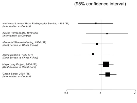
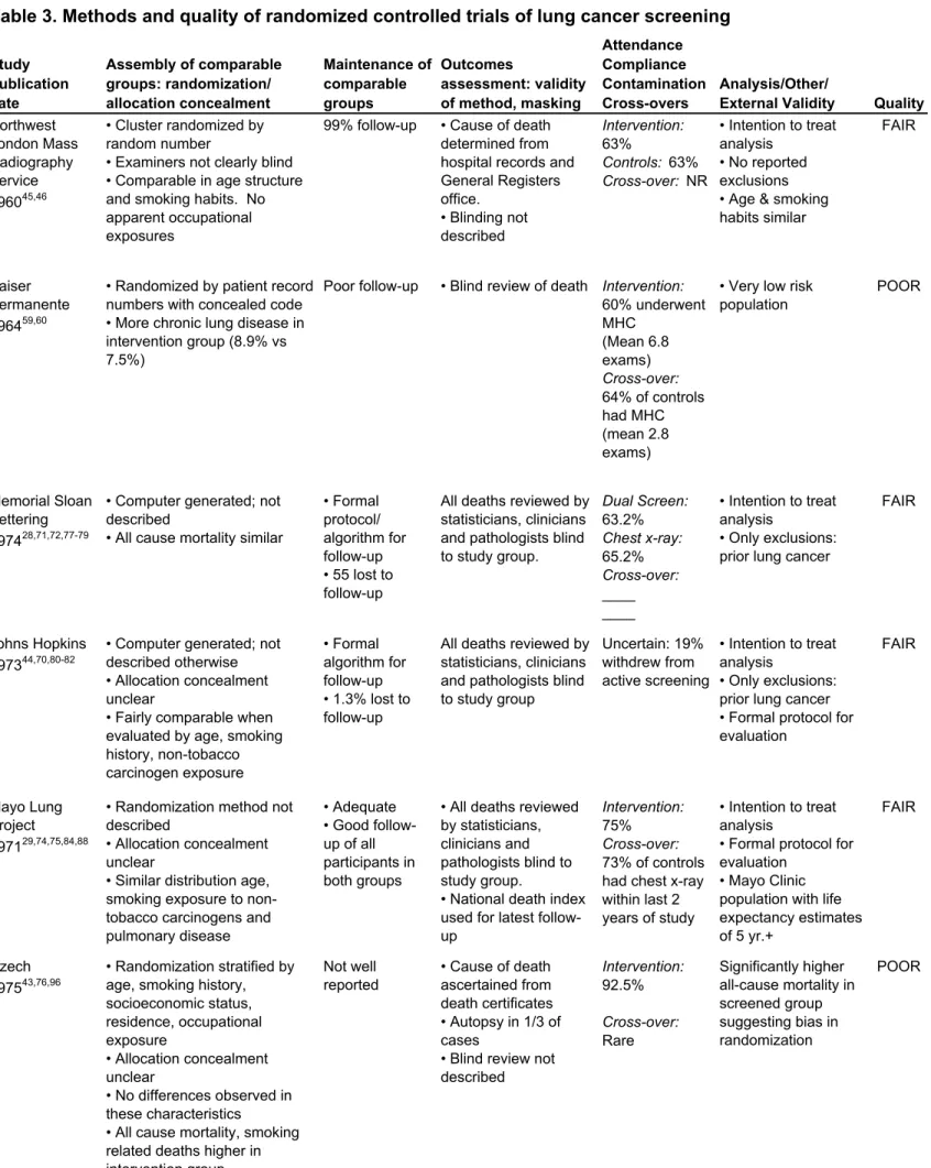
Tables 2 and 3 summarize the controlled trials of lung cancer screening and their methods and quality. Figure 2 shows the relative risks and confidence intervals of the randomized trials. In the 1960s, a controlled nonrandomized trial of chest x-ray screening every 6 months involving approximately 55,000 men older than 40 years was conducted by the Northwest London Mass Radiography Service.45, 46 In this cluster-randomized trial, 29,723 male factory workers from 75 randomly identified firms were offered chest x-ray every 6 months and compared to 25,300 controls from other factories offered screening at baseline and at 3 years. After 3 years, the annual mortality rate from lung cancer in the intervention group was 0.7/1,000; the rate was 0.8/1,000 in the control population, not different statistically.
The Kaiser-Permanente Multiphasic Evaluation Study was a randomized trial designed to determine whether or not encouraging middle-aged people to have annual multiphasic health checkups (MHC) would reduce mortality “from a group of diseases hypothesized in advance to have a fatal outcome preventable or postponable through periodic MHCs.”60 The MHC
consisted of several screening tests including chest x-ray. More than 10,700 members of Kaiser’s large health maintenance organization aged 35-54 of average risk (17% smokers) were randomized by medical record number into a study group (n=5,156) who were encouraged to undergo the MHC annually, and a control group (n=5,557) who were not advised regarding the
MHC but who could receive one if they requested it. Approximately 60% of the study group and 20% of the control group underwent the MHC annually. There were 44 lung cancer deaths in the study group and 42 in the control group (death rates per 1,000 for the 16-year follow-up period of 8.6 and 7.6 respectively)—not a statistically significant difference.
Three National Cancer Institute-sponsored randomized controlled trials of lung cancer screening in male smokers were conducted in the United States in the 1970s28, 44, 70-75 and a fourth in Czechoslovakia.43, 76 Those conducted at the Memorial Sloan Kettering (MSK) Cancer Center28, 71, 72, 77-79 and at Johns Hopkins (JH) University44, 70, 80-83 were identical in design and were conducted to evaluate the incremental benefit of adding sputum cytology to annual chest x-ray. Each was rated of fair quality based on USPSTF criteria. The reasons for each quality score are shown in Table 1. Of the 20,400 male smokers (at least 20 pack years of smoking) age 45 and above who volunteered for these 2 studies, 10,194 were randomized into a “dual screen” group that was offered screening with annual chest x-ray and sputum cytology every 4 months for 5 years, and 10,233 to a chest x-ray group that was offered annual chest x-ray screening for 5 years. Each group was followed for 5-8 years.
In the MSK study, the baseline (prevalence) screen identified 30 (6.0/1,000) lung malignancies in the “dual screen” group and 23 (4.6/1,000) in the chest x-ray group.71 Average 5-year survival of the prevalence cases among the dual screen patients was 48% and among those in the chest x-ray group, 37%. Following the prevalence screen, 114 subsequent (incident) lung cancers were identified in the dual screen group and 121 in the annual x-ray group, with 33 and 32 cases, respectively, diagnosed in the 2 years following screening. Combining the incidence and prevalence tumors, 144 lung cancers were detected in each group28, 72, 79; 40% of all lung cancers detected were stage I. Survival of both groups was approximately 35% at 5 years. This
compares with an average 5-year survival for lung cancer in the general population at that time of 10%. The mortality rate was 2.7/1,000 person-years in both the chest x-ray and dual screen groups.
In the JH study, the prevalence screen identified 39 malignancies in the dual screen and 40 in the chest x-ray group.44, 82 Prevalence varied significantly with age and reached 2.2% in individuals above age 65. Fifty-six percent of the cancers in the prevalence cases were resected and 5-year survival was 33% and 59% for the chest x-ray and dual screen groups, respectively.44 After 8 years of follow-up, 194 incidence cancers were identified in the dual screen and 202 in the chest x-ray group. Survival at 8 years was approximately 20% for both groups, with a
mortality rate of 3.4/1000 person-years in the dual screen group and 3.8/1,000 person-years in the chest x-ray group, not statistically significant differences. These rates were similar to community lung cancer mortality rates at the time.82
The authors of these studies concluded that adding sputum cytology to annual chest x-ray screening had no benefit in reducing lung cancer mortality. It is notable that the minimum screening intensity in these studies involved annual chest x-ray over a period of 6 years, and that the observed 5-year survival of 35% in both groups was higher than the average lung cancer survival at the time the study was conducted, and continues to be higher than current usual 5-year survival. However, the absence of a mortality benefit among trial participants in the Johns Hopkins study when compared to the population at the time argues against an important screening benefit and suggests survival was prolonged relative to the community because of screening biases, particularly lead-time and length bias. In a recent Cochrane review, the results of the Memorial Sloan-Kettering and Johns Hopkins studies were pooled using a random effects
model and showed a trend toward reduced lung cancer mortality in the intervention group (RR 0.88; 95% CI, 0.74-1.03).41
The first trial to evaluate the value of intense screening with chest x-ray was the Mayo Clinic Lung Project (MLP) involving 10,933 male smokers age 45 and above.29, 74, 75, 84-91 All participants underwent a prevalence screen with sputum cytology and chest x-ray; 91 cancers were identified (prevalence 0.8%) with resectability rates of 54% and 5-year survival of 40%, which was more than twice the survival of an age-similar Mayo Clinic comparison group with lung cancer. Almost half the prevalence cases were stage I or II and among these cases, 5-year survival was 70%.29, 73, 75
After the prevalence screen, 9,211 men were randomized to either a study group (n = 4,618) and screened with chest x-ray and pooled 3-day sputum cytology every 4 months for 6 years, or to a control group (n=4,593) and advised to have annual chest x-ray and sputum cytology. During the study period, 206 incidence cases of lung cancer were identified in the experimental group and 160 incidence cases identified in the control group. Resectability of the cases was 46% in the experimental group and 32% in the control group, with 5-year survival of 33% in the experimental group and 15% in the control group. After 6 years of follow-up, there were 115 lung cancer deaths in the control group and 122 in the study group, with death rates of 3.0/1,000 patient-years in the control group and 3.2/1,000 patient-years in the study group, not significantly different. After 20.5 years of follow-up the lung cancer death rates were 4.4 (95% CI, 3.9-4.9) and 3.9 (95% CI, 3.5-4.4) per 1,000 person-years in the intervention and control groups, respectively.88
The Mayo Lung Project was the first individually randomized controlled trial to
in determining current public health policy. Although it is rated of fair quality by USPSTF criteria, there are several limitations of the study. 1. A prevalence screen detected 91 cases (0.8%). Thus, there was no completely unscreened control group. Also, these cases were followed separately and not evaluated in the randomized comparison. Thus, any effect of these cases on mortality could not be determined. 2. Nearly half of the control subjects obtained annual chest x-rays during the course of the study, with one-third of the malignancies in the control group discovered by screening chest x-ray. Seventy-three percent of the controls received chest x-rays during the study’s last 2 years. 3. Compliance of the intervention group was 75%, reducing the study’s power.73 4. Assuming full compliance of the study group, the study was underpowered from the beginning with a 48% chance of detecting a 20% reduction in mortality.73 Based on screening of the control group, the reduced compliance of the intervention group, the initial study power, and the relatively short duration of follow-up, 1 analysis of the first study results reflecting 6 years of follow-up determined that it would have less than a 20% probability of showing a benefit from screening.74 However, in the most recent MLP follow-up, Marcus et al. effectively demonstrate that with a greater length of follow-up, a major reduction in lung cancer mortality was not missed due to low power.88, 92
The incidence of lung cancer in the experimental group in the Mayo Clinic Study was approximately 22% higher than in the control group. Even 3 years after the end of the trial, there were 46 more malignancies in the intervention group than in the control group.73 It is unlikely that the increase in incidence was a consequence of radiation from the screening x-rays, based on radiation exposure literature.93 Strauss et al. has suggested that this may be due to non-random distribution of important lung cancer risk factors between the control and intervention group.94 This issue was evaluated by Marcus89 and the distribution of several potential lung cancer risk
factors was not found to vary significantly between the intervention and control groups. Although little detailed information is provided, there is evidence on review of the MLP publications that not all patients were asymptomatic,29, 73 which could alter the findings of the screening study if patients with symptoms were disproportionately enrolled in the intervention arm of the study. However, there is no evidence to support this. Another possibility is that the higher incidence of lung cancer in the screened population may represent the diagnosis of insignificant lung cancers, e.g., overdiagnosis. This issue is complex and also relevant to the CT studies and is discussed below.34, 50, 94, 95
The most recent randomized controlled trial was conducted in Czechoslovakia43, 76, 96 where 6,364 male smokers ages 40-64 received chest x-ray and sputum cytology as a prevalence screen; 19 lung cancer cases were identified (3.0/1,000). After exclusion of the prevalence cases, 3,172 men were randomized to the study group and 3,174 to the control group. The study group received chest x-ray and sputum cytology every 6 months for 3 years and the control group received a single chest x-ray and sputum cytology at the end of the screening period. In the next 3 years of follow-up, chest x-rays were administered annually to subjects in both groups. In the first 3 years of the study, 36 lung cancers were identified in the experimental group and 19 in the control group. After 6 years, there were 108 malignancies identified in the experimental group and 82 in the control group, with 85 lung cancer deaths in the study group and 67 in the control group, rates that were not statistically different. A problem in randomization is suggested by the finding of significantly higher total all-cause, cancer, and smoking-related mortality in the
intervention group (341 versus 291 deaths), over the entire 6-year study period.76 This study is of poor quality, based on USPSTF criteria, and we consider the results likely to be invalid.
All cause mortality was calculated in the recent Cochrane review and data were available from the Erfurt County, Czechoslavakian, MSK, MLP, and Kaiser studies. Pooled analysis comparing frequent chest x-ray screening with less frequent screening, excluding the Czech trial, identified a relative risk of 0.97 (95% CI, 0.94-1.01) using a fixed effects model.41
In summary, 2 fair-quality randomized trials evaluating intense screening among high risk males with sputum cytology have shown no benefit of adding cytology to annual chest x-ray. Two fair-quality trials among high risk men comparing intense chest x-rays with less intense chest x-rays showed no benefit. Finally, a fair-poor randomized trial of multiple procedures in a low lung cancer risk population showed no benefit of annual chest x-ray.
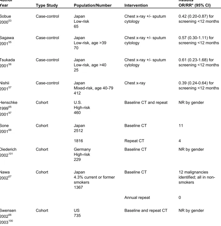
Case-Control Studies
Six case-control studies were identified in our search, 5 conducted in Japan and 1 in Berlin. The five Japanese studies are all of fair quality and the German study of poor quality, based on USPSTF criteria; the studies’ methods and findings are summarized in Table 4. In the 1950s a case-control study based on a tuberculosis screening program in which chest x-ray was offered every 1-2 years to all adults in the former German Democratic Republic showed no association between periodic chest x-ray and reduced lung cancer mortality.52 This study’s quality is rated poor, based on poor smoking assessment among controls, no report of response quality rates and use of hospital controls.
A 1992 case-control study from Japan35 reported data from 15 municipal lung cancer screening programs where participants were screened using chest x-ray, and in some areas, sputum cytologic tests. In general, the population offered screening consisted of high-risk men (smoking exposure) and low-risk women. The cases were comprised of 273 patients (208 men, 65 women) with fatal lung cancer. Cases’ screening histories were compared with those of 1,269
control subjects matched by sex, age, smoking status, and type of health insurance. The odds ratio for dying from lung cancer was lower at 0.72 (95% CI, 0.50-1.03) for those screened with chest x-ray +/- sputum within 12 months of diagnosis. For those screened between 12 and 24 months prior to the diagnosis, the odds ratio was 0.83 (95% CI, 0.56-1.23). The odds ratio for women screened within 12 months of diagnosis was 0.42 (95% CI, 0.20-0.87) (Table 4).
In a recent case-control study performed in Japan,54 the study group was comprised of 193 persons dying of lung cancer (158 men and 35 women), aged 40-74, and holding national health insurance. Three controls for each case were selected randomly from living national health insurance holders matched by residence, gender, and year of birth (n=579). The screening histories of the cases and controls were reviewed dating from the time of diagnosis of the case. After adjusting for smoking history, the odds of dying from lung cancer for participants screened within 12 months of diagnosis compared with non-screened individuals was 0.54 (95% CI, 0.34-0.85). The odds ratio for screening in the 12-24 months prior to diagnosis was 0.64 (95% CI, 0.30-0.97); no significant benefit was observed for screening occurring over 24 months prior to the diagnosis of lung cancer (OR 0.59; 95% CI, 0.30-1.15) (Table 4).
A case-control study conducted in the Miyagi Prefecture in Japan,55 where mass screening with chest x-ray and sputum cytology had been conducted since 1982, was of similar design to the above Japanese studies and involved 328 cases of fatal lung cancer diagnosed after 1990 among individuals aged 40-79 (70 low-risk women, 258 mixed-risk men). Each case was matched by gender, age, municipality, and smoking history, to 6 controls. The odds ratio for screening within 12 months was 0.54 (95% CI, 0.41-0.73) and 0.62 (95% CI, 0.42-0.92) for screening within 24 months. The odds ratio was 0.64 (95% CI, 0.36-1.14) for screening within
36 months. Screening 36-48 months prior to diagnosis showed no benefit. Among women (all “low-risk”), the odds ratio for screening within 12 months was 0.57 (95% CI, 0.30-1.11).
Another case-control study conducted among holders of National Health Insurance in the Nigata Prefecture of Japan,56 with similar methods among 25 low-risk women and 149 high-risk men with fatal lung cancer, identified reduced risk of fatal lung cancer among individuals screened within 12 months (OR 0.40; 95% CI, 0.27-0.59). Unlike 2 of the other Japanese case-control studies, the benefit did not extend past 12 months. Among women (all “low risk”), the odds ratio was 0.61 (95% CI, 0.23-1.68).
Finally, a case-control study was conducted among Japanese individuals in the Okayama Prefecture where population-based lung cancer screening had been conducted. Four hundred and twelve individuals with fatal lung cancer aged 40-79 were matched with 2-10 controls by gender, age, and district. After adjustment for smoking exposure, the odds ratio for screening within 12 months was 0.59 (95% CI, 0.46-0.74). Among women of mixed smoking status, the odds ratio associated with screening within 12 months was 0.39 (95% CI, 0.24-0.64), lower than among men (RR 0.67; 95% CI, 0.51-0.87).
In summary, 5 fair quality case-control studies among high to average risk men and low risk women, suggest a screening benefit. All are limited by lack of control for occupational exposures and family history and, as discussed below, the findings must be interpreted cautiously due to lack of randomization and possible healthy screenee bias.
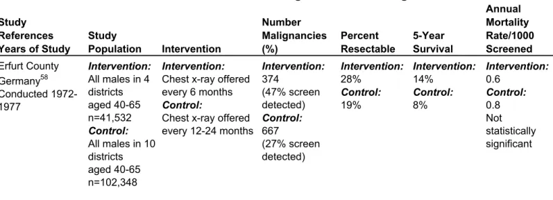
Controlled, Non-randomized Studies
A study conducted in Germany between 1972 and 1977 compared chest x-ray screening every 6 months in 41,532 males in 4 districts with chest x-ray screening every 18 months in 102,348 men in 10 districts. After 10 years, lung cancer mortality rates were similar at 0.6/1,000
and 0.8/1,000 in the intervention and control groups, respectively, not statistically different.58 The details of this study are described in Table 5.
Uncontrolled Studies
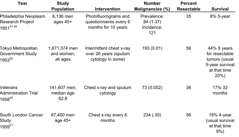
Four non-randomized, uncontrolled studies of lung cancer screening were conducted in the 1950s and 1960s. Two showing no survival benefit with screening when compared to usual population survival were the Philadelphia Pulmonary Neoplasm Research Project47, 48 and the Veterans Administration Trial.49 The Tokyo Metropolitan Government Study50 and the South London Cancer Study51 evaluated periodic chest x-ray screening and showed improved survival from lung cancer when compared to population rates at the time. Details of these studies are shown in Table 6. Because they do not have control groups they receive little emphasis in this review and were not rated in quality.
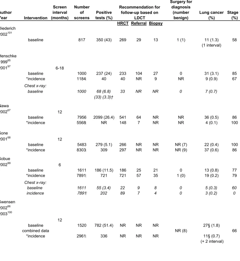
Lung Cancer Screening with CT
Several recent cohort studies, all without control groups, have evaluated screening for lung cancer with low dose CT. The details of these studies are shown in Table 7. In 1999, a study by Henschke et al. reported findings from the baseline screen of the Early Lung Cancer Action Project (ELCAP).65 The goals of this uncontrolled study were to compare low-dose CT and chest x-ray (in the same subjects) and to determine how nodule size affects survival. The study involved 1,000 symptom-free volunteers age 60 and above, with at least 10 pack years of smoking, and no prior malignancy, who were evaluated as medically fit for surgery and who each underwent chest x-ray and low-dose CT. Each CT and chest x-ray were read independently by chest radiologists; all nodules were radiographically characterized and evaluated according to standardized protocols, including high-resolution CT (HRCT), biopsy, or short-term follow-up. The study results are summarized in Table 7. The study details are provided here also given the
current intense public interest in screening CT, based largely on this trial and because the study contributes important information about the test characteristics of current chest x-ray screening.
The population was 46% female, had a median age of 67, and median pack years of 45. Approximately 14% of the entire population had been exposed to asbestos. Baseline (prevalence screen) chest x-ray identified 68 individuals with non-calcified nodules (NCN), of which 33 were confirmed by low-dose CT scan; 7 were malignant, and all were resectable. Baseline (prevalence screen) low-dose CT identified 233 individuals with 1 to 6 NCN. High-resolution CT scan was recommended to 233 and 183 complied. Among them, 63 were negative and 16 had biopsies recommended, with 13 malignancies identified. One hundred and four other individuals with abnormal high-resolution CT were followed with repeat CT and from these, 14 more biopsies were recommended; all revealed malignancy. Of the 30 recommended biopsies, which included 9 video assisted thorascopic surgeries (VATS), 27 malignancies were identified, of which 26 were resectable and 23 were stage I disease. Four other lung cancers were also diagnosed based on non-nodule CT abnormatlities. Thus, the prevalence of lung cancer following a baseline low-dose CT and follow-up of abnormalities was 3.1%. These findings are shown in Table 7.
Six to eighteen months after the baseline exam, 841 underwent a first repeat screen and 461 underwent a first repeat (incidence) exam. Based on the timing of the incidence exam the investigators estimate there were 1184 “annual” examinations. Among the original 1,000 alive and without cancer, 2 developed symptom-detected lung cancer (interval cancer). Thirty of the 1,184 (2.5%) individuals undergoing annual screening required further evaluation. Two of these 30 died prior to workup and of the remaining 28, 12 nodules resolved over 1 month, 8 were followed with HRCT, and 9 underwent biopsy resulting in 7 lung cancer diagnoses (6 stage IA).97
The significant findings from this study were that the prevalence of lung cancer was relatively high, and that low-dose CT has the ability to detect malignant tumors 4 times more commonly than chest x-ray, and stage I tumors 6 times more frequently than chest x-ray, suggesting that the sensitivity of chest x-ray compared to low-dose CT is 25% for detecting malignancy. There are no mortality data available yet on the ELCAP cohort; however, based on data from lung cancer survival in the usual practice setting, the expected 5-year survival of the 85% of patients with stage I non-small cell lung cancer is estimated at 65-70%10, 98 and possibly higher based on the size distribution within the stage I carcinomas detected on the initial screen. The ELCAP study is also helpful in describing the screening test accuracy of chest x-ray since CT is usually the definitive test for evaluating an abnormal chest x-ray. The test characteristics of chest x-ray based on this study and using CT as a gold standard are shown in Table 8.
Three LDCT studies have been conducted in Japan involving large numbers of both high- and low-risk men and women. Each study used different protocols but each included chest x-ray and sputum cytology. The details of these studies are described in Table 7. One study involved 5,483 prevalence and 8,303 incidence screens and detected 59 primary lung cancers among men and women aged 40-74 with LDCT; 1 lung cancer was detected with sputum cytology only. Fifty-five out of 60 were stage I at diagnosis; no survival or mortality data have been
published.61, 68 Interestingly, the prevalence (baseline) screen identified more malignancies in non-smokers than smokers (0.44% versus 0.40%, respectively). Incident screens revealed more tumors in smokers than non-smokers, suggesting different biological properties of the tumors in smokers and non-smokers.
Another study included 742 participants who had been screened biennially with chest x-ray and sputum cytology for a number of years and 940 new participants (n=1,611) who
underwent LDCT screening exams beginning in 1993. These participants were current smokers, age 40 and above, and recruited from the general population (1,415 male, 196 female). Among 1,611 baseline screens, 11.5% had abnormal CTs, 3.4% abnormal chest x-rays, and 0.8%
abnormal sputum cytology from which 13 lung cancers (11 Stage I) were diagnosed based on CT and one lung cancer was diagnosed with sputum cytology only. 7,891 repeat screens resulted in 721 (9.1%) abnormal nodules, 719 HRCT and 35 biopsies, with 19 lung cancer diagnoses (18 Stage I) based on CT findings; 3 lung cancers were detected with sputum cytology only.62, 99 Among the 56 biopsies performed because of abnormalities on LDCT, 20 VATS procedures were performed resulting in 14 lung cancer diagnoses. Finally, another Japanese cohort study involved 7,956 Hitachi employees (1,637 women, 6,319 men) aged 50-69 where chest CT was conducted as part of annual health examinations.67 In this study, the prevalence screen identified 2,099 individuals with abnormal nodules from which 541 underwent HRCT, and 64 were further evaluated; 24 malignancies were identified in men and 12 in women. The annual incidence screen among 5,568 individuals identified 4 new malignancies. Thoracotomy, (including VATS) was performed in 57 patients resulting in 40 diagnoses of lung cancer.
A LDCT study conducted at the Mayo Clinic involved 1,520 men and women aged 50 or older with 20 or more pack years of smoking.66, 69, 100 Each participant underwent a baseline (prevalence) screen and from this screen, 782 (51%) individuals had 1 or more non-calcified nodules and 26 (1.7%) were diagnosed with primary lung cancer; one individual was diagnosed with lung cancer with sputum cytology only. Among 1,464 individuals from this cohort who underwent an annual incidence screen,(2,916 screens), 191 (13%) individuals were found to have new nodules and 10 new diagnoses of lung cancer were made (6.7/1000). There were also 2 interval and 2 cancers diagnosed with sputum cytology. Of the 50 individuals with malignancies
identified, 36 were non-small cell lung cancer (NSCLC) of which 31 (86%) were resected for cure; 8 patients underwent surgery for benign disease in this study; there were also 2 interval cancers and 2 cancers diagnosed with sputum cytology only (1 at baseline and 1 at incidence screen). It is possible that the high number of nodules in this study is because of the Mayo Clinic’s Midwest location and higher rates of histoplasmosis in this region.
A German study101 involving 817 asymptomatic volunteers age 40 and above with at least 20 pack years of smoking was conducted between November 1995 and July 1999. The median age was 53, and 229 of the 817 were women. All underwent LDCT and each CT was read by 1 of 2 radiologists. All non-calcified nodules greater than 10 mm were considered potentially malignant and evaluated with HRCT or follow-up in 3, 6, 12, and 24 months. Non-calcified nodules less than 10 mm were followed with repeat low dose CT. The prevalence findings included 350 individuals with non-calcified nodules and HRCT was recommended. Of the 350, 81 were re-evaluated at 3 months and 269 underwent HRCT, which identified 32 nodules in 29 individuals. Seventeen individuals (18 nodules) had morphology suggesting benign causes and were followed; 1 of these grew over 24 months and was diagnosed as stage I adenocarcinoma. Twelve underwent biopsy (including 8 VATS procedures) and malignancy was diagnosed in 10 with one additional interval lung cancer; 6 were stage IA. After an average of 2.7 years of follow-up, 6 are alive without evidence of recurrence. In total, 13 individuals underwent biopsy and 11 malignancies were diagnosed.
In summary, 6 cohort studies evaluating screening with low dose CT have shown that CT is significantly more sensitive than chest x-ray for identifying non-calcified nodules, that the false positive rates are high but significantly reduced with non-invasive follow-up (CT or time) and that the majority of patients undergoing biopsy are diagnosed with lung cancer, usually at an
early stage. Importantly, no conclusions about the overall impact of this procedure on reducing lung cancer mortality can be made in the absence of randomization and a control group with mortality as an outcome.
Lung Cancer Screening Among Women
Lung cancer is the leading cause of cancer-related death among women in the United States.102Most lung cancer in women is attributed to smoking.102 However, women have substantial exposure to passive smoking and a significant proportion of lung cancer in non-smoking women is attributed to passive non-smoking.12 In addition, although it is controversial, some studies suggest that for any level of smoking, women are at higher risk of developing cancer than men.3, 14, 15 For unknown reasons, women also tend to develop adenocarcinoma of the lung disproportionately to men,10, 15, 23 and adenocarcinoma is also found more commonly among non-smokers.23 This cell type tends to occur peripherally10, 103 and may be more apt to be detected with chest x-ray and/or CT than other cell types. As a consequence, radiologic imaging and screening for lung cancer may perform differently and may actually be better among women. Unfortunately, no randomized trials of specific lung cancer screening have included women. The only data evaluating screening among women and including control populations come from 4 Japanese case-control studies evaluating screening among primarily non-smoking women (passive smoking not assessed). These studies are summarized in Table 9 and show odds ratios for screening conducted within 12 months of lung cancer diagnosis 0.39-0.61, 2 studies
statistically significant; however, interpretation is limited by the screening biases discussed in this review. Five studies of LDCT have included women; mortality data are not yet available. In addition, randomized trials of lung cancer screening with chest x-ray and/or CT involving women are currently underway.
Summary
1. Five fair-quality Japanese case-control studies show or suggest benefit with chest x-ray screening among men with smoking exposure and women without direct smoking exposure or of mixed risk. There is a suggestion of a gradient of benefit over time in 3 of these studies. Interpretation of these studies is limited by potential screening biases.
2. Two trials evaluated sputum cytology and although neither showed benefit, a recent Cochrane review pooled the findings and found a suggestion of benefit (RR 0.88; 95% CI, 0.74-1.03), though the finding was not statistically significant.
3. Two individually randomized controlled trials of lung cancer screening in high risk men evaluated the role of screening chest x-ray; however, only 1 is of sufficient quality to evaluate the findings and it showed no benefit of intense chest x-ray screening (every 4-6 months) over intermittent chest x-ray screening. One randomized trial of multiphasic screening among relatively young average risk
individuals that included chest x-ray showed no benefit, but was compromised by low power, contamination of the control group, and poor compliance. One cluster
randomized trial suggested benefit but was not statistically significant.
4. All lung cancer screening specific randomized trials involved the use of a prevalence screen at the beginning of the study. Consequently, there were no completely
5. No randomized controlled trials have evaluated lung cancer screening among women; 4 case-control studies from Japan suggest there may be benefit of screening with chest x-ray.
6. Several recent uncontrolled studies of low dose CT have shown that:
a. LDCT is significantly more sensitive than chest x-ray for identifying lung cancer.
b. LDCT identifies a significantly higher proportion of small (low-stage, resectable) lung cancers than chest x-ray.
c. There are high rates of false positive findings in the LDCT studies, and many patients undergo further studies, as well as invasive procedures, to
discriminate benign from malignant disease.
d. While survival is anticipated to be longer among the individuals with smaller tumors, the effectiveness of screening cannot be evaluated in the absence of control groups given the biases associated with screening, particularly lead-time and length bias.
Discussion
The personal and public health importance of lung cancer in the United States and worldwide is enormous, and even a small benefit associated with screening could save many lives. However, the outcomes of screening, as shown in this report, are mixed, with some lower grades of evidence (case-control studies) showing benefit and higher grade evidence (randomized controlled trials) not showing benefit and possibly showing harm. Unfortunately, none of the existing randomized trials answer the question faced by clinicians, which is, should patients be screened for lung cancer at all?
The case-control studies from Japan give some support to chest x-ray screening for lung cancer. The studies were methodologically sound and possibly demonstrate a gradient in screening effectiveness by length of interval of screening prior to diagnosis, suggesting a dose-response benefit. An alternative explanation for this gradient is that it is measuring a gradient of healthy screenee behavior. The studies also provide information about the interval of time in which screening might be effective (< 24 months) and the length of lung cancer’s detectable pre-clinical stage. Although case-control studies are not considered the gold standard in evaluating screening efficacy and effectiveness, several authors believe they can be a useful and efficient method of evaluating a screening method.27, 104, 105 The adoption of flexible sigmoidoscopy as a recommended screening modality for colon cancer screening has been based largely on data from well-conducted case-control studies.106, 107 Unfortunately, it is very difficult to overcome the possibilities of volunteer/healthy screenee bias in case-control studies, even well-conducted ones, that might bias the study toward benefit, since those choosing screening may differ from those not being screened in factors which of themselves influence lung cancer mortality.108
Although the LDCT studies indicate earlier stage lung cancer can be detected, drawing conclusions from the uncontrolled CT studies is difficult because of the methodologic biases discussed above that could significantly affect interpretation of the findings. Thus, the implications of the CT studies are uncertain due to lack of controls and mortality data. It is possible, based on the stage distribution of the detected malignancies, that survival may be prolonged and mortality reduced. However, because of lead-time and length bias, survival may be prolonged, but mortality unchanged. Randomized trials of LDCT with mortality as an outcome will be needed to definitively evaluate this issue.
The hope of benefit from lung cancer screening is high, however, the implications of screening, especially in the absence of clear-cut benefit, are also great. Evaluating harm or potential harm associated with screening for lung cancer is difficult. One approach to this issue is to evaluate the outcomes of screening. The best data about outcomes from chest x-ray screening come from the recent CT studies since data from the chest x-rays trials accumulated prior to the use of CT for evaluation of x-ray abnormalities, and many patients underwent thoracotomy or biopsy who currently would not. These data are displayed in Table 7, which shows positive test rates and the diagnostic outcomes associated with chest x-rays in a screening setting. Based on the CT studies, most chest x-ray abnormalities are resolved or found to be false positives when evaluated by CT. In a Japanese study, 1,611 individuals underwent CT and chest x-ray and of 23 positive baseline chest x-rays, 22 individuals underwent high-resolution CT, 8 eventually
underwent biopsy, and 3 were diagnosed with lung cancer.99 In the ELCAP study, 68 individuals had abnormal chest x-rays at baseline, of which 33 were identified on LDCT and referred for further work-up, from which 7 lung cancers were identified. No patient underwent thoracotomy for diagnosis.65
In the CT studies, the false positive rate is the number of patients with low-dose CTs requiring further evaluation or follow-up who do not have cancer. Using this criterion, the false positive rates in the CT studies range from 5 to 50% in prevalence screens and 3-12% in
incidence screens, with most abnormalities resolved on high-resolution CT. Among the LDCT studies, 4.8-14.5% of patients undergoing HRCT are referred for biopsy, from which most (63-90%) are diagnosed with cancer. For comparison, in US and European clinical practices,
approximately half of patients undergoing surgical biopsy of indeterminate nodules subsequently receive a benign diagnosis.66 In some studies, false positive rates associated with CT screening
appear to decrease when the screened cohorts are periodically rescreened.68, 97 Based on data from 3 studies which reported rates of thoracotomy, among 3,928 patients screened, 2 underwent thoracotomy from which one was diagnosed with cancer and 83 malignancies were diagnosed. In the current practice setting, Positron Emission Tomography (PET) scans are used as a non-invasive means of discriminating between malignant and nonmalignant lesions.109 In one study, PET scans had 94% accuracy in identifying malignant and non-malignant lesions.109 With PET scans, many patients who might have undergone biopsy or thoracotomy in the past might avoid invasive procedures.
All of the individuals with false positive results experience a period of time potentially associated with high anxiety and concern, and for those pursuing further evaluation, the cost and risk associated with it. If there is no benefit to having a false positive screening exam, then many suffer so that few may gain. If benefit can come from being a false positive, then this also needs to be considered. In addition to risk (physical and emotional), there also is the added expense of evaluating the false positive exams, which is significant. Although the false positive rate is high in the lung cancer screening studies, the meaning of a false positive lung cancer screening study (either chest x-ray or CT) to a patient may be different than for other types of false positive cancer screening tests, as the patients potentially have some control over their subsequent risk and may be able to more effectively modify their high-risk behavior. Data from the ELCAP study suggest that CT scan results in combination with smoking cessation counseling improved
smoking cessation rates among all participants.65 In addition, the ELCAP data show that an abnormal CT finding is associated with nearly two-fold greater odds of decreased smoking or cessation among current smokers (62% reduced or quit rates among those with positive scans compared with 45% rates among those with negative scans).110 It is reasonable to assume that an
abnormal screening chest x-ray might also influence smoking behavior. However, it is also possible that these rates are higher because the participants are volunteers and/or have formed relationships with the investigators. It is also possible that individuals seeking screening will be reassured by a negative screen and will continue smoking.
An important issue in lung cancer screening is the question of over-diagnosis (and potential over-treatment). The prevalence of lung cancer in the asymptomatic, older, healthy, high-risk ELCAP population was high (3.1%), suggesting that there is a significant pre-clinical pool of lung cancer in high-risk populations. High prevalence has also been shown among
individuals being evaluated for lung reduction surgery.111 Supporting over-diagnosis is data from the Mayo Lung Project showing increased rates of early tumors in the intensely screened group compared to the control group, without a change in numbers of advanced tumors or subsequent mortality rates, suggesting diagnosis of a pool of indolent tumors. It can be argued, particularly in conjunction with the increased number of lung cancer cases identified in the intervention group of the Mayo Clinic study that this high prevalence reflects the detection of tumors that would not progress in the patient’s lifetime. Arguments against an important role for over-diagnosis in lung cancer are based on autopsy and clinical studies. One autopsy series involving 3,286 necropsies identified unsuspected lung cancer in 0.8% of patients who had died of a multitude of causes, suggesting a low rate of clinically unrecognized lung cancer.95 However, autopsy may
underestimate rates of lung cancer when compared to CT, since the lungs are not always thinly sectioned with autopsy.112 Moreover, whether autopsy data are generalizable to living
populations is questionable, particularly given selection biases for autopsy.
In the MLP, Marcus et al88 reason that the disparity between the finding of prolonged survival among intervention cases compared to control cases, and no difference in mortality
between the 2 groups, largely results from lead-time or length-bias and/or over-diagnosis. To evaluate this, the authors consider survival rates from the time of randomization (eliminating lead-time bias), and an increase in survival rates persists in the absence of a mortality benefit. The implication of this finding is that length bias and over-diagnosis account for the disparate findings. An editorial by Black accompanying the most recent update of the MLP113 makes a clear argument for the potential of over-diagnosis in lung cancer screening and the implications of over-diagnosis among an elderly population. Although the higher lung cancer mortality rate among the intervention group in the MLP was not statistically significant, a major concern is that the increase in mortality rates might not be due to chance and may be a consequence of
screening, e.g., more individuals undergo evaluation and treatment in the screened group with its attendant risk, resulting in a true increase in mortality. Alternatively, an increase in lung cancer mortality rates among screened individuals may be a consequence of misclassification of cause of death or “sticking-diagnosis bias,” meaning that there is a propensity to label any diagnosed malignancy as the cause of death, regardless of the tumor’s clinical course, in the absence of autopsy data.114 Whether a strong case for over-diagnosis should be made on the basis of current data is uncertain. However, it is possible that with an increasingly sensitive detection tool, such as LDCT, over-diagnosis may be demonstrated to occur. The issue of over-diagnosis is
particularly relevant to the harm associated with lung resection for cancer where there is significant mortality and morbidity associated with treatment. More data are needed to definitively evaluate this issue.
Another potential harm in screening is false negative findings with possible false reassurance. The best estimate of the rate of false negative chest x-rays comes from the CT studies where false negative rates as high as 75% have been shown. Clinical series of chest x-ray
suggest retrospective identification of lung cancer ranges from 12-90%.115, 116 While CT is considered the gold standard for evaluating nodules, it has also has been shown to have false negative rates (e.g., nodules identified retrospectively).66 The clinical implications of false negative exams on CT have not been reported to our knowledge. However, lung cancers missed on chest x-ray have been shown to delay diagnosis and are an important source of malpractice claims.116 The potential for false reassurance certainly exists, particularly if screenees believe that they are undergoing a definitive examination.
The rate of complications associated with biopsy is not described in the CT studies. The known potential complications depend on the type of biopsy performed. The morbidity and/or mortality associated with thoracotomy for true positive tests is also difficult to evaluate.
Complication rates from studies among symptomatic patients are very likely greater than among asymptomatic individuals in screening programs directed at those judged healthy enough to undergo surgery. Thus, it is most appropriate to evaluate these rates from screening studies if the data are available. Unfortunately they are not. In general, studies from symptomatic patients suggest that the more lung tissue removed the greater the morbidity and mortality. Overall, mortality rates range from 1.3 to 11.6% among several series reviewed, with lower mortality among patients undergoing smaller resections.36, 117-121For example, the mortality rate among individuals undergoing lobectomy in one series was 0.6% compared with 5.7% among those undergoing pneumonectomy.120 Co-morbidity also affects surgical risks; in one US series the rates of operative mortality for resection of stage one non-small cell lung cancer ranged from 0.7 to 6.8% depending on the level of co-morbidity.122 The volume of surgery also has been shown to be associated with operative mortality,119 with rates of 3% in high volume centers compared with 6% in low volume centers.123 The morbidity reported among several series of thoracotomy ranges
between 8.8% and 44%, again dependent on the extent of the resection, the volume of the center and the co-morbidity of the patient.36, 117, 118, 120, 121, 123
The current standard of clinical practice in the United States is that most patients are not screened for lung cancer.124 Based on evidence from the older studies discussed above,
particularly the randomized controlled trials, there are no professional organizations that
currently recommend routine lung cancer screening. The second U.S. Preventive Services Task Force did not recommend routine screening of asymptomatic persons for lung cancer with chest x-ray or sputum cytology and gave this a D recommendation, meaning that there is fair evidence to support the recommendation that screening should not be performed.1 However, because conclusions about lung cancer screening have been based on limited data and no trials have compared screening with no screening, or screening among women, the issue is being re-evaluated. There is enough uncertainty in the field that in 1992, the National Cancer Institute funded a study to evaluate the usefulness of routine chest x-ray screening for lung cancer in both men and women as part of the large Prostate, Lung, Ovarian, and Colorectal Cancer Study (PLCO). This trial involves over 100,000 men and women aged 55-74 who are randomized to receive either 4 annual anterior-posterior chest x-rays or usual care. Data from this study should be available in 2010. The National Lung Screening Trial (NLST), cosponsored by the American College of Radiology Imaging Network (ACRIN) and the Lung Screening Study, sponsored by the Division of Cancer Prevention at the National Cancer Institute, will evaluate screening CT. Ten screening centers derived from the PLCO centers were involved along with 20 ACRIN sites. In the study, men and women aged 55 to 74, with at least 30-pack years of smoking who were not enrolled in the PLCO, were randomized to either a base-line spiral CT with two annual follow-ups, or a base-line chest x-ray with two annual follow-ups. Early data from this trial suggest that
there has been overwhelming interest by the public and because of high enrollment; some
evidence may be available by the year 2005 if there is a significant mortality reduction associated with screening. It is notable that there is not a placebo (non-screened) group enrolled in either of these trials.125
New technologies may also contribute to the early detection of lung cancer and
potentially, screening for lung cancer. Some being investigated include: immunocytochemical analysis of sputum with monoclonal antibodies,126 and identification of genetic mutations,127 abnormal DNA methylation,128, 129 abnormal patterns of immunostaining and other molecular changes.130-133 There are several other potential targets in sputum, bronchial fluid and expired air that may have a role in early lung cancer detection and are currently being investigated.134, 135
In summary, there is 1 fair quality individually randomized study of intense chest x-ray screening showing no benefit, as well as the possibility of over-diagnosis, and one cluster randomized study suggesting possible benefit with reduced morality, which was not statistically significant. There are also non-randomized studies of lung cancer screening with chest x-ray that suggest benefit to the screened populations. In addition, pooled data from a Cochrane analysis suggest there may be benefit associated with sputum cytologic screening. There are important methodological limitations to all of these studies. The studies of LDCT have demonstrated that lung cancer can be diagnosed at a significantly earlier stage than currently occurs in clinical practice. However, whether this finding will translate to a mortality benefit is unclear. Critical information will come from the current randomized controlled trials of screening CT. Given the uncertainty associated with chest x-ray screening, it is unfortunate that there are not unscreened control groups in the Lung Screening Study and the NLST; fortunately, data will be available on chest x-ray screening from the PLCO trial in the next 5-8 years. In the meantime, however, other
approaches for evaluation of screening might be considered, such as rigorously conducted case-control studies of chest x-ray and/or screening CT, since the results of randomized case-controlled trials will take years to complete, and lung cancer deaths continue to be major personal and public health issues world-wide. In addition, it is hopeful that new methods of screening for lung cancer will be developed and refined.135 Even a small decrease in lung cancer mortality
References
1. U.S. Preventive Services Task Force. Guide to Clinical Preventive Services.2nd ed. Washington, DC: Office of Disease Prevention and Health Promotion; 1996.
2. Strauss GM. Bronchiogenic carcinoma. In: Textbook of pulmonary diseases, 6th ed. Philadelphia, PA: Lippincott-Raven Publishers; 1998.
3. Zang EA, Wynder EL. Differences in lung cancer risk between men and women: examination of the evidence. J Natl Cancer Inst 1996;88(3-4):183-92.
4. American Cancer Society. Cancer Facts and Figures 2003. Available at:
http://www.cancer.org/downloads/STT/CAFF2003PWSecured.pdf. Accessed March 11, 2004.
5. Strauss GM. Screening for lung cancer: An evidence-based synthesis. Surg Oncol Clin N Am 1999;8(4):747-74, viii.
6. Osann KE. Lung cancer in women: the importance of smoking, family history of cancer, and medical history of respiratory disease. Cancer Res 1991;51(18):4893-7.
7. Tockman MS, Anthonisen NR, Wright EC, Donithan MG. Airways obstruction and the risk for lung cancer. Ann Intern Med 1987;106(4):512-8.
8. Hole DJ, Watt GC, Davey-Smith G, Hart CL, Gillis CR, Hawthorne VM. Impaired lung function and mortality risk in men and women: findings from the Renfrew and Paisley prospective population study [see comments]. BMJ 1996;313(7059):711-5; discussion 715-6.
9. Skillrud DM, Offord KP, Miller RD. Higher risk of lung cancer in chronic obstructive pulmonary disease: a prospective, matched, controlled study. Ann Intern Med
1986;105(4):503-7.
10. Nesbitt JC, Lee JL, Komaki R, Roth JA. Cancer of the lung. In: Holland JF, Bast RC, Jr., Morton DL, Frei E, III, Kufe DW, Weichselbaum RR, editors. Cancer Medicine.
Baltimore: William & Wilkins; 1997.
11. Trichopoulos D, Mollo F, Tomatis L, Agapitos E, Delsedime L, Zavitsanos X, et al. Active and passive smoking and pathological indicators of lung cancer risk in an autopsy study. JAMA 1992;268(13):1697-701.
12. Fontham ET, Correa P, Reynolds P, Wu-Williams A, Buffler PA, Greenberg RS, et al. Environmental tobacco smoke and lung cancer in nonsmoking women. A multicenter study [published erratum appears in JAMA 1994 Nov 23-30;272(20):1578]. JAMA