PROSPECTIVE ANALYSIS OF PRE-OPERATIVE PROGNOSTIC INDICATORS OF VITAL PULP THERAPY ON MATURE PERMANENT TEETH
William S. Yeung
A thesis submitted to the faculty at the University of North Carolina at Chapel Hill in Partial fulfillment of the requirements for the degree of Masters of Science in the Department of
Endodontics in the School of Dentistry.
Chapel Hill 2017
Approved by:
Asma Khan
Peter Tawil
iii ABSTRACT
William S. Yeung: Prospective Analysis of Pre-operative Prognostic Indicators of Vital Pulp Therapy on Mature Permanent Teeth
(Under the direction of Asma Khan)
Vital pulp therapy (VPT) is a conservative treatment option for exposed pulps with the goal
of preserving the vitality of the pulp (1). Historically, VPT with calcium hydroxide had
inconsistent success rates when used for carious exposures. Since that time, newer materials have
resulted in a more favorable success rate for this treatment option. Factors in addition to the
material type may affect the outcome that previous studies with small sample sizes did not identify
(2, 3). This prospective, observational study aims to examine the correlation between the outcome
of vital pulp therapy and pre-operative factors such as patients’ gender, age, tooth type, and the
site of pulp exposure. Written informed consent was obtained from asymptomatic patients
experiencing a pulp exposure on a vital, mature permanent tooth. Caries removal was completed
and hemorrhage was controlled with sodium hypochlorite. Mineral trioxide aggregate was placed
on the exposed pulp and the tooth was restored permanently. The patients were followed up with
a series of phone calls and in person examinations at 6 months and 1 year postoperatively. The
data was analyzed by logistic regression using a generalized linear model to evaluate associations
between pre-operative factors and outcome of treatment. A total of 73 patients were treated with
ages between 15 and 78 years old. 51 patients were available for 6 month recall with a success rate
of 79%. This study found that vital pulp therapy on mature, permanent teeth was a viable treatment
iv
To Mom, Dad, & Sally.
v
TABLE OF CONTENTS
LIST OF TABLES ... vi
LIST OF FIGURES ... vii
REVIEW OF LITERATURE ... 1
MANUSCRIPT ... 14
Section 1.1 Introduction ... 14
Section 1.2 Materials and Methods ... 15
Section 1.3 Capping Procedure ... 15
Section 1.4 Follow up... 16
Section 1.3 Statistical Analysis ... 17
Section 1.4 Results ... 17
Section 1.5 Discussion ... 23
Conclusion ... 26
DISCUSSION ... 28
vi
LIST OF TABLES
vii
LIST OF FIGURES
Figure 1: Subject Flow ... 19
Figure 2: Age Group Distribution ... 20
Figure 3: Gender Distribution ... 20
Figure 4: Tooth Types Treated ... 21
Figure 5: Proximal Surface Involvement ... 21
1
REVIEW OF LITERATURE
Section 1.1 Introduction
Endodontic disease manifests as a variety of signs and symptoms. If untreated, it can lead
to devastating outcomes with intense pain and swelling. Despite the different presentations, most
endodontic diseases share the common etiology of microbial infection (4, 5). The most common
route for infection is through carious lesions which permit microorganisms from the oral cavity to
infect the pulp. Other routes of infection can be trauma, or mechanical exposures.
Pulpal exposures can occur under three different scenarios: caries, trauma, or mechanical
causes. Caries, a common chronic disease, allows for bacteria from the oral environment to
penetrate through the tooth’s enamel and dentin into the pulp (6). Nearly 80% of dentists report
encountering caries to the pulp at least once per month in their practice (7). Traumatic exposures
are not as commonly encountered, making up only 10% of total dental traumas experienced (8).
These can occur during physical accidents where the coronal part of the tooth becomes chipped in
a fashion that exposes the pulp. Mechanical exposures are the least frequently encountered. These
occur during a dental procedure where healthy tooth structure is removed erroneously, exposing
the pulp. This study focuses on treating carious exposures which is the most commonly
encountered by clinicians. Regardless of the method of pulpal exposure, all three scenarios allow
for microbial invasion into a delicately formed tissue.
Microbial invasion of the pulp can elicit a dual immune response: innate and adaptive
2
flow of dentinal fluid, infiltration of phagocytes, and chemical mediators (9-11). Adaptive
immunity plays a role if innate immunity is unable to resolve the microbial infection.
Intrapulpal pressure causes an outward flow of dentinal tubule fluid which assists in the
diffusion of noxious elements away from the pulp: a protective response. This underscores one of
the arguments for maintaining a healthy vital pulp since non-vital teeth do not have this outward
pressure. Non-vital teeth have increased invasion of noxious stimuli compared to their vital
counterparts (12). In addition to fluid flow, the composition of the fluid aids in pulpal defense.
Dentinal fluid has been found to contain immunoglobulins that change in intensity as caries
progression advances (13). As caries forms and progresses, increased levels of immunoglobulins
are detected in the dentinal tubules (14).
The cells that function as part of the pulp’s innate immunity consists of neutrophils,
macrophages, and dendritic cells. The primary goal of these cells is to phagocytize foreign bodies.
Neutrophils are mainly for short-term responses. They phagocytize bacteria and their byproducts
by engulfing them and digesting them into small peptides. Neutrophils and chemokines are
attracted to the area by lipopolysaccharides on gram negative cell walls. They recognize foreign
bodies via antibodies as well as bacterial cell walls. Macrophages are recruited to the site relatively
slower than neutrophils but also engulf and digest antigens as well. Macrophages are developed
from monocytes in the blood and recognize glycoproteins on the surface of bacteria. Dendritic
cells function as antigen presenting cells which are used to activate T and B cells. They are derived
from hematopoietic stem cells. Immature dendritic cells possess phagocytic activity while mature
dendritic cells have less ability to phagocytize.
Chemical mediators such as cytokines, chemokines, arachidonic acid metabolites, nitric
3
Cytokines are proteins that are produced mainly by immune cells and assist in regulation. They
have three main categories: proinflammatory, Th1, and Th2. Proinflammatory cytokines are
produced mainly by macrophages and cause elevation in temperatures which limits bacterial
growth and promotes adaptive immunity. Th1 cytokines are produced by Th1 type helper cells.
Their function includes assistance in macrophage and natural killer cell and antiviral activity while
also downregulation of Th2 type reactions. Th2 cytokines are utilized in the activation, growth,
and differentiation of B cells as well as inhibition of macrophage activation. The main function of
chemokines is to induce exudation and translocation of leukocytes. Arachidonic acid metabolites
are involved in different inflammatory and homeostastic processes while nitric oxide regulates the
synthesis of various chemical mediators. Neuropeptides such as Substance P and calcitonin
gene-related peptides function to regulate vasculature and control local blood flow. In the event where
innate immunity is insufficient to defend the pulp, the pulp’s other defense, adaptive immunity,
comes into play (10).
Unlike innate immunity, adaptive immunity is more specific toward exogenous antigens
and is composed primarily of T cells and B cells. T cells are lymphocytes that are activated to
secrete cytokines and differentiate into various other cells. They require antigen presenting cells
to capture and present the antigens to function. In the pulp, B cells along with immature dendritic
cells bind to antigens and present them to T cells (15). The B cells are formed in the bone marrow
and differentiate to become plasma cells to function as antibody producing cells. As the carious
lesion approaches the pulp, the number of B cells in the pulp increases significantly. Their main
function is to produce antibodies which then bind to the antigens tagging the antigen for other parts
4
If the pulpal irritant is not removed, irreversible damage ensues and the pulp progresses to
necrosis. It was once believed that the body’s response to chronic inflammation increases fluid
movement into the pulp as a part of the immune response. This increase in fluid was thought to
increase the pressure inside the root canal system due to its non-compliant environment. The
increase in pressure would eventually lead to strangulation of the vessels at the apex causing
overall necrosis (16). Newer findings have refuted this, showing that the tissue pressure only rises
in the immediate area. It is now believed that local necrosis occurs in an area immediately beneath
the source of inflammation. If not treated, then the area of necrosis progresses gradually throughout
the pulp and into the periapical tissues causing apical periodontitis (9).
The goal of endodontic treatment is to diagnose, prevent and/or treat apical periodontitis.
There are two main treatment options to accomplish this goal: root canal therapy and vital pulp
therapy. Traditionally, the standard method of approach to accomplish is root canal therapy. Root
canal therapy is commonly achieved by chemo-mechanical debridement while maintaining aseptic
technique (17). It is a predictable and effective treatment for teeth with vital pulps with a success
rate of around a 90%. Despite these high success rates, it is not without its own drawbacks (18).
With the average cost of root canal therapy in private practice ranging from $700 to over
$1,000, over 80% of dentists consider it to be a barrier to treatment for the general public (7). In
addition to this high cost, there are risks associated with the procedure. Despite attempts to
thoroughly clean the root canal system, micro CT studies have shown that nearly 40% of the
surfaces remain untouched by instruments (19, 20). In the areas that are instrumented, errors can
create perforations which can lower the prognosis of the treatment. Instrument failures can also
leave segments of broken instruments that block the root canals from being cleaned out effectively.
5
necrosis on mature permanent teeth. In the event of pulpal necrosis, it is currently not within our
capability to regenerate this complex tissue. While vast amounts of efforts have been carried out
to revitalize a necrotic pulp, a simpler solution may be to preserve and sustain a vital pulp.
Vital pulp therapy is a procedure aimed at preserving the vitality of the pulp. Although root
canal treatment can prolong the tooth’s survival, there are indications that the loss of the pulp can
decrease the survival time of the tooth (21). Some theories have pointed to the damping properties
of the pulp and minor proprioceptive functions that can be lost along with the pulp (22, 23). As
stresses traverse the tooth, the pulp acts as a kind of shock absorber resulting in less forces overall.
This allows the tooth to dissipate more strain than a non-vital tooth. Mechanoreceptors in the
periodontal ligament monitor touch and pressure forces subjected by the tooth. No such structures
are present in the pulp, however, non-vital teeth have higher pain thresholds that vital teeth. This
can lead to more forces subjected by non-vital teeth. These extra forces can lead to early failure of
the tooth. When appropriate, vital pulp therapy can be utilized to retain the pulp’s vitality and
maintain these benefits.
Vital pulp therapy includes procedures such as full coronal pulpotomies, partial
pulpotomies, and pulp capping. A full coronal pulpotomy is a procedure to completely remove the
coronal portion of a vital pulp to the orifice level. The pulp in the radicular portion is left untouched
and a medicament is placed at the canal orifice(s). In an immature permanent tooth with a diseased
coronal pulp, a pulpotomy can be used to promote continuing development and formation of the
tooth’s root (24). An immature tooth lacks a constriction at the apex which is challenging to
hermetically seal with conventional root canal therapy. Since the pulp is necessary for continued
6
Maintaining the pulp’s vitality is preferred in these cases so that the root apices can continue
development.
Partial pulpotomy is a procedure for teeth with exposed pulps. It involves the aseptic
removal of a small section of the pulp and dentin immediately surrounding the exposure site. The
goal is to remove irreversibly inflamed tissue as well microbial organisms while leaving healthy,
clean tissue (25). Afterwards, hemorrhage from the site is controlled with an irrigant and dried.
The partial pulpotomy creates a space for a pulp capping procedure to be carried out.
There are two kinds of pulp caps: indirect and direct. Indirect pulp capping is a technique
for cases of deep carious lesions without direct exposure of the root canal system. It is based on
the belief that there are two zones of dentin: the infected outer layer, and affected inner layer (26).
The infected layer is between the carious lesion and the affected layer is closer to the pulp. When
the infected layer is removed, it is believed that the affected dentin has the ability to remineralize.
This is facilitated by placement of a medicament over the remaining dentin to reduce the bacteria.
Indirect pulp capping is controversial with some studies reporting repair in up to 99% of cases
while others report high failures that increase with time (27, 28). Direct pulp capping is a procedure
where the pulp is exposed but covered with a medicament that allows for healing and repair while
being protected from further noxious stimulus. Historically, direct pulp caps were placed were
placed on tissue where inflammation was still present (29). This led to inflammation beneath the
pulp caps and low success rates (30). Over the years, this technique has changed to improve the
outcome for vital pulp therapy.
Vital pulp therapy gained popularity in the 1960s for traumatic pulpal exposure (25).
Historically, this treatment has only been popular for traumatic pulpal exposures (31). If a pulp
7
undergoes what is called a carious pulp exposure. In the past, vital pulp therapy on carious
exposures have been considered controversial and conventional endodontic therapy has been
recommended in many cases (31). This was due in part to the lower success rates of direct pulp
capping compared to conventional root canal treatment which was reported to be 64% and 83%
respectively (32). Since then, the advent of newer materials and treatment protocols have raised
success rates of vital pulp therapy to levels comparable to root canal therapy in specific cases (25,
33, 34).
Previous direct pulp capping protocols involved placement of medicaments directly on
inflamed and contaminated pulpal tissue (31). Newer protocols involve attempts to disinfect or
remove the superficial inflamed tissue via partial pulpotomy prior to placement of a direct pulp
cap (25, 34, 35). Histology and clinical studies support evidence that only localized irreversible
damage occurs initially (36). Removal of this superficial layer of inflamed pulp and contaminated
dentin has been associated with better outcome (31). This new protocol along with better capping
materials have brought back interest vital pulp therapy as a viable treatment.
Many materials have been used for direct pulp caps. These have included hydrophilic
resins, zinc oxide eugenol and antibiotics mixed with glucocorticoids (37, 38). In the 1930s, a
compound known as calcium hydroxide was discovered to be an effective material to cover a pulp
exposure site with relatively high success rates (39). Calcium hydroxide is a white, crystalline,
soluble salt that dissociates into calcium and hydroxyl ions when placed in a solution (40). In the
first week after placement of calcium hydroxide on the pulp, a zone of pulpal necrosis is formed
adjacent to the capping material while the rest of the pulp remains vital with few inflammatory
cells (41). The zone of necrosis consists of three layers. The most superficial zone is formed due
8
consisted of edema and necrosis caused by chemical injury from the medicament. The tissue and
plasma partly neutralizes the hydroxyl ions from the calcium hydroxide leading to a weaker effect
in the deepest layer (38). The alkaline effect from calcium hydroxide is of short duration. In vitro
testing of the medicament on cell cultures showed that the pH drops to a range conducive for cell
growth after 24 hours and did not have any lasting negative effects on cell proliferation. There is
initial inflammation followed by migration and proliferation of pulpal cells to areas adjacent to the
necrotic zone. New collagen is laid down after 4 days with increased DNA synthesis in the cells
indicating increased cellular activity. This activity leads to formation of a calcific barrier known
as a dentin bridge.
The dentin bridge formed by calcium hydroxide has been extensively studied (42, 43). It
is not entirely known how the dentin bridge is formed however several theories exist. The high
alkalinity environment produced by calcium hydroxide creates a favorable environment for the
activation an enzyme, alkaline phosphatase, used for mineralization (44). Even though calcium
ions are a component of the dentin bridge, it is not from calcium hydroxide. In a transmission
electron microscope study, it was found that mineralization begins in the deepest layer of necrosis
after 7 days (45). Degenerated pulp cells were seen near the necrotic zone with more collagen
located more apically. Matrix vesicles like the ones that assist in mineralization of cartilage and
bone were observed in the area. After 1 month, an irregular bone-like barrier was found with
pre-dentin-like tissue incorporated into the bridge. After 3 months, the barrier had two layers: the
coronal layer consisted of irregular dentin-like tissue with irregular tubules while the deeper layer
had pre-dentin characteristics with collagen fibrils. These two layers make up the dentin bridge.
Although the dentin bridge is not an indication of healing, it does serve as a barrier to microleakage
9
One characteristic that has been scrutinized are structural defects in the dentin bridge
known as tunnel defects. These defects increase the permeability of the dentin bridge and allow
for infection due to microleakage leading to inflammation of the pulp (46). Despite this drawback
calcium hydroxide in the form of a powder or paste is still used in many direct pulp capping
procedures. Although it was once the gold standard medicament, its association with bacterial
leakage and tunnel defects have led to the development of newer materials.
Tricalcium silicate based materials such as mineral trioxide aggregate (MTA) were first
approved by the FDA in 1998 (47). Mineral trioxide aggregate is a variation of Portland cement
and was found to have success rates comparable or better than calcium hydroxide (47, 48).
Tricalcium silicates are typically composed of tricalcium silicate and dicalcium with varying
amounts of silicate, tricalcium aluminate, tetracalcium aluminoferrite, gypsum and bismuth oxide
(49). It typically comes in a powder form. Hydration of the powder turns the material into a gel
which solidifies into a hard structure. Like calcium hydroxide, tricalcium silicates have been
shown to induce dentinal bridge formation but at a faster rate with less pulpal side effects (50).
Although the exact mechanism by which it induces dentinal bridge formation is not known, it is
theorized that the tricalcium oxide component reacts with fluids from the pulp to form calcium
hydroxide and then dentinal bridge formation ensues in a similar manner as calcium hydroxide
(51). Unlike calcium hydroxide, tricalcium silicates are not associated with a high incidence of
tunnel defects in the dentinal bridge (52). This results in better resistance to microleakage. Unlike
calcium hydroxide, it functions well in the presence of blood. Tricalcium silicates have replaced
calcium hydroxide as the gold standard in pulp capping material (53). Today, both materials are
10
procedure itself has changed very little with one of the largest difficulties being diagnosis of the
pulp status.
Accurate diagnosis of the pulpal status is paramount to the success of any endodontic
treatment and direct pulp caps are no different. The status of the pulp is a major factor in
determining whether the treatment will be successful or not. In teeth where there is progressive
inflammation, this will lead to total necrosis of pulp. These cases are regarded as irreversibly
inflamed pulps and should not be treated with vital pulp therapy. If the pulp is still able to recover
from the inflammation after treatment, then the pulp is classified as reversible pulpitis.
Historically, this diagnosis has been difficult to determine clinically (54, 55). The gold standard of
diagnosing the pulpal status is histological examination of the pulpal tissue. Unfortunately, this is
not practical in a clinical setting. Instead of histology, dentists use five methods to obtain
information to form a preoperative diagnosis of the pulp. These methods include patient symptoms,
radiographic exam, thermal testing, electric pulp testing, and percussion testing.
Patient symptoms and tests such as percussion and palpation are an integral part of
endodontic diagnosis. Percussion testing involves the application of a slight tapping force to each
tooth to evaluate for tenderness. This test can be used to detect signs of apical periodontitis in
conjunction with symptoms the patient may have (56). Palpation testing, on the other hand, can
provide valuable information indicating whether symptoms have spread to the overlying
periodontium.
Thermal testing involves placing either a hot or cold stimulus on the surface of the tooth to
evaluate for a response form the patient. Even though the mechanism by which temperature is
transmitted through dentine are not completely understood, it is believed that at some point,
11
microscopy have shown that the dentin is porous. The neuron is the most basic cell of the nervous
system and is able to transmit electrical signals known as nerve impulses to other cells (57). Its
mechanism is achieved by changes in the cell membrane’s ability to separate positive and negative
ions resulting in a resting potential. Stimulation of the neuron will cause a depolarization in the
cell membrane along the length of the neuron known as an action potential thus lending to the
cell’s ability to transmit the nerve impulse to the next cell. The presence of nerve fibers in dentinal
tubules has been detected but it does not extend throughout the entire length of the tubule (58). In
addition to the nerve endings, cells known as odontoblasts that formed the dentin of the tooth
remain in the periphery of the pulp. These cell have processes that extend into the dentinal tubules
(59). One theory is direct stimulation of the nerve fibers from the thermal stimulus. It states that
the temperature from the thermal testing travels through the tooth and eventually reaches the nerve
where the nerve senses the temperature change and transmits the signals further to the brain. It has
been noted that the sensory response to thermal stimulation occurs before there is a temperature
change in the region of the nerve fibers (60). Another theory is the odontoblast acts as a transducer
between the thermal stimulus and the nerve endings that transmit the signal to the brain. However,
studies have indicated odontoblasts are not physically capable of transmissions of these kinds (61).
The most widely accepted theory is the hydrodynamic theory. This theory predicts that the
movement of fluids through the dentinal tubules stimulate the nerve endings that are located some
distance away from the dentinal tubules (58). Temperature change and air flow across exposed
dentinal tubules can cause movement of fluid. The hydrodynamic theory answers the question as
to why the part of the dentine containing no nerve fibers can be sensitive to thermal stimuli as well
as why there is a response to temperature when the temperature change has not yet reached the
12
The electric pulp test is another method that can measure pulp vitality (62). This method
consists of conducting an electrical current to the tooth being evaluated. Vital receptors inside the
pulp can detect the current and transmit sensations to the brain. Non-vital pulps lack these vital
receptors therefore should produce no response. This method can be a useful supplement to thermal
testing but unfortunately cannot be used if there is not any dentin or enamel exposed on the tooth
being tested.
Radiographic examination can also be useful in diagnosis of endodontic pathology.
Endodontic pathology with necrotic pulps can be associated with radiolucencies on periapical
radiographs if enough bone loss has occurred (63, 64). Lack of a periapical radiolucency does not
necessarily indicate a healthy pulp and presence of a periapical radiolucency does not always
indicate endodontic pathology. However this is still a very important diagnostic test that is a part
of an endodontic evaluation (65).
In most cases when a carious pulp exposure is encountered, the patient is offered two
options: root canal therapy or extraction. As mentioned previously, root canal therapy can be very
costly for the patient causing many to become focused on the high initial cost of the procedure and
lead them to choose extraction (66). Extraction of the tooth can lead to a short term financial gain
but can become very costly down the road. It can be very costly to replace the missing tooth and
lead to morbidities such as nonfunctional edentulous spaces and compromise the integrity of the
dental arch. Vital pulp therapy can have a far reach with a low cost especially in underserved areas
but is rarely offered as an option. This can change if the procedure becomes more predictable by
identifying pre-operative factors that influence the outcome.
There are indications that factors such as the patient’s gender, age, and exposure site may
13
into the topic, most of them are retrospective and have small sample sizes (1-3, 33, 67, 68). A
prospective investigation of these factors can provide more evidence on the matter. By providing
clinicians the information on which factors are associated with the outcome of the procedure, they
can make a better clinical decision as well as have a more thorough discussion with the patient.
This can lead to better and more efficient care of the patient. We hypothesize that the patient’s age,
gender, tooth type, and exposure site are pre-operative factors that influence the outcome of vital
14
MANUSCRIPT
Section 1.1 Introduction
Vital pulp therapy such as direct pulp caps on mature, permanent teeth have been shown
to be successful with recalls up to 9 years postoperatively (33). It is a cost effective, minimally
invasive procedure that saves time, and effort for both the clinician and the patient (69). The
procedure was once only commonly accepted in traumatic pulp exposures. However, most
clinicians treat carious pulpal exposures with conventional root canal therapy (31). This is due in
part to the erratic long term success rates reported for the procedure ranging from 13% to 97% (33,
68). Successful treatment relies on treating reversible pulpal inflammation, removing the source
of the inflammation, and preventing future microbial leakage. There has been mounting interest in
the topic with the advent of newer materials that are more biocompatible and improves the seal
against future leakage (39).
Calcium hydroxide was once the gold standard with its ability to prevent bacterial growth
and high pH (70). It initially induces a zone of necrosis in the pulp and stimulates formation of a
dentinal bridge. Unfortunately, calcium hydroxide has been associated with bacterial leakage with
pores in the dentinal bridge known as tunnel defects (50). Tunnel defects allow for leakage of
microbial elements leading to pulpal inflammation (42). Tricalcium silicate based materials such
as mineral trioxide aggregate (MTA) have since replaced calcium hydroxide as the gold standard
material. Like calcium hydroxide, these newer materials are able to induce hard-tissue formation
but studies have indicated better biocompatibility with thicker dentinal bridge formation that have
15
penetration and remain effective in the presence in blood make it a superior material to calcium
hydroxide. Recent studies comparing the two materials show better outcome with tricalcium
silicates compared to calcium hydroxide (2, 3).
While the introduction of tricalcium silicates has resulted in better outcomes, it is yet to be
determined whether there are other factors that affect the outcome of vital pulp therapy. Other
factors have been suggested however most of these studies are retrospective or have small sample
sizes (2). The objective of this prospective, observational study is to evaluate the association
between pre-operative factors associated with the outcome of direct pulp capping.
Section 1.2 Materials and Methods
This prospective study was approved by the Office of Human Ethics at our institution.
Written informed consent was obtained from each participant. Patient demographics, history of
odontogenic pain, treatment notes, and postoperative data including symptoms, and follow-up
examinations were recorded.
The inclusion criteria were healthy men and women (aged 15 years or older) undergoing
routine treatment who experienced a vital pulp exposure on a mature, permanent tooth during
caries removal. Confirmation of a vital pulp was established visually after caries removal.
Exclusion criteria includes history of pain indicative of irreversible pulpitis (e.g. patient presents
with or reports history of spontaneous or exaggerated pain from the tooth), inability to obtain
hemostasis at the exposure site, and medically compromised patients ASA Class 3 or higher.
Section 1.3 Capping Procedure
The procedure was carried out with a standardized protocol using calibrated clinicians.
Each case was isolated prior to the procedure and caries removal was standardized using Sable™
16
applied to the exposure site with a sterile cotton pellet slightly moistened with approximately
4.125% dilution of sodium hypochlorite for 60 seconds which was then removed and the site dried
gently with an indirect gentle spray of air from the air water syringe. If hemorrhage was not
controlled from the exposure site, a 1 mm diameter diamond round bur in a high speed handpiece
was used to prepare 1 mm circumferentially into and around the exposure site to remove additional
inflamed pulpal tissue. Pressure from a fresh sterile cotton pellet was applied and the site was dried
again. This cycle could be repeated up to a total of 3 times to control any hemorrhage if needed. If
the hemorrhage was not controlled after the 3 cycles, the patient was excluded from the study.
White MTA Angelus® (Angelus, Londrina, Brazil) was prepared by following the manufacturers
recommendation. It was transported using either an amalgam carrier or Dovgan MTA carrier. The
exposure site was sealed with a layer of MTA approximately 3 mm thick. The MTA was then
covered with a layer of Vitrebond™ Plus (3M™, St. Paul, MN, USA) and the tooth was restored
immediately. A periapical radiograph of the tooth was taken and the patient was given verbal
expectations about post-operative symptoms and follow-up.
Section 1.4 Follow up
Each patient was followed up with a phone call at 24 hours to collect data on postoperative
symptoms and any analgesics used. Pain levels were evaluated using a standardized questionnaire.
Data included a rating of pain on an 11-point numerical scale and information on the quality of the
pain. Additionally, data regarding analgesic intake including the type, quantity, and frequency was
obtained. The same telephone questionnaire was repeated at 1 week and 3 months post-operatively.
At 6 months and 1 year, each patient was asked to return for a clinical exam involving
17
pulp testing) as well as a periapical radiograph to evaluate for any pathology. The examination
was conducted by study investigators who were calibrated.
Section 1.3 Statistical Analysis:
Sample size analysis was based on prior studies on vital pulp therapy (71). The main
outcome variable for this study was success or failure at 6 months. Data was analyzed using the
“R” function “glm” for generalized linear model with family (success/failure) as “binomial”.
Success was defined as a tooth that exhibited a positive response to the pulp sensibility testing
without evidence of irreversible pulpitis or pulp necrosis. The procedure was considered a failure
if additional treatment such as root canal therapy or extraction was indicated.
The data was also examined for association between selected peri-operative predictors and
post- treatment pain. For size of the exposure, the data was log transformed and linear regression
was fit with the predictor and outcome variable. All the statistical analysis was performed in R
statistical software (version 3.2.3, www.cran.r-project.org).
Section 1.4 Results:
Over a period of 15 months between 2015 and 2017, a total of 73 healthy men and women
were enrolled (Figure 1). 71 of the 73 patients were available for the 24-hour follow-up and
1-week follow-up. 55 out of 68 patients were available for 3-month phone calls. 51 out of 59 patients
were available for the 6-month clinical follow-up and 23 out of 36 patients were available for the
1 year follow-up. The patients were between the ages of 15-78 years old (Figure 2). The mean age
was 46.7 years old with a standard deviation of 18.8. 32 subjects were 40 years old or younger and
41 subjects were over the age of 40. 10 subjects were between 70 and 80 years old. Subjects
consisted of 24 males and 49 females (Figure 3). Teeth that were treated consisted of 19 incisors,
18
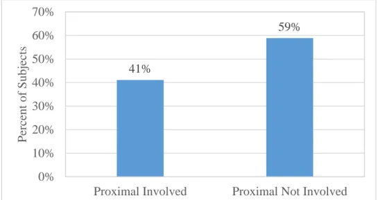
had exposure sites that involved the proximal surface of the tooth while 43 did not (Figure 5).
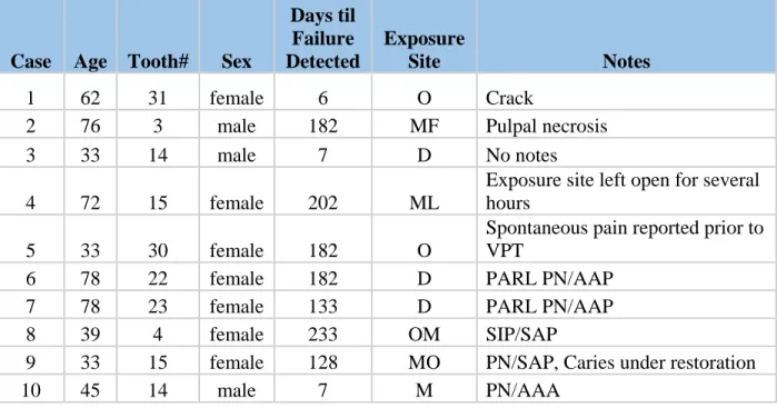
Failures were documented with notes indicating suspected cause of failure as well as the timespan
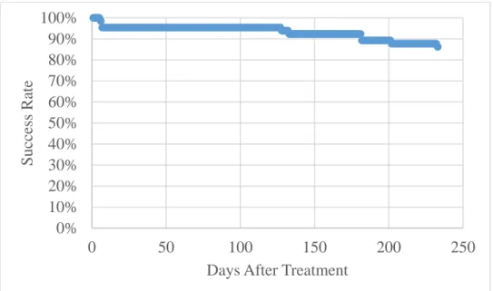
between when the VPT was completed and when the failure was detected (Table 1). 3 failures
were detected at 1 week, the next 2 failures did not occur until 4 months postoperatively. 2 failures
were detected at 6 months and 2 more at 7 and 8 months.
Using the generalized linear model with predictors – pain at 24 hours, pain at 1 week and
pain at 3 months, tooth type and patient’ age, pain was found to be significantly associated with
failure at 3 months (p=0.028). We also noted marginal significance between patients age and
failure (p=0.059) with failure rates being higher in older patients. On analyzing our data for an
association between exposure size and post-treatment pain, a marginal significance (p=0.0596)
19
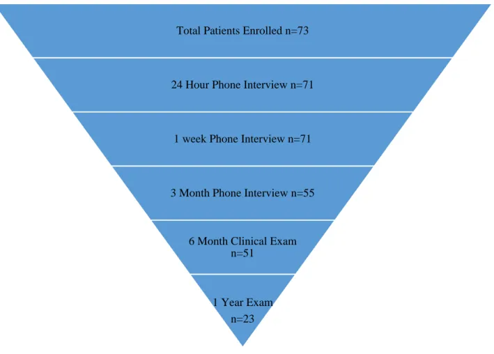
Figure 1: Subject Flow. 73 total subjects participated in the pulp capping procedure. 71 were available for 24-hour follow-up and 1-week follow-up. 55 patients were available for 3-month phone calls. 51 returned for a 6-month in person exam and 23 subjects were available for the 1-year exam.
Total Patients Enrolled n=73
24 Hour Phone Interview n=71
1 week Phone Interview n=71
3 Month Phone Interview n=55
6 Month Clinical Exam n=51
20
Figure 2: Age Group Distribution. Ages of subjects ranged from 15 to 78 years old with 32 subjects 40 years old and younger, and 41 subjects older than 40 years old.
Figure 3: Gender Distribution. A majority of patients were female with 33% (n=24) male and (n=49) 67% female.
12%
10%
25%
14%
13%
17%
14%
0% 5% 10% 15% 20% 25% 30%
15-20 20-30 30-40 40-50 50-60 60-70 70-80
Perc
ent of
Subject
s
Age
33%
67%
21
Figure 4: Tooth Types Treated. All tooth types were evaluated with a majority of cases being molars. There were 20 anterior teeth, 19 premolars, and 34 molars treated over the course of this study.
Figure 5: Proximal Surface Involvement. 30 cases had pulp exposures involving the proximal surface while 43 cases did not.
27% 26% 47% 0% 10% 20% 30% 40% 50% 60%
Anterior Premolar Molar
Perc
ent of
Subject
s
Type of Tooth
41% 59% 0% 10% 20% 30% 40% 50% 60% 70%
Proximal Involved Proximal Not Involved
Perc
ent of
Subject
22
Figure 6: Success Related to Time. Showing the number of success as related to time. 3 failures were detected at 1 week, the next 2 failures did not occur until 4 months postoperatively. 2 failures were detected at 6 months and 2 more at 7 and 8 months.
0% 10% 20% 30% 40% 50% 60% 70% 80% 90% 100%
0 50 100 150 200 250
Success
Rate
23 Case Age Tooth# Sex
Days til Failure Detected
Exposure
Site Notes
1 62 31 female 6 O Crack
2 76 3 male 182 MF Pulpal necrosis
3 33 14 male 7 D No notes
4 72 15 female 202 ML
Exposure site left open for several hours
5 33 30 female 182 O
Spontaneous pain reported prior to VPT
6 78 22 female 182 D PARL PN/AAP
7 78 23 female 133 D PARL PN/AAP
8 39 4 female 233 OM SIP/SAP
9 33 15 female 128 MO PN/SAP, Caries under restoration
10 45 14 male 7 M PN/AAA
Table 1: Distribution of Teeth. Details showing each failed case including any notes regarding findings during treatment. PARL=Periapical radiolucency, PN=Pulpal necrosis, AAP=Acute apical periodontitis, SIP=Symptomatic irreversible pulpitis, SAP=Symptomatic apical periodontitis, AAA=Acute apical abscess.
Section 1.5 Discussion:
Many clinicians believe that vital pulp therapy for teeth with carious exposures has a low
success rate (31). This is due in part to prior studies done with calcium hydroxide (68). Recent
studies based on tricalcium silicates report higher success rates in the range of 80%-90% (2, 34,
35). The purpose of the current study was to build upon prior studies to examine factors associated
with outcome of VPT when capped with tricalcium silicates. This study was not designed to
evaluate success rates but instead it focused on preoperative factors that may associate with
outcome.
The capping material we used in this study was MTA Angelus. While there are many
24
Studies comparing different formulations of MTA did not find any significant differences in the
pulpal responses (72). MTA Angelus has the beneficial therapeutic effects of traditional MTA with
the added benefit of a faster setting time. Traditional MTA takes up to 4 hours to set while MTA
Angelus was reported to set in 15 minutes (39). The quicker setting time allowed for placement of
the pulp cap and restoration in one appointment rather than two appointments thus removing the
risk of leakage through the temporary restoration.
Inability to obtain hemostasis from the exposure site was one of the exclusion criteria in
this study. Excess bleeding was used as a surrogate measure of inflammation based on prior studies
which report a 40% increase in blood flow in inflamed pulps (73). Systemic factors such as
medications, herbs, or systemic disease can cause excess bleeding as well. Medications and herbs
such as clopidogrel, rivaroxaban, and ginseng are becoming more popular. Systemic conditions
such as vitamin K deficiency is a factor as well. These can cause dramatic effects on the flow of
blood through the body (74). While these conditions can lead to increased bleeding, there are
factors that can reduce bleeding.
The local anesthetic commonly used prior to dental treatment contains epinephrine, a
vasoconstrictor. Epinephrine from the local anesthetic can affect regional blood vessels leading to
a reduction in blood flow of up to 40% compared to anesthetics without epinephrine (75).
Additionally, the patient’s age can affect blood flow. In older teeth, there is a general decrease in
the number of blood vessels (76). The size of the pulp chamber reduces in size with deposits of
calcifications in the root and coronal pulp. These factors as well as a partially necrotic pulp can
lead to less blood flow in the region. Even though we used hemorrhage as a measure of
25
This study evaluated tooth type and site of the exposure as a possible factor of outcome.
Since the size and configuration of each pulp chamber is different from one tooth type to another,
the two factors may influence the pulp’s ability to heal as suggested by several studies (77, 78).
The thickness of pulp immediately beneath the pulpal exposure site can vary. This variation can
play a role in the healing potential. For example, mandibular anterior teeth have narrow pulp
chambers mesiodistally compared to the labiolingual dimension. A pulp exposure on the proximal
surface would affect the pulp in an area that is thinner than a pulp exposure facially or lingually.
Although this is the case for mandibular incisors, it is quite the opposite in maxillary incisors.
Maxillary incisors typically have pulp chambers which are wider mesiodistally while being
narrower labiolingually. Additionally, as we move posteriorly, it becomes more difficult to isolate
and access certain areas. Interproximal areas in posterior teeth are typically significantly more
difficult to treat than the facial of an anterior tooth. This could affect the clinician’s ability to
properly adapt restorations or medicaments to the area thereby affecting the outcome.
The results of this study suggest that none of the preoperative factors examined are
correlated with failure of the treatment. Like many other studies, age was evaluated as a possible
factor influencing the outcome however we were unable to find any correlation. Other factors
examined including site of exposure as well as tooth type did not affect the outcome. There could
be three possibilities for this explanation. The number of subjects may not have been sufficent, the
number of failures may not have been at a level high enough to detect a difference, or there simply
may not be a significant association between these factors and outcome.
A goal of the procedure is to maintain the vitality of a healthy pulp. Obtaining the correct
pulpal diagnosis can be challenging (55, 79). A correct diagnosis is crucial in order to provide the
26
used to reproduce symptoms and help distinguish between reversible pulpitis, irreversible pulpitis,
and pulpal necrosis. This distinction is paramount since vital pulp therapy will only be effective in
reversible pulpitis and not the other two. It was traditionally believed that in cases of carious
exposures on mature permanent teeth, the pulp is irreversibly inflamed and there is little chance of
healing. Histologically, previous studies have indicated that although there is a pulpal exposure,
only the area immediately adjacent to the exposure site has irreversible damage (36). The other
areas have the potential to heal. In several of our cases that resulted in failure, some multi-rooted
teeth were found to have developed partial necrosis (Table 1). The tissue in one root canal was
necrotic while the rest still had vital tissue. This reinforces the notion that the pulp can
compartmentalize itself and try to separate diseased from healthy pulp tissue. Standard diagnostic
terms and techniques used today have limitations in practice. A diagnostic technique that may have
future potential is chairside molecular assessment of pulpitis.
We are not yet able to fully comprehend the changes of the pulp at the molecular level. We
know that specific mediators have been associated with inflammation such as irreversible pulpitis.
These mediators include elevation in CGRP, Substance P, Neurokinin A, TNF-α, bradykinin,
Prostaglandin E2 and F2 (80, 81). Although there are many other inflammatory mediators,
measuring a few key markers may potentially predict the outcome of vital pulp therapy (82). It is
possible that in the future instead of relying on the subjective endodontic testing we use today, we
can rely on a chairside sampling of mediators from the pulp for a clear diagnosis.
Conclusion:
Based on our findings, we confirmed that vital pulp therapy is a viable treatment. Age
27
moderate postoperative pain be managed with analgesics first, before additional treatment. If
28 DISCUSSION
Many clinicians believe that vital pulp therapy for teeth with carious exposures has a low
success rate (31). This is due in part to prior studies done with calcium hydroxide (68). Recent
studies based on tricalcium silicates report higher success rates in the range of 80%-90% (2, 34,
35). The purpose of the current study was to build upon prior studies to examine factors associated
with outcome of VPT when capped with tricalcium silicates. This study was not designed to
evaluate success rates but instead it focused on preoperative factors that may associate with
outcome.
The capping material we used in this study was MTA Angelus. While there are many
different formulations of tricalcium silicates, MTA is the most extensively studied to date (39).
Studies comparing different formulations of MTA did not find any significant differences in the
pulpal responses (72). MTA Angelus has the beneficial therapeutic effects of traditional MTA with
the added benefit of a faster setting time. Traditional MTA takes up to 4 hours to set while MTA
Angelus was reported to set in 15 minutes (39). The quicker setting time allowed for placement of
the pulp cap and restoration in one appointment rather than two appointments thus removing the
risk of leakage through the temporary restoration.
In many studies on VPT with MTA, the treatment was completed using a 2-step protocol
to confirm setting of the MTA (3, 34, 35, 83, 84). The first visit consisted of caries removal,
capping the pulp with MTA, placing a cotton pellet moistened with saline, and sealing the tooth
with a temporary restoration. After allowing several days for setting, the patient would return to
29
the tooth is then restored permanently. Temporary materials like the ones used for the 2-step
protocols have the potential to leak (85). Leakage from an inadequate seal violates one of the goals
of vital pulp therapy which is to prevent future leakage. Requiring a second visit is also time
consuming for the patient as well as the clinician. This study utilized a one-step approach to
treatment. Immediate sealing of the tooth with a permanent restoration has indicated better
outcome (3, 68). We did not believe setting of the MTA was a concern. Studies indicate moisture
content in the dentin as well as from the pulp provides sufficient moisture for setting of the material
(86). Prior studies utilizing a 2-step protocol indicated no difficulty in setting of the MTA
suggesting that a 2-step protocol is unnecessary (1, 34).
Inability to obtain hemostasis from the exposure site was one of the exclusion criteria in
this study. Excess bleeding was used as a surrogate measure of inflammation based on prior studies
which report a 40% increase in blood flow in inflamed pulps (73). Systemic factors such as
medications, herbs, or systemic disease can cause excess bleeding as well. Medications and herbs
such as clopidogrel, rivaroxaban, and ginseng are becoming more popular. Systemic conditions
such as vitamin K deficiency is a factor as well. These can cause dramatic effects on the flow of
blood through the body (74). While these conditions can lead to increased bleeding, there are
factors that can reduce bleeding.
The local anesthetic commonly used prior to dental treatment contains epinephrine, a
vasoconstrictor. Epinephrine from the local anesthetic can affect regional blood vessels leading to
a reduction in blood flow of up to 40% compared to anesthetics without epinephrine (75).
Additionally, the patient’s age can affect blood flow. In older teeth, there is a general decrease in
the number of blood vessels (76). The size of the pulp chamber reduces in size with deposits of
30
lead to less blood flow in the region. Even though we used hemorrhage as a measure of
inflammation, many factors can influence this characteristic.
This study evaluated tooth type and site of the exposure as a possible factor of outcome.
Since the size and configuration of each pulp chamber was different from one tooth type to another,
the two factors may influence the pulp’s ability to heal as suggested by several studies (77, 78).
The pulp thickness beneath the pulpal exposure site can vary. This variation can play a role in the
healing potential. For example, mandibular anterior teeth have very narrow pulp chambers
mesiodistally compared to the labiolingual dimension. A pulp exposure on the proximal site could
affect the pulp in an area that is thinner than a pulp exposure facially or lingually. Although this is
the case for mandibular incisors, it is quite the opposite in maxillary incisors. Maxillary incisors
typically have pulp chambers which are wider mesiodistally while being narrower labiolingually.
Additionally, as we move posteriorly, it becomes more difficult to isolate and access certain areas.
Interproximal areas in posterior teeth are typically significantly more difficult to treat than the
facial of an anterior tooth. This could affect the clinician’s ability to properly adapt restorations or
medicaments to the area thereby affecting the outcome.
Long term follow-up periods provide valuable information on treatment success. However,
this study only evaluates follow-up at 6 months and 1 year. Most failures of vital pulp therapy have
been suggested to occur in the first 3 months after the procedure and most will remain relatively
stable afterwards until 18 months (2). Many cases in this study that failed had the failures detected
in the first several days when the patient came back in severe pain. Failures at the 6 month
in-person examination were patients that only had mild or no discomfort. There was not enough data
31
underscore the importance of an in-person examination for follow-up at 6 months to ensure the
treatment was successful.
A previous study reported high success rates of the VPT for up to 9 years (33). This
indicates that vital pulp therapy can have long term results provided the treatment is rendered in
ideal circumstances. In that study, the procedures were performed by a single operator who was a
specialist. Preoperative testing was performed and all teeth treated had no previous restorations.
The study was also conducted on a younger age group with the mean age of 16 years old. Although
this study was well controlled, it does not reflect the reality of dental care.
Our study was designed in a way to reflect the realities of a dental clinic. In many cases, a
pulpal exposure was not evident until caries removal had already begun. In these cases, the patient
was already anesthetized rendering pulpal testing unavailable. This scenario reflects real world
dentistry where unanticipated pulp exposures occur without prior pulp testing. Although pulp
testing was unavailable, a thorough history of symptoms was reviewed with each patient. This
could still be accomplished after anesthesia is administered.
Vital pulp therapy could have a profound impact on the quality of life of patients especially
those in underserved areas. As mentioned previously, most practitioners recommend either root
canal therapy or extraction as options for carious pulpal exposures. Both have high costs overall
while vital pulp therapy is often unmentioned. This is an enormous disservice to patients especially
in clinics serving lower income patients. Vital pulp therapy can make a tremendous impact in these
areas where root canal therapy is not affordable by many patients and extraction is commonly
elected. Vital pulp therapy can satisfy the goal of preventing apical periodontitis and maintaining
32
The results of this study suggest that none of the preoperative factors examined are
correlated with the treatment’s outcome. Like many other studies, age was evaluated as a possible
factor influencing the outcome however we were unable to find any significant correlation. Other
factors examined including site of exposure as well as tooth type did not affect the outcome of the
procedure as well. There could be three possibilities for this explanation. The number of subjects
may not have been adequate, the number of failures may not have been at a level high enough to
detect a difference, or there simply may not be a significant association between these factors and
outcome. Future studies could add more to the literature by including more cases as well as for a
longer time period.
A goal of the procedure is to maintain the vitality of a healthy pulp. Obtaining the correct
pulpal diagnosis can be challenging (55, 79). A correct diagnosis is crucial in order to provide the
appropriate treatment plan and prognosis of the tooth. Standard endodontic tests used today are
used to reproduce symptoms and help distinguish between reversible pulpitis, irreversible pulpitis,
and pulpal necrosis. This distinction is paramount since vital pulp therapy will only be effective in
reversible pulpitis and not the other two. It was traditionally believed that in cases of carious
exposures on mature permanent teeth, the pulp is irreversibly inflamed and there is little chance of
healing. Histologically, previous studies have indicated that although there is a pulpal exposure,
only the area immediately adjacent to the exposure site has irreversible damage (36). The other
areas have the potential to heal. In several of our cases that resulted in failure, some multi-rooted
teeth were found to have developed partial necrosis (Table 1). The tissue in one root canal was
necrotic while the rest still had vital tissue. This reinforces the notion that the pulp can
33
terms and techniques used today have limitations in practice. A diagnostic technique that may have
future potential is chairside molecular assessment of pulpitis.
Our study measures success by both clinical and radiographic signs. One study that is
commonly quoted as evidence vital pulp therapy reports a 10-year success rate of 13% (68).
Interestingly, it considers success only to be in teeth that respond to both thermal testing and
electric pulp testing. This may be too strict a measure of success because clinicians rarely require
a positive response to both tests in order to rule our disease (36). Our study, like many others, only
requires a positive response to either cold or EPT. If the tooth responds to one or the other, it
indicates that there is vital tissue which is one of the goals.
We are not yet able to fully comprehend the changes of the pulp at the molecular level. We
know that specific mediators have been associated with inflammation such as irreversible pulpitis.
These mediators include elevation in CGRP, Substance P, Neurokinin A, TNF-α, bradykinin,
Prostaglandin E2 and F2 (80, 81). Although there are many other inflammatory mediators,
measuring a few key markers may potentially predict the outcome of vital pulp therapy (82). It is
possible that in the future instead of relying on the subjective endodontic testing we use today, we
34
REFERENCES
1. Caliskan MK, Guneri P. Prognostic factors in direct pulp capping with mineral trioxide aggregate or calcium hydroxide: 2- to 6-year follow-up. Clin Oral Investig 2016.
2. Cho SY, Seo DG, Lee SJ, Lee J, Lee SJ, Jung IY. Prognostic factors for clinical outcomes according to time after direct pulp capping. J Endod 2013;39(3):327-331.
3. Mente J, Hufnagel S, Leo M, Michel A, Gehrig H, Panagidis D, et al. Treatment outcome of mineral trioxide aggregate or calcium hydroxide direct pulp capping: long-term results. J Endod 2014;40(11):1746-1751.
4. Kakehashi S, Stanley HR, Fitzgerald RJ. The Effects of Surgical Exposures of Dental Pulps in Germ-Free and Conventional Laboratory Rats. Oral Surg Oral Med Oral Pathol 1965;20:340-349.
5. Moller AJ, Fabricius L, Dahlen G, Ohman AE, Heyden G. Influence on periapical tissues of indigenous oral bacteria and necrotic pulp tissue in monkeys. Scand J Dent Res
1981;89(6):475-484.
6. Fluoridation USDoHaHSFPoCW. U.S. Public Health Service Recommendation for Fluoride Concentration in Drinking Water for the Prevention of Dental Caries. Public Health Reports 2015;130.
7. Richardson S. Access to Endodontic Care in North Carolina Public Health and Medicaid Settings. 2012.
8. Ritwik P, Massey C, Hagan J. Epidemiology and outcomes of dental trauma cases from an urban pediatric emergency department. Dent Traumatol 2015;31(2):97-102.
9. Hahn CL, Liewehr FR. Update on the adaptive immune responses of the dental pulp. J Endod 2007;33(7):773-781.
10. Hahn CL, Liewehr FR. Innate immune responses of the dental pulp to caries. J Endod 2007;33(6):643-651.
35
12. Nagaoka S, Miyazaki Y, Liu HJ, Iwamoto Y, Kitano M, Kawagoe M. Bacterial invasion into dentinal tubules of human vital and nonvital teeth. J Endod 1995;21(2):70-73.
13. Knutsson G, Jontell M, Bergenholtz G. Determination of plasma proteins in dentinal fluid from cavities prepared in healthy young human teeth. Arch Oral Biol 1994;39(3):185-190.
14. Hahn CL, Best AM. The pulpal origin of immunoglobulins in dentin beneath caries: an immunohistochemical study. J Endod 2006;32(3):178-182.
15. Lanzavecchia A. Receptor-mediated antigen uptake and its effect on antigen presentation to class II-restricted T lymphocytes. Annu Rev Immunol 1990;8:773-793.
16. Van Hassel HJ. Physiology of the human dental pulp. Oral Surg Oral Med Oral Pathol 1971;32(1):126-134.
17. Peters OA PC. Cleaning and Shaping of the Root Canal System. Pathways of the Pulp. 10th Edition 2011:283-348.
18. Marquis VL, Dao T, Farzaneh M, Abitbol S, Friedman S. Treatment outcome in endodontics: the Toronto Study. Phase III: initial treatment. J Endod 2006;32(4):299-306.
19. Peters OA, Schonenberger K, Laib A. Effects of four Ni-Ti preparation techniques on root canal geometry assessed by micro computed tomography. Int Endod J 2001;34(3):221-230.
20. de Chevigny C, Dao TT, Basrani BR, Marquis V, Farzaneh M, Abitbol S, et al. Treatment outcome in endodontics: the Toronto study--phase 4: initial treatment. J Endod 2008;34(3):258-263.
21. Caplan DJ, Cai J, Yin G, White BA. Root canal filled versus non-root canal filled teeth: a retrospective comparison of survival times. J Public Health Dent 2005;65(2):90-96.
22. Ou KL, Chang CC, Chang WJ, Lin CT, Chang KJ, Huang HM. Effect of damping properties on fracture resistance of root filled premolar teeth: a dynamic finite element analysis. Int Endod J 2009;42(8):694-704.
36
24. Goldstein S, Sedaghat-Zandi A, Greenberg M, Friedman S. Apexification & apexogenesis. N Y State Dent J 1999;65(5):23-25.
25. Cvek M. A clinical report on partial pulpotomy and capping with calcium hydroxide in permanent incisors with complicated crown fracture. J Endod 1978;4(8):232-237.
26. Dannenberg JL. Pedodontic endodontics. Dent Clin North Am 1974;18(2):367-377.
27. Nirschl RF, Avery DR. Evaluation of a new pulp capping agent in indirect pulp therapy. ASDC J Dent Child 1983;50(1):25-30.
28. Stanley HR. Criteria for standardizing and increasing credibility of direct pulp capping studies. Am J Dent 1998;11 Spec No:S17-34.
29. Ørstavik D, Pitt Ford TR. Essential endodontology : prevention and treatment of apical periodontitis. Oxford ; Malden, MA: Blackwell Science; 1998.
30. Cohen S, Burns RC. Pathways of the pulp. 6th ed. St. Louis: Mosby; 1994.
31. Ward J. Vital pulp therapy in cariously exposed permanent teeth and its limitations. Aust Endod J 2002;28(1):29-37.
32. Strindberg LZ. The dependence of the results of pulp therapy on certain factors; an analytic study based on radiographic and clinical follow-up examinations. [Tr. from the Swedish manuscript]. Stockholm,; 1956.
33. Bogen G, Kim JS, Bakland LK. Direct pulp capping with mineral trioxide aggregate: an observational study. J Am Dent Assoc 2008;139(3):305-315; quiz 305-315.
34. Marques MS, Wesselink PR, Shemesh H. Outcome of Direct Pulp Capping with Mineral Trioxide Aggregate: A Prospective Study. J Endod 2015;41(7):1026-1031.
35. Jang Y, Song M, Yoo IS, Song Y, Roh BD, Kim E. A Randomized Controlled Study of the Use of ProRoot Mineral Trioxide Aggregate and Endocem as Direct Pulp Capping Materials: 3-month versus 1-year Outcomes. J Endod 2015;41(8):1201-1206.
37
37. Watts A, Paterson RC. The response of the mechanically exposed pulp to prednisolone and triamcinolone acetonide. Int Endod J 1988;21(1):9-16.
38. Zander HA, Glass RL. The healing of phenolized pulp exposures. Oral Surg Oral Med Oral Pathol 1949;2(6):803-810.
39. Komabayashi T, Zhu Q, Eberhart R, Imai Y. Current status of direct pulp-capping materials for permanent teeth. Dent Mater J 2016;35(1):1-12.
40. Rasmussen P, Mjor IA. Calcium hydroxide as an ectopic bone inductor in rats. Scand J Dent Res 1971;79(1):24-30.
41. Tronstad L. Reaction of the exposed pulp to Dycal treatment. Oral Surg Oral Med Oral Pathol 1974;38(6):945-953.
42. Cox CF, Subay RK, Ostro E, Suzuki S, Suzuki SH. Tunnel defects in dentin bridges: their formation following direct pulp capping. Oper Dent 1996;21(1):4-11.
43. Goldberg F, Massone EJ, Spielberg C. Evaluation of the dentinal bridge after pulpotomy and calcium hydroxide dressing. J Endod 1984;10(7):318-320.
44. Witherspoon DE, Small JC, Harris GZ. Mineral trioxide aggregate pulpotomies: a case series outcomes assessment. J Am Dent Assoc 2006;137(5):610-618.
45. Schroder U. Effects of calcium hydroxide-containing pulp-capping agents on pulp cell migration, proliferation, and differentiation. J Dent Res 1985;64 Spec No:541-548.
46. Murray PE, Hafez AA, Smith AJ, Cox CF. Hierarchy of pulp capping and repair activities responsible for dentin bridge formation. Am J Dent 2002;15(4):236-243.
47. Torabinejad M, Hong CU, McDonald F, Pitt Ford TR. Physical and chemical properties of a new root-end filling material. J Endod 1995;21(7):349-353.
48. Song M, Kang M, Kim HC, Kim E. A randomized controlled study of the use of ProRoot mineral trioxide aggregate and Endocem as direct pulp capping materials. J Endod
38
49. Camilleri J. Mineral trioxide aggregate: present and future developments. Endodontic Topics 2015;32(1):31-46.
50. Ford TR, Torabinejad M, Abedi HR, Bakland LK, Kariyawasam SP. Using mineral trioxide aggregate as a pulp-capping material. J Am Dent Assoc 1996;127(10):1491-1494.
51. Holland R, de Souza V, Nery MJ, Otoboni Filho JA, Bernabe PF, Dezan Junior E. Reaction of rat connective tissue to implanted dentin tubes filled with mineral trioxide aggregate or calcium hydroxide. J Endod 1999;25(3):161-166.
52. Torabinejad M, Rastegar AF, Kettering JD, Pitt Ford TR. Bacterial leakage of mineral trioxide aggregate as a root-end filling material. J Endod 1995;21(3):109-112.
53. Martell B, Chandler NP. Electrical and dye leakage comparison of three root-end restorative materials. Quintessence Int 2002;33(1):30-34.
54. Dammaschke T. The history of direct pulp capping. J Hist Dent 2008;56(1):9-23.
55. Dummer PM, Hicks R, Huws D. Clinical signs and symptoms in pulp disease. Int Endod J 1980;13(1):27-35.
56. Garfunkel A, Sela J, Ulmansky M. Dental pulp pathosis. Clinicopathologic correlations based on 109 cases. Oral Surg Oral Med Oral Pathol 1973;35(1):110-117.
57. Seltzer S. Hypothetic mechanisms for dentine sensitivity. Oral Surg Oral Med Oral Pathol 1971;31(3):388-399.
58. Brannstrom M. The hydrodynamics of the dental tubule and pulp fluid: its significance in relation to dentinal sensitivity. Annu Meet Am Inst Oral Biol 1966;23:219.
59. Corpron RE, Avery JK. Ultrastructure of odontoblasts in dentinal tubules of mice. J Dent Res 1971;50(2):511.
60. Trowbridge HO, Franks M, Korostoff E, Emling R. Sensory response to thermal stimulation in human teeth. J Endod 1980;6(1):405-412.
39
62. Degering CI. Physiologic evaluation of dental-pulp testing methods. J Dent Res 1962;41:695-700.
63. Bender IB, Seltzer S. Roentgenographic and direct observation of experimental lesions in bone: II. 1961. J Endod 2003;29(11):707-712; discussion 701.
64. Bender IB, Seltzer S. Roentgenographic and direct observation of experimental lesions in bone: I. 1961. J Endod 2003;29(11):702-706; discussion 701.
65. Eversole LR, Stone CE, Strub D. Focal sclerosing osteomyelitis/focal periapical osteopetrosis: radiographic patterns. Oral Surg Oral Med Oral Pathol 1984;58(4):456-460.
66. McDougal RA, Delano EO, Caplan D, Sigurdsson A, Trope M. Success of an alternative for interim management of irreversible pulpitis. J Am Dent Assoc 2004;135(12):1707-1712.
67. EW H, Stanley H, J C, H S. Direct pulp capping treatment : a long-term follow-up. JADA 1978;97(4):5.
68. Barthel CR, Rosenkranz B, Leuenberg A, Roulet JF. Pulp capping of carious exposures: treatment outcome after 5 and 10 years: a retrospective study. J Endod 2000;26(9):525-528.
69. Maryniuk GA, Haywood VB. Placement of cast restorations over direct pulp capping procedures: a decision analytic approach. J Am Dent Assoc 1990;120(2):183-187.
70. Pitt Ford TR. Pulpal response to a calcium hydroxide material for capping exposures. Oral Surg Oral Med Oral Pathol 1985;59(2):194-197.
71. Matsuo T, Nakanishi T, Shimizu H, Ebisu S. A clinical study of direct pulp capping applied to carious-exposed pulps. J Endod 1996;22(10):551-556.
72. Kang CM, Sun Y, Song JS, Pang NS, Roh BD, Lee CY, et al. A randomized controlled trial of various MTA materials for partial pulpotomy in permanent teeth. J Dent 2016.