Jennifer Ann Rutan
A dissertation submitted to the faculty of the University of North Carolina at Chapel Hill in partial fulfillment of the requirements for the degree of Doctor of Philosophy in the Department of Microbiology and Immunology
Chapel Hill 2008
Approved by:
Jennifer Ann Rutan: Regulation of autoreactive B cells during innate immune responses (Under the direction of Dr. Barbara J. Vilen)
B cells to respond to threats while potentially autoreactive B cells remain quiescent. We have recently described a mechanism where antigen-experienced B cells are regulated by IL-6 and sCD40L released by dendritic cells (DCs) and macrophages (MΦs) during innate immune responses. Naïve B cells are unaffected by these factors, permitting a robust immune response in the absence of autoimmunity. IL-6/sCD40L-mediated repression is dependent on ERK activation and “repressed” antigen-experienced cells
To Annie, Bullet, Jack, Star, and Fancy, for their comfort and undying loyalty.
I would like to thank my advisor, Dr. Barbara Vilen, for the training she has provided in the practice of science, critical analysis of data, writing of manuscripts, and management of a laboratory and its members. I have learned a great deal from my time in her laboratory, and I appreciate the additional writing and editing opportunities that she provided. I am also grateful to my other graduate school mentors, particularly the
members of my thesis committee, Drs. Steve Clarke, Jeff Frelinger, Larry Arnold, and Jenny Ting, as well as Dr. Zhi Liu. I would also like to thank the past and present members of the Vilen Lab for their scientific and moral support as well as their friendship, especially Heather Mueller Rauscher, Jin Kim, Mileka Gilbert, Diane Carnathan, Michelle Kilmon, Bianca Trollinger, Nikki Wagner, Stephanie Carmicle-Davis, Shannon Jones, and Sang-Ryul Lee. You have all taught me about the importance of good life choices.
My family has always supported me in my educational pursuits, and I would like to thank them for their love and encouragement. In particular, I would like to thank my parents for their sacrifices, my brother for his optimism and humor, my grandparents for their unconditional faith in me, and Uncle Joe for being my scientific role model (and giving me my first microscope!). I love you all.
TABLE OF CONTENTS
LIST OF TABLES………...………...x
LIST OF FIGURES……….………..…….…………xi
LIST OF ABBREVIATIONS AND SYMBOLS………..xii
CHAPTER I: Introduction………...1
1.1. Learning tolerance: a B cell’s story…...………..2
1.2. A short history of Sm………...………3
1.3. Arresting and silencing Sm-specific B cells………...…….5
1.4. Failed tolerance: Sm-specific autoantibodies in a murine model of SLE...….9
1.5. Apoptotic cells: dangerous in death………...………11
1.6. Selective repression: new treatments for SLE?...14
1.7. Summary………..……….……….…15
1.8. References…………..……….………...17
1.9. Figures………32
CHAPTER II: ERK prevents differentiation of autoreactive B cells during innate immune responses………33
2.1. Abstract………...……...34
2.2. Introduction………...…….35
2.5. Results….………...………...….43
2.5. Discussion………...………...48
2.6. References…………...………...52
2.7. Figures………57
LIST OF TABLES
LIST OF FIGURES
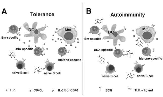
Figure 1.1. DCs and MΦs repress antibody secretion from
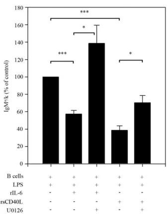
autoreactive B cells via IL-6 and sCD40L.……….…….…32 Figure 2.1. IL-6 and sCD40L selectively repress Ig secretion by
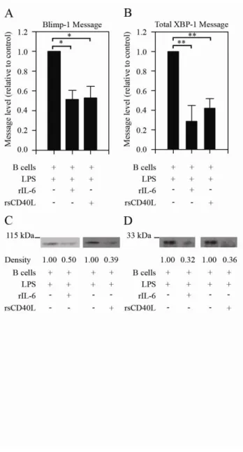
chronically antigen-experienced B cells………..57 Figure 2.2. IL-6 and sCD40L treatment causes a decrease in levels
of transcription factors involved in plasma cell differentiation…………...59 Figure 2.3. IL-6- and sCD40L-mediated repression is dependent on
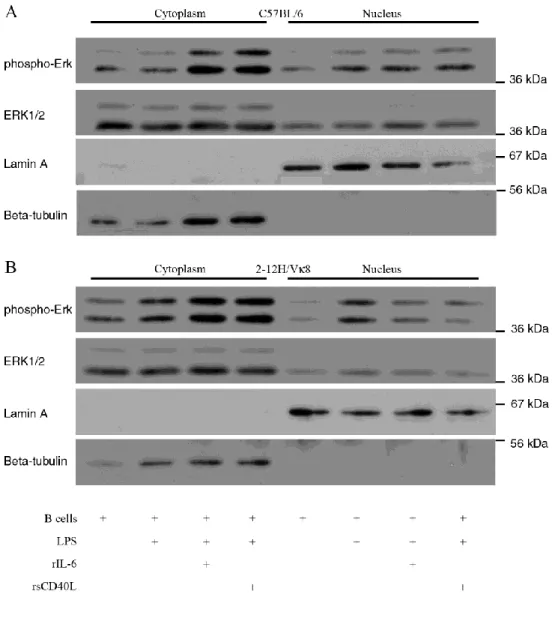
Erk activation………...60 Figure 2.4. Chronically antigen-experienced 2-12H/Vκ8 B cells exhibit
higher basal phosphorylation of Erk than naïve C57BL/6 B cells………...61 Figure 2.5. IL-6 and sCD40L alter the subcellular localization of
LIST OF ABBREVIATIONS AND SYMBOLS
BCR B cell receptor
BLIMP-1 B lymphocyte induced maturation protein 1 BLyS B lymphocyte stimulator
BMT nonmyeloablative bone marrow transplant
DC dendritic cell
dsDNA double-stranded DNA DTT dithiothreitol
ERK extracellular signal-related kinase
FcR Fc receptor
FO follicular
HEL hen egg lysozyme
HSCT nonmyeloablative hematopoietic stem cell transplant
IC immune complex
Ig immunoglobulin
IL-4 interleukin-4
IL-6 interleukin-6
IL-6R interleukin-6 receptor (CD126) KSR kinase suppressor of Ras
LPS lipopolysaccharide
MAPK mitogen-activated protein kinase MEF mouse embryonic fibroblast
MΦ macrophage
MZ marginal zone
NES nuclear export signal NLS nuclear localization signal
PAMP pathogen-associated molecular pattern PI3-K phosphatidylinositol 3-kinase
PMSF phenylmethanesulfonyl fluoride
RF rheumatoid factor
sCD40L soluble CD40L
SLE systemic lupus erythematosus
Sm Smith antigen
snRNA small nuclear RNA
snRNP small nuclear ribonucleoproteins ssRNA single-stranded RNA
Tg transgenic
Introduction
This review was originally published in Immunologic Research. Vilen, B.J., and Rutan, J.A.
The regulation of autoreactive B cells during innate immune responses. Immunologic Research. 2008;41(3):295-309
1.1 Learning tolerance: a B cell’s story
A diverse B cell repertoire is critical in combating pathogens, but inherent in generating diversity is the threat of autoimmunity. In the bone marrow, central tolerance mechanisms such as deletion or receptor editing remove high affinity autoreactive B cells before they exit to the periphery (1-10). Those that escape are subject to receptor
revision (8, 11-14), peripheral deletion (15, 16), or a shortened lifespan because they fail to enter B cell follicles (17-22). In rare cases, autoreactive B cells are fully functional but indifferent to their specific antigen (23-25). Finally, many low affinity autoreactive B cells are maintained in an unresponsive state known as anergy. Anergic B cells do not receive sufficient activation signals to differentiate into plasma cells or secrete
immunoglobulin (Ig) in response to antigenic or mitogenic stimulation (26-29). Their proliferative responses to B cell receptor (BCR) or toll-like receptor (TLR) signaling as well as their lifespans vary in different models (18, 30-35). Some anergic B cells transduce BCR-derived signals (30, 32, 36-40), while others exhibit desensitized BCRs (30, 33, 41). Quiescence is dependent on chronic exposure to self-antigen and occupancy of the BCR (42, 43). Furthermore, ERK activation is critical in sustaining anergy during polyclonal stimulation by TLR ligands (27, 28). Recently, it was demonstrated that anergy can be mediated by DCs and MΦs. In the presence of TLR ligands, DCs and MΦs release IL-6 and sCD40L that selectively represses Ig secretion from autoreactive B
The affinity and avidity of the antigen-BCR interaction determines whether developing B cells will be deleted, edited, anergized, or ignored (46). Self-reactive B cells that survive these developmental checkpoints tend to bind self-antigens with low affinity and they remain anergic in the absence of costimulation by cognate T cells. Many of the early transgenic (Tg) models expressed BCRs with high affinities.
However, concerns were raised that these models may not adequately reflect how bona fide low affinity self-antigens are regulated (1, 26). To study the tolerance mechanisms regulating low-affinity B cells, new Ig Tg models were generated that expressed BCRs specific for double-stranded (ds) DNA, single-stranded (ss) DNA, rheumatoid factor (RF), insulin, and Smith antigen (Sm).
1.2 A short history of Sm
Sm antigens are conserved proteins that are indispensable in RNA splicing. In 1966, Sm was identified as a unique autoantigen, the first non-histone target of
autoantibodies in systemic lupus erythematosus (SLE) patients (47). Named for 15-year-old Stephanie Smith, antibodies to Sm became one of the diagnostic criteria for SLE (48), detectable in 5-30% of SLE patients (48, 49). Later studies correlated anti-Sm titers with kidney disease (50-53).
pre-mRNA in the 5’ splice site region (56). This interaction stabilizes the commitment
complex formed when U1 snRNP binds the pre-mRNA substrate (57). The remaining snRNPs, U2 and U4/U5/U6 (the triple snRNP) bind to pre-mRNA, rearrange to form the splicesome, and remove introns from the transcript (58).
Self-antigens composed of protein and DNA or RNA can co-ligate the BCR with TLRs, potentially overcoming tolerance mechanisms and activating autoreactive B cells. For example, concomitant ligation of the BCR and TLR9 by chromatin:IgG immune complexes activates RF-specific B cells in the absence of T cell help or overt TLR stimulation (38, 59, 60). Signaling through both the BCR and TLR9 is necessary to activate NF-κB in these B cells (37). Recently, RNA-IgG immune complexes were shown to activate RF-specific B cells via TLR7 (39). TLR7 binds ssRNA, a viral antigen that acts as a sensor of infection as well as a component of the U1 snRNP splicing
complex, a known autoantigen in SLE (61, 62). Aberrantly high expression of TLR7, or an increased burden of immune complexes and/or apoptotic cells, aggravates disease in lupus-prone mice. For instance, the gene duplication of TLR7 in Yaa mice results in hyperactive B cells, exacerbation of disease in lupus-prone models, and shifts
1.3 Arresting and silencing Sm-specific B cells
Sm-specific autoantibodies are a hallmark of both human and murine lupus. To identify the mechanisms that regulate Sm-specific B cells, the 2-12H Tg mice were generated (35, 65). In this model, an Ig heavy chain, 2-12H, was identified from an Sm-specific hybridoma derived from an MRL/lpr mouse. The 2-12H chain pairs with a variety of light chains, giving rise to B cells specific for Sm and/or ss-DNA. B cells from the 2-12H model express BCRs of multiple affinities that develop and are regulated on a non-autoimmune background.
Tolerance to Sm is dependent on several cell types. B cells are the most obvious suspects in SLE since disease pathology is mediated by autoantibodies. In vivo, Sm-specific B cells are regulated since 2-12H Tg mice have low titers of anti-Sm antibodies (35, 66). However, ex vivo non-subsetted 2-12H B cells (uncontaminated by DCs and MΦs) are activated by TLR stimulation (LPS, CpG, dsRNA) in vitro but their Ig
secretion is lower than that of C57BL/6 controls (35, 67). The follicular (FO) B cell subset is repressed by DCs and MΦs secreting IL-6 and sCD40L, while secretion by the MZ B cell subset is partially repressed, but only by MΦs and sCD40L (45). Some MZ B
cells and peritoneal B-1 cells ignore endogenous levels of Sm, but an increase in the number of apoptotic cells can activate peritoneal and MZ B cells (66, 68, 69). Sm-specific B cells arrested at the pre-plasma cell stage, interrupting plasma cell differentiation and preventing Ig secretion (32)
this mouse are distributed among splenic transitional, FO, and MZ subsets, as well as the peritoneal B-1 subset (70). 2-12H/Vκ4 B cells are anergic and all subsets are
hyporesponsive to LPS in vitro. Additionally, MZ B cells exhibit a block in BCR signaling (70). LPS-stimulated 2-12H/Vκ4 B cells are repressed by IL-6 and sCD40L (unpublished data). To study low-affinity anti-Sm responses, the 2-12H/Vκ8 Tg mouse was created (32). In this model, only transitional and FO B cells are present and these cells are regulated by anergy (32). As in the previous anti-Sm models, 2-12H/Vκ8 B cells are susceptible to IL-6- and sCD40L-mediated repression (44, 45).
T cells are implicated in SLE and Sm-specific T cells are present in the repertoires of both normal and autoimmune mice (71). Sm-specific T cells in 2-12H Tg mice are anergic and do not proliferate in response to B cells presenting Sm (71). Anergic T cells are also unable to upregulate CD40L and provide costimulation to their cognate B cells (72). Anti-Sm B cells do not secrete Ig in vivo (35, 65), perhaps because they are deprived of T cell costimulation. However, in autoimmune situations, autoreactive T cells induce class-switching and somatic hypermutation of anti-Sm B cells, resulting in high levels of pathogenic high-affinity IgG autoantibodies (73, 74). Paradoxically, anti-Sm B cells are required to tolerize anti-Sm-specific T cells from C57BL/6 mice, but they activate Sm-specific T cells from MRL/lpr mice (71, 75, 76). This indicates that although T cells are necessary for the development of autoantibodies and disease, they are also regulated by autoreactive B cells in normal individuals.
DCs and MΦs regulate innate and adaptive immune responses by tolerizing or
drive adaptive immune response. The activation of DCs during innate immune responses induces the secretion of IL-6 that promotes immunity by releasing CD4+ T-helper cells from their inhibitory functions (77). This promotes polyclonal activation of naive B cells, the production of neutralizing antibody and the clearance of the invading pathogen.
In addition to regulating T cells, DCs and MΦs affect the fate of B cells. They activate naïve B cells by secreting type I interferon, IL-6, and B lymphocyte stimulator (BLyS) (78-80). They also repress Ig secretion by B cells that have been chronically exposed to antigen. Our laboratory showed that DCs and MΦs regulate autoantibody production, in part through their secretion of IL-6 and sCD40L (44, 45). Repression is selective in that naïve B cells (not chronically exposed to antigen) are unaffected by the presence of IL-6 and sCD40L while Ig secretion by autoreactive B cells is repressed (Figure 1A). Coupled with the data showing that IL-6 de-represses regulatory T cells, a mechanism emerges explaining how the pleiotropic affects of IL-6 produced by DCs and MΦs simultaneously promotes immunity and represses autoimmunity during innate
immune responses.
Signal transduction through many cell surface receptors influences neighboring receptors. For example, crosstalk between the IFN-αβ and IL-6 receptor (IL-6R) signaling pathways augment transcription factor binding and gene expression in mouse embryonic fibroblasts (MEFs) (81). Similarly, stimulation of B cells with sCD40L, IL-4, and LPS reprograms the BCR signaling pathway, enhancing ERK activation and
bypassing the requirement for phosphatidylinositol 3-kinase (PI3-K) (82-86). The
outcome of 6R and CD40 signal transduction. On a molecular level, the ability of IL-6 and sCD40L to repress Ig secretion reflects diminished BLIMP-1 and XBP-1 mRNA and protein levels. These data indicate that regulation occurs upstream of transcriptional activation. In support of this, pharmacologically inhibiting MEK restores LPS-induced Ig secretion. This suggests that the ability of IL-6/sCD40L to repress TLR4-induced Ig secretion is MEK/ERK-dependent (unpublished data).
Susceptibility of B cells to IL-6/sCD40L requires that B cells be chronically exposed to antigen, consistent with a central role for the BCR in tolerance. High affinity neo-antigens direct a unique tolerance scheme compared to low-affinity
self-antigens. Anergic B cells from high affinity models are characterized by elevated phospho-ERK (42, 87). In addition, the binding of high affinity antigen to the BCR and constitutive MEK/ERK activation is sufficient to repress TLR4 and TLR9-induced Ig secretion (27, 28). In the low affinity Sm model (2-12H/Vκ8) basal phospho-ERK levels are comparable to those in the HEL model. However, unlike the HEL model, the binding of soluble SmD or snRNPs coupled with elevated phospho-ERK levels does not repress TLR-induced Ig secretion (unpublished data). This indicates that ERK is only part of the “BCR-derived” signals that regulate innate immune responses in vivo. Although B cells
The finding that DCs secrete IL-6 while MΦs secrete IL-6 and sCD40L suggests that the anatomical location within the secondary lymphoid organs might dictate how autoreactive B cells are regulated. Marginal zone B cells are solely repressed by sCD40L while FO B cells are repressed by IL-6 and sCD40L (45). This specificity may result from the anatomic location, since different subsets of DCs and MΦs localize to specific regions of the spleen. For example, B cells are retained in the marginal zone by MΦs (88) and they are regulated by MΦ-derived sCD40L. However, upon activation and differentiation into pre-plasma cells, they may become susceptible to repression by IL-6 secreted by DCs within the periarteriolar lymphoid sheath (PALS). Thus, depending on the anatomical location of the autoreactive B cell, DCs and/or MΦs can repress
autoantibody production during innate immune responses.
The presence of class-switched autoantibodies in MRL/lpr mice suggests a breakdown in tolerance within the adaptive immune response. In MRL/lpr mice where somatic hypermutation and isotype switch recombination are blocked (AID-/-), lupus-like symptoms such as glomerulonephritis, proteinuria, and immune complex deposition are ameliorated (94). Early studies showed a critical role for T cells in disease because thymectomized MRL/lpr mice failed to develop lupus-like disease (95). Subsequently, it was shown that defects in central deletion and the number and function of T-regulatory cells allow CD4+ T-helper cells to activate autoreactive B cells, induce terminal
differentiation and autoantibody production (96-101).
Dysregulation of the innate immune system is apparent in MRL/lpr mice (39). Immune complexes containing RNA or chromatin stimulate RF-specific B cells through TLR7 and TLR9 to secrete anti-Sm and anti-chromatin (38, 39). Consistent with a role for TLRs, autoantibody responses were reduced in MyD88-/-/ MRL/lpr mice (39) and disease was ameliorated (102). TLR7-/-/ MRL/lpr mice exhibit reduced autoantibody titer and gene duplication of TLR7 shifts autoantibody specificities toward RNA, exacerbating disease (63, 64). This reveals TLR7 as a key receptor in promoting autoimmunity when tolerance is overcome (63, 103). Like TLR7-/- mice, TLR9-/-/ MRL/lpr mice exhibited lower titers of anti-DNA autoantibodies. However unlike TLR7-/- mice, they remain plagued by accelerated kidney disease and increased mortality (103). These data suggest that TLR9 induces anti-DNA responses but also has an anti-inflammatory effect, possibly through its induction of regulatory T cells (104, 105). The function of regulatory T cells in TLR9-/-/MRL/lpr mice is impaired, potentially allowing autoreactive cells to
tremendous and opposing effects on autoantibody production and disease in MRL/lpr mice.
Defects in DC/MΦ-mediated tolerance are evident in lupus-prone mice. Our
studies indicate that secretion of IL-6 and sCD40L by DCs/MΦs, as well as
reprogramming of IL-6R and CD40 in autoreactive B cells, promote tolerance during innate immune responses. This implies that defects in either the secretion of IL-6/sCD40L or the selective response of autoreactive B cells to these factors regulate autoantibody production. Our studies indicate that DCs and MΦs from MRL/lpr mice
are unable to repress autoreactive B cells (Figure 1B). Defects in DC/MΦ-mediated repression are coincident with diminished secretion of IL-6 and sCD40L and failure to sustain IL-6 mRNA production and activation of the IκB/NFκB pathways (107). Similarly, we found that IL-6 and sCD40L fail to repress B cells from lupus-prone mice (unpublished data). The finding that lupus-prone mice harbor defects in DCs/MΦs and B cells suggests the influence of an environmental stimulus.
1.5 Apoptotic cells: dangerous in death
of self-antigens on apoptotic cells is necessary to develop autoantibodies. For instance, autoantibodies to cytoplasmic but not nuclear antigens develop when nuclear
fragmentation is blocked in apoptotic cells (115). Therefore, it is imperative that scavenger cells such as MΦs and DCs quickly clear apoptotic cells and their associated self-antigens to minimize inflammatory and autoimmune responses.
Apoptotic cells are bound and cleared by a variety of receptors on DCs and MΦs. Ingestion of apoptotic cells reduces the secretion of proinflammatory cytokines by DCs and MΦs, inhibiting their maturation and controlling the activation of T cells and
autoimmune responses to apoptotic cells (116-121). Decreased clearance of apoptotic cells and defects in phagocytosis by MΦs in SLE patients and lupus-prone mice may lead to autoimmunity (69, 108, 122-124). The increase in apoptotic cells and antigens could dysregulate DCs and MΦs, triggering a chronic anti-inflammatory response that
downregulates the production of cytokines important for B cell tolerance (45, 107). If apoptotic cells fail to be cleared efficiently, they may become necrotic and expel self-antigens and TLR ligands that are opsonized by autoantibodies, forming immune complexes (125, 126). Like apoptotic cells, immune complexes (ICs) present self-antigens in a multimeric form and enhance phagocytosis by DCs and MΦs (127, 128). While apoptotic cells stimulate anti-inflammatory responses, ICs bind to Fc
receptors (FcRs) and elicit inflammatory responses from MΦs and DCs (128). Individual FcRs have distinct activating and inhibitory functions to ensure balance in a normal immune system. Activating FcRs include FcγRI and FcγRIII, which phagocytose ICs and provoke inflammatory responses from DCs and MΦs (129-131). These receptors are
Co-ligation of TLR9 and FcγRIII by chromatin:IC induces more DC activation than Co-ligation of FcγRIII alone, indicating that TLRs synergize with FcR signaling (135). In contrast to the activating FcRs, FcγRIIB is an inhibitory FcR with various functions depending on cell type. It modulates MΦ phagocytosis, DC maturation, BCR signaling, IgG secretion,
and the expansion of autoreactive IgG-producing B cells (134, 136-145). Autoantibodies and glomerulonephritis develop in certain strains of mice deficient in FcγRIIB, affirming its role in preventing autoreactive B cell activation (146, 147). Additionally, antigen internalized by DCs through FcγRIIB, but not FcγRI or FcγRIII, is presented in its native form and activates antigen-specific B cells (148). Immune complexes and FcRs have important roles in the immune system, but their dysregulation can result in autoantibody secretion and nephritis.
Clearance of apoptotic cells and their associated autoantigens is crucial in
preventing activation of B cells, DCs, MΦs, and T cells. When mice are immunized with apoptotic cells or exhibit a defect in apoptotic cell clearance, Sm-specific antibodies are detectable in the serum and MZ and B-1 B cells are inappropriately activated (66, 68, 69). This suggests that recognition of autoantigens on apoptotic cells can drive B cell terminal differentiation (66, 68). Impaired clearance of apoptotic cells in Faslpr (69) and merkd (149, 150) mice prevents BCR-mediated reprogramming of IL-6R and CD40
1.6 Selective repression: new treatments for SLE?
Systemic lupus erythematosus (SLE) is primarily a B cell-mediated autoimmune disease, with symptoms arising from autoantibody deposition and inflammation in target organs, such as the kidneys, skin, and brain. Until recently, treatments for SLE were dependent on immunosuppression, which depresses immune function and causes dangerous side effects such as opportunistic infections (151). Therapies that target specific cell types or biological processes are now being developed, with the hopes that they will be more powerful and have less harmful side effects. For instance, chimeric anti-CD20 (rituximab), depletes peripheral B cells and reduces the severity of SLE symptoms in many patients (152). However, a subset of patients is resistant to rituximab treatment (151). Additionally, rituximab recently failed a late-stage study when its efficacy in achieving a clinical response was no greater than a placebo (153). In murine studies of human CD20, B cells in autoimmune-prone strains were refractory to depletion by rituximab (154), compared to non-autoimmune-prone strains. Other biological agents being studied or developed target complement activation, B cell-T cell interactions, cytokines, TLRs, interferon, or direct removal of antibodies from circulation. However, developing therapies to target each lupus-related autoantigen would be cumbersome and slow. A more efficient approach would be a therapy that selectively targeted autoreactive B cells through a common trait to restore tolerance. In previous studies, we determined that DC- and MΦ-mediated repression via IL-6 and sCD40L is effective on B cells of multiple specificities. These data suggest that any B cell chronically exposed to antigen would be susceptible to DC/MΦ-mediated tolerance. We are currently determining
innate immune responses. Preliminary data indicate that Sm-specific B cells adoptively transferred into chimeric mice lacking IL-6 and sCD40L become activated and secrete autoantibodies (unpublished data). This is consistent with our model indicating that DCs/MΦs and their secreted products regulate autoreactive B cells during innate immune responses. Future experiments will examine if tolerance can be restored in lupus-prone mice reconstituted with a mix of autoimmune and non-autoimmune hematopoietic stem cells. If DC/MΦ-mediated tolerance is found to be defective in SLE patients, future therapies could target their in vivo activation or introduce normal DCs/MΦs to reinstate B cell tolerance of newly emerging B cells following B cell depletion therapy. One
approach would be nonmyeloablative bone marrow transplant (BMT) or
nonmyeloablative hematopoietic stem cell transplant (HSCT) to promote mixed chimerism. This may reinstate tolerance by providing a pool of DCs/MΦs that repress autoreactive B cells. Such an approach has shown promise in controlling B cell mediated autoimmune disease in humans and mouse models (155-160), resulting in remission of rheumatoid arthritis in a human patient (158) and reduction in lupus-like disease in mice (155, 157).
1.7 Summary
immune complexes, and provoke inflammatory responses from DCs and MΦs. Fortunately, DCs and MΦs specifically repress autoreactive B cells during TLR
1.8 References
1. Nemazee, D. A., and K. Burki. 1989. Clonal deletion of B lymphocytes in a transgenic mouse bearing anti-MHC class I antibody genes. Nature 337:562. 2. Lang, J., M. Jackson, L. Teyton, A. Brunmark, K. Kane, and D. Nemazee. 1996.
B cells are exquisitely sensitive to central tolerance and receptor editing induced by ultralow affinity, membrane-bound antigen. J Exp Med 184:1685.
3. Halverson, R., R. M. Torres, and R. Pelanda. 2004. Receptor editing is the main mechanism of B cell tolerance toward membrane antigens. Nat Immunol 5:645. 4. Hippen, K. L., B. R. Schram, L. E. Tze, K. A. Pape, M. K. Jenkins, and T. W.
Behrens. 2005. In vivo assessment of the relative contributions of deletion, anergy, and editing to B cell self-tolerance. J Immunol 175:909.
5. Retter, M. W., and D. Nemazee. 1998. Receptor editing occurs frequently during normal B cell development. J Exp Med 188:1231.
6. Casellas, R., T. A. Shih, M. Kleinewietfeld, J. Rakonjac, D. Nemazee, K.
Rajewsky, and M. C. Nussenzweig. 2001. Contribution of receptor editing to the antibody repertoire. Science 291:1541.
7. Ait-Azzouzene, D., L. Verkoczy, J. Peters, A. Gavin, P. Skog, J. L. Vela, and D. Nemazee. 2005. An immunoglobulin C kappa-reactive single chain antibody fusion protein induces tolerance through receptor editing in a normal polyclonal immune system. J Exp Med 201:817.
8. Nemazee, D., and M. Weigert. 2000. Revising B Cell Receptors. J. Exp. Med. 191:1813.
9. Gay, D., T. Saunders, S. Camper, and M. Weigert. 1993. Receptor editing: an approach by autoreactive B cells to escape tolerance. J Exp Med 177:999. 10. Tiegs, S. L., D. M. Russell, and D. Nemazee. 1993. Receptor editing in
self-reactive bone marrow B cells. J Exp Med 177:1009.
11. Han, S., S. R. Dillon, B. Zheng, M. Shimoda, M. S. Schlissel, and G. Kelsoe. 1997. V(D)J recombinase activity in a subset of germinal center B lymphocytes. Science 278:301.
13. Han, S., B. Zheng, D. G. Schatz, E. Spanopoulou, and G. Kelsoe. 1996. Neoteny in lymphocytes: Rag1 and Rag2 expression in germinal center B cells. Science 274:2094.
14. Hikida, M., M. Mori, T. Takai, K. Tomochika, K. Hamatani, and H. Ohmori. 1996. Reexpression of RAG-1 and RAG-2 genes in activated mature mouse B cells. Science 274:2092.
15. Russell, D. M., Z. Dembic, G. Morahan, J. F. A. P. Miller, K. BUrki, and D. Nemazee. 1991. Peripheral deletion of self-reactive B cells. 354:308.
16. Kench, J. A., D. M. Russell, and D. Nemazee. 1998. Efficient Peripheral Clonal Elimination of B Lymphocytes in MRL/lpr Mice Bearing Autoantibody
Transgenes. J. Exp. Med. 188:909.
17. Cyster, J. G., S. B. Hartley, and C. C. Goodnow. 1994. Competition for follicular niches excludes self-reactive cells from the recirculating B-cell repertoire. Nature 371:389.
18. Cyster, J. G., and C. C. Goodnow. 1995. Antigen-induced exclusion from follicles and anergy are separate and complementary processes that influence peripheral B cell fate. Immunity 3:691.
19. Schmidt, K. N., and J. G. Cyster. 1999. Follicular exclusion and rapid elimination of hen egg lysozyme autoantigen-binding B cells are dependent on competitor B cells, but not on T cells. J Immunol 162:284.
20. Ekland, E. H., R. Forster, M. Lipp, and J. G. Cyster. 2004. Requirements for follicular exclusion and competitive elimination of autoantigen-binding B cells. J Immunol 172:4700.
21. Paul, E., A. Nelde, A. Verschoor, and M. C. Carroll. 2007. Follicular exclusion of autoreactive B cells requires Fc{gamma}RIIb. Int. Immunol. 19:365.
22. Mandik-Nayak, L., S. Seo, A. Eaton-Bassiri, D. Allman, R. R. Hardy, and J. Erikson. 2000. Functional consequences of the developmental arrest and follicular exclusion of anti-double-stranded DNA B cells. J Immunol 164:1161.
23. Aplin, B. D., C. L. Keech, A. L. de Kauwe, T. P. Gordon, D. Cavill, and J. McCluskey. 2003. Tolerance through Indifference: Autoreactive B Cells to the Nuclear Antigen La Show No Evidence of Tolerance in a Transgenic Model J Immunol 171:5890.
Reproduce an Apparent Paradox to the Clonal Tolerance Theory. J Immunol 166:1463.
25. Liu, X., and T. Manser. 2005. Antinuclear Antigen B Cells That Down-Regulate Surface B Cell Receptor during Development to Mature, Follicular Phenotype Do Not Display Features of Anergy In Vitro. J Immunol 174:4505.
26. Goodnow, C. C., J. Crosbie, S. Adelstein, T. B. Lavoie, S. J. Smith-Gill, R. A. Brink, H. Pritchard-Briscoe, J. S. Wotherspoon, R. H. Loblay, K. Raphael, and et al. 1988. Altered immunoglobulin expression and functional silencing of self-reactive B lymphocytes in transgenic mice. Nature 334:676.
27. Rui, L., C. G. Vinuesa, J. Blasioli, and C. C. Goodnow. 2003. Resistance to CpG DNA-induced autoimmunity through tolerogenic B cell antigen receptor ERK signaling. Nat Immunol 4:594.
28. Rui, L., J. I. Healy, J. Blasioli, and C. C. Goodnow. 2006. ERK signaling is a molecular switch integrating opposing inputs from B cell receptor and T cell cytokines to control TLR4-driven plasma cell differentiation. J Immunol 177:5337.
29. Nossal, G. J., and B. L. Pike. 1980. Clonal anergy: persistence in tolerant mice of antigen-binding B lymphocytes incapable of responding to antigen or mitogen. Proc Natl Acad Sci U S A 77:1602.
30. Noorchashm, H., A. Bui, H.-L. Li, A. Eaton, L. Mandik-Nayak, C. Sokol, K. M. Potts, E. Pure, and J. Erikson. 1999. Characterization of anergic anti-DNA B cells: B cell anergy is a T cell-independent and potentially reversible process. Int. Immunol. 11:765.
31. Acevedo-Suarez, C. A., C. Hulbert, E. J. Woodward, and J. W. Thomas. 2005. Uncoupling of anergy from developmental arrest in anti-insulin B cells supports the development of autoimmune diabetes. J Immunol 174:827.
32. Borrero, M., and S. H. Clarke. 2002. Low-affinity anti-Smith antigen B cells are regulated by anergy as opposed to developmental arrest or differentiation to B-1. J Immunol 168:13.
33. Benschop, R. J., K. Aviszus, X. Zhang, T. Manser, J. C. Cambier, and L. J. Wysocki. 2001. Activation and anergy in bone marrow B cells of a novel immunoglobulin transgenic mouse that is both hapten specific and autoreactive. Immunity 14:33.
35. Santulli-Marotto, S., M. W. Retter, R. Gee, M. J. Mamula, and S. H. Clarke. 1998. Autoreactive B Cell Regulation: Peripheral Induction of Developmental Arrest by Lupus-Associated Autoantigens. Immunity 8:209.
36. Nguyen, K. A., L. Mandik, A. Bui, J. Kavaler, A. Norvell, J. G. Monroe, J. H. Roark, and J. Erikson. 1997. Characterization of anti-single-stranded DNA B cells in a non-autoimmune background. J Immunol 159:2633.
37. Busconi, L., J. W. Bauer, J. R. Tumang, A. Laws, K. Perkins-Mesires, A. S. Tabor, C. Lau, R. B. Corley, T. L. Rothstein, F. E. Lund, T. W. Behrens, and A. Marshak-Rothstein. 2007. Functional Outcome of B Cell Activation by
Chromatin Immune Complex Engagement of the B Cell Receptor and TLR9. J Immunol 179:7397.
38. Leadbetter, E. A., I. R. Rifkin, A. M. Hohlbaum, B. C. Beaudette, M. J.
Shlomchik, and A. Marshak-Rothstein. 2002. Chromatin-IgG complexes activate B cells by dual engagement of IgM and Toll-like receptors. Nature 416:603. 39. Lau, C. M., C. Broughton, A. S. Tabor, S. Akira, R. A. Flavell, M. J. Mamula, S.
R. Christensen, M. J. Shlomchik, G. A. Viglianti, I. R. Rifkin, and A. Marshak-Rothstein. 2005. RNA-associated autoantigens activate B cells by combined B cell antigen receptor/Toll-like receptor 7 engagement. J. Exp. Med. 202:1171. 40. Acevedo-Suarez, C. A., D. M. Kilkenny, M. B. Reich, and J. W. Thomas. 2006.
Impaired Intracellular Calcium Mobilization and NFATc1 Availability in Tolerant Anti-Insulin B Cells. J Immunol 177:2234.
41. Cooke, M. P., A. W. Heath, K. M. Shokat, Y. Zeng, F. D. Finkelman, P. S. Linsley, M. Howard, and C. C. Goodnow. 1994. Immunoglobulin signal
transduction guides the specificity of B cell-T cell interactions and is blocked in tolerant self-reactive B cells. J Exp Med 179:425.
42. Gauld, S. B., R. J. Benschop, K. T. Merrell, and J. C. Cambier. 2005.
Maintenance of B cell anergy requires constant antigen receptor occupancy and signaling. Nat Immunol 6:1160.
43. Goodnow, C. C., J. Crosbie, H. Jorgensen, R. A. Brink, and A. Basten. 1989. Induction of self-tolerance in mature peripheral B lymphocytes. Nature 342:385. 44. Kilmon, M. A., J. A. Rutan, S. H. Clarke, and B. J. Vilen. 2005. Low-affinity,
45. Kilmon, M. A., N. J. Wagner, A. L. Garland, L. Lin, K. Aviszus, L. J. Wysocki, and B. J. Vilen. 2007. Macrophages prevent the differentiation of autoreactive B cells by secreting CD40 ligand and interleukin-6. Blood 110:1595.
46. Kouskoff, V., S. Famiglietti, G. Lacaud, P. Lang, J. E. Rider, B. K. Kay, J. C. Cambier, and D. Nemazee. 1998. Antigens Varying in Affinity for the B Cell Receptor Induce Differential B Lymphocyte Responses. J. Exp. Med. 188:1453. 47. Tan, E. M., and H. G. Kunkel. 2006. Pillars Article: Characteristics of a Soluble
Nuclear Antigen Precipitating with Sera of Patients with Systemic Lupus Erythematosus. J. Immunol. 1966. 96: 464-471. J Immunol 176:1297.
48. Tan, E. M., A. S. Cohen, J. F. Fries, A. T. Masi, D. J. McShane, N. F. Rothfield, J. G. Schaller, N. Talal, and R. J. Winchester. 1982. The 1982 revised criteria for the classification of systemic lupus erythematosus. Arthritis Rheum 25:1271. 49. Clotet, B., J. Guardia, C. Pigrau, E. Lience, C. Murcia, R. Pujol, and R. Bacardi.
1984. Incidence and clinical significance of anti-ENA antibodies in systemic lupus erythematosus. Estimation by counterimmunoelectrophoresis. Scand J Rheumatol 13:15.
50. Martinez-Cordero, E., E. Martinez-Miranda, M. C. Negrete-Garcia, A. Padilla, and D. E. Aguilar Leon. 1992. Anti-dsDNA and Sm autoantibodies in systemic lupus erythematosus. Clin Rheumatol 11:341.
51. Gripenberg, M., A. M. Teppo, and C. Friman. 1991. Antibodies to Sm and SS-A demonstrated by enzyme immunoassay. Correlation to clinical manifestations and disease activity in patients with systemic lupus erythematosus. Rheumatol Int 11:209.
52. Barada, F. A., Jr., B. S. Andrews, J. S. t. Davis, and R. P. Taylor. 1981.
Antibodies to Sm in patients with systemic lupus erythematosus. Correlation of Sm antibody titers with disease activity and other laboratory parameters. Arthritis Rheum 24:1236.
53. Lopez-Longo, F. J., C. M. Gonzalez Fernandez, M. Rodriguez Mahou, R. Grau Simo, I. Monteagudo Saez, A. C. Meno Garcia, and L. Carreno Perez. 1997. [Clinical expression of systemic lupus erythematosus with U1-RNP and anti-Sm antibodies]. Rev Clin Esp 197:329.
55. Stark, H., P. Dube, R. Luhrmann, and B. Kastner. 2001. Arrangement of RNA and proteins in the spliceosomal U1 small nuclear ribonucleoprotein particle. 409:539.
56. Zhang, D., N. Abovich, and M. Rosbash. 2001. A Biochemical Function for the Sm Complex. Molecular Cell 7:319.
57. Seraphin, B., and M. Rosbash. 1989. Identification of functional U1 snRNA-pre-mRNA complexes committed to spliceosome assembly and splicing. Cell 59:349. 58. Staley, J. P., and C. Guthrie. 1998. Mechanical devices of the spliceosome:
motors, clocks, springs, and things. Cell 92:315.
59. Marshak-Rothstein, A., L. Busconi, C. M. Lau, A. S. Tabor, E. A. Leadbetter, S. Akira, A. M. Krieg, G. B. Lipford, G. A. Viglianti, and I. R. Rifkin. 2004. Comparison of CpG s-ODNs, chromatin immune complexes, and dsDNA fragment immune complexes in the TLR9-dependent activation of rheumatoid factor B cells. J Endotoxin Res 10:247.
60. Rifkin, I. R., E. A. Leadbetter, B. C. Beaudette, C. Kiani, M. Monestier, M. J. Shlomchik, and A. Marshak-Rothstein. 2000. Immune complexes present in the sera of autoimmune mice activate rheumatoid factor B cells. J Immunol 165:1626. 61. Eric L. Greidinger, R. W. H. 2001. The appearance of U1 RNP antibody
specificities in sequential autoimmune human antisera follows a characteristic order that implicates the U1-70 kd and Bprime/B proteins as predominant U1 RNP immunogens. Arthritis & Rheumatism 44:368.
62. Crozat, K., and B. Beutler. 2004. TLR7: A new sensor of viral infection. Proceedings of the National Academy of Sciences 101:6835.
63. Deane, J. A., P. Pisitkun, R. S. Barrett, L. Feigenbaum, T. Town, J. M. Ward, R. A. Flavell, and S. Bolland. 2007. Control of toll-like receptor 7 expression is essential to restrict autoimmunity and dendritic cell proliferation. Immunity 27:801.
64. Pisitkun, P., J. A. Deane, M. J. Difilippantonio, T. Tarasenko, A. B. Satterthwaite, and S. Bolland. 2006. Autoreactive B cell responses to RNA-related antigens due to TLR7 gene duplication. Science 312:1669.
65. Santulli-Marotto, S., Y. Qian, S. Ferguson, and S. H. Clarke. 2001. Anti-Sm B Cell Differentiation in Ig Transgenic MRL/Mp-lpr/lpr Mice: Altered
Differentiation and an Accelerated Response. J Immunol 166:5292.
Maintained by Differentiation to B-1 and Governed by B Cell Receptor Signaling Thresholds. J Immunol 166:2412.
67. Chuanlin Ding, L. W., Hayma AL-Ghawi, Jose Marroquin, Mark Mamula, Jun Yan,. 2006. Toll-like receptor engagement stimulates anti-snRNP autoreactive B cells for activation. European Journal of Immunology 36:2013.
68. Qian, Y., H. Wang, and S. H. Clarke. 2004. Impaired clearance of apoptotic cells induces the activation of autoreactive anti-Sm marginal zone and B-1 B cells. J Immunol 172:625.
69. Qian, Y., K. L. Conway, X. Lu, H. M. Seitz, G. K. Matsushima, and S. H. Clarke. 2006. Autoreactive MZ and B-1 B-cell activation by Faslpr is coincident with an increased frequency of apoptotic lymphocytes and a defect in macrophage clearance. Blood 108:974.
70. Diz, R., S. K. McCray, and S. H. Clarke. 2008. BCR affinity and B cell subset identity integrate to define the effectiveness, affinity threshold, and mechanism of anergy. J Immunol In press.
71. Yan, J., and M. J. Mamula. 2002. B and T cell tolerance and autoimmunity in autoantibody transgenic mice. Int Immunol 14:963.
72. Bowen, F., J. Haluskey, and H. Quill. 1995. Altered CD40 ligand induction in tolerant T lymphocytes. Eur J Immunol 25:2830.
73. Craft, J., S. Peng, T. Fujii, M. Okada, and S. Fatenejad. 1999. Autoreactive T cells in murine lupus: origins and roles in autoantibody production. Immunol Res 19:245.
74. Peng, S. L., S. Fatenejad, and J. Craft. 1996. Induction of nonpathologic, humoral autoimmunity in lupus-prone mice by a class II-restricted, transgenic alpha beta T cell. Separation of autoantigen-specific and -nonspecific help. J Immunol
157:5225.
75. Chan, O., and M. J. Shlomchik. 1998. A new role for B cells in systemic autoimmunity: B cells promote spontaneous T cell activation in MRL-lpr/lpr mice. J Immunol 160:51.
76. Yan, J., B. P. Harvey, R. J. Gee, M. J. Shlomchik, and M. J. Mamula. 2006. B cells drive early T cell autoimmunity in vivo prior to dendritic cell-mediated autoantigen presentation. J Immunol 177:4481.
77. Pasare, C., and R. Medzhitov. 2003. Toll Pathway-Dependent Blockade of
78. Jego, G., A. K. Palucka, J. P. Blanck, C. Chalouni, V. Pascual, and J. Banchereau. 2003. Plasmacytoid dendritic cells induce plasma cell differentiation through type I interferon and interleukin 6. Immunity 19:225.
79. Balazs, M., F. Martin, T. Zhou, and J. Kearney. 2002. Blood dendritic cells interact with splenic marginal zone B cells to initiate T-independent immune responses. Immunity 17:341.
80. Craxton, A., D. Magaletti, E. J. Ryan, and E. A. Clark. 2003. Macrophage- and dendritic cell--dependent regulation of human B-cell proliferation requires the TNF family ligand BAFF. Blood 101:4464.
81. Mitani, Y., A. Takaoka, S. Kim, Y. Kato, T. Yokochi, N. Tanaka, and T.
Taniguchi. 2001. Cross talk of the interferon-alpha/beta signalling complex with gp130 for effective interleukin-6 signalling. Genes to Cells 6:631.
82. Dye, J. R., A. Palvanov, B. Guo, and T. L. Rothstein. 2007. B Cell Receptor Cross-Talk: Exposure to Lipopolysaccharide Induces an Alternate Pathway for B Cell Receptor-Induced ERK Phosphorylation and NF-{kappa}B Activation. J Immunol 179:229.
83. Guo, B., and T. L. Rothstein. 2005. B Cell Receptor (BCR) Cross-Talk: IL-4 Creates an Alternate Pathway for BCR-Induced ERK Activation That Is Phosphatidylinositol 3-Kinase Independent. J Immunol 174:5375.
84. Guo, B., D. Blair, T. C. Chiles, C. A. Lowell, and T. L. Rothstein. 2007. Cutting Edge: B cell receptor (BCR) cross-talk: the IL-4-induced alternate pathway for BCR signaling operates in parallel with the classical pathway, is sensitive to Rottlerin, and depends on Lyn. J Immunol 178:4726.
85. Mizuno, T., and T. L. Rothstein. 2005. B cell receptor (BCR) cross-talk: CD40 engagement creates an alternate pathway for BCR signaling that activates I kappa B kinase/I kappa B alpha/NF-kappa B without the need for PI3K and
phospholipase C gamma. J Immunol 174:6062.
86. Mizuno, T., and T. L. Rothstein. 2005. B cell receptor (BCR) cross-talk: CD40 engagement enhances BCR-induced ERK activation. J Immunol 174:3369. 87. Healy, J. I., R. E. Dolmetsch, L. A. Timmerman, J. G. Cyster, M. L. Thomas, G.
R. Crabtree, R. S. Lewis, and C. C. Goodnow. 1997. Different Nuclear Signals Are Activated by the B Cell Receptor during Positive Versus Negative Signaling. Immunity 6:419.
89. Andrews, B., R. Eisenberg, A. Theofilopoulos, S. Izui, C. Wilson, P. McConahey, E. Murphy, J. Roths, and F. Dixon. 1978. Spontaneous murine lupus-like
syndromes. Clinical and immunopathological manifestations in several strains. J. Exp. Med. 148:1198.
90. Tan, E. M. 1989. Antinuclear antibodies: diagnostic markers for autoimmune diseases and probes for cell biology. Adv Immunol 44:93.
91. Lamoureux, J. L., L. C. Watson, M. Cherrier, P. Skog, D. Nemazee, and A. J. Feeney. 2007. Reduced receptor editing in lupus-prone MRL/lpr mice. J. Exp. Med. 204:2853.
92. Mandik-Nayak, L., S.-j. Seo, C. Sokol, K. M. Potts, A. Bui, and J. Erikson. 1999. MRL-lpr/lpr Mice Exhibit a Defect in Maintaining Developmental Arrest and Follicular Exclusion of Anti-double-stranded DNA B Cells. J. Exp. Med. 189:1799.
93. Culton, D. A., B. P. O'Conner, K. L. Conway, R. Diz, J. Rutan, B. J. Vilen, and S. H. Clarke. 2006. Early preplasma cells define a tolerance checkpoint for
autoreactive B cells. J Immunol 176:790.
94. Jiang, C., J. Foley, N. Clayton, G. Kissling, M. Jokinen, R. Herbert, and M. Diaz. 2007. Abrogation of Lupus Nephritis in Activation-Induced Deaminase-Deficient MRL/lpr Mice. J Immunol 178:7422.
95. Hang, L., A. N. Theofilopoulos, R. S. Balderas, S. J. Francis, and F. J. Dixon. 1984. The effect of thymectomy on lupus-prone mice. J Immunol 132:1809. 96. Peng, S. L., M. P. Madaio, D. P. Hughes, I. N. Crispe, M. J. Owen, L. Wen, A. C.
Hayday, and J. Craft. 1996. Murine lupus in the absence of alpha beta T cells. J Immunol 156:4041.
97. Jevnikar, A. M., M. J. Grusby, and L. H. Glimcher. 1994. Prevention of nephritis in major histocompatibility complex class II-deficient MRL-lpr mice. J Exp Med 179:1137.
98. Koh, D. R., A. Ho, A. Rahemtulla, W. P. Fung-Leung, H. Griesser, and T. W. Mak. 1995. Murine lupus in MRL/lpr mice lacking CD4 or CD8 T cells. Eur J Immunol 25:2558.
100. Valencia, X., C. Yarboro, G. Illei, and P. E. Lipsky. 2007. Deficient
CD4+CD25high T Regulatory Cell Function in Patients with Active Systemic Lupus Erythematosus. J Immunol 178:2579.
101. Philpott, K. L., J. L. Viney, G. Kay, S. Rastan, E. M. Gardiner, S. Chae, A. C. Hayday, and M. J. Owen. 1992. Lymphoid development in mice congenitally lacking T cell receptor alpha beta-expressing cells. Science 256:1448.
102. Sadanaga, A., H. Nakashima, M. Akahoshi, K. Masutani, K. Miyake, T. Igawa, N. Sugiyama, H. Niiro, and M. Harada. 2007. Protection against autoimmune
nephritis in MyD88-deficient MRL/lpr mice. Arthritis Rheum 56:1618.
103. Christensen, S. R., J. Shupe, K. Nickerson, M. Kashgarian, R. A. Flavell, and M. J. Shlomchik. 2006. Toll-like receptor 7 and TLR9 dictate autoantibody
specificity and have opposing inflammatory and regulatory roles in a murine model of lupus. Immunity 25:417.
104. Moseman, E. A., X. Liang, A. J. Dawson, A. Panoskaltsis-Mortari, A. M. Krieg, Y. J. Liu, B. R. Blazar, and W. Chen. 2004. Human plasmacytoid dendritic cells activated by CpG oligodeoxynucleotides induce the generation of CD4+CD25+ regulatory T cells. J Immunol 173:4433.
105. Obermeier, F., U. G. Strauch, N. Dunger, N. Grunwald, H. C. Rath, H. Herfarth, J. Scholmerich, and W. Falk. 2005. In vivo CpG DNA/toll-like receptor 9
interaction induces regulatory properties in CD4+CD62L+ T cells which prevent intestinal inflammation in the SCID transfer model of colitis. Gut 54:1428. 106. Wu, X., and S. L. Peng. 2006. Toll-like receptor 9 signaling protects against
murine lupus. Arthritis Rheum 54:336.
107. Gilbert, M. R., D. G. Carnathan, P. C. Cogswell, L. Lin, A. S. Baldwin, Jr., and B. J. Vilen. 2007. Dendritic cells from lupus-prone mice are defective in repressing immunoglobulin secretion. J Immunol 178:4803.
108. Munoz, L. E., U. S. Gaipl, S. Franz, A. Sheriff, R. E. Voll, J. R. Kalden, and M. Herrmann. 2005. SLE--a disease of clearance deficiency? Rheumatology 44:1101. 109. Casiano, C. A., S. J. Martin, D. R. Green, and E. M. Tan. 1996. Selective
cleavage of nuclear autoantigens during CD95 (Fas/APO-1)-mediated T cell apoptosis. J Exp Med 184:765.
111. Utz, P. J., M. Hottelet, P. H. Schur, and P. Anderson. 1997. Proteins phosphorylated during stress-induced apoptosis are common targets for autoantibody production in patients with systemic lupus erythematosus. J Exp Med 185:843.
112. Cocca, B. A., A. M. Cline, and M. Z. Radic. 2002. Blebs and Apoptotic Bodies Are B Cell Autoantigens. J Immunol 169:159.
113. Casciola-Rosen, L., G. Anhalt, and A. Rosen. 1994. Autoantigens targeted in systemic lupus erythematosus are clustered in two populations of surface structures on apoptotic keratinocytes. J. Exp. Med. 179:1317.
114. Radic, M., T. Marion, and M. Monestier. 2004. Nucleosomes Are Exposed at the Cell Surface in Apoptosis. J Immunol 172:6692.
115. Frisoni, L., L. McPhie, S.-A. Kang, M. Monestier, M. Madaio, M. Satoh, and R. Caricchio. 2007. Lack of Chromatin and Nuclear Fragmentation In Vivo Impairs the Production of Lupus Anti-Nuclear Antibodies. J Immunol 179:7959.
116. Fadok, V. A., D. L. Bratton, A. Konowal, P. W. Freed, J. Y. Westcott, and P. M. Henson. 1998. Macrophages that have ingested apoptotic cells in vitro inhibit proinflammatory cytokine production through autocrine/paracrine mechanisms involving TGF-beta, PGE2, and PAF. J Clin Invest 101:890.
117. Huynh, M. L., V. A. Fadok, and P. M. Henson. 2002. Phosphatidylserine-dependent ingestion of apoptotic cells promotes TGF-beta1 secretion and the resolution of inflammation. J Clin Invest 109:41.
118. Voll, R. E., M. Herrmann, E. A. Roth, C. Stach, J. R. Kalden, and I. Girkontaite. 1997. Immunosuppressive effects of apoptotic cells. Nature 390:350.
119. Stuart, L. M., M. Lucas, C. Simpson, J. Lamb, J. Savill, and A. Lacy-Hulbert. 2002. Inhibitory Effects of Apoptotic Cell Ingestion upon Endotoxin-Driven Myeloid Dendritic Cell Maturation. J Immunol 168:1627.
120. Sen, P., M. A. Wallet, Z. Yi, Y. Huang, M. Henderson, C. E. Mathews, H. S. Earp, G. Matsushima, A. S. Baldwin, Jr., and R. M. Tisch. 2007. Apoptotic cells induce Mer tyrosine kinase-dependent blockade of NF-kappaB activation in dendritic cells. Blood 109:653.
122. Licht, R., J. W. C. Dieker, C. W. M. Jacobs, W. J. M. Tax, and J. H. M. Berden. 2004. Decreased phagocytosis of apoptotic cells in diseased SLE mice. Journal of Autoimmunity 22:139.
123. Baumann, I., W. Kolowos, R. E. Voll, B. Manger, U. Gaipl, W. L. Neuhuber, T. Kirchner, J. R. Kalden, and M. Herrmann. 2002. Impaired uptake of apoptotic cells into tingible body macrophages in germinal centers of patients with systemic lupus erythematosus. Arthritis Rheum 46:191.
124. Martin Herrmann, R. E. V., Otmar M. Zoller, Manuela Hagenhofer, Botond B. Ponner, Joachim R. Kalden,. 1998. Impaired phagocytosis of apoptotic cell material by monocyte-derived macrophages from patients with systemic lupus erythematosus. Arthritis & Rheumatism 41:1241.
125. Ren, Y., and J. Savill. 1998. Apoptosis: the importance of being eaten. Cell Death Differ 5:563.
126. Májai, G., G. Petrovski, and L. Fésüs. 2006. Inflammation and the apopto-phagocytic system. Immunology Letters: Signals and Signal Processing in the Immune System 104:94.
127. Frisoni, L., L. Mcphie, L. Colonna, U. Sriram, M. Monestier, S. Gallucci, and R. Caricchio. 2005. Nuclear Autoantigen Translocation and Autoantibody
Opsonization Lead to Increased Dendritic Cell Phagocytosis and Presentation of Nuclear Antigens: A Novel Pathogenic Pathway for Autoimmunity? . J Immunol 175:2692.
128. Angelo A. Manfredi, P. R., Giacomo Galati, Silvia Heltai, Enrica Bozzolo, Laura Soldini, Jean Davoust, Genesio Balestrieri, Angela Tincani, Maria Grazia
Sabbadini,. 1998. Apoptotic cell clearance in systemic lupus erythematosus: I. Opsonization by antiphospholipid antibodies. Arthritis & Rheumatism 41:205. 129. Gerber, J. S., and D. M. Mosser. 2001. Stimulatory and inhibitory signals
originatingfrom the macrophage Fc[gamma] receptors. Microbes and Infection 3:131.
130. Barnes, N., A. L. Gavin, P. S. Tan, P. Mottram, F. Koentgen, and P. M. Hogarth. 2002. Fc[gamma]RI-Deficient Mice Show Multiple Alterations to Inflammatory and Immune Responses. Immunity 16:379.
132. Bergtold, A., A. Gavhane, V. D'Agati, M. Madaio, and R. Clynes. 2006. FcR-Bearing Myeloid Cells Are Responsible for Triggering Murine Lupus Nephritis. J Immunol 177:7287.
133. Clynes, R., N. Calvani, B. P. Croker, and H. B. Richards. 2005. Modulation of the immune response in pristane-induced lupus by expression of activation and inhibitory Fc receptors. Clin Exp Immunol 141:230.
134. Clynes, R., C. Dumitru, and J. V. Ravetch. 1998. Uncoupling of Immune Complex Formation and Kidney Damage in Autoimmune Glomerulonephritis. Science 279:1052.
135. Boule, M. W., C. Broughton, F. Mackay, S. Akira, A. Marshak-Rothstein, and I. R. Rifkin. 2004. Toll-like Receptor 9-Dependent and -Independent Dendritic Cell Activation by Chromatin-Immunoglobulin G Complexes. J. Exp. Med. 199:1631. 136. Daeron, M., S. Latour, O. Malbec, E. Espinosa, P. Pina, S. Pasmans, and W. H.
Fridman. 1995. The same tyrosine-based inhibition motif, in the intracytoplasmic domain of Fc gamma RIIB, regulates negatively BCR-, TCR-, and FcR-dependent cell activation. Immunity 3:635.
137. Takai, T., M. Ono, M. Hikida, H. Ohmori, and J. V. Ravetch. 1996. Augmented humoral and anaphylactic responses in Fc gamma RII-deficient mice. Nature 379:346.
138. Ono, M., S. Bolland, P. Tempst, and J. V. Ravetch. 1996. Role of the inositol phosphatase SHIP in negative regulation of the immune system by the receptor Fc(gamma)RIIB. Nature 383:263.
139. Coggeshall, K. M. 2000. Positive and negative signaling in B lymphocytes. Curr Top Microbiol Immunol 245:213.
140. Brauweiler, A., I. Tamir, S. Marschner, C. D. Helgason, and J. C. Cambier. 2001. Partially distinct molecular mechanisms mediate inhibitory FcgammaRIIB
signaling in resting and activated B cells. J Immunol 167:204.
141. Bolland, S., and J. V. Ravetch. 1999. Inhibitory pathways triggered by ITIM-containing receptors. Adv Immunol 72:149.
142. Clatworthy, M. R., and K. G. C. Smith. 2004. Fc{gamma}RIIb Balances Efficient Pathogen Clearance and the Cytokine-mediated Consequences of Sepsis. J. Exp. Med. 199:717.
Peripheral Tolerance by Inhibiting Effector T Cell Responses. J Immunol 178:6217.
144. Fukuyama, H., F. Nimmerjahn, and J. V. Ravetch. 2005. The inhibitory Fcgamma receptor modulates autoimmunity by limiting the accumulation of
immunoglobulin G+ anti-DNA plasma cells. Nat Immunol 6:99.
145. Brownlie, R. J., K. E. Lawlor, H. A. Niederer, A. J. Cutler, Z. Xiang, M. R. Clatworthy, R. A. Floto, D. R. Greaves, P. A. Lyons, and K. G. C. Smith. 2008. Distinct cell-specific control of autoimmunity and infection by Fc{gamma}RIIb. J. Exp. Med. 205:883.
146. Bolland, S., and J. V. Ravetch. 2000. Spontaneous autoimmune disease in Fc(gamma)RIIB-deficient mice results from strain-specific epistasis. Immunity 13:277.
147. McGaha, T. L., M. C. I. Karlsson, and J. V. Ravetch. 2008. Fc{gamma}RIIB Deficiency Leads to Autoimmunity and a Defective Response to Apoptosis in Mrl-MpJ Mice. J Immunol 180:5670.
148. Bergtold, A., D. D. Desai, A. Gavhane, and R. Clynes. 2005. Cell surface recycling of internalized antigen permits dendritic cell priming of B cells. Immunity 23:503.
149. Camenisch, T. D., B. H. Koller, H. S. Earp2, and G. K. Matsushima. 1999. A Novel Receptor Tyrosine Kinase, Mer, Inhibits TNF-{alpha} Production and Lipopolysaccharide-Induced Endotoxic Shock. J Immunol 162:3498.
150. Cohen, P. L., R. Caricchio, V. Abraham, T. D. Camenisch, J. C. Jennette, R. A. Roubey, H. S. Earp, G. Matsushima, and E. A. Reap. 2002. Delayed apoptotic cell clearance and lupus-like autoimmunity in mice lacking the c-mer membrane tyrosine kinase. J Exp Med 196:135.
151. Sfikakis, P. P., J. N. Boletis, and G. C. Tsokos. 2005. Rituximab anti-B-cell therapy in systemic lupus erythematosus: pointing to the future. Curr Opin Rheumatol 17:550.
152. Maria J. Leandro, J. C. E., Geraldine Cambridge, Michael R. Ehrenstein, David A. Isenberg,. 2002. An open study of B lymphocyte depletion in systemic lupus erythematosus. Arthritis & Rheumatism 46:2673.
154. Ahuja, A., J. Shupe, R. Dunn, M. Kashgarian, M. R. Kehry, and M. J. Shlomchik. 2007. Depletion of B Cells in Murine Lupus: Efficacy and Resistance. J Immunol 179:3351.
155. Jones, O. Y., A. Steele, J. M. Jones, Y. Marikar, Y. Chang, A. Feliz, R. A. Cahill, and R. A. Good. 2004. Nonmyeloablative Bone Marrow Transplantation of BXSB Lupus Mice Using Fully Matched Allogeneic Donor Cells from Green
Fluorescent Protein Transgenic Mice. J Immunol 172:5415.
156. Burt, R. K., S. Slavin, W. H. Burns, and A. M. Marmont. 2002. Induction of tolerance in autoimmune diseases by hematopoietic stem cell transplantation: getting closer to a cure? Blood 99:768.
157. Wang, B., Y. Yamamoto, N. S. El-Badri, and R. A. Good. 1999. Effective treatment of autoimmune disease and progressive renal disease by mixed bone-marrow transplantation that establishes a stable mixed chimerism in BXSB recipient mice. Proceedings of the National Academy of Sciences 96:3012. 158. Richard K. Burt, Y. O., Larissa Verda, Kathleen Quigley, Mary Brush, Kimberly
Yaung, Laisvyde Statkute, Ann Traynor, Walter G. Barr,. 2004. Induction of remission of severe and refractory rheumatoid arthritis by allogeneic mixed chimerism. Arthritis & Rheumatism 50:2466.
159. Flierman, R., H. J. Witteveen, E. I. H. van der Voort, T. W. J. Huizinga, R. R. P. de Vries, W. E. Fibbe, R. E. M. Toes, and J. M. van Laar. 2005. Control of systemic B cell-mediated autoimmune disease by nonmyeloablative conditioning and major histocompatibility complex-mismatched allogeneic bone marrow transplantation. Blood 105:2991.
1.9 Figures
2.2 Introduction
The innate immune system pioneers the response to pathogens, promoting an inflammatory response and activation of a variety of immune cells. B cell activation and immunoglobulin (Ig) secretion are crucial for an effective early response and efficient clearance of pathogens. A broad swath of B cell specificities is critical in combating an array of pathogens during both the innate and adaptive immune responses, but the threat of autoimmunity is inherent in this diversity. Breaches in B cell tolerance can result in autoimmune diseases such as systemic lupus erythematosus (SLE). Since innate immune responses are inherently non-specific, it seems that these responses must incorporate tolerance mechanisms that distinguish between naïve and chronically
antigen-experienced B cells. We recently identified a tolerance mechanism that selectively represses Ig secretion by autoreactive B cells during innate immune responses. We find that chronically antigen-experienced B cells are maintained in an unresponsive state by dendritic cells and macrophages that secrete IL-6 and/or sCD40L in response to innate stimuli binding Toll-like receptors (TLRs). DC/MΦ-mediated tolerance is reversible in vitro; removal of dendritic cells and macrophages allow previously unresponsive B cells
to secrete Ig in response to TLR ligands. Treating de-repressed B cells with dendritic cells, macrophages, recombinant IL-6 (rIL-6) or recombinant soluble sCD40L (rsCD40L) restores the repressed phenotype. The repressive effect of IL-6 and sCD40L is limited to chronically antigen-experienced B cells and naïve B cells remain competent to secrete Ig (1-3). This suggests that chronic B cell receptor (BCR) ligation in vivo affects the outcome of IL-6 receptor (IL-6R) and CD40 signal transduction. In effect, these
IL-4, and LPS reprogram the IL-6R and the BCR, respectively (4-9). Our finding that DCs and MΦs selectively regulate autoreactive B cells provides a cellular explanation for the
ability of the immune system to mount polyclonal responses to pathogens without promoting a break in tolerance.
The molecular basis for the selective repression of antigen-experienced B cells during innate immune responses remains unclear. However, others have shown that BCR signaling opposes TLR-induced Ig secretion by antigen-experienced B cells and that this repression is mediated by ERK activation. In this report, we show that inhibiting
2.3 Materials and Methods Mice
2-12H/V Tg and 2-12H Tg mice were created as previously described (10, 11). 2-12H/
V mice were created and provided by Stephen Clarke (manuscript in press). C57BL/6
mice were purchased from The Jackson Laboratory (Bar Harbor, ME). All micewere 8 to 16 weeks old at the time of analysis. Animals weremaintained in an accredited animal facility at University ofNorth Carolina (Chapel Hill, NC).
Reagents
Recombinant IL-6 (rIL-6) was purchased from BD Biosciences (San Diego, CA) and Peprotech (Rocky Hill, NJ). Recombinant soluble CD40L (rsCD40L) was from R&D Systems (Minneapolis, MN). Fluorochrome- or biotin- labeled antibodies specific for CD9, CD19, CD3, CD11b, and CD11c were purchased from BD Biosciences.
Antibodies specific for phospho-ERK and total ERK1/2 were purchased from Cell Signaling Technologies. Anti-BLIMP-1 was purchased from Novus Biologicals
(Littleton, CO). Antibodies specific for XBP-1, lamin A, and beta-tubulin were obtained from Santa Cruz Biotechnology, Inc. (Santa Cruz, CA). Horseradish
obtained from Millipore (Billerica, MA). Protease inhibitors (phenylmethanesulfonyl fluoride (PMSF), dithiothreitol (DTT), aprotinin, alpha-1-antitrypsin, and leupeptin), and LPS from E. coli 055:B5 was purchased from Sigma (St. Louis, MO), and LPS from E. coli 0111:B4 was purchased from Invivogen (San Diego, CA). The MEK1 inhibitor
U0126 was purchased from Calbiochem (San Diego, CA) and resuspended in dimethyl sulfoxide (DMSO, Sigma).
B cell purification
Splenic B cells were isolated by negative selection (StemCell Technologies, Vancouver, Canada). B cells were 90-95% pure (with fewer than 10% dendritic cells and
macrophages), as determined by flow cytometry. Marginal zone (MZ) B cells were depleted from C57BL/6 splenocytes by supplementing the EasySep antibody mixture with biotinylated anti-CD9.
LPS stimulation
Purified B cells (1 x 10⁵/per well) were cultured in the presence or absence of Sigma LPS (30ug/mL) for four days. Optimal amounts of rIL-6 (20 ng/mL), rsCD40L (25-75
ng/mL), and U0126 (0.5 μM) were added at the initiation of the cultures. Cell-free
culture supernatants were harvested and IgMa/ or total IgM secretion was measured by
ELISA
IgMa/from 2-12H/V8 and2-12H/V4 B cell culture supernatants was captured with
rat anti-mouse (187.1) and detected with anti-IgMa-biotin (HB100-biotin), and
streptavidin-alkaline phosphatase (Sigma) as described (11). Total IgM from C57Bl/6 B cell culture supernatants was captured with rat anti-mouse IgM (33-60) and detected with rat anti-mouse IgM-biotin (B7.6) and streptavidin-alkaline phosphatase. IgMa from 2-12H cell culture supernatants was captured with rat anti-mouse IgMa (RS3.1) and detected with anti-IgMa-biotin (HB100-biotin) and streptavidin-alkaline phosphatase.
The standard for all ELISAs consisted of purified mouse IgMa/TEPC 183, Sigma).
Data were plotted as percent of control, which was calculated as the percent secretion relative to cultures of LPS-stimulated purified B cells.
Flow cytometry
Single-cell suspensions of splenocytes were prepared and red blood cellswere lysed using TAC lysis solution (1 M Tris, 0.15 M ammoniumchloride, and 0.1 M EDTA). All staining was done in phosphate-buffered saline with 2% Fetal Clone II (Hyclone, Logan, UT). Fc receptors were blocked with anti-CD16/32 (2.4G2) for 5 minat room
Real-time PCR
Total RNA was extracted from 1-2 x 10⁶ B cells using TRIZOL (Invitrogen) as
recommended by the manufacturer. cDNA was prepared with random hexamers and M-MLV reverse transcriptase (Invitrogen, Carlsbad, CA) andanalyzed in triplicate real-time PCR reactions. For relative quantification of message levels of Xbp-1, and secreted IgM, primers were designed using Primer3 (http://frodo.wi.mit.edu) and verified by
dissociation analysis as well as gel electrophoresis and cloning and sequencing of PCR products (Table 2.1). Primers for Blimp-1 and 18s RNA have been described ((12, 13). ABsolute™ QPCR SYBR®
Green ROX (500nM) Mix (ABgene, Epsom, UK) was used with the following conditions: 2 min at 50ºC, 10 min at 95 °Cfor initial denaturing, followed by 40 cycles of 95 °C for15 s and 62 °C for 1 min, ending with a dissociation step of 95ºC for 15 s, 62ºC for 20 s, and 97ºC for 15 s to ensure that single products were amplified. Reactionswere performed on an ABI PRISM 7000 Sequence Detection System (Applied Biosystems, Foster City, CA). Quantification of transcripts relative to 18s RNA transcriptswas determined using the Ct method (14) and expressed as
Table 2.1 Real-time PCR primers and their sequences.
Primer Sequence
Blimp-1 forward 5’-TGTTGGATCTTCTCTTGGAAAA-3’ Blimp-1 reverse 5’-GTGTAAAGTAGACTGCCTTGA-3’
Xbp-1 (total) forward 5’-ACACGCTTGGGAATGGACAC’-3’ Xbp-1 (total) reverse 5’-CCATGGGAAGATGTTCTGGG-3’
18s forward 5’-TCAAGAACGAAAGTCGGAGGTT-3’
18s reverse 5’-GGACATCTAAGGGCATCACAG-3’
Message levels of IL-6 receptor and GAPDH RNA were measured by real-time PCR using ABI On-Demand TaqMan primers and FAM-labeled probes (Applied Biosystems) according to the manufacturer’s directions. Fluorescence was measured in triplicate
real-time reactions by an ABI Prism 7000 SDS thermocycler and relative CT values were obtained from ABI Prism 7000 SDS software. Using the ΔΔCT method, fold-induction
values of IL-6 receptor were calculated relative to 18s RNA.
Immunoblotting
Purified splenic B cells were either lysed ex vivo or cultured with LPS alone (30 μg/mL) or in combination with rIL-6 (20 ng/mL) or rsCD40L (optimal dose of 25 or 75 ng/mL) for three days. 3 x 10⁶ cells were lysed in buffer containing 1% Igepal CA-630 (formerly known as NP-40, Sigma) and protease and phosphatase inhibitors
PVDF membrane in a semi-dry blotting apparatus, and immunoblotted for phospho-ERK and total ERK or BLIMP-1 and XBP-1. HRP-conjugated antibodies and ECL or ECL+ (GE Healthcare) were used to detect proteins.
Nuclear and cytoplasmic extraction
Purified splenic B cells were stimulated with LPS alone or in combination with rIL-6 or rsCD40L for 30 minutes. 15 x 106 cells were disrupted by hypotonic lysis and
cytoplasmic and nuclear fractions were prepared as described previously (15, 16) with modifications. Protease and phosphatase inhibitors (described above, plus DTT) were added to both cytoplasmic and nuclear extract buffers. Protein concentrations were determined by the Bradford method (Bio-Rad) (17). Cytoplasmic (5μg) and nuclear (10μg) extracts were separated by SDS-PAGE and immunoblotted as above with
phospho-ERK, total ERK, lamin A, and beta-tubulin.
Statistical analysis
2.4 Results
IL-6 and sCD40L regulate immunoglobulin secretion by antigen-experienced B cells
B cells expressing the 2-12H transgene recognize the self antigen Sm with a range of affinities depending on the light chain that is expressed. 2-12H/Vκ8 B cells bind Sm with low affinity and fail to respond to LPS stimulation in ex vivo splenic cultures. These anergic B cells comprise an excellent system to study the regulation of B cells specific for a bona fide self-antigen. We previously reported that splenic 2-12H/Vκ8 B cells are repressed by IL-6 and sCD40L produced by dendritic cells and macrophages in response to LPS (2, 3). In LPS-stimulated cultures of low-affinity 2-12H/Vκ8 B cells, rIL-6 and rsCD40L repressed 47% and 63% of Ig secretion, respectively (Figure 1A). This mechanism of regulation is not limited to low-affinity Sm-specific B cells. A range of BCR affinities for Sm is apparent in 2-12H mice due to their unrestricted light chain (10). LPS-induced Ig secretion from 2-12H B cells is repressed 34% by rIL-6 and 54% by rsCD40L (Figure 1B). 2-12H/Vκ4 B cells bind Sm with a moderate affinity and are also susceptible to rIL-6- and rsCD40L-mediated repression (47% and 35%, respectively, Figure 1C). In contrast, naïve C57BL/6 B cells (Figure 1D) are not significantly
repressed by these cytokines, indicating that continuous exposure to self-antigen in vivo is necessary to “reprogram” the IL-6 receptor and CD40 to repress Ig secretion. The
level of IL-6 receptor (CD126) message is not significantly different between antigen-experienced and naïve B cells (Figure 1E), suggesting that the specificity of this
signaling and is increased on other anergic B cells that have presumably encountered antigen and are primed for T cell costimulation (18, 19). Therefore, higher expression of CD40 on 2-12H/Vκ8 B cells could be due to antigen experience in vivo and this increase in expression, along with receptor reprogramming, could potentially explain the increased susceptibility of 2-12H/Vκ8 cells to sCD40L-mediated repression.
IL-6 and sCD40L regulate transcription factors involved in plasma cell differentiation
Since IL-6 and sCD40L repress Ig secretion, we examined several transcription factors involved in plasma cell differentiation. BLIMP-1 is often referred to as the “master regulator” of plasma cell differentiation and immunoglobulin secretion (20).