Toll-Like Receptor 3 Signaling via TRIF Contributes to a Protective
Innate Immune Response to Severe Acute Respiratory Syndrome
Coronavirus Infection
Allison L. Totura,aAlan Whitmore,bSudhakar Agnihothram,c*Alexandra Schäfer,cMichael G. Katze,dMark T. Heise,a,b Ralph S. Barica,c
Departments of Microbiology and Immunology,aGenetics,band Epidemiology,cUniversity of North Carolina—Chapel Hill, Chapel Hill, North Carolina, USA; Department of Microbiology, University of Washington, Seattle, Washington, USAd
*Present address: Food and Drug Administration, National Center for Toxicology Research, Jefferson, Arizona, USA.
ABSTRACT Toll-like receptors (TLRs) are sensors that recognize molecular patterns from viruses, bacteria, and fungi to initiate innate immune responses to invading pathogens. The emergence of highly pathogenic coronaviruses severe acute respiratory syndrome coronavirus (SARS-CoV) and Middle East respiratory syndrome coronavirus (MERS-CoV) is a concern for global public health, as there is a lack of efficacious vaccine platforms and antiviral therapeutic strategies. Previously, it was shown that MyD88, an adaptor protein necessary for signaling by multiple TLRs, is a required component of the innate immune response to mouse-adapted SARS-CoV infectionin vivo. Here, we demonstrate that TLR3ⴚ/ⴚ, TLR4ⴚ/ⴚ, and TRAMⴚ/ⴚmice are more sus-ceptible to SARS-CoV than wild-type mice but experience only transient weight loss with no mortality in response to infection. In contrast, mice deficient in the TLR3/TLR4 adaptor TRIF are highly susceptible to SARS-CoV infection, showing increased weight loss, mortality, reduced lung function, increased lung pathology, and higher viral titers. Distinct alterations in inflamma-tion were present in TRIFⴚ/ⴚmice infected with SARS-CoV, including excess infiltration of neutrophils and inflammatory cell types that correlate with increased pathology of other known causes of acute respiratory distress syndrome (ARDS), including influenza virus infections. Aberrant proinflammatory cytokine, chemokine, and interferon-stimulated gene (ISG) signaling pro-grams were also noted following infection of TRIFⴚ/ⴚmice that were similar to those seen in human patients with poor disease outcome following SARS-CoV or MERS-CoV infection. These findings highlight the importance of TLR adaptor signaling in gen-erating a balanced protective innate immune response to highly pathogenic coronavirus infections.
IMPORTANCE Toll-like receptors are a family of sensor proteins that enable the immune system to differentiate between “self” and “non-self.” Agonists and antagonists of TLRs have been proposed to have utility as vaccine adjuvants or antiviral com-pounds. In the last 15 years, the emergence of highly pathogenic coronaviruses SARS-CoV and MERS-CoV has caused significant disease accompanied by high mortality rates in human populations, but no approved therapeutic treatments or vaccines cur-rently exist. Here, we demonstrate that TLR signaling through the TRIF adaptor protein protects mice from lethal SARS-CoV disease. Our findings indicate that a balanced immune response operating through both TRIF-driven and MyD88-driven path-ways likely provides the most effective host cell intrinsic antiviral defense responses to severe SARS-CoV disease, while removal of either branch of TLR signaling causes lethal SARS-CoV disease in our mouse model. These data should inform the design and use of TLR agonists and antagonists in coronavirus-specific vaccine and antiviral strategies.
Received16 April 2015Accepted20 April 2015Published26 May 2015
CitationTotura AL, Whitmore A, Agnihothram S, Schaefer A, Katze MG, Heise MT, Baric RS. 2015. Toll-like receptor 3 signaling via TRIF contributes to a protective innate immune response to severe acute respiratory syndrome coronavirus infection. mBio 6(3):e00638-15. doi:10.1128/mBio.00638-15.
EditorW. Ian Lipkin, Columbia University
Copyright© 2015 Totura et al. This is an open-access article distributed under the terms of theCreative Commons Attribution-Noncommercial-ShareAlike 3.0 Unported license, which permits unrestricted noncommercial use, distribution, and reproduction in any medium, provided the original author and source are credited.
Address correspondence to Ralph S. Baric, [email protected].
T
he recent emergence of highly pathogenic severe acute respi-ratory syndrome (SARS; pandemic in 2002 to 2004), Middle East respiratory syndrome (MERS; Arabian Peninsula epidemic from 2012 to the present), and porcine epidemic diarrhea (PEDV; United States porcine epidemic from 2013 to the present) coro-navirus (CoV) infections is indicative of a reoccurring global pub-lic health vulnerability (1–3). At the end of the SARS-CoV pan-demic, of the 8,096 cases confirmed by the WHO, 774 patients died from SARS, a mortality rate of slightly less than 10% (4). Tencoronavi-ruses, have observed that bats harbor myriad novel and potentially emergent coronaviruses with unknown pathogenic potential, in-dicating that coronavirus spillover into human and livestock pop-ulations may continue (6). Despite the importance of SARS-CoV and MERS-CoV as public health threats, there are currently no available antivirals against these pathogens, with current evidence suggesting that the antiviral drugs ribavirin and interferon (IFN) are not efficacious in ameliorating SARS or MERS infections (7– 9). While research on MERS-CoV is still in the nascent stages, efforts to develop a vaccine against SARS-CoV have been hindered by the challenges of vaccine-induced immune pathology as well as the likely need for cross-protection against highly variable and antigenically distinct coronaviruses with unknown emergence and pathogenic potential (10–12).
SARS-CoV and MERS-CoV are phylogenetically and antigeni-cally distinct members of the Coronaviridae family (1, 2). Pathogen-associated molecular patterns (PAMPs) that differenti-ate between viral and host molecules likely traffic within similar locations in coronavirus-infected host cells and may be detected by similar classes of cellular sensors. Innate immune sensors rec-ognize PAMPs specific to viruses and other invading pathogens, triggering transcriptional changes in host cell signaling programs to establish an antiviral state that suppresses viral replication effi-ciency. Respiratory virus infections are potentially devastating global health concerns, as evidenced by emerging highly patho-genic 1918 and 2009 H1N1, H5N1, and H7N9 influenza A viruses (IAV), as well as the SARS-CoV and MERS-CoV epidemics (13). The human lung has critical functions in gas exchange and represents a large and complex but highly vulnerable mucosal surface that inter-faces with multiple microorganisms in the environment. Lung cells, including type II pneumocytes and ciliated cells of the airway epithe-lium, are the primary targets of SARS-CoV and IAV infection in the lung (13, 14). When these cells are exposed to pathogens, innate im-mune signaling cascades are initiated by pattern recognition recep-tors (PRRs), which include multiple classes of cellular sensors distrib-uted at cellular membranes and within the cytosol to ensure maximal detection of viruses at multiple stages of the replication cycle, includ-ing viral entry and genome replication (15).
Toll-like receptors (TLRs) are membrane-bound PRRs that detect molecular patterns associated with viruses, bacteria, and fungi at the plasma membrane and within endosomes. TLR3 has been implicated in the detection of many RNA viruses and in altering the pathogenesis of airway disease resulting from respira-tory virus infections such as IAV, respirarespira-tory syncytial virus (RSV), and rhinovirus infections (16–18). Basal levels of TLR3 expression are detectable in lung tissues such as in human alveolar cells and bronchial epithelial cells, as well as in various immune cell populations (19). In cells, TLR3 is anchored to the membrane of endosomes, where it recognizes double-stranded RNA (dsRNA) motifs from invading pathogens (20). After binding the dsRNA motif, TLR3 dimerizes and recruits the TRIF adaptor pro-tein (21, 22). TRIF recruitment to the endosome results in signal-ing to activate transcription factors, includsignal-ing IRF3 and NF-B (23). In addition to TLR3-specific signaling, TRIF has also been described as an adaptor for signaling by DDX1/DDX21/DHX36 complexes as well as an adaptor for TLR4 signaling (22, 24).
TLR4 is expressed at low basal levels in bronchial epithelial cells and alveolar cells, and expression increases upon infiltration of inflammatory cells in response to insults such as viral infections (25, 26). TLR4 signals through either MyD88 or TRIF using two
sorting adaptors: MAL (for MyD88-dependent signaling) and TRAM (for TRIF-dependent signaling) (27). The TLR4/TRAM/ TRIF signaling cascade has been previously implicated in the ex-acerbation of acute respiratory distress syndrome (ARDS) caused by influenza virus infections and acid damage models (28). Con-troversially, TLR4 has been identified as potentially mediating im-munopathogenesis of influenza virus, and TLR4 antagonist Erito-ran has been proposed as an immunomodulatory therapeutic for influenza virus infections (29, 30). The role of TLR4 in highly pathogenic coronavirus infections is unclear, although C3H/HeJ mice that are naturally deficient in TLR4 are more susceptible to mouse hepatitis virus (MHV) infection than C3H/HeN mice with wild-type TLR4 signaling capability (31). TLR signaling via TRIF leads to the activation of type I interferons (IFN-␣and IFN-), proinflammatory cytokines (IL-6, TNF, IFN-␥, and CCL5), and interferon-stimulated genes (ISGs) (RSAD2, IFIT1, and CXCL10) (19, 22). These effector molecules have defined importance in the context of ARDS and respiratory virus infections (13, 32).
TLR agonists and antagonists have been proposed as compounds with broad-spectrum therapeutic potential against a number of re-spiratory infections in the context of antiviral drugs and vaccine ad-juvants (29, 33–35). Both the TLR3 agonist poly(I:C) and the TLR4 agonist lipopolysaccharide (LPS) are protective against SARS-CoV infection in mice when administered prophylactically, although poly(I:C) is more effective than LPS (33). In addition, treatment with poly(I:C), a TLR3 agonist which signals independently of MyD88, has protective effects in mouse models of infections by highly pathogenic coronavirus species, including group 2c (MERS-like) coronaviruses (36). There is a need to understand how TLR signaling and effector networks may regulate coronavirus pathogenesis, given the diverse pool of zoonotic precursors with potential for spillover into human and livestock populations. Previous data from our laboratory had indicated a protective role for the TLR adaptor protein MyD88, which facilitates downstream signaling through a large number of TLRs, in our mouse model of SARS-CoV disease (37). Here, we pres-ent evidence that MyD88-independpres-ent signaling operating through TLR3 and TLR4 via the TRIF adaptor protein exerts a powerful pro-tective cell-intrinsic defense network in response to SARS-CoV infec-tion and disease.
RESULTS
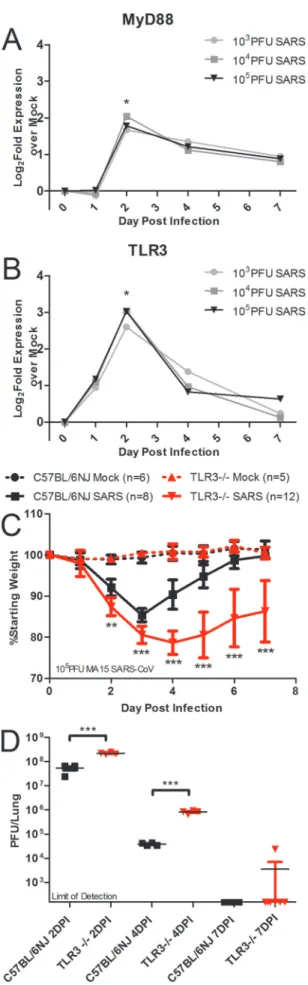
MyD88 transcripts were significantly upregulated at the doses of 103, 104, and 105PFU at day 2 postinfection and met the fold change threshold for categorization as representing a differentially expressed gene at that time point but not at day 1, 4, or 7 postin-fection (Fig. 1A). SARS-CoV-infected mice had a RNA expression profile for TLR3 similar to that for MyD88: it was differentially expressed at day 2 postinfection with SARS-CoV at the doses of 103, 104, and 105PFU of SARS-CoV (Fig. 1B). Based on the simi-larities in gene expression of MyD88 and TLR3, the ranking of the genes by network integration analyses, and the known protective role of MyD88 in SARS-CoV infection, we hypothesized that TLR3 signaling may also be involved in the protective innate im-mune response to SARS-CoV infection. TLR3 signaling occurs in a MyD88-independent manner via the TRIF adaptor protein, so these data indicate that at least two discrete TLR signaling path-ways are involved in the host response to SARS-CoV infection.
To test the hypothesis that TLR3 has a protective role in SARS-CoV infection of mice, 10-week-old female TLR3⫺/⫺mice and wild-type C57BL/6NJ mice were infected intranasally with 105 PFU of recombinant mouse-adapted SARS-CoV (rMA15– CoV) to observe differences in the pathogenesis of SARS-CoV disease. Wild-type mice infected with SARS-SARS-CoV experi-enced transient weight loss that peaked on day 3 postinfection, but all of the wild-type mice began to recover from weight loss on day 4 postinfection and recovered fully from weight loss by 6 to 7 days postinfection (Fig. 1C). TLR3⫺/⫺mice lost a greater average per-centage of their starting weight, with statistically significant differ-ences in TLR3⫺/⫺weight loss, than wild-type mice on days 2 to 7 postinfection (Fig. 1C; **,P⬍0.01; ***,P⬍0.001). Titers ob-served in the lungs of TLR3⫺/⫺mice infected with SARS-CoV were about 4-fold higher than those observed with wild-type mice at day 2, and titers observed in the lungs of TLR3⫺/⫺mice infected with SARS-CoV were about 20-fold higher than those observed with wild-type mice at 4 postinfection (Fig. 1D; ***,P⬍0.001). On day 7 postinfection, one TLR3⫺/⫺mouse had detectable virus in the lungs, while the rest of the TLR3⫺/⫺mice and all of the C57BL/6NJ mice had no detectable virus in the lungs (Fig. 1D). Additionally, TLR3⫺/⫺mice showed increased SARS-CoV disease measured by aberrant lung function parameters and histopathol-ogy compared to wild-type mice (see Fig. S1 in the supplemental material).
TLR3 regulates downstream responses of several key proin-flammatory cytokines, and TLR3 can also regulate the induction of type I interferon and downstream signaling by ISGs (Fig. 2A). Surprisingly, microarray analysis of host gene expression showed
FIG 1 Two discrete TLR pathways regulate SARS-CoV pathogenesis. (A and
B) Profiles from microarray analysis of MyD88 (A) and TLR3 (B) RNA expres-sion results in 20-week-old C57BL/6J mice infected with 103, 104, or 105PFU of SARS-CoV indicate that differential levels of gene expression occurred at day 2 postinfection (a single asterisk [*] indicates differential expression determined by a⬎1.5 log2-foldincrease in expression compared to the results seen with mock infections;P⬍0.05). (C and D) Infection of TLR3⫺/⫺and C57BL/6NJ
mice with SARS-CoV showed significantly greater weight loss in TLR3⫺/⫺
mice than in wild-type mice (**,P⬍0.01; ***,P⬍0.001, by nonparametric Mann-Whitney test, where values indicate the mean percent starting weight and error bars indicate standard deviation) (C), and viral titers were signifi-cantly higher in the TLR3⫺/⫺mice than in wild-type mice (***,P⬍0.001 [by
few alterations in gene expression downstream of TLR3 in com-parisons of TLR3⫺/⫺mice to wild-type mice (Fig. 2B to I). There was no change in the levels of IL-6 or TNF, two proinflammatory cytokines downstream of TLR3 signaling (Fig. 2B and C). CCL5 and IFN-␥were differentially expressed in TLR3⫺/⫺mice com-pared to type mice, with higher gene expression in the wild-type mice than in the TLR3⫺/⫺mice by greater than a 1.5-fold change difference on day 4 postinfection (Fig. 2D and E; *,⬎ 1.5-fold change). No differences were observed in expression of IFN-, a type I interferon (Fig. 2F), or of RSAD2 (Fig. 2G), CXCL10 (Fig. 2H), and IFIT1 (Fig. 2I), three ISGs. Analysis of microarray results for genes differentially expressed in wild-type C57BL/6NJ mice compared to TLR3⫺/⫺mice indicated differ-ences in a number of other genes at days 2 and 4 postinfection (see Table S1 in the supplemental material).
Toll-like receptor adaptor TRIF has a protective role in the host response to SARS-CoV. Because TLR3 utilizes the TRIF adaptor for downstream signaling programs, we infected TRIF⫺/⫺ mice and wild-type C57BL/6J mice intranasally with 105PFU of rMA15–SARS-CoV to determine the role of TRIF in SARS-CoV pathogenesis. TRIF⫺/⫺mice experienced typical early weight loss and then continued to lose weight on days 4 through 6 postinfec-tion, when wild-type mice were recovering from weight loss (Fig. 3A; ***,P⬍0.001). All of the TRIF⫺/⫺mice approached 70% of their starting weight on day 6 postinfection, when the experiment was ended according to our humane endpoint animal protocols. At days 2 and 4 postinfection, significantly higher viral loads were observed in the lungs of TRIF⫺/⫺mice than in wild-type mice (Fig. 3B; ***,P⬍0.001). By day 6 postinfection, wild-type mice had cleared virus to levels below the limit of detection of the plaque assay, but there was still detectable virus in the lungs of TRIF⫺/⫺mice (Fig. 3B; ***,P⬍0.001). Additionally, at 6 days postinfection, the lungs of TRIF⫺/⫺mice infected with SARS-CoV had severe hemorrhage encompassing the entire lung tissue, while little if any hemorrhage was observed in the lungs of wild-type mice (Fig. 3C; ***,P⬍0.001). On the basis of these observations, TRIF⫺/⫺mice had more-severe SARS-CoV clinical disease signs than TLR3⫺/⫺mice, leading to a lethal phenotype from SARS-CoV infection in the TRIF⫺/⫺mice (Fig. 1 and 3).
To determine if the increased susceptibility of TRIF⫺/⫺mice to SARS-CoV infection affects lung function, whole-body plethys-mography was used to measure changes in lung function in TRIF⫺/⫺mice compared to wild-type mice over the course of the SARS-CoV infection (Fig. 3D to F). TRIF⫺/⫺mice had signifi-cantly higher levels of enhanced pause (PENH) on days 2 to 6 postinfection (Fig. 3D; *,P⬍0.05; ***,P⬍0.001) indicative of airway hyperresponsiveness and infection-associated airway ob-struction (40, 41). Lower values of the ratio ofPENHto midtidal
FIG 2 TLR3⫺/⫺mice show few alterations in cytokine and IFN signaling
responses to SARS-CoV infection compared to wild-type mice. (A) TLR3 is a pattern recognition receptor that recognizes viral pathogen-associated
molec-(Continued)
Figure Legend Continued
ular patterns and initiates antiviral signaling programs of IFNs, cytokines, and chemokines via the TRIF adaptor molecule. (B to I) RNA expression profiles of cytokines and ISGs downstream of TLR3 signaling measured by microarray analysis of IL-6 (B), TNF (C), CCL5 (D), IFN-␥(E), IFN-(F), RSAD2 (G), CXCL10 (H), and IFIT1 (I) from TLR3⫺/⫺or C57BL/6NJ mice infected with
105PFU of SARS-CoV normalized to the corresponding mock-infected, PBS-inoculated TLR3⫺/⫺or C57BL/6NJ mice (n⫽4 to 5 mice per group).
Differ-entially expressed genes indicated by a single asterisk (*) showed a⬎1.5-fold change in expression levels between wild-type and knockout mice, withP⬍
expiratory flow (EF50) (TPEF/Te[RPEF]) in TRIF⫺/⫺mice persisted throughout SARS-CoV infection, while wild-type miceRPEF val-ues recovered to basal levels by day 4 postinfection (Fig. 3E; ***,
P⬍0.001). The EF50was significantly higher in TRIF⫺/⫺mice than in wild-type mice on days 1 to 6 postinfection with SARS-CoV (Fig. 3F; *,P⬍0.05; **,P⬍0.01), which is consistent with previous data in SARS-CoV infection models and studies of hy-poxic conditions (42, 43). These measures indicate that major changes in lung function occurred in the TRIF⫺/⫺mice infected with SARS-CoV, potentially due to changes in large-airway debris and denudation, as indicated by the results of histological analysis (see Fig. S2A to I in the supplemental material).
To determine if differences in viral titers in TRIF⫺/⫺mice in-fected with SARS-CoV compared to wild-type mice resulted in increased viral spread or were associated with infection of differ-ent cell types, we evaluated lung sections stained by immunohis-tochemistry (IHC) specific for the SARS-CoV nucleocapsid pro-tein (Fig. 4). Significantly more viral antigen was present in the lungs of TRIF⫺/⫺mice than in those of wild-type mice at day 2 postinfection, consonant with the higher viral loads quantified by plaque assay (Fig. 4A and F; *,P⬍0.05). In the large airways of TRIF⫺/⫺ mice infected with SARS-CoV, no difference in the amount of viral antigen present was observed (Fig. 4B and E), but infected cells in both the C57BL/6J and TRIF⫺/⫺mice were mor-phologically consistent with ciliated airway epithelial cells, a pri-mary target of SARS-CoV in humans. Interestingly, significantly more viral antigen staining was observed in the parenchyma of the lungs of TRIF⫺/⫺mice than in those of C57BL/6J mice infected with SARS-CoV (Fig. 4A and E; **,P⬍0.01). Alveolar spaces showed the presence of viral antigen in cells morphologically con-sistent with type II pneumocytes, another primary target cell of SARS-CoV (Fig. 4C). Observation of the immunohistochemistry did not reveal any evidence that SARS-CoV infection of TRIF⫺/⫺ mice occurs in cell types other than those infected in C57BL/6J mice.
Due to the differences in weight loss and survival between TRIF⫺/⫺and TLR3⫺/⫺mice infected with SARS-CoV, we hypoth-esized that TLR4 may also contribute to signaling through TRIF in response to SARS-CoV infection. To determine the role of TLR4 in the pathogenesis of SARS-CoV, we infected TLR4⫺/⫺mice and wild-type C57BL6/J mice intranasally with 105PFU of rMA15– SARS-CoV. TLR4⫺/⫺mice lost a greater percentage of their start-ing weight, with statistically significant differences in weight loss compared to wild-type mice on days 3 to 7 postinfection, but began recovering by 7 days postinfection (Fig. 5A; *,P⬍0.05; ***,
P⬍0.001). TLR4⫺/⫺mice had significantly higher titers of virus in the lungs than wild-type mice infected with SARS-CoV at days 2 (3-fold difference; ***,P⬍0.001) and 4 (4-fold difference; *,
P⬍0.05) postinfection but had cleared the virus by day 7 postin-fection, similarly to wild-type mice (Fig. 5C). Because TLR4 can signal in either a MyD88-dependent or TRIF-dependent manner via the usage of the sorting adaptor MAL or TRAM, respectively, we infected 10-week-old female TRAM⫺/⫺mice and wild-type C57BL6/J mice intranasally with 105PFU of SARS-CoV in order to discriminate between the effects of MyD88-dependent and TRIF-dependent signaling. TRAM⫺/⫺mice infected with SARS-CoV lost significantly more weight than wild-type mice (Fig. 5B; ***,
P⬍0.001). TRAM⫺/⫺mice infected with SARS-CoV demon-strated prolonged weight loss through 10 days postinfection, while wild-type mice recovered from weight loss by day 6 postinfection
FIG 3 TRIF⫺/⫺mice are highly susceptible to SARS-CoV infection. (A to C)
TRIF⫺/⫺mice infected with SARS-CoV have significantly greater weight loss (**,
P⬍0.01; ***,P⬍0.001 [by a nonparametric Mann-Whitney test, where values indicate mean percent starting weight and error bars indicate standard deviation]) (A), viral titers (***,P⬍0.001 [by unpaired Student’sttest]) (B), and lung hem-orrhage scores (scored from 0 to 4; ***,P⬍0.001 [by unpaired Student’sttest]) (C) than C57BL/6J mice infected with SARS-CoV over a 6-day course of infection. (D to F) Whole-body plethysmography analysis showed that SARS-CoV-infected TRIF⫺/⫺mice (solid red line) have alterations in lung functions compared to
SARS-CoV-infected C57BL/6J mice (solid black line), including enhanced pause (PENH) (D),TPEF/Teratio (RPEF) (E), and midtidal expiratory flow (EF50) (F) levels over the course of 6 days (dashed red line, TRIF⫺/⫺mock-infected mice; dashed
(Fig. 5B). TRAM⫺/⫺mice had titers that were 3-fold higher than those seen with wild-type mice infected with SARS-CoV on day 2 postinfection (**,P⬍0.01) but not on day 4 postinfection and cleared the virus by day 7 postinfection similarly to wild-type mice, despite lack of recovery from weight loss (Fig. 5D). The alterations in lung function measured by whole-body plethys-mography that were observed in TLR4⫺/⫺and TRAM⫺/⫺mice infected with SARS-CoV were similar to those observed in
wild-FIG 4 Increased presence of viral antigen in the lungs of TRIF⫺/⫺mice. (A to
C) Immunohistochemistry was used to stain for SARS-CoV nucleocapsid an-tigen in the lungs of TRIF⫺/⫺SARS-CoV-infected mice (first column in panels
A to C) and C57BL/6J SARS-CoV-infected mice (second column in panels A to C) on day 2 postinfection with SARS-CoV. TRIF⫺/⫺mock-inoculated mice
and C57BL/6J mock-inoculated mice were also evaluated as negative controls and showed no viral antigen staining (data not shown). (D to F) Immunohis-tochemistry lung sections from day 2 postinfection were scored for the pres-ence of SARS nucleocapsid antigen in the large airways (D) and lung paren-chyma (E) and for overall staining (F). Sections were scored in a blinded manner, and scores were evaluated for significance by an unpaired Student’s
ttest (*,P⬍0.05; **,P⬍0.01; NS, not significant).
FIG 5 TLR4⫺/⫺and TRAM⫺/⫺mice are more susceptible to SARS-CoV
infection than wild-type mice. (A) TLR4⫺/⫺, TRAM⫺/⫺, and wild-type mice
were infected intranasally with 105PFU of SARS-CoV. Weight loss was mea-sured each day postinfection, and TLR4⫺/⫺mice lost significantly more weight
than wild-type mice on days 3 to 7 postinfection (*,P⬍0.05; ***,P⬍0.001 [by nonparametric Mann-Whitney test; values indicate mean percent starting weight and error bars indicate standard deviation]). (B) TRAM⫺/⫺mice lost
significantly more weight than wild-type mice on days 2 to 10 postinfection (***,P⬍0.001 [by nonparametric Mann-Whitney test, values indicate the mean percent starting weight and error bars indicate standard deviation]). (C) Virus titer in the lung was measured by plaque assay, and TLR4⫺/⫺mice had
significantly higher virus titers in the lungs at days 2 and 4 postinfection but had cleared the virus by day 7 postinfection, similarly to wild-type mice. (D) TRAM⫺/⫺ mice had significantly higher virus titers in the lungs at day 2
postinfection as measured by plaque assay, but the differences were not signif-icant at day 4 postinfection, and both TRAM⫺/⫺mice and wild-type mice had
no detectable virus in the lungs by day 7 postinfection (*,P⬍0.05; ***,P⬍
type mice (see Fig. S3 in the supplemental material), supporting our hypothesis that TLR4/TRAM/TRIF signaling has a modest protective effect against SARS-CoV infection.
Aberrant cellular and signaling responses in TRIFⴚ/ⴚmice in response to SARS-CoV infection.Because TRIF acts as a TLR adaptor protein that leads to activation of transcription factors that transcribe cytokines and chemokines, we hypothesized that differences in chemokines downstream of TRIF⫺/⫺ signaling would be altered in TRIF⫺/⫺mice compared to wild-type mice. Measurements of proinflammatory cytokines IL-6, TNF, and IFN-␥and chemokines CCL2, CCL3, CCL5, CCL7, and CCL8 showed reduced expression of all these genes at day 2 postinfec-tion in TRIF⫺/⫺mice, while these transcripts are highly induced in wild-type mice infected with SARS-CoV (Fig. 6A to D [*,P⬍
0.05]; see also Fig. S4A to D in the supplemental material [***,P⬍
0.001]). At day 4 postinfection, significantly more expression of IL-6, TNF, CCL5, and IFN-␥was observed in TRIF⫺/⫺mice, while expression was diminished in wild-type animals infected with SARS-CoV (Fig. 6A to D; *,P⬍0.05; ***,P⬍0.001). On day 4 postinfection, there was no significant difference in the levels of chemokines CCL2, CCL3, CCL7, and CCL8 in the TRIF⫺/⫺and wild-type mice infected with SARS-CoV (Fig. S4A to D). In con-trast, increased protein levels of IFN-were observed in TRIF⫺/⫺ mice on day 2 and day 4 postinfection compared to wild-type mice infected with SARS-CoV (Fig. 6E; *,P⬍0.05); this result was likely due to the increased viral loads in the lungs of TRIF⫺/⫺mice at those time points. Expression of ISGs downstream of IFN, in-cluding RSAD2, CXCL10, and IFIT1, was greatly reduced in TRIF⫺/⫺mice infected with SARS-CoV on day 2 postinfection compared to wild-type mice (Fig. 6F to H; *,P⬍0.05; **,P⬍
0.01; ***,P⬍0.001) but was induced to very high levels in the TRIF⫺/⫺mice infected with SARS-CoV on day 4 postinfection while the ISG response was waning in wild-type mice (*,P⬍0.05; **,P⬍0.01).
Because differences in inflammation were observed in TRIF⫺/⫺ mouse lung sections stained with hematoxylin and eosin (H&E) (see Fig. S2 in the supplemental material) and because differences in chemokines that recruit inflammatory cells were observed in TRIF⫺/⫺mice compared to wild-type mice (Fig. 6; see also Fig. S4 in the supplemental material), we hypothesized that differences in infiltrating immune cell populations would be observed in TRIF⫺/⫺mice compared to wild-type mice infected with SARS-CoV. Both wild-type and TRIF⫺/⫺lungs from mice infected with SARS-CoV had larger numbers of cells due to infiltrating cell pop-ulations than lungs from mock-inoculated mice, but there were no significant differences in the overall numbers of infiltrating cells of TRIF⫺/⫺lungs on day 4 or 6 postinfection compared to those of lungs from wild-type mice infected with SARS-CoV (Ta-ble 1). There were significantly more neutrophils in the lungs of TRIF⫺/⫺mice infected with SARS-CoV than in lungs of wild-type mice on day 4 postinfection (Table 1; *,P⬍0.05); however, by day 6 postinfection there were no longer significant differences in trophils. The levels of chemokines that are chemotactic for neu-trophil recruitment (CXCL1, CXCL2, and CXCL3) were signifi-cantly lower on day 2 postinfection in TRIF⫺/⫺mice infected with SARS-CoV but were higher in TRIF⫺/⫺mice on day 4 postinfec-tion, coinciding with the increased recruitment of neutrophils in the TRIF⫺/⫺mice infected with SARS-CoV (Fig. S4E to G; *,P⬍ 0.05; **,P⬍0.01; ***,P⬍0.05).
Differences in monocytic subpopulations were observed on
days 4 and 6 postinfection with SARS-CoV (Table 1). There were significantly more Ly6Chi inflammatory monocytes in the TRIF⫺/⫺mice infected with SARS-CoV on day 4 postinfection (Table 1; *,P⬍0.05), but there was no difference between wild-type and TRIF⫺/⫺mice in the Ly6Clopopulation of monocytes on day 4 postinfection. In contrast, there was no difference in the levels of Ly6Chimonocytes between TRIF⫺/⫺and wild-type mice infected with SARS-CoV on day 6 postinfection, but wild-type mice infected with SARS-CoV had significantly more Ly6Clo reg-ulatory monocytes on day 6 postinfection (Table 1; *,P⬍0.05). On day 4 postinfection, there was no significant difference in the number of alveolar macrophages or plasmacytoid dendritic cells (pDCs) between the lungs of TRIF⫺/⫺and wild-type mice infected with SARS-CoV, but at day 6 postinfection there were significantly more alveolar macrophages and pDCs in the lungs of TRIF⫺/⫺ mice infected with SARS-CoV than in the lungs of wild-type mice (Table 1; *,P⬍0.05; ***,P⬍0.001). No statistically significant differences were observed in populations of eosinophils or monocyte-derived DCs or in CD11bposDC populations between TRIF⫺/⫺mice and wild-type mice infected with SARS-CoV (data not shown).
The total numbers of lymphocytes were not significantly dif-ferent between TRIF⫺/⫺mice and wild-type mice at day 4 or day 6 postinfection (Table 1). There were no differences in the number of T cells in the lungs of TRIF⫺/⫺mice and wild-type mice infected with SARS-CoV on day 4 postinfection, but on day 6 postinfec-tion, there were significantly more T cells in the lungs of wild-type mice infected with SARS-CoV than in the lungs of TRIF⫺/⫺mice (Table 1; *,P⬍0.05). There were no significant differences in the number of CD4⫹T cells on day 4 or day 6 postinfection or the number of CD8⫹T cells on day 4 postinfection, but there were significantly more CD8⫹T cells observed in wild-type mice at day 6 postinfection than in TRIF⫺/⫺mice (Table 1; **,P⬍0.01). Aberrant SARS-CoV disease responses were observed in the TRIF⫺/⫺mice that led to a lethal phenotype, including increased weight loss, lack of viral clearance, alterations in lung function and pathology, changes in infiltrating cell populations, and aberrant cellular signaling programs, all indicating that TRIF is a master regulator in the protective innate immune response to SARS-CoV disease.
DISCUSSION
increase in proinflammatory cytokine and ISG signaling on day 4 postinfection (Fig. 6). One explanation of these data may be that an initial lack of innate immune response in TRIF⫺/⫺mice in-fected with SARS-CoV leads to higher viral titers, which in turn leads to compensatory innate immune signaling resulting in the induction of cytokine, chemokine, and ISG expression at day 4 postinfection. Consistent with these data, TRIF⫺/⫺mice infected intranasally with herpes simplex virus (HSV-1) have increased mortality rate, significantly greater viral titers in the brain, and increased production of type I IFN (46). In contrast, TRIF⫺/⫺ mice infected with IAV were not significantly different from wild-type mice in mortality, but one study found that MyD88⫺/⫺mice were more susceptible than wild-type mice, while another found there was no difference between the responses of MyD88⫺/⫺mice and wild-type mice infected with IAV (16, 47). These data indicate that, although both TLR adaptors (MyD88 and TRIF) are vitally important to a protective immune response to SARS-CoV, differ-ences exist between the cellular signaling programs induced by highly pathogenic respiratory infections caused by coronaviruses and influenza viruses that should be considered prior to the ad-ministration of therapeutic regimes (48).
The differences in viral pathogenesis between MyD88⫺/⫺and TRIF⫺/⫺mice also include major differences in infiltrating cell populations resulting from SARS-CoV infection. MyD88⫺/⫺mice had significantly fewer inflammatory monocytes and macro-phages at day 2 postinfection than wild-type mice infected with SARS-CoV, but no cellular populations measured were signifi-cantly different at day 4 postinfection (37). In addition, despite similarities in infiltrating cell populations in MyD88⫺/⫺and
wild-type mice infected with SARS-CoV on day 4 postinfection, a lack of cytokine and chemokine signaling persisted, indicating a likely deficiency in the activation of signaling programs of these cells. In TRIF⫺/⫺mice, however, many differences in infiltrating cell pop-ulations were observed at day 4 and day 6 postinfection, but the increase in transcription of proinflammatory cytokines indicates that a lack of TRIF does not inhibit cell signaling programs by these cell populations. Rather, the large induction of IFN- ex-pression on day 2 postinfection and the presence of significantly more viral antigen at early times postinfection likely drive the increased stimulation of infiltrating cell types in TRIF⫺/⫺mice, contributing to aberrant cellular responses.
The accumulation of neutrophils in TRIF⫺/⫺mice on day 4 postinfection with SARS-CoV correlates with increased amounts of neutrophil recruitment chemokines CXCL1 and CXCL2 (IL-8 rodent homologs) and increased levels of proinflammatory cyto-kines such as TNF and IL-6, mirroring the neutrophil infiltration and cellular responses of ARDS patients (reviewed in reference 49). Similarly indicative of lethal pathogenesis of respiratory vi-ruses, infection of mice with highly pathogenic strains of influenza virus, including 1918 H1N1 and H5N1 IAV, resulted in signifi-cantly more recruitment of neutrophils (at levels similar to the levels seen in TRIF⫺/⫺mice infected with SARS-CoV) than was observed following infection with seasonal IAV strains of low pathogenicity (50). There is evidence that neutrophils infiltrating the pulmonary compartment produce robust amounts of CXCL10, contributing to the pathogenesis of ARDS from IAV infection, and the induction of large levels of CXCL10 was ob-served in TRIF⫺/⫺mice infected with SARS-CoV on day 4
postin-FIG 6 Aberrant proinflammatory cytokine, interferon, and interferon-stimulated gene signaling responses in TRIF⫺/⫺mice infected with SARS-CoV. (A to D)
RNA expression profiles of cytokines and chemokines downstream of TRIF and TLR signaling programs measured by qPCR analysis of IL-6 (A), TNF (B), CCL5 (C), and IFN-␥(D) from TRIF⫺/⫺mice (red bars) or wild-type C57BL/6J mice (black bars) infected with 105PFU of SARS-CoV normalized to mock-infected TRIF⫺/⫺or wild-type mice (n⫽4 mice per group) at day 2 and day 4 postinfection. (E) TRIF⫺/⫺mice infected with SARS-CoV have significantly higher protein
levels of IFN-measured by ELISA on day 2 and day 4 postinfection in lung homogenates. (F to H) RNA expression profiles of ISGs measured by qPCR analysis of RSAD2 (F), CXCL10 (G), and IFIT1 (H) in TRIF⫺/⫺mice (red bars) or wild-type C57BL/6J mice (black bars) infected with 105PFU of SARS-CoV normalized to mock-infected TRIF⫺/⫺mice or wild-type mice at day 2 and day 4 postinfection. Significant differences between groups were evaluated by an unpaired
Student’sttest, bar graphs show the mean normalized fold change on the day postinfection, and the error bars indicate 1 standard deviation from the mean (*,
P⬍0.05, **,P⬍0.01; ***,P⬍0.001; NS, not significant).
TABLE 1 Quantitation of infiltrating cells in the lungs of TRIF⫺/⫺and C57BL/6J mice following SARS-CoV infectiona
Cell category
No. of cells (⫻104)
Day 4 Day 6
Mock SARS Mock SARS
WT TRIF⫺/⫺ WT TRIF⫺/⫺ WT TRIF⫺/⫺ WT TRIF⫺/⫺
Total 1,430 1,490 13,100 16,500 813 1,620 7,240 7,880
Neutrophil 54.6 49.9 272 892* 35.4 45.8 44.5 252
Monocyte 91.4 111 1,860 2,470 85.8 110 1,360 877
Ly6Chi Mono 11.0 24.3 250 1,070* 47.9 27.9 394 276
Ly6Clo Mono 80.8 87.6 1,620 1,430 28.9 63.5 951 466*
Alveolar Mac 66.1 80.5 178 338 34.0 59.4 119 229*
pDC 1.27 4.86 13.2 18.7 4.74 5.92 5.81 36.4***
Viable lymphocyte 1,080 1,080 10,800 10,800 586 1,110 5,950 5,000
Total T cells 180 287 1,930 1,800 120 294 1,910 972*
CD4⫹T cells 67.6 108 689 625 51.4 159 693 476
CD8⫹T cells 71.2 119 622 642 52.4 118 1,000 398**
fection, coinciding with the influx of neutrophils (32). In influ-enza virus infection, infiltration of Ly6Chi monocytes resulting from IFN induction contributes to resistance of influenza virus infection, while significantly more Ly6Chimonocytes were ob-served in the more susceptible TRIF⫺/⫺mice on day 4 postinfec-tion in our model (51). This inflammatory monocyte populapostinfec-tion can differentiate into macrophage and pDC subsets, which were observed in significantly higher numbers in TRIF⫺/⫺mice in-fected with SARS-CoV on day 6 postinfection (Table 1; reviewed in reference 52).
Because TLR3 senses double-stranded RNAs, an intermediate nucleic acid species present during acute viral infections, it could be predicted that loss of TLR3 signaling would negatively impact the host and alter cellular signaling programs after SARS-CoV infection. Although TLR3⫺/⫺mice infected with SARS-CoV ex-perienced greater weight loss, higher viral titers, and more signif-icant alterations in lung function over the course of infection (Fig. 1C and D; see also S1A to C in the supplemental material), relatively few changes in TRIF-dependent downstream cellular signaling programs resulted from the absence of TLR3 (Fig. 2B to I), indicating that additional pathways may compensate for the absence of TLR3 in SARS-CoV infection. Other innate immune sensors, such as MDA5, that detect dsRNA and that have been implicated in the sensing of coronaviruses may be important for compensatory effects in the absence of TLR3 (53). Among the few changes that were observed in TRIF-dependent signaling path-ways in TLR3⫺/⫺mice on day 4 postinfection, IFN-␥expression was significantly higher in wild-type mice than in TLR3⫺/⫺mice; however, absence of the IFN-␥receptor has minimal impact on SARS-CoV pathogenesis in a similar mouse model (54). The rel-evance of changes in CCL5 is difficult to infer, as CCL5 and other related chemokines signal through multiple receptors, including CCR1 and CCR5, the absence of which modestly increases SARS-CoV pathogenesis in a mouse model (37). In contrast to our re-sults with SARS-CoV, TLR3⫺/⫺mice are less susceptible to H3N2 and H5N1 IAV, with a decreased mortality rate compared to lethal infection of wild-type mice, but there is no difference in the sur-vival of TLR3⫺/⫺mice compared to wild-type mice infected with a lethal dose of p2009 H1N1 IAV (16, 55). The phenotype of TLR3⫺/⫺mice in West Nile virus (WNV) mouse models is some-what controversial, with one group showing a modest increase in WNV-induced mortality with no differences in type I IFN levels in TLR3⫺/⫺mice, while another group showed that TLR3⫺/⫺mice have less susceptibility to WNV and reduced proinflammatory cytokine responses compared to wild-type mice (56, 57).
Our study results indicate that TLR4 is also involved in medi-ating the pathogenesis of SARS-CoV infection, and it is likely that TLR4 signaling occurs in a TRIF-dependent manner through the sorting adaptor TRAM, as TRAM⫺/⫺mice recapitulate features of increased SARS-CoV pathogenesis similarly to TLR4⫺/⫺mice, in-cluding increased weight loss, similar alterations in lung parame-ters, and higher viral titers at early times postinfection. The in-volvement of TRAM signaling indicates that the TRIF adaptor protein mediates a large part of TLR4 signaling in response to SARS-CoV. The studies presented here indicated that TLR3 and TLR4 individually mediate a portion of the TRIF-dependent TLR signaling necessary for survival of SARS-CoV in our mouse model. These are among the first observations of the role of indi-vidual TLRs in contributing to protection from SARS-CoV dis-ease. Interestingly, the absence of either TLR3 or TLR4 does not
lead to lethal SARS-CoV disease similar to that seen with TRIF⫺/⫺ mice infected with SARS-CoV, likely because an absence of signal-ing via a ssignal-ingle TLR may be compensated for by senssignal-ing of viral PAMPs by other PRRs.
In other models of acute lung injury, TLR4⫺/⫺ mice and TRIF⫺/⫺mice are less susceptible to lung injury mediated by the introduction of acid and IAV into the lung (28). Imai et al. ob-served that oxidized phospholipids, putative PAMPs potentially contributing to acute lung injury by activating TLR4 and signaling through TRIF, were present in ARDS patient samples as a conse-quence of the presence of infectious diseases such as H5N1 IAV and SARS-CoV infections (28). The finding that TLR4⫺/⫺mice are resistant to acute lung injury via IAV and acid models is in contrast to our findings here that TLR4⫺/⫺mice have significantly more disease resulting from SARS-CoV infection than wild-type mice (Fig. 5). One explanation for these conflicting data is that activation of TLR4 by oxidized phospholipid PAMPs may be det-rimental in acid injury and IAV infection mouse models but that, in the case of SARS-CoV infection, the benefits of TLR4 sensing of PAMPs may outweigh the damaging effects of cellular signaling programs resulting from sensing of oxidized phospholipids by TLR4. Additionally, TLR4⫺/⫺mice are less susceptible to influ-enza virus infection and an immunomodulatory approach using a TLR4 antagonist was proposed to have ameliorative properties for the treatment of influenza virus infections (29). These findings indicate that protective signaling via TLR4/TRAM/TRIF may be a unique feature in the pathogenesis of coronaviruses compared to that of other respiratory pathogens such as influenza viruses and that different cellular sensors recognize pathogens with similar clinical features and infecting similar cell types.
Despite the various outcomes in host survival and morbidity in SARS-CoV, WNV, and IAV infection models, the commonality is that mice deficient in TLR signaling have increased viral loads in the infected tissues, demonstrating that the initial recognition of viral PAMPs by TLRs is necessary for controlling viral replication and that the increased presence of viral antigen could partially drive downstream phenotypes in these systems (Fig. 1D and 5C) (16, 56, 57). Neither MyD88⫺/⫺mice nor TRIF⫺/⫺mice infected with SARS-CoV efficiently cleared the virus by day 6 postinfec-tion, but both showed increased signs of disease, ultimately lead-ing to death of the TLR adaptor knockout mice from SARS-CoV infection. Our observations support previous findings indicating that signaling through TRIF is critical for CD8⫹T cell expansion, a key component of adaptive immunity for viral clearance (58, 59). In contrast to the TLR adaptor knockout mice, RAG1⫺/⫺ mice with no mature T cells fail to clear SARS-CoV but show no signs of increased disease, as defined by weight loss, indicating that the lack of clearance of virus alone is not responsible for the dis-ease phenotypes seen in the TRIF⫺/⫺and MyD88⫺/⫺mice (37). In generating a protective immune response to highly pathogenic coronavirus infections, our findings indicate that not only the activation of an adaptive response but also the proper activation of a balanced innate immune response through both adaptor arms of TLR-mediated signaling is required for viral clearance.
especially when both components are critical to the host response, such as in SARS-CoV infection. Our data support the idea that innate immune responses are important for the antiviral state of cells, immune cell recruitment, and expansion of adaptive im-mune responses. Comparison of these data to those from other models of highly pathogenic respiratory virus infection (particu-larly influenza virus infection) indicates that although these vi-ruses may be detected by similar pathways, the result of that sens-ing can lead to differences in disease outcome which should be considered in the design and administration of vaccine and anti-viral therapeutics.
MATERIALS AND METHODS
Viruses, cells, and plaque assay.A mouse model of the recombinant mouse-adapted rMA15–SARS-CoV virus used in this study has been pre-viously described (37, 60). As prepre-viously described, virus stocks were propagated on Vero E6 cells (60). Plaque assays to quantify virus in viral stocks and to enumerate the number of viruses in the lower left lobe of lungs from mice were performed in Vero E6 cells, with a limit of detection of 100 PFU (12). Experiments with rMA15–SARS-CoV were performed in a certified biosafety level 3 laboratory, using a class II biological safety cabinet. Laboratory workers were equipped with high-efficiency particu-late air (HEPA)-filtered powered air-purifying respirators (PAPRs), Tyvek suits, hoods, aprons, booties, and personal protective equipment.
Ethics statement.The ethical treatment of mice in this study was in accordance with the guidelines of The University of North Carolina (UNC) at Chapel Hill Institutional Animal Care and Use Committee (IA-CUC), and approved documentation registered under protocol no. 11-213 was used in the experiments described. The guidelines for care of the mice are from theGuide for the Care and Use of Laboratory Animals, 8th ed., published by the U.S. National Institutes of Health (NIH). All animal housing and care was conducted according to Institutional Animal Care and Use Committee (IACUC)-approved protocols of the University of North Carolina, Chapel Hill (NIH/PHS Animal Welfare Assurance no. a3410-01).
Animals.Animals were maintained in HEPA-filtered Sealsafe cages (Techniplast) during experiments performed with rMA15–SARS-CoV. The following strains of age-matched female mice were obtained from Jackson Laboratory: C57B/6NJ (stock no. 005304), TLR3⫺/⫺(stock no.
009675), C57BL/6J (stock no. 000664), and TRIF⫺/⫺(stock no. 005307).
At 10 weeks of age, mice were anesthetized with a mixture of ketamine and xylazine and inoculated intranasally with either 50 l of phosphate-buffered saline (PBS) (for mock-inoculated controls) or 105 PFU of
rMA15–SARS-CoV–PBS. Animals were weighed daily, and lung tissues from days 2, 4, and 7 postinfection (C57BL/6NJ and TLR3⫺/⫺mice) or
days 2, 4, and 6 postinfection (C57BL/6J and TRIF⫺/⫺mice) were
col-lected for downstream analyses by plaque assay, histology, and RNA anal-ysis.
Flow cytometry.On days 4 and 6 postinfection, whole lungs were harvested from groups of 3 to 4 mock-treated wild-type and TRIF⫺/⫺
mice and 4 to 5 SARS-CoV-infected wild-type and TRIF⫺/⫺mice. Whole
lungs were prepared for flow cytometry analysis by collagenase digestion and tissue disruption into cell suspensions as previously described (11). Antibody staining panels were used to stain cell preparations (see Text S1 in the supplemental material). Analysis was performed with Summit soft-ware (Beckman-Coulter) for sorting into defined subpopulations (see Text S1).
Hemorrhage scores, histological analysis, and immunohistochem-istry.Scores for gross hemorrhage were recorded by observation of the lung during necropsy using a scale ranging from a value of zero, indicating no hemorrhage, to a value of 4, indicating severe hemorrhage in all lobes of the lung, as previously described (38). For histological analysis, the entire right lobe of lungs from infected or mock-treated wild-type and knockout mice was fixed in 10% formalin, embedded in paraffin, and prepared in 5-m-thick sections for hematoxylin and eosin (H&E)
stain-ing by the UNC Histopathology core facility. For immunohistochemistry (IHC), formalin-fixed and paraffin-embedded histology samples from C57BL/6J and TRIF⫺/⫺ mice were sectioned (thickness, 5 m) and
stained for viral antigen using a commercially available polyclonal SARS-CoV anti-nucleocapsid antibody (Imgenex) following the manufacturer’s protocols (61). Slides for IHC and H&E histology were scored in a blinded manner (n⫽4 to 5 mice per group) for metrics of inflammation, and images were captured using an Olympus BX41 microscope with an Olym-pus DP71 camera.
Whole-body plethysmography.Lung function was measured by un-restrained whole-body plethysmography using IACUC-approved proto-cols as has been previously described (42). Briefly, animals were intro-duced into randomized individual plethysmography chambers following calibration according to protocols of the manufacturer (Buxco). After a 30-min acclimation period, data on lung function parameters were col-lected during a 5-min measurement period. Data were analyzed by Fine-point software (Buxco) for established metrics of airway hyperresponsive-ness and virus infection-associated airway obstruction, including enhanced pause (PENH), tidal midexpiratory flow (EF50), and ratio ofTPEF
toTe(RPEF).PENHis an empirical measure calculated byTe⫺Tr
Tr *PEF
PIF, where PEF is the peak expiratory flow, PIF is the peak inspiratory flow,Te
represents the time of expiration, andTrrepresents the relaxation time to
36% of the peak expiration pressure (62).PENHhas been controversially
linked to airway hyperresponsiveness, obstruction, and bronchoconstric-tion but has been used in several viral models of airway infecbronchoconstric-tion, where increases in thePENHvalue correlate with increased lung pathology
fol-lowing respiratory viral infection (40, 42, 62–64). EF50indicates the flow
rate at 50% of the tidal volume.RPEFis calculated byTPEF
Te
, whereTPEFis
the time to peak expiratory flow andTeis the time of expiration, and the
ratio may be interpreted as an indication of the shape of the exhalation curve.
Real-time qPCR analysis.The upper left lobe of rMA15–SARS-CoV-infected mice was stored in RNAlater solution (Life Technologies) for 48 h at 4°C and then stored at⫺80°C. Lung sections were thawed and then homogenized in TRIzol (Life Technologies) for 60 s at 6,000 rpm using a Magna Lyzer instrument (Roche). Following chloroform-isopropanol ex-traction of RNA from TRIzol homogenates, cDNA was generated by re-verse transcription-PCR (RT-PCR) using a SuperScript III First-Strand synthesis kit (Life Technologies). Quantitative PCR (qPCR) was per-formed using TaqMan gene expression assays (Life Technologies) for cy-tokines or chemokines normalized to 18-s expression. For each cytokine or chemokine, expression from groups of 4 rMA15–SARS-CoV-infected mice was normalized to mock (PBS)-inoculated mice (either wild-type or knockout mice). Normalized fold change was calculated using the thresh-old cycle (⌬⌬CT) method as has been previously described (11).
ELISA. Levels of IFN-were quantified using a VeriKine mouse IFN-enzyme-linked immunosorbent assay (ELISA) kit (RD Systems) according to manufacturer protocols. The lower left lobe of lungs from TRIF⫺/⫺or C57BL/6J mice from the mock-inoculated or
SARS-CoV-infected group (n⫽4 each group) was homogenized in 1 ml of PBS using a Magna Lyzer instrument (Roche). One hundred microliters of cleared homogenate was used for the ELISA assay sample. A seven-point standard curve was prepared using the manufacturer’s protocol, and interferon titers in the samples were determined by plotting the standards using a 4-parameter fit. Optical densities were read at an absorbance of 450 nm.
Differentially expressed gene identification.Differentially expressed gene targets were selected from data collected in a previously described study performed with transcriptomics data banked at NCBI Gene Expres-sion Omnibus (accesExpres-sion no. gse33266) (38). Briefly, that study was per-formed using microarray analysis of RNA from lungs of 20-week-old fe-male C57BL/6J mice infected with doses of 102, 103, 104, or 105PFU of
model was used to determine differential expression (DE) for each tran-script, requiring an absolute log2(fold change) value of⬎1.5 as well as an adjusted false-discovery-rate (FDR)Pvalue of⬍0.05. In a separate study, TLR3⫺/⫺lung homogenates in TRIzol were analyzed by microarray, and
the full data set has been deposited in the National Center for Biotechnol-ogy Information (NCBI) Gene Expression Omnibus (GEO) database and is available at GEO accession number GSE68820.
SUPPLEMENTAL MATERIAL
Supplemental material for this article may be found athttp://mbio.asm.org/
lookup/suppl/doi:10.1128/mBio.00638-15/-/DCSupplemental.
Text S1, DOCX file, 0.03 MB. Figure S1, TIF file, 1.6 MB. Figure S2, TIF file, 2.1 MB. Figure S3, TIF file, 0.1 MB. Figure S4, TIF file, 0.7 MB. Table S1, DOCX file, 0.03 MB.
ACKNOWLEDGMENTS
We acknowledge the Systems Virology Center (NIAID contract no. HHSN272200800060C) for transcriptomics data used from a previous study.
This work was supported by NIH grant no. U19 AI100625 to R.S.B. and M.T.H. and NIH grant no. U19 AI106772 to R.S.B.
REFERENCES
1.Drosten C, Günther S, Preiser W, van der Werf S, Brodt H-R, Becker
S, Rabenau H, Panning M, Kolesnikova L, Fouchier RA, Berger A, Burguière A-M, Cinatl J, Eickmann M, Escriou N, Grywna K, Kramme S, Manuguerra J-C, Müller S, Rickerts V, Stürmer M, Vieth S, Klenk
H-D, Osterhaus ADME, Schmitz H, Doerr HW. 2003. Identification of
a novel coronavirus in patients with severe acute respiratory syndrome. N Engl J Med348:1967–1976.http://dx.doi.org/10.1056/NEJMoa030747.
2.Zaki AM, van Boheemen S, Bestebroer TM, Osterhaus AD, Fouchier
RA. 2012. Isolation of a novel coronavirus from a man with pneumonia in Saudi Arabia. N Engl J Med367:1814 –1820.http://dx.doi.org/10.1056/
NEJMoa1211721.
3.Stevenson GW, Hoang H, Schwartz KJ, Burrough ER, Sun D, Madson
D, Cooper VL, Pillatzki A, Gauger P, Schmitt BJ, Koster LG, Killian
ML, Yoon KJ. 2013. Emergence of porcine epidemic diarrhea virus in the
United States: clinical signs, lesions, and viral genomic sequences. J Vet Diagn Invest25:649 – 654.http://dx.doi.org/10.1177/1040638713501675. 4.WHO. 31 December 2003, posting date. Summary of probable SARS cases with onset of illness from 1 November 2002 to 31 July 2003. WHO, Ge-n e v a , S w i t z e r l a Ge-n d . h t t p : / / w w w . w h o . i n t / c s r / s a r s / c o u n t r y /
table2004_04_21/en/.
5. Ohnmeiss DD. 2015. ECDC Communicable Disease Threats Report
(CDTR). European Center for Disease Control.
6.Anthony SJ, Epstein JH, Murray KA, Navarrete-Macias I,
Zambrana-Torrelio CM, Solovyov A, Ojeda-Flores R, Arrigo NC, Islam A, Ali Khan S, Hosseini P, Bogich TL, Olival KJ, Sanchez-Leon MD, Karesh
WB, Goldstein T, Luby SP, Morse SS, Mazet JAK, Daszak P, Lipkin WI.
2013. A strategy to estimate unknown viral diversity in mammals. mBio
4:e00598-13.http://dx.doi.org/10.1128/mBio.00598-13.
7.Al-Tawfiq JA, Momattin H, Dib J, Memish ZA. 2014. Ribavirin and
interferon therapy in patients infected with the Middle East respiratory syndrome coronavirus: an observational study. Int J Infect Dis20:42– 46.
http://dx.doi.org/10.1016/j.ijid.2013.12.003.
8.Stockman LJ, Bellamy R, Garner P. 2006. SARS: systematic review of
treatment effects. PLoS Med 3:e343.http://dx.doi.org/10.1371/
journal.pmed.0030343.
9.Falzarano D, de Wit E, Martellaro C, Callison J, Munster VJ, Feldmann
H. 2013. Inhibition of novelcoronavirus replication by a combination of interferon-␣2b and ribavirin. Sci Rep3:1686.http://dx.doi.org/10.1038/
srep01686.
10. Ge X-Y, Li J-L, Yang X-L, Chmura AA, Zhu G, Epstein JH, Mazet JK,
Hu B, Zhang W, Peng C, Zhang Y-J, Luo C-M, Tan B, Wang N, Zhu Y,
Crameri G, Zhang S-Y, Wang L-F, Daszak P, Shi Z-L. 2013. Isolation
and characterization of a bat SARS-like coronavirus that uses the ACE2 receptor. Nature503:535–538.http://dx.doi.org/10.1038/nature12711.
11. Bolles M, Deming D, Long K, Agnihothram S, Whitmore A, Ferris M,
Funkhouser W, Gralinski L, Totura A, Heise M, Baric RS. 2011. A
double-inactivated severe acute respiratory syndrome coronavirus vac-cine provides incomplete protection in mice and induces increased eosin-ophilic proinflammatory pulmonary response upon challenge. J Virol85:
12201–12215.http://dx.doi.org/10.1128/JVI.06048-11.
12. Deming D, Sheahan T, Heise M, Yount B, Davis N, Sims A, Suthar M,
Harkema J, Whitmore A, Pickles R, West A, Donaldson E, Curtis K,
Johnston R, Baric R. 2006. Vaccine efficacy in senescent mice challenged
with recombinant SARS-CoV bearing epidemic and zoonotic spike vari-ants. PLoS Med3:e525.http://dx.doi.org/10.1371/journal.pmed.0030525.
13. Short KR, Kroeze EJ, Fouchier RA, Kuiken T. 2014. Pathogenesis of
influenza-induced acute respiratory distress syndrome. Lancet Infect Dis
14:57– 69.http://dx.doi.org/10.1016/S1473-3099(13)70286-X.
14. Chow K, Hsiao C, Lin T, Chen C, Chiou S. 2004. Detection of severe
acute respiratory syndrome associated coronavirus in pneumocytes of the lung. Am J Clin Pathol 121:574 –580. http://dx.doi.org/10.1309/
C0EDU0RAQBTXBHCE.
15. Yoo J-K, Kim TS, Hufford MM, Braciale TJ. 2013. Viral infection of the
lung: host response and sequelae. J Allergy Clin Immunol132:1263–1276.
http://dx.doi.org/10.1016/j.jaci.2013.06.006.
16. Le Goffic R, Balloy V, Lagranderie M, Alexopoulou L, Escriou N, Flavell
R, Chignard M, Si-Tahar M. 2006. Detrimental contribution of the
Toll-like receptor (TLR)3 to influenza A virus–induced acute pneumonia. PLoS Pathog2:e53.http://dx.doi.org/10.1371/journal.ppat.0020053.
17. Rudd BD, Smit JJ, Flavell RA, Alexopoulou L, Schaller MA, Gruber A,
Berlin AA, Lukacs NW. 2006. Deletion of TLR3 alters the pulmonary
immune environment and mucus production during respiratory syncytial virus infection. J Immunol176:1937–1942.http://dx.doi.org/10.4049/
jimmunol.176.3.1937.
18. Wang Q, Miller DJ, Bowman ER, Nagarkar DR, Schneider D, Zhao Y,
Linn MJ, Goldsmith AM, Bentley JK, Sajjan US, Hershenson MB. 2011.
MDA5 and TLR3 initiate pro-inflammatory signaling pathways leading to rhinovirus-induced airways inflammation and hyperresponsiveness. P L o S P a t h o g 7 :e 1 0 0 2 0 7 0 . h t t p : / / d x . d o i . o r g / 1 0 . 1 3 7 1 /
journal.ppat.1002070.
19. Guillot L, Le Goffic R, Bloch S, Escriou N, Akira S, Chignard M,
Si-Tahar M. 2005. Involvement of Toll-like receptor 3 in the immune
response of lung epithelial cells to double-stranded RNA and influenza A virus. J Biol Chem 280:5571–5580. http://dx.doi.org/10.1074/
jbc.M410592200.
20. Alexopoulou L, Holt AC, Medzhitov R, Flavell RA. 2001. Recognition of
double-stranded RNA and activation of NF-[kappa]B by Toll-like recep-tor 3. Nature413:732–738.http://dx.doi.org/10.1038/35099560.
21. Oshiumi H, Matsumoto M, Funami K, Akazawa T, Seya T. 2003.
TICAM-1, an adaptor molecule that participates in Toll-like receptor 3-mediated interferon-[beta] induction. Nat Immunol4:161–167.http://
dx.doi.org/10.1038/ni886.
22. Yamamoto M, Sato S, Hemmi H, Hoshino K, Kaisho T, Sanjo H,
Takeuchi O, Sugiyama M, Okabe M, Takeda K, Akira S. 2003. Role of
adaptor TRIF in the MyD88-independent Toll-like receptor signaling p a t h w a y . S c i e n c e 3 0 1 :6 4 0 – 6 4 3 . h t t p : / / d x . d o i . o r g / 1 0 . 1 1 2 6 / science.1087262.
23. Yamamoto M, Sato S, Mori K, Hoshino K, Takeuchi O, Takeda K,
Akira S. 2002. Cutting edge: a novel Toll/IL-1 receptor
domain-containing adapter that preferentially activates the IFN-promoter in the Toll-like receptor signaling. J Immunol 169:6668 – 6672.http://
dx.doi.org/10.4049/jimmunol.169.12.6668.
24. Zhang Z, Kim T, Bao M, Facchinetti V, Jung SY, Ghaffari AA, Qin J,
Cheng G, Liu Y-J. 2011. DDX1, DDX21, and DHX36 helicases form a
complex with the adaptor molecule TRIF to sense dsRNA in dendritic c e l l s . I m m u n i t y 3 4 :8 6 6 – 8 7 8 . h t t p : / / d x . d o i . o r g / 1 0 . 1 0 1 6 /
j.immuni.2011.03.027.
25. Guillot L, Medjane S, Le-Barillec K, Balloy V, Danel C, Chignard M,
Si-Tahar M. 2004. Response of human pulmonary epithelial cells to
lipo-polysaccharide involves Toll-like receptor 4 (TLR4)-dependent signaling pathways: evidence for an intracellular compartmentalization of TLR4. J Biol Chem279:2712–2718.http://dx.doi.org/10.1074/jbc.M305790200.
26. Monick MM, Yarovinsky TO, Powers LS, Butler NS, Carter AB,
Gud-mundsson G, Hunninghake GW. 2003. Respiratory syncytial virus
up-regulates TLR4 and sensitizes airway epithelial cells to endotoxin. J Biol Chem278:53035–53044.http://dx.doi.org/10.1074/jbc.M308093200.