DYNAMIC MODULATION OF IN VIVO SEROTONIN SIGNALING
Elyse Cathleen Dankoski
A dissertation submitted to the faculty at the University of North Carolina at Chapel Hill in partial fulfillment of the requirements for the degree of Doctor of Philosophy in the
Curriculum in Neurobiology.
Chapel Hill 2014
Approved by:
R. Mark Wightman
Regina M. Carelli
Sabrina Burmeister
Paul Manis
ii
© 2014
iii
ABSTRACT
Elyse C. Dankoski: Dynamic Modulation of In Vivo Serotonin Signaling (Under the direction of Prof. R. Mark Wightman)
Serotonin signaling influences many neural processes and is highly implicated in the
etiology and treatment of depression. Understanding how in vivo elements regulate serotonin
signaling may provide insight into serotonergic function in the depressed brain. This work
presents a modification to fast-scan cyclic voltammetric methods that enables rapid, spatially
resolved, and chemically selective measurement of in vivo serotonin signaling in the substantia
nigra pars reticulata. We used this modified technique to investigate the dynamics of serotonin
neurotransmission. Initial investigations found that serotonin release can be evoked by electrical
stimulations in vivo, but it is greatly attenuated in comparison to serotonin evoked in brain slices.
We hypothesized that feedback from intact inhibitory circuitry restricted serotonin signaling in
vivo. A comparison of in vivo dopamine and serotonin release revealed a critical role for the
serotonin transporter (SERT) in limiting serotonin signaling. This process is likely involved in
the therapeutic effects of selective serotonin reuptake inhibitors (SSRIs), which are common
pharmacotherapies for depression. Further studies found that acute SSRI treatment uniquely
enhances serotonin signaling by disrupting inhibitory feedback to the dorsal raphe nucleus. We
hypothesized that this action plays an important role in the 3-6 week lapse between SSRI
treatment onset and clinical efficacy. Indeed, in a study of chronic SSRI treatment, we found that
long-term reductions in SERT function result in enduring enhancement of serotonin signaling. In
iv
activity likely drives both ambient and transient serotonin concentrations in a coordinated,
equilibrated manner. Our finding that the introduction of mild stress can significantly impact the
effects of SSRIs implicates a more complex network of elements in the modulation of serotonin
v
vi
ACKNOWLEDGEMENTS
Thank you to my mentor, Mark Wightman. My experience working with you has made
me a better problem solver, writer, critical thinker, and an aspiring PBR connoisseur. You had
confidence in my ability to work as an independent scientist, to make mistakes, and to learn from them, even when I didn’t have confidence in myself. This was a training experience I feel lucky
to have had.
I am sincerely grateful for the intellectual contribution and moral support of all my
current and former labmates in the Wightman lab during my doctoral work. Above all, thank you
to Dr. Nina Owesson-White, Dr. Anna Belle, Dr. Zoe McElligott, Jenny Ariansen, and Meg Fox
for many hours of science mixed with fun (and sometimes just fun).
Many collaborators contributed to the research that made these chapters and publications
a reality. I would especially like to acknowledge the contributions of Dr. Thorfinn Riday, Dr.
Kara Agster, and Dr. Sheryl Moy.
7 years ago, I had no clue how to pursue a graduate education in science. For the
inspiration, information, and confidence to follow my interest in neuroscience toward a Ph.D, I
thank Dr. Paul Gold, Dr. Donna Korol, Dr. Gene Robinson, and the ISNI Program at the
University of Illinois at Urbana-Champaign. For mentorship and guidance throughout my
undergraduate studies, I thank Dr. Steve Pruett-Jones and Dr. Philip Lloyd at the University of
vii
Finally, I thank my family for their support and dedicate this dissertation to them. To my parents, who encouraged me to live “the life of the mind.” To my grandfather, whose love of
knowledge has always been an inspiration to me. To Rylan, whose continual support, kindness,
and friendship make every day a good day.
viii
TABLE OF CONTENTS
LIST OF ABBREVIATIONS ... xii
CHAPTER 1: MONITORING SEROTONIN SIGNALING WITH FSCV ... 1
Introduction ... 1
1. Fast-scan cyclic voltammetry of serotonin... 2
2. Electrical stimulation... 9
3. Release ... 11
4. Uptake ... 17
5. Future Directions ... 25
Conclusion ... 27
Support ... 28
CHAPTER 2: VOLTAMMETRIC DETECTION OF 5-HYDROXYTRYPTAMINE RELEASE IN THE RAT BRAIN ... 34
Introduction ... 34
Experimental Methods ... 38
Results and Discussion ... 42
Conclusion ... 51
CHAPTER 3: SIMULTANEOUS SEROTONIN AND HISTAMINE RELEASE FOLLOWING MEDIAL FOREBRAIN BUNDLE STIMULATION ... 61
Introduction ... 61
Materials and Methods ... 63
Results ... 68
Discussion ... 72
CHAPTER 4: DIFFERENTIAL MECHANISMS IN THE REGULATION OF IN VIVO DOPAMINE AND SEROTONIN RELEASE ... 85
Introduction ... 85
Methods ... 87
ix
Discussion ... 93
Conclusions ... 97
Supplemental Methods ... 98
Support ... 101
CHAPTER 5: THE DORSAL RAPHE NUCLEUS MEDIATES SSRI-INDUCED FACILITATION OF SEROTONIN SIGNALING ... 107
Introduction ... 107
Materials and Methods ... 108
Results ... 111
Discussion ... 115
Support ... 119
CHAPTER 6: FACILITATION OF SEROTONIN SIGNALING BY SSRIs IS ATTENUATED BY SOCIAL ISOLATION ... 123
Introduction ... 123
Materials and Methods ... 125
Results ... 128
Discussion ... 134
Supplemental Methods ... 137
Support ... 140
CHAPTER 7: CONCLUSION ... 148
Summary of findings ... 148
Discussion and Future Directions ... 154
Concluding Remarks ... 156
x
LIST OF FIGURES
Figure 1.1 In vitro calibration of microelectrodes ... 29
Figure 1.2 Illustration of a carbon-fiber microelectrode in SNr ... 30
Figure 1.3 A model predicts serotonin release and uptake at different frequencies. ... 31
Figure 1.4 Sample trace of in vivo serotonin release and uptake. ... 32
Figure 1.5 A synopsis of the findings presented in this article. ... 33
Figure 2.1 Comparison of waveforms for detection of dopamine and serotonin ... 53
Figure 2.2 Comparison of cyclic voltammograms obtained in vivo vs. in vitro ... 54
Figure 2.3 Nafion-modified microelectrodes are more sensitive to 5-HT. ... 55
Table 2.1 Comparison of Responses of Bare vs. Nafion-Modified ... 56
Figure 2.4 Response of bare and Nafion-modified electrodes to common electronegative species... 57
Figure 2.5 Determination of lp and D5-HT for a Nafion-coated carbon-fiber microelectrode using flow injection analysis ... 58
Figure 2.6 Electrochemical and anatomical validation of 5-HT detection. ... 59
Figure 2.7 Pharmacological validation of 5-HT signal with GBR 12909 and citalopram ... 60
Figure 3.1 Histology of Stimulating and Carbon-Fiber Electrode Placements in the MFB and SNpr ... 79
Figure 3.2 5-HT Color Plot, CV and Time Profile of Response in SNpr with DRN and MFB Stimulation. ... 80
Figure 3.3 Comparison of In vivo Signal to a Mixed In vitro 5-HT and Histamine Signal .... 81
Figure 3.4 Effect of Varying Scan Rate and Upper Potential Limit on Histamine Response In vitro. ... 82
Figure 3.5 Pharmacological Characterization of Histamine with SKF 91488... 83
Figure 3.6 A Comparison of 5-HT Experimental Data and Kinetic Analysis Between DRN and MFB Stimulation ... 84
xi
Figure 4.2 Comparison of dopamine and serotonin release evoked by
varying stimulation parameters. ... 103
Figure 4.3 Effects of manipulating dopamine and serotonin synthesis, packaging and release. ... 104
Figure 4.4 Effects of uptake and MAO inhibitors on dopamine and serotonin release. ... 105
Figure 4.5 Dual uptake and MAO inhibition disregulates serotonin signaling. ... 106
Figure 5.1 Serotonin release is not frequency-dependent in vivo ... 120
Figure 5.2 Inhibiting SERT induces frequency-dependent serotonin release ... 121
Figure 5.3 DRN mediates enhancement of serotonin signaling by SSRIs ... 122
Figure 6.1 Voltammetric determination of serotonin release in the substantia nigra pars reticulata of the mouse brain. ... 141
Figure 6.2 Effects of cCIT or VEH treatment on marble-burying and OF assays in pair- and single-housed mice ... 142
Table 6.1 Maximal serotonin release measured in SNpr ... 143
Figure 6.3. Comparison of serotonin release and uptake evoked by electrical stimulation of the DRN ... 144
Figure 6.4 Comparison of electrically-evoked serotonin release following acute CIT challenge ... 145
Figure 6.S1. Comparison of liquid consumption before and during CIT/VEH treatment and tissue content analysis. ... 146
xii
LIST OF ABBREVIATIONS
[5-HT]max maximal evoked serotonin amplitude
[5-HT]p serotonin released per stimulus pulse
5-HIAA 5-Hydroxyindoleacetic acid; a metabolite of serotonin
5-HT 5-Hydroxytryptamine, serotonin
CIT citalopram; a selective serotonin reuptake inhibitor
DRN dorsal raphe nucleus
FSCV fast-scan cyclic voltammetry
Ki Inhibition constant denoting affinity of ligand for target
KM Michaelis constant for half maximal activity
MAOI monoamine oxidase inhibitor
MFB medial forebrain bundle
SERT serotonin transporter
SNpr substantia nigra pars reticulata
SSRI selective serotonin reuptake inhibitor
t1/2 decay time between peak and half-maximal amplitude
1
CHAPTER 1: MONITORING SEROTONIN SIGNALING WITH FSCV 1
Introduction
The neurotransmitter serotonin (also called 5-hydroxytryptamine or 5-HT) can be
found in nearly every region of the central nervous system. Its functions are as diverse as the
areas it innervates, and it is a complex component of many psychiatric disorders. This
pervasive involvement in brain-wide neurocircuitry is supported by an exceptionally large
family of receptors whose collective functional scope enables the multifarious actions of
serotonin throughout the brain (Barnes & Sharp, 1999). Much of our knowledge about
serotonin comes from studies investigating its actions via these receptors, which remain the
target of many pharmacotherapies involving serotonergic signaling. However, the release and
uptake dynamics of serotonin precipitate its downstream effects, and exploring how these
dynamics are modulated has provided key insights to the serotonin system and its therapeutic
potential.
A number of techniques have been used to characterize serotonin signaling in the
brain. In vivo microdialysis has provided key insights into natural and
pharmacologically-induced fluctuations in ambient extracellular levels of serotonin. However, even recent
advancements in microdialysis sampling rates provide markedly lower temporal resolution
than required to examine individual release and uptake events (Schultz & Kennedy, 2008).
1 This chapter previously appeared as an article in Frontiers in Integrative Neuroscience. The original citation is
2
Electrophysiological measurements can infer some properties of neurotransmitter release by
measuring postsynaptic response, and this method works well for neurotransmitters like
glutamate and GABA, whose ligands effect instantaneous changes in ionic current or
membrane potential. However, most serotonin receptors in the brain are G-protein coupled
and activate intracellular cascades over time periods of 400 ms or more, resulting in
postsynaptic effects that are too slow or heterogeneous to reveal information about small, fast
changes in concentration. Rigorous characterization of serotonin signaling requires a
technique that operates on the same temporal and spatial scales as its release and uptake
processes.
Electroanalytical techniques, which combine chemical selectivity with high temporal
resolution, are often used in brain tissue to monitor small, fast changes in neurotransmitter
concentrations concurrent with release and uptake. Serotonin signaling has been studied
using several electroanalytical techniques, including differential pulse voltammetry and
chronoamperometry (for a review, see (Stamford, 1985)). Among these techniques, fast-scan
cyclic voltammetry (FSCV) is the best combination of temporal and chemical sensitivity for
measuring endogenous changes in serotonin concentration in brain tissue. This article will
review the findings of voltammetric studies and discuss their contribution to current
understanding of the mechanisms modulating serotonin release and uptake.
1. Fast-scan cyclic voltammetry of serotonin
FSCV is an electrochemical technique that detects changes in endogenous
neurotransmitter levels rapidly enough to distinguish release and uptake events in brain
tissue. The monoamine neurotransmitters dopamine, norepinephrine, and serotonin are
3
evaluate changes in neurotransmitter concentration, FSCV measures the current generated by
the oxidation of a neurotransmitter. Oxidation is driven by a potential waveform applied to a
carbon-fiber sensor. The current generated is proportional to the concentration of analyte at
the carbon surface, so the current-to-concentration relationship can be quantified by
calibrating microelectrodes in authentic standards before or after experimental use. Chemical
selectivity, or the ability to identify the neurotransmitter being measured, is facilitated by
analyzing the plot of generated current vs. applied potential. This current-voltage curve is
termed the cyclic voltammogram. Monoamines oxidize and reduce at predictable potentials,
and their cyclic voltammograms have a characteristic shape that is easy to recognize. An
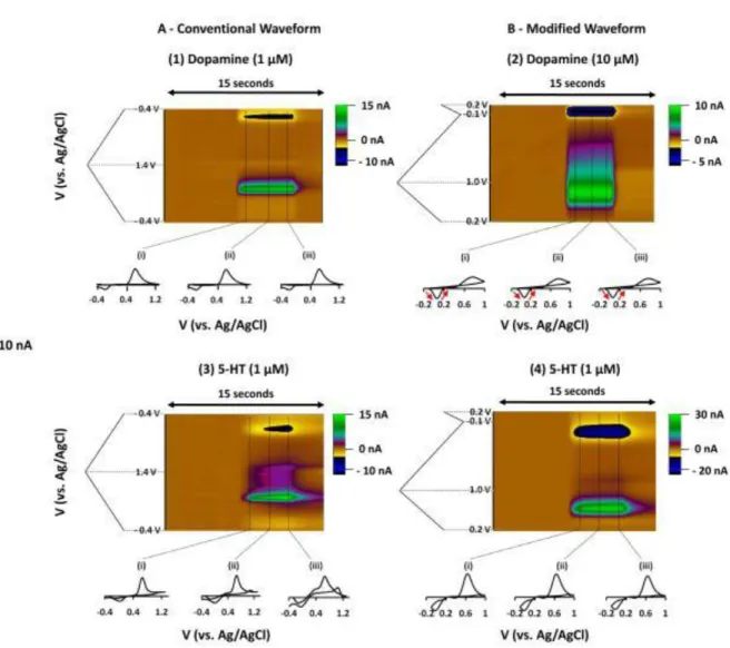
example of a voltage waveform, cyclic voltammograms, and in vitro calibration is shown in Figure 1.1. The “fast-scan” in the technique’s name refers to the potential waveform, which
is applied rapidly and repeatedly, producing up to 10 cyclic voltammograms per second. The
carbon-fiber microelectrode sensors used in FSCV have small dimensions (5 x 100 m), and
this small size enables sampling from as few as 100 synapses at a time, with the electrode
targeted to a discrete brain region. Thus, FSCV is a technique for which temporal and spatial
scales of data collection are compatible with monitoring neurotransmission.
1.1. Brain regions with measurable serotonin release
In brain slices, changes in serotonin concentration can be evoked using local
electrical stimulation in brain regions containing serotonergic neurons or their axonal
projections. The dorsal raphe nucleus (DRN), a tiny hub in the core of the medulla, contains
4
Voltammetric measurements detect serotonin efflux from both axonal and
somatodendritic sites in this region because a subset of serotonergic neurons synapse locally.
Although axonal serotonin release is prevalent throughout the central nervous system,
experiments employing FSCV are typically constrained to brain regions dense with
serotonergic terminals and limited interference from other neurotransmitters and metabolites.
These studies predominantly take place in the substantia nigra, a midbrain region composed
of the pars compacta, packed with dopamine-synthesizing neurons, and the pars reticulata
(SNpr), a networked relay region that includes the densest serotonergic projections from the
DRN to any forebrain region (9 x 10⁶ sites per mm3) (Moukhles et al., 1997). In the SNpr,
serotonin is the predominant electroactive neurotransmitter evoked by electrical stimulations,
frequently observed in the absence of somatodendritic dopamine release (Cragg, Hawkey, &
Greenfield, 1997). However, Moukhles et al. (1997) reported that serotonergic processes
form synaptic junctions at a high rate in the SNpr than any other brain region. It should be
considered, therefore, that serotonin dynamics described in this region may be dissimilar to
the dynamics in other brain regions, including the cerebral cortex, neostriatum, and
hippocampus, where a majority of serotonin terminals form non-junctional synapses
(Descarries et al., 1990). Serotonin efflux has also been described using FSCV in brain slices
containing the suprachiasmatic nucleus (SCN) and ventral lateral geniculate nucleus (vLGN),
hypothalamic and thalamic areas with similarly robust serotonergic innervation.
Serotonin measurements in in vivo FSCV experiments have taken place exclusively in
the SNpr. Thick vasculature and meninges above the DRN make targeting this region in the
intact brain with a fragile carbon-fiber microelectrode difficult. Other serotonergic regions of
5
significant chemical interference from other monoamines. However, recent advancements in
neuronal stimulation technology may help circumvent this problem, and these potential
future directions will be discussed in more detail in the conclusion of this article.
1.2. Electrochemical identification
Electrochemical methods, including FSCV, lack absolute chemical specificity. Some
chemical species, particularly those with similar structure, can interfere with detection of the
desired substance by oxidizing at similar or identical potentials. Therefore, voltammetric
measurements rely on five criteria for identification of endogenously released substances:
First, cyclic voltammograms obtained under experimental conditions must have high
correlation with cyclic voltammograms of the authentic compound. Second, presence of the
neurotransmitter must be validated by independent chemical identification, such as
microdialysis, tissue content analysis, or radioligand binding in the targeted brain region. The
third criterion requires precise anatomical positioning of the sensor into the brain region of
interest. Fourth, observed release should follow known physiological properties for the
neurotransmitter and target brain region. Finally, identification of the released substance is
dependent on pharmacological validation.
O’Connor and Kruk (1991a) systematically addressed the criteria for electrochemical
validation in the first published report of endogenous serotonin measured in rat brain slices
using FSCV (O'Connor & Kruk, 1991a). The cyclic voltammogram obtained from
electrically-evoked serotonin is highly correlated to the one obtained after adding known
concentrations of serotonin to the bath solution. Stimulation trains (500 ms in duration)
6
are located, and SCN, a region dense in serotonin projections from the DRN (Fuxe, 1965).
The evoked concentrations measured in both brain regions was completely and reversibly
abolished by removal of calcium from the buffer solution or addition of sodium-channel
blocker tetrodotoxin, complying with known physiological properties of exocytotic release.
RO 4-1284, an irreversible vesicular monoamine transporter 2 (VMAT2) inhibitor,
attenuated release, confirming that observed release was vesicular in nature. Inhibition of
monoamine oxidase had no effect on stimulated efflux, ruling out interference from
serotonin’s metabolite, 5-HIAA. Finally, the clearance rate of serotonin in the DRN and SCN
could be decreased after application of citalopram, a selective serotonin uptake inhibitor, but
not by benztropine, a norepinephrine uptake inhibitor, to bath solution. Similar procedures
validate the identity of serotonin detected in subsequent experiments by this and other
groups.
Bunin and Wightman (1998) later investigated an aspect of serotonin’s physiological
release properties that had not been addressed by initial voltammetric characterizations. The
dimensions of carbon-fiber microelectrodes are considerably larger than the synaptic cleft
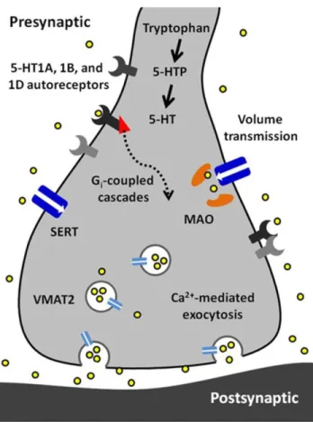
into which neurotransmitters are released (Figure 1.2). Consequently, FSCV detects
extracellular, not intrasynaptic, changes in concentration, and its measurements are limited to
the neurotransmitter concentration that diffuses into the extrasynaptic space following
release. A number of neuromodulators diffuse beyond the synaptic space to reach their
receptors and transporters in a process called volume transmission (Fuxe et al., 2010), and
prior evidence from non-voltammetric techniques implicated serotonin as a volume
neurotransmitter. Ultrastructural studies of serotonergic terminals throughout the brain
7
This terminal architecture, together with reports that expression of serotonin transporters and
receptors occurs primarily on extrasynaptic regions of neuronal processes (Kia, Brisorgueil,
Hamon, Calas, & Verge, 1996; Zhou, Tao-Cheng, Segu, Patel, & Wang, 1998), is indicative
of volume transmission. In light of this information, Bunin and Wightman (1998)
hypothesized that electrically-evoked serotonin should reach the extracellular space via
diffusion, without buffering from uptake and receptor binding sites. This was found to be the
case for both somatodendritic and terminal release, where the concentration of serotonin
evoked per stimulation pulse during 20-pulse trains was equivalent to the concentration
evoked by a single pulse (Bunin & Wightman, 1998). Therefore, the authors concluded that
serotonin concentrations measured by voltammetry reflect physiological volume transmission
from the synapse to its extrasynaptic targets.
1.3. Technical considerations
Since O’Connor and Kruk’s first report of voltammetric detection of serotonin,
several modifications have been implemented to adapt and improve the use of FSCV for
novel applications. The voltage potential waveform (-1 to +1.4 to -1) used by the Stamford
and Kruk labs, as well as others, in serotonin studies cited throughout this review was
adjusted by Jackson and Wightman (1995) to improve temporal resolution. This modification
to an N-shaped waveform, which scans from +0.2 to +1.0 to -0.1 back to +0.2 (Figure 1.1A),
was designed to reduce serotonin adsorption to the electrode surface, as this slows electrode response times. It also avoids fouling reactions of serotonin’s oxidative and reductive
byproducts, improving electrode sensitivity and stability over time (Jackson, Dietz, &
8
times and enabled more accurate measurements of release and uptake rates, facilitating closer
examination of the kinetic parameters of serotonin release.
Improvement of carbon-fiber microelectrode sensors has been another ongoing
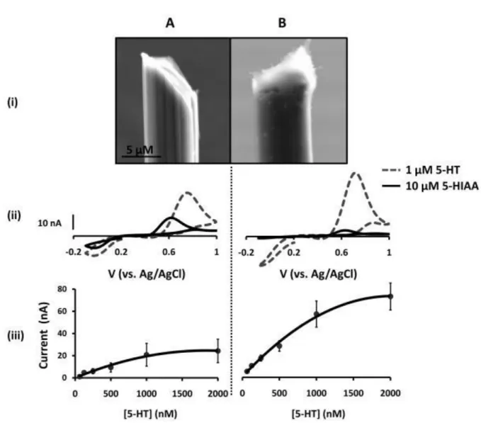
adaptation to voltammetric measurements of serotonin. Brazell and Adams (1987) first
reported that dip-coating a carbon-fiber microelectrode in Nafion, a cation-selective polymer,
improves serotonin and dopamine detection (Brazell et al., 1987; Jackson et al., 1995). Nafion enhances serotonin detection in two ways: first, by directly increasing the electrode’s
sensitivity to (positively-charged) serotonin, and second, by reducing its sensitivity to interfering anionic species such as uric acid and serotonin’s metabolites. Years later, the
success of the first in vivo voltammetric measurements of endogenous serotonin
concentrations in a rat owe their success to the enhanced sensitivity and temporal resolution
facilitated by Nafion-coated sensors and the modified voltage potential waveform (Hashemi,
Dankoski, Petrovic, Keithley, & Wightman, 2009).
It is important to note that many labs continued their investigations of serotonin
release without adopting either modification. Because each study reports on
electrically-evoked changes in serotonin concentration, which are derived from in vitro calibrations,
comparing findings between labs is not considered an issue within this review. It is important
to note that these calibrations do not take into account the deleterious effects of electrode
fouling that may be appreciably different over the course an experiment depending on the
waveform used. Regardless of waveform choice, however, demonstration of a linear
relationship between concentration applied and the current evoked establish the suitability of
9
2. Electrical stimulation
Many of the optimal electrical stimulation parameters for evoking somatodendritic
and terminal serotonin in brain slices are consistent with previously established physiological
principles. Serotonergic fibers are not myelinated and, like other unmyelinated fibers, are
maximally excited by wider stimulation pulse widths, up to 2 ms in length (Anden, Fuxe, &
Ungerstedt, 1967; Bunin, Prioleau, Mailman, & Wightman, 1998; Merrill, Wall, & Yaksh,
1978; Millar, Stamford, Kruk, & Wightman, 1985). The amplitude of evoked serotonin
concentration is also strongly dependent on increases in stimulation intensity (up to 380 A)
and number of pulses in a stimulation train. Maximal release amplitudes are also positively
correlated with increasing frequency, up to 100 Hz (Bunin & Wightman, 1998; Iravani &
Kruk, 1997; O'Connor & Kruk, 1991b), although a detailed investigation by John, et al.
(2006) found that electrically-evoked concentrations were less sensitive to stimulation
frequencies above 30 Hz (John, Budygin, Mateo, & Jones, 2006). These constrained ranges
of frequency dependence could reflect limitations in vesicular availability, but Aghajanian et
al. (1990) has posited that processes in terminal regions store enough serotonin to sustain
long, high frequency release (Aghajanian, Sprouse, Sheldon, & Rasmussen, 1990). Although
serotonergic neurons are typically thought to fire at a rate of 0.5-5 Hz, burst-firing in the
DRN has been measured at a rate of 100 Hz (Aghajanian, Wang, & Baraban, 1978; Hajos,
Gartside, Villa, & Sharp, 1995; Vandermaelen & Aghajanian, 1983). Differences in the range
of frequency sensitivity between studies may therefore reflect dynamic, physiological
fluctuations and could point to yet another regulatory component within the serotonin
system. Future investigation of the mechanisms influencing frequency dependence would be
10
Although voltammetric studies of serotonin have used a wide array of stimulation
parameters, one type has been used repeatedly in the studies reviewed in this article.
Pseudo-one-pulse (El Yacoubi et al.) stimulations consist of 5-10 pulses applied at 100-200 Hz and
are shorter than 100 ms in duration. They are designed to approximate a single electrical
impulse but evoke more consistent efflux. In brain slice experiments, POP stimulations are often used to avoid creating an endogenous “tone” at receptors, which facilitates more direct
investigation of selective agonists and antagonists effects on autoreceptor-mediated
modulation of release (Limberger, Trout, Kruk, & Starke, 1991; Thienprasert & Singer,
1993).
Endogenous serotonin concentrations have been evoked in vivo using electrical
stimulation of the DRN as well as the medial forebrain bundle (MFB). A subset of
serotonergic neurons that project to the SNr also send axon collaterals to forebrain structures
via the MFB (van der Kooy & Hattori, 1980). Electrical stimulation of these collaterals
excites SNr-projecting neurons in a retrograde direction, eliciting serotonin in the desired
region (Hashemi, Dankoski, Wood, Ambrose, & Wightman, 2011). While targeting a
stimulation electrode to the MFB is less challenging than targeting the DRN, this stimulation
site can also be used to evoke neurotransmitter release in many brain regions. This may have
indirect effects on serotonin signaling, complicating interpretation of data. Many optimal
stimulation parameters are consistent between in vitro and in vivo measurements, including
pulse width, stimulation intensity, and stimulation length. However, the concentration of
serotonin evoked in the SNr is remarkably lower than predicted by brain slice measurements,
prompting curiosity about the potential for serotonergic regulatory mechanisms that require
11
3. Release
Local electrical stimulations of serotonin terminals in brain slices typically evoke
concentration changes in the 100 nM range. In vivo, however, serotonin concentrations
evoked in the SNpr rarely reach 100 nM, even after pharmacological manipulations
(Hashemi et al., 2012). In vivo serotonin release, measured in an intact brain, is presumably
limited by negative feedback from somatodendritic and terminal autoreceptors as well as
inhibitory neurotransmitters that are released concurrently, which may account for some of
the disparity in release amplitudes. In brain slices, concentration flux coincides with onset of
the stimulation pulse train and this rising phase reaches its maximum within milliseconds of the stimulation’s end. Serotonin evoked in vivo tends to overshoot the duration of
stimulation. The overshoot is partially an effect of the broader area of release sites activated
by a remote stimulation location, but is also due to limited diffusion rates through a Nafion
polymer coating that is applied to enhance sensitivity in vivo (Hashemi et al., 2009).
As mentioned in a previous section, electrically-evoked serotonin concentrations
measured in brain slices are sensitive to stimulation frequency. A proposal by Wightman et
al. (1988) explains this observation: more uptake occurs in the time between stimulation
pulses during low frequency stimulations, which limits the summation of extracellular
neurotransmitter concentration (R. M. Wightman et al., 1988). Jennings et al. (2010)
hypothesized that shifts in uptake rate associated with differential serotonin transporter
expression would predictably alter this frequency dependence. Mice with either gain or loss
of SERT expression both displayed significantly lower sensitivity to stimulation frequency
than their wild-type littermates. Furthermore, in wild-type mice, a selective serotonin
12
Cragg, 2010). These findings underscore the importance of SERT in establishing a
functional, dynamic equilibrium between release and uptake that enables coherent serotonin
signaling.
Time-resolved measurements with FSCV also enable examination and comparison of
the kinetic parameters of serotonin transmission. Neurotransmitter uptake is assumed to
follow Michaelis-Menten dynamics, and uptake as well as concentration evoked per stimulus
pulse can be calculated using a modified model of enzyme kinetics. Figure 1.3 shows the
equations used to model (i) uptake and (ii) release and representative signals predicted for
stimulations of varying frequency. In brain slice preparations, the concentration evoked per
stimulation pulse ([5-HT]pulse) was found to be 100 ± 20 nM in DRN, and significantly lower
in the SNpr, at 55 ± 7 nM. Differences in [5-HT]pulse are proportional to differences in tissue
content between the two brain regions, indicating that local stores of serotonin may influence
the concentration that can be evoked by electrical stimulation (Bunin et al., 1998). In vivo
[5-HT]pulse in the SNpr is much lower, comparatively: 1.5 nM per pulse using DRN stimulation,
and 1.1 nM per pulse from the MFB. Figure 1.4 shows an averaged recording of in vivo
serotonin signals in the SNr; note that the concentration evoked is strikingly lower than
predicted by the model in Figure 1.3. Given that both Bunin et al. (1998) and Hashemi et al.
(2011) conducted experiments in the SNpr, the nearly 50-fold difference cannot be attributed
to differences in tissue content. Instead, this discrepancy between brain slice and in vivo
preparations suggests powerful regulatory mechanisms acting on serotonin release in vivo
which may depend on intact circuitry.
Hashemi et al. (2012) investigated mechanisms that may limit in vivo
13
efflux in the SNpr and nucleus accumbens, respectively. The dopamine system serves as a
good basis for comparison with the serotonin system because the two monoamines share
parallel features in the mechanisms controlling their synthesis, release, modulation, uptake,
and metabolic degradation. Inhibition of the monoamine synthesis enzyme aromatic amino
acid decarboxylase and VMAT2 considerably decreased the concentration of evoked
dopamine to 18% and 6% of control amplitudes, respectively, but affected serotonin to a
much lesser extent (48% and 72%, respectively). Serotonin efflux was also resistant to short
term depression after repeated stimulation pulse trains, while dopamine efflux was attenuated
by 38% after 20 stimulations. This suggests that a relatively small proportion of the available
vesicular serotonin is mobilized for release by each electrical stimulation train, a finding
which may partially explain the low concentrations observed in vivo.
3.1. Modulation by autoreceptors
Three subtypes of serotonin receptors, all 5-HT1-type, are expressed on serotonergic
axons, soma, and dendrites and function as autoreceptors that provide inhibitory feedback.
5-HT1-type receptors are found throughout the brain as autoreceptors, expressed on
pre-synaptic serotonin terminals, and also as heteroreceptors, expressed on post-pre-synaptic targets.
The most well-studied autoreceptors, 5-HT1A, 1B, and 1D are seven transmembrane,
G-protein coupled receptors (GPCRs). 5-HT1B and 1D autoreceptors negatively couple to
adenylyl cyclase (Yocca & Maayani, 1990). 5-HT1A heteroreceptors throughout the brain
also inhibit adenylyl cyclase activity, but autoreceptors in the DRN apparently function
through a different Gi-coupled mechanism (Clarke, Yocca, & Maayani, 1996). In vivo studies
of these autoreceptors are challenging because even highly-selective drugs inadvertently
14
high levels in the same brain region as the autoreceptor. Intact circuitry thus makes it difficult
to extricate direct effects of autoreceptor activity from indirect regulation by heteroreceptors.
Voltammetric measurements in brain slices avoid some of the problems associated
with 5-HT1-type receptor pharmacology. In slices, the absence of spontaneous activity in
serotonergic cells , due either to separation from cell bodiesin a terminal slice or to
elimination of noradrenergic inputs in a DRN slice, results in loss of endogenous serotonin
tone (Judge & Gartside, 2006). Therefore, these experiments avoid tonic activation of
autoreceptors and can also avoid transient autoreceptor activity, when appropriate, using POP
stimulations. This provides an opportunity to study the timing and function of these receptors in relative isolation. O’Connor and Kruk (1991b) showed that the non-selective autoreceptor
antagonist methiothepin did not affect the concentration of serotonin evoked by POP
stimulations, but increased serotonin elicited by longer stimulations. Further exploration with
stimulations of varying frequency and duration determined that activation of autoreceptors
requires a stimulation period of at least 400 ms (O'Connor & Kruk, 1991b). This time frame
is comparable to the activation window for dopamine autoreceptors in striatal and limbic
regions. Phillips et al. (2002) found that the activation delay observed for dopamine
autoreceptors reflects timing of intracellular cascades added to the rate of neurotransmitter
diffusion in a given brain area (P. E. Phillips, Hancock, & Stamford, 2002).
5-HT1A, 1B, and 1D receptors are expressed at high levels in the DRN, where they
negatively influence neuronal firing rate and extracellular levels of serotonin (Adell, Celada,
& Artigas, 2001; Moret & Briley, 1997; Pineyro, Castanon, Hen, & Blier, 1995; Sprouse &
Aghajanian, 1987). Voltammetric studies corroborate the inhibitory functions of all three
15
amplitude of electrically-evoked serotonin release (Davidson & Stamford, 1995b; Hopwood
& Stamford, 2001). Although its heteroreceptor analogues are prominently expressed in
limbic regions, 5-HT1A autoreceptors are only expressed in the DRN and median raphe
nucleus (Verge et al., 1985). Serotonin levels in forebrain terminal regions are affected by
5-HT1A-mediated changes in DRN unit activity (Casanovas, Lesourd, & Artigas, 1997; Kreiss
& Lucki, 1994), but only 5-HT1B and 1D autoreceptors are expressed locally to functionally
inhibit release in these regions. Voltammetric measurements in the SCN and vLGN confirm
absence of 5-HT1A autoreceptor function in these terminal regions. 5-HT1B and 1D
receptors, and not 5-HT1A receptors, negatively influence serotonin efflux in vLGN brain
slices (Davidson & Stamford, 1996; O'Connor & Kruk, 1992).
The 5-HT1A receptor may be the trump card in this family of autoreceptors: 5-HT1A
receptor mRNA is expressed in nearly 100% of serotonergic cells and up to 15% of
GABAergic interneurons in the DRN (H. E. Day et al., 2004). This receptor robustly
regulates both neuronal firing rates and extracellular serotonin levels in the DRN (Hjorth &
Sharp, 1991; Sprouse & Aghajanian, 1987). Voltammetric measurements find that
antagonists for 5-HT1A and 1B have a supra-additive effect when administered together:
increase in serotonin efflux is greater when both receptors are blocked than would be
expected given the effect of each antagonist alone (Roberts & Price, 2001). In addition, the
effects of 5-HT1B receptor antagonists on serotonin efflux are overpowered by 5-HT1A
receptors unless they are also blocked, suggesting that these receptors compensate for
reductions of 5-HT1B activity. Given these results, it is suggested that 5-HT1A and 1B
receptors exhibit a functional interaction that is facilitated by proximal expression sites on
16
play a role in depression and anxiety-related disorders (Ohno, 2010). Use of FSCV in future
studies could meaningfully contribute to our understanding of how 5-HT1A
receptor-mediated modulation of serotonin release plays a role in the etiology and treatment of these
disorders.
Much speculation has occurred regarding the explanation for seemingly parallel
functions of 5-HT1B and 1D autoreceptors. Both receptors are expressed in most
serotonergic brain regions and have superficially redundant effects. One theory posits that
these autoreceptors differ in their affinity for serotonin: one high affinity and the other low
affinity. However, it has since been demonstrated that their affinities are nearly identical
(Boess & Martin, 1994). More likely, the two receptors are expressed in different anatomical
locations, and thus provide site-specific regulation of serotonin release, e.g. dendritic vs.
axonal localizations in the DRN. Stamford et al. (2000) have reviewed the evidence
supporting this hypothesis (Stamford, Davidson, McLaughlin, & Hopwood, 2000).
The SNr expresses the highest concentration of 5-HT1B autoreceptors and
heteroreceptors in the murine brain (Pazos & Palacios, 1985). 5,7-HT-induced lesions of
serotonin neurons reduced 5-HT1B expression level by 37%, presumably due to degradation
of serotonin terminals (Verge et al., 1986); this suggests that over 1/3 of 5-HT1B receptors
expressed in the SNr could function as autoreceptors. Heterosynaptic function of 5-HT1B
receptors on presynaptic sites in the SNr has been well-documented and may yield important
therapeutic findings (Sari, 2004), but its functionality as an autoreceptor in the SNr remains
controversial. Iravani and Kruk (1997) found no effects of 5-HT1B receptor antagonists on
electrically-evoked serotonin concentrations in SNr slice preparations (Iravani & Kruk,
short-17
term depression of serotonin efflux. In paired stimulation trains, the concentration of
serotonin evoked by the second stimulation (S2) reached 30% of that evoked by the first
stimulation (S1) when there was a 1 second delay between S1 and S2. Antagonists of
5-HT1B receptors relieved this depression by up to 20% (Threlfell, Greenfield, & Cragg,
2010). 5-HT1B autoreceptors are thus apparently functional in the SNpr, although their
modulatory effects may be less robust than in other brain regions.
It is possible that the role of autoreceptors could be better elucidated by in vivo
voltammetric studies, where endogenous serotonin tone is undisturbed and autoreceptor
function is closer to normal physiological levels. However, a limited number of studies currently address the effects of serotonin’s autoreceptors in vivo. In practice, it is difficult to
selectively target 5-HT1-type receptors on serotonin terminals when pharmacologically
indistinct 5-HT1-type heteroreceptors are expressed throughout the brain. As with in vivo
microdialysis, the direct roles of the autoreceptor would be difficult to extricate from indirect
modulation by in situ circuitry. Recent technological advancements in iontophoresis enable
spatially-resolved, quantitative drug delivery at the site of voltammetric measurements (Herr
& Wightman, 2013). Future studies using FSCV combined with this drug-delivery method have great potential to answer important questions about serotonin’s autoreceptors.
4. Uptake
Serotonin clearance is achieved primarily via active transport. Its transporter, SERT,
is a member of the Na+/Cl- transporter family, which includes dopamine, norepinephrine,
GABA, and glutamate transporters (Bennett, Logan, & Snyder, 1973; Iversen, 1974). SERT
displays high affinity for serotonin in the nanomolar concentration range (Blakely et al.,
18
significant target of research efforts for decades, owing to their widespread use as
antidepressant medications. Given acutely, SSRIs exert striking effects on the serotonin
system: they elevate extracellular serotonin levels in the DRN (Bel & Artigas, 1992), which
in turn decreases rate of cell firing due to activation of 5-HT1A autoreceptors (Chaput, de
Montigny, & Blier, 1986; Gartside, Umbers, Hajos, & Sharp, 1995). However, in therapeutic
practice, SSRIs relieve depressive symptoms only after a chronic period of 3-6 weeks. It is
during this period that the effects of transport inhibition on serotonin transmission become
less clear. FSCV provides an ideal method for deciphering the effects of SSRIs because it can
distinguish between changes in released serotonin and changes in rate of uptake.
Electrically-evoked changes in serotonin concentration are cleared from the
extrasynaptic space within seconds of stimulation termination. The term t½ is often used to
compare rates of clearance; t½ is the time elapsed between peak concentration of
neurotransmitter and its decay to half this amplitude. Across brain regions, brain slice and in
vivo voltammetric measurements report similar values of t1/2 ranging from approximately 1 to
3 s (Davidson & Stamford, 2000; Hashemi et al., 2012; Iravani, Muscat, & Kruk, 1999;
O'Connor & Kruk, 1994). Rates of neurotransmitter clearance may positively correlate with
the density of transporter sites in a given brain region: Bunin et al. (1998) report clearance
rates of 1,300 ± 20 nM/s in the DRN and 570 ± 70 nM/s in the SNr, and quantitative
autoradiographic studies report two to four-fold greater SERT binding levels in the DRN
(Kovacevic, Skelin, & Diksic, 2010; Kovachich, Aronson, Brunswick, & Frazer, 1988).
However, some comparisons of SERT density across brain regions do not support this
conclusion, particularly in species other than rat, so more thorough investigation of the
19
influencing uptake rate, brain slice studies in mice that either lack or overexpress SERT have
demonstrated a negative correlation between transporter expression level and concentration
of serotonin evoked by electrical stimulation (Jennings et al., 2010; John et al., 2006). The
disparity observed between clearance rates in the DRN and SNpr is conspicuously
proportional to differences Bunin et al. (1998) reported in release rates. This suggests a
consistent relationship between transporter expression levels, uptake rates, and release rate.
Modeling serotonin signaling kinetics in more brain regions could confirm whether this
relationship holds true throughout the brain.
SSRIs decrease rate of neurotransmitter clearance while increasing the maximum
amplitude of electrically-evoked serotonin concentrations. In brain slices, SERT inhibition
slows clearance (measured as an increase in t½) by 150-700%. This wide spread of responses
may be attributable to experimental variability between studies, particularly differences in
stimulation parameters. Indeed, in vivo studies of SSRI effects in the SNr using identical
stimulation parameters report comparable changes in t½ using MFB and DRN stimulation
sites (increasing by 324% and 306%, respectively)(Hashemi et al., 2012; Hashemi et al.,
2009). SSRIs also increase evoked serotonin concentrations by 200%-450% in SNr brain
slices, and up to 410% in vivo (Hashemi et al., 2012; Iravani et al., 1999; John et al., 2006). In this case, the intensity of the SSRI’s effect is associated with different stimulation
frequencies or pulse number. Structure and selectivity differences between SERT inhibitors
may also contribute to variable responses between voltammetric studies; however,
differences between SSRIs have not been specifically investigated using FSCV. Serotonin
efflux in SNr brain slices has been modeled to describe the effects of an SSRI, fluoxetine, on
20
Vmax and [5-HT]p may be a more effective way to contrast the effects of various SERT
inhibitors on serotonin signaling in future studies in brain slices and in vivo. Thorough
comparison of these effects could inform clinical usage of these pharmacotherapies.
4.1. Autoreceptors mediate some effects of acute uptake inhibition
In addition to their inhibitory influence on release, serotonin’s autoreceptors appear to
modulate response to SERT inhibition. A number of studies report that autoreceptor
antagonists can potentiate the rise in extracellular serotonin levels elicited by SSRIs (Artigas,
Perez, & Alvarez, 1994; Hjorth, 1993), and 5-HT1A autoreceptors also mediate reduction of
firing rate by SSRIs in the DRN (Gartside et al., 1995). The concentration change evoked by
POP stimulations, deliberately rapid enough to avoid creating an endogenous tone, typically
does not activate autoreceptors and is thus not affected by their antagonists. However, in
brain slices of the DRN, paroxetine-induced increases in serotonin efflux were potentiated by
5-HT1A and 1B/D receptor antagonists (Davidson & Stamford, 1995a). Therefore, it is
hypothesized that SERT inhibition causes an increase in extracellular serotonin levels
sufficient to activate autoreceptors, even in brain slices. This produces an inhibitory tone,
such that autoreceptor antagonists can further unmask SSRI-induced increases in release.
5-HT1B and 1D autoreceptors appear to similarly potentiate the effects of SSRIs in distal brain
regions, as the Stamford lab also reports increases in paroxetine’s effects in the vLGN when
co-administered with 5-HT1B and 1D receptor antagonists (Davidson & Stamford, 1997b).
The interaction between regulation of release and uptake functions may also be an important
detail in understanding how chronic uptake inhibition functions in treating depressive
21
4.2. Chronic uptake inhibition
The gap between onset of acute physiological effects and the therapeutic efficacy
achieved in a chronic treatment period implies that SSRI-induced increases in serotonin
levels are not directly producing antidepressant effects. Instead, elevated serotonin levels
may influence long-term changes in serotonin signaling and its downstream targets to relieve
symptoms of depression (Blier, de Montigny, & Chaput, 1987).The effects of long-term
SERT inhibition are conflicting: some find increases in extracellular serotonin levels, and
some find no changes. Associated with these outcomes are variable reports of autoreceptor
desensitization or hypersensitization of 5-HT1A and 1B autoreceptors (Bosker,
Klompmakers, & Westenberg, 1995; Bosker, van Esseveldt, Klompmakers, & Westenberg,
1995; Chaput, de Montigny, et al., 1986; R. Invernizzi, Belli, & Samanin, 1992; R.
Invernizzi, Bramante, & Samanin, 1995; Moret & Briley, 1996). Studies examining the
effects of chronic SSRI treatment using FSCV have produced more consistent findings.
FSCV measurements of serotonin signaling after 21 days of SSRI exposure reveal
that rate of clearance, measured by t½ , is unchanged by this treatment. This lack of change is
intriguingbecause radioligand binding studies report brain-wide reductions in SERT density
after chronic inhibition (Kovacevic et al., 2010). It may reflect compensation by other
clearance mechanisms, such as low affinity serotonin transporters. High and low affinity
transport systems have been described for other monoamine neurotransmitters (Hagan,
Schenk, & Neumaier; Iversen, 1974; Stamford, Kruk, & Millar, 1990; Stamford, Kruk,
Millar, & Wightman, 1984). Although studies suggest that these transporters may play an
important role in serotonin signaling (Daws, 2009), there are presently no FSCV studies
22
transporters in modulating serotonin signaling, particularly following chronic SSRI
treatment, would be interesting to investigate using FSCV.
Long-term SSRI treatment increases stimulation-evoked serotonin concentrations in
the DRN and other brain regions by 20-100%, depending on the experiment and brain region
studied (Davidson & Stamford, 1998, 2000; O'Connor & Kruk, 1994).These findings concur
with the results of Schoups and De Potter (1986), who found that electrically-evoked release
of tritiated serotonin (3[H]5-HT) in the hypothalamus increased after 21 days of SSRI
treatment (Schoups et al., 1986). Although increases in serotonin efflux are observed after
acute SERT inhibition, these can be explained by changes in rate of uptake. However, t½ was
not altered in any voltammetric investigation of long-term SSRI treatment. Therefore,
increases in evoked concentrations induced by chronic treatment must rely on another
mechanism. Changes in other aspects of release may contribute to this effect, for example:
the quantity or composition of serotonin stored in vesicles, regulation of intracellular
calcium, or excitability of the synaptic membrane. In-depth exploration of these mechanisms
has not yet been explored using voltammetric methods.
Alterations in 5-HT1A autoreceptors contribute to the effects of chronic SSRIs on
serotonin signaling. Under normal conditions, activated 5-HT1A receptors inhibit serotonin
release and neuronal firing rates, and chronic SSRI treatment may modify this activity.
Selective suppression of 5-HT1A autoreceptors can produce antidepressant behavioral effects
in the absence of SSRIs (Bortolozzi et al., 2012). Many investigations have described
functional desensitization of 5-HT1A receptors after chronic SERT inhibition, but to varying
degrees across brain regions (Bosker et al., 2001; Cremers et al., 2000; Kreiss & Lucki, 1994,
23
serotonin release and uptake and neuronal firing rates in the DRN of rats treated with water
or paroxetine for 21 days. Paroxetine-treated rats had significantly higher serotonin release
rates but exhibited no differences in firing rate. Interestingly, application of a 5-HT1A
receptor agonist revealed that firing rate was less sensitive, and release amplitude more
sensitive, to this manipulation. Contradictory findings of 5-HT1A receptor sensitivity was
not a total surprise: prior studies found similar desensitization of 5-HT1A receptors in the
control of firing rate after chronic paroxetine treatment (Blier, de Montigny, & Chaput, 1988,
1990; Chaput, de Montigny, et al., 1986), and O’Connor and Kruk (1994) had previously
reported sensitization of 5-HT1A receptors controlling release amplitude. The dichotomous
effect of chronic SSRIs on 5-HT1A receptor sensitization indicates a functional distinction
between the receptors mediating neuronal firing and those controlling release. Given the
complex effects of chronic SSRIs on 5-HT1A autoreceptors in the DRN, it would be
interesting to see how these changes translate to serotonin release in an intact brain.
Currently, however, no studies employing FSCV have examined the effects chronic SERT
inhibition in vivo.
5-HT1B and 1D receptors also desensitize after chronic SERT inhibition, although
the extent to which this occurs appears to vary between brain regions. O’Connor and Kruk
(1994) reported desensitization of 5-HT1B receptors in SCN after chronic treatment with
fluoxetine. In contrast, the Stamford lab found no changes in the sensitivity of 5-HT1B
receptors in the vLGN, instead finding desensitization of 5-HT1D receptors after chronic
paroxetine. This inconsistency may reflect differences in autoreceptor expression in the SCN
and vLGN, or result from difficulty in selectively targeting the 5-HT1B receptor
24
their study). Additionally, while O’Connor and Kruk (1994) found no desensitization of
5-HT1B receptors in the DRN, Davidson and Stamford (2000) later demonstrated that 5-5-HT1B
receptor desensitization was apparent only when the 5-HT1A autoreceptor is antagonized
(Davidson & Stamford, 2000). This adds further weight to the conjecture that 5-HT1A and
1B receptors functionally interact in the DRN.
4.3. Monoamine oxidase
Metabolic degradation of serotonin by the enzyme monoamine oxidase (MAO) also
contributes to serotonin clearance, especially in the developing brain (Cases et al., 1998;
Cases et al., 1995). However, MAO inhibition in brain slices has no reported effect on
release amplitudes or uptake (O'Connor & Kruk, 1991a), a finding used to confirm absence of serotonin’s metabolites from the voltammetric signal. Owesson and Stamford (2002)
showed a greater role of MAO in regulating serotonin efflux using transgenic mice lacking
MAO-A expression. MAO-A is the isoenzyme that preferentially degrades norepinephrine,
epinephrine, dopamine, and serotonin, and mice lacking this enzyme have decreased
neuronal firing rates in the DRN and increased extracellular serotonin levels (Evrard et al.,
2002). In brain slices of the DRN, MAO-A-deficient mice displayed significantly greater
serotonin efflux and reduced clearance rates compared to wild-type controls. Additionally,
the effects of citalopram were smaller and radioligand binding showed significantly lower
expression of SERT in these mice (Owesson et al., 2002). This suggests that serotonin
signaling is subject to regulation by MAO under the right experimental conditions. In vivo
work supports this idea, as a recent study has shown that MAO inhibitors dramatically
increase serotonin efflux in the SNpr (Hashemi et al., 2012). MAO inhibition also has a much
25
role for metabolic degradation in the regulation of serotonin transmission compared to other
monoaminergic systems.
5. Future Directions
Most of the studies reviewed in this article focused on describing the role of
autoreceptors and transporters in modulating serotonin signaling throughout the brain.
However, signaling is also considerably influenced by many other neurotransmitter systems,
including norepinephrine, glutamate, GABA, and a number of neuroendocrine modulators. These external influences are highly implicated in serotonin’s involvement in a number of
psychiatric disorders and, while they have been investigated by other techniques, their
functions have not been fully described using subsecond voltammetric measurements.
Evaluating the effects of external modulatory mechanisms on the subsecond dynamics of
serotonin signaling could provide important clues about their role in neurological disorders.
Ongoing methodological developments continue to progress voltammetric
measurements beyond the current experimental limits. While electrochemical techniques
have been optimized for serotonin detection (Lama, Charlson, Anantharam, & Hashemi,
2012), multi-electrode arrays are being developed which would enable measurements of
multiple neurotransmitters in multiple locations simultaneously. Additionally, iontophoretic
methods adapted for FSCV now enable localized, quantitative drug delivery, enabling
investigation of recording-site specific effects in vivo. FSCV can also be paired with
concurrent electrophysiological measurements to couple information about neurotransmitter
release to single-unit responses of post-synaptic neurons. Iontophoretic and
electrophysiological methods have already been applied to voltammetric studies of dopamine
26
Wightman, 2011), and the Wightman group is currently working to adapt these methods for
serotonin detection.
It has previously been challenging to selectively study serotonin’s autoreceptors in
vivo because homologous receptors are expressed throughout the brain. However, many
novel drug-delivery and transgenic methods have been developed to avoid this type of
complication. DREADDs, designer receptors with exogenous ligands, have been used to
target specific G-protein-activated cascades in serotonergic neurons (Dong, Allen, Farrell, &
Roth, 2010). A light-activated 5-HT1A receptor has been generated that can be expressed
only on serotonergic neurons (Oh, Maejima, Liu, Deneris, & Herlitze, 2010). Furthermore,
transgenic mice and rats offer many opportunities to study signaling in models of
neurological disorders and targeted deletions. The effects of SERT deletion or
overexpression on serotonin signaling have been investigated in brain slices of the SNr but
not in an in vivo preparation. Many conditional knockout mouse models, which avoid confounding developmental effects, are now available for serotonin’s transporter and
receptors. These techniques could lead to more selective targeting and better characterization of serotonin’s receptors and their downstream effectors in combination with voltammetric
measurements.
Voltammetric measurements have, until recently, been limited to brain regions with
high levels of the neurotransmitter of interest and limited presence of other electroactive
compounds. This is because electrical stimulations indiscriminately excite all proximal nerve
terminals. Use of optogenetic stimulation circumvents this barrier by enabling selective
excitation of a specific population of neurons. Channelrhodopsin-2-mediated serotonin efflux
27
The light-evoked efflux is vesicular and subject to regulation by synthesis and uptake
transport in a manner that is similar to mammalian serotonin release (Borue, Condron, &
Venton, 2010; Borue, Cooper, Hirsh, Condron, & Venton, 2009). Selective stimulation of
serotonergic neurons in a mammalian model would permit measurements in brain regions
with significant interference from other electroactive neurotransmitters, such as the
hippocampus.
Finally, while voltammetric measurements of serotonin have presently only occurred
in brain slices and anaesthetized animals, an exciting future direction for research will be
monitoring serotonin signaling in an awake, freely moving animal. FSCV has been used to
measure endogenous dopamine and norepinephrine release in freely moving animals, and this
research has led to groundbreaking information coupling real-time neurotransmission to
specific facets of behaviors. Many questions remain about serotonin’s role in both basic and
complex nervous system processes, and coupling FSCV to relevant behavioral paradigms
may yield important clues about its function.
Conclusion
Serotonin signaling is an important component in the etiology and treatment of many
neurological disorders. By combining sub-second temporal resolution with nanomolar
sensitivity to concentration changes, FSCV has revealed a great deal about dynamic
serotonin transmission. These findings are summarized by the illustration in Figure 1.5.
Studies using voltammetric methods have emphasized the importance of
autoreceptor-mediated inhibitory feedback mechanisms in normal signaling as well as response to SSRIs.
Further, recent in vivo measurements suggest that intact brain circuitry supports the
28
The work presented in this dissertation describes some of the mechanisms that control
serotonin release and uptake in an in vivo preparation. In chapter 2, we present modifications
to existing voltammetric methods which enable detection of endogenous serotonin release in
an intact brain for the first time. In chapter 3, we model this release, finding that it is
significantly smaller than concentrations evoked in brain slices. Chapter 4 describes how the
similar biochemical components of dopaminergic and serotonergic systems differentially
modulate release of the two neurotransmitters, and the work suggests that SERT plays a
significant role in regulating both release and uptake. In chapter 5, we further elucidate this
role, showing how SERT inhibitors can potentiate serotonin release by dysregulating the
control over serotonin signaling that occurs at a neuronal level. Finally, in chapter 6, we
show how long-term SERT inhibition, modeled after chronic antidepressant therapies, causes
enduring plasticity in the regulation of serotonin signaling. Importantly, this effect can be
attenuated by ongoing stress throughout SSRI treatment. Taken together, chapters 5 and 6
suggest that acute responses to SERT inhibition may predicate the therapeutic effects of
long-term treatment. Non-serotonergic neurochemical responses to stress may modify
serotonin signaling following acute and chronic SSRIs in a way that influences chronic
therapeutic outcomes. Future work will focus on how environmental stress is transduced by
the serotonergic system into adaptive behavioral responses.
Support
The authors wish to thank the Electronics Facility at University of North Carolina for
their contribution to selected studies cited in this review. Our work was funded by the
29
Figure 1Figure 1.1 In vitro calibration of microelectrodes
30
Figure 2Figure 1.2 Illustration of a carbon-fiber microelectrode in SNr
31
Figure 3Figure 1.3 A model predicts serotonin release and uptake at different frequencies.
32
Figure 4Figure 1.4 Sample trace of in vivo serotonin release and uptake.
33
Figure 5Figure 1.5 A synopsis of the findings presented in this article.
34
CHAPTER 2: VOLTAMMETRIC DETECTION OF 5-HYDROXYTRYPTAMINE
RELEASE IN THE RAT BRAIN 2
Introduction
5-Hydroxytryptamine (5-HT), or serotonin, is an electroactive indole that acts as an
important neurotransmitter in the brain. It has been the focus of considerable research efforts
over the last 30 years. Despite its implication in several neurological disorders such as
depression and anxiety (Aghajanian & Wang, 1978), for which serotonin-altering
medications are widely prescribed, little is known about its dynamics in the brain.
Limitations in analytical techniques for in vivo 5-HT detection come from a combination of
low endogenous levels and rapid uptake of 5-HT from the extracellular space.
We have previously reported on the use of fast-scan cyclic voltammetry for in vivo
dopamine monitoring (May, Kuhr, & Wightman, 1988; Stamford et al., 1984). Both
dopamine and 5-HT undergo reversible 2-electron oxidations that yield characteristic and
distinguishable cyclic voltammograms. However, unlike dopamine, the oxidative
electrochemistry of 5-HT is complex, yielding many oxidation side-products which adsorb to
the carbon surface on the conventional positive sweeping scan (Wrona & Dryhurst, 1990;
Wrona, Lemordant, Lin, Blank, & Dryhurst, 1986). Even at the high scan rates (400 Vs-1)
used to detect dopamine with fast scan cyclic voltammetry, these oxidation side-products still
2 This chapter previously appeared as an article in Analytical Chemistry. The original citation is as follows:
35
polymerize and produce films that quickly and irreversibly foul the carbon surface. This
fouling has been reported to be minimized on diamond microelectrode surfaces since the
diamond surface expresses significantly less oxygen groups when compared to the carbon
surface.6 These microelectrodes have been used to provide real-time monitoring of 5-HT in
guinea ileum where the endogenous 5-HT levels are relatively high (10-20 μM) (Bertrand,
Hu, Mach, & Bertrand, 2008; Patel, Bian, Quaiserova-Mocko, Galligan, & Swain, 2007).
Neurochemical measurements with diamond microelectrodes however, remain a challenge in
part due to the relatively large size of the microelectrodes (~76 μm diameter) (Park et al.,
2008). 76 μm is larger than inter-capillary distance in the rat brain (~30 μm); in brain tissue,
such devices with dimensions larger than inter-capillary distance have been shown to cause
tissue damage (Silvani et al., 2004). Even modest tissue damage caused by these
microelectrodes may impede the measurements of synaptic 5-HT overflow that carbon fiber microelectrodes (~5 μm) are able to detect.
In previous work, we modified the electrochemical waveform that is routinely used
for dopamine monitoring with carbon fiber microelectrodes to minimize the majority of side
reactions using Nafion-modified carbon-fiber disc microelectrodes (Jackson et al., 1995). We
showed the feasibility of this modification for in vivo use via a model described by Stamford
et al. (1990), where striatal dopaminergic cells are forced to release high levels of 5-HT by
arresting dopamine production and pre-loading dopamine neurons with a 5-HT precursor
(5-HTP) (Bull et al., 1990). Although this was not a physiological situation, it demonstrated that
fast-scan cyclic voltammetry can be applied to 5-HT measurements. This modified waveform
also enables stable detection of 5-HT in tissue slice preparations in experiments that last up
36
Jones, 2007a). However, despite these studies, there remain no in vivo reports of the use of
this approach for endogenous 5-HT detection.
A major physiological difference between tissue slice preparations and the intact
brain is the concentration of extracellular metabolites such as 5-hydoxyindole acetic acid
(5-HIAA). This metabolite is present at 200-1000 times the basal concentration of 5-HT in
serotonin containing regions in the intact brain whereas in tissue slices, extracellular species
such as ascorbic acid, and by the same reasoning 5-HIAA, wash out (Bell, McIlwain, &
Thomas, 1956; Rice, 1999; Ross & Stenfors, 1997; Schenk, Miller, Gaddis, & Adams, 1982).
5-HIAA is an indole with electrochemical oxidation properties similar to 5-HT. Given the
high and persistent baseline extracellular concentrations of 5-HIAA, we hypothesize that in
vivo microelectrode implantation in a 5-HT rich brain region, even in the absence of 5-HT
release, will lead to a quick and profound deterioration of the microelectrode due to electrode
fouling by 5-HIAA.
Because 5-HIAA is negatively-charged in solution, cation exchange polymers such as
Nafion can be deposited onto the microelectrode surface to limit its access. Carbon-fiber
microelectrode discs can be dip-coated with Nafion in a straightforward process due to
electrostatic properties of silica groups on the surface of the glass capillary (Baur, Kristensen,
May, Wiedemann, & Wightman, 1988; Jackson et al., 1995). However, the small surface area
of the discs makes them unsuitable for in vivo use: the microelectrode must have a larger
surface area to sample enough release sites to record a substantial signal since these sites are
not uniformly distributed throughout the tissue. Cylindrical carbon-fiber microelectrodes