Transient activation of mucosal effector immune responses by
resident intestinal bacteria in normal hosts is regulated by
interleukin-10 signalling
Cong Wu,1,2,3 R. Balfour Sartor,2,3 Kehe Huang1 and
Susan L. Tonkonogy2,4
1College of Veterinary Medicine, Nanjing
Agricultural University, Nanjing, China,
2Center for Gastrointestinal Biology and
Dis-ease, University of North Carolina at Chapel Hill, Chapel Hill, NC,3Division of
Gastroenterology and Hepatology, Department of Medicine, University of North Carolina at Chapel Hill, Chapel Hill, NC, and4College of Veterinary Medicine, North Carolina State University, Raleigh, NC, USA
doi:10.1111/imm.12612
Received 11 December 2015; revised 7 April 2016; accepted 21 April 2016.
Correspondence: Susan L. Tonkonogy, Col-lege of Veterinary Medicine, North Carolina State University, 1060 William Moore Drive, Raleigh, NC 27607, USA. Email:
[email protected] and Kehe Huang, College of Veterinary Medicine, Nanjing Agricultural University, Nanjing 210095, China. Email: [email protected] Senior author: Susan L. Tonkonogy
Summary
Interleukin-10 (IL-10) is a key regulator of mucosal homeostasis. In the cur-rent study we investigated the early events after monoassociating germ-free
(GF) wild-type (WT) mice with anEscherichia coli strain that we isolated
previously from the caecal contents of a normal mouse housed under
speci-fic pathogen-free conditions. Our results show that interferon-c (IFN-c)
secreted by mesenteric lymph node (MLN) cells from both IL-10 deficient
mice and WT mice, stimulatedex vivowithE. colilysate, was dramatically
higher at day 4 after monoassociation compared with IFN-csecreted by cells
from GF mice withoutE. colicolonization. Production of IFN-crapidly and
progressively declined after colonization of WT but not IL-10-deficient
mice. TheE. colilysate-stimulated WT MLN cells also produced IL-10 that
peaked at day 4 and subsequently declined, but not as precipitously as
IFN-c. WT cells that express CD4, CD8 and NKp46 produced IFN-c; WT
CD4-positive cells and B cells produced IL-10. Recombinant IL-10 added to E. coli-stimulated MLN cell cultures inhibited IFN-c secretion in a
dose-dependent fashion. MLN cells from WT mice treatedin vivowith
neutraliz-ing anti-IL-10 receptor antibody produced more IFN-c compared with
MLN cells from isotype control antibody-treated mice. These findings show
that a residentE. colithat induces chronic colitis in monoassociated
IL-10-deficient mice rapidly but transiently activates the effector immune system in normal hosts, in parallel with induction of protective IL-10 produced by
B cells and CD4+cells that subsequently suppresses this response to mediate
mucosal homeostasis.
Keywords: germ-free mice; interleukin-10; resident bacteria.
Introduction
The gastrointestinal tract harbours a complex set of microbes, particularly in the large intestine, which is inhabited by approximately 10–100 trillion bacteria com-prised of more than 1000 different species.1–5 Bacteria colonize the intestinal lumen after birth and interact with the host through antigens and adjuvants that stimulate either pathogenic or protective immune responses.1,6
Interleukin-10 (IL-10) is a regulatory cytokine that is produced by a wide variety of cell types including T cells, B cells, macrophages, dendritic cells and epithelial cells.7–12
This cytokine is a key inhibitor of antigen-presenting cell function and so suppresses effector T-cell responses and helps to maintain mucosal homeostasis.12–14Interleukin-10 is associated with immunological tolerance and regulatory T-cell responses.15–17The critical role of IL-10 in maintain-ing intestinal immune regulation is demonstrated by the observation that conventionally housed IL-10-deficient and IL-10-receptor-deficient mice spontaneously develop chronic intestinal inflammation.18,19In parallel, IL-10 pro-duced by T cells is necessary for inhibition of colitis in various murine colitis models.20In addition to T cells, IL-10-secreting B cells activated by resident bacteria induce
regulatory type 1 T cells, suppress effector T cells, and inhi-bit colitis in several murine model systems.21,22Soon after IL-10-deficient mice became available, we re-derived them into germ-free (GF) conditions and observed that the GF cohort, unlike the conventional IL-10-deficient mice, did not develop intestinal inflammation or activation of the gut-associated immune system.23These results demonstrate the importance of resident intestinal microorganisms in the development of intestinal inflammation in IL-10-deficient mice.
Numerous publications report IL-10-mediated inhibi-tion of intestinal inflammainhibi-tion and investigators have iden-tified pivotal molecules in the IL-10 signalling pathway;14,24,25 however, most of these studies focus on chronic inflammation. Few have investigated the immune response at very early time-points after exposure to resi-dent intestinal bacteria. In a previous study,13we examined the early stages of immune activation in experiments designed to determine the role of T cell-derived IL-10 com-pared to antigen-presenting-cell-derived IL-10 in regulating colitis. Our kinetic analysis of cytokine production after adoptive transfer of T cells to specific pathogen-free (SPF) immunodeficient hosts demonstrated the rapid induction of interferon-c(IFN-c) in response to components of resi-dent intestinal microorganisms. This response was subse-quently down-regulated in the presence but not in the absence of IL-10-producing antigen-presenting cells. These results are particularly interesting because the adop-tive transfer model permits evaluation of the parameters of T-cell responses as they develop during homeostatic prolif-eration. El Aidyet al.26,27 observed similarly rapid induc-tion of cytokine gene expression after GF wild-type (WT) mice were colonized with complex murine resident intesti-nal microbiota. We now report results that confirm and extend the previous studies, showing that IFN-cand IL-10 were both produced rapidly after GF WT mice were monoassociated with a single intestinal resident bacteria designatedEscherichia coliNC101 that we isolated from the caecum of an SPF WT mouse. Interferon-c subsequently declined; IL-10 also declined, but not as precipitously as IFN-c. The IFN-cis produced by CD4-positive, CD8-posi-tive and NKp46-posiCD8-posi-tive cells while CD4-posiCD8-posi-tive cells and B cells produce IL-10. In vitro addition of recombinant murine IL-10 suppressed E. coli -induced IFN-c produc-tion, while blocking the IL-10 receptor (IL-10R) in vivo enhanced IFN-c production, confirming that IL-10 is involved in down-regulating the early immune response to a residentE. coliin normal hosts.
Materials and methods
Mice
Interleukin-10-deficient mice on a 129S6/SvEv back-ground, and WT 129S6/SvEv mice were maintained in
GF conditions in flexible film isolators at the National Gnotobiotic Rodent Resource Center at the University of North Carolina, Chapel Hill and the Gnotobiotic Core of the Center for Gastrointestinal Biology and Disease (CGIBD) at North Carolina State University. We confirm GF status each time we remove mice from isolators for an experiment. All mice used were older than 8 weeks of age. Mice were monoassociated with E. coli NC101 by swabbing the mouth and anus with an overnight bacterial culture and were subsequently euthanized at different time-points after initial bacterial colonization. Animal use protocols were approved by the University of North Car-olina at Chapel Hill and the North CarCar-olina State Univer-sity Institutional Animal Care and Use Committees.
Bacterial strain and lysates
The murine E. coli strain designated E. coli NC101 was originally isolated from WT mice housed in SPF condi-tions. Lysates were prepared from E. coli cultures grown in brain–heart infusion broth as described previously.28 Protein concentrations were determined using a standard assay (Bio-Rad Laboratories, Hercules, CA) and sterility of the lysates was confirmed by culturing on brain–heart infusion agar plates. Lysates were divided into aliquots and stored at 80°.
Mesenteric lymph node cell cultures
Mesenteric lymph nodes (MLN) were collected from individual mice, teased gently and the cell suspension was passed through a 40-lm filter (BD Falcon, Franklin Lakes, NJ). Unseparated MLN cells (4 9105 cells per well) were stimulated with 1 or 10lg/ml E. coli NC101 lysate or cultured in medium alone in 96-well flat-bot-tom plates (Costar, Corning, NY) for 72 hr at 37° in a humidified incubator with 5% CO2. Culture medium
was RPMI-1640 (Cellgro, Manassas, VA) supplemented with 5% heat inactivated fetal calf serum, 2 mM L
-gluta-mine, 1 mM sodium pyruvate, 005 mM
2-mercaptoetha-nol and 50lg/ml gentamicin. Culture supernatants were collected for cytokine analysis and frozen in aliquots at
20°.
For evaluation of cytokine-producing cells, MLN cells were stimulated overnight with 10lg/ml E. coli NC101 lysate. Cells were then re-stimulated with 100 ng/ml PMA (Sigma-Aldrich, St Louis, MO) plus 1lg/ml ionomycin (Tocris Bioscience, Minneapolis, MN) for 5 hr. Golgi stop (BD Biosciences, San Diego, CA) was added during the last 4 hr.
Recombinant IL-10 treatment and IL-10R blockade
MLN cultures at 0, 250 pg/ml, 500 pg/ml, 1000 pg/ml, or 2000 pg/ml.
To block IL-10 signalling in vivo, 05 mg per mouse of anti-IL-10R-specific antibody (clone 1B1.3A; BioXCell, West Lebanon, NH) or purified rat IgG1 isotype control antibody (clone HRPH; BioXCell) was injected intraperi-toneally 1 day before, 2 days after and 5 days after monoassociation with E. coli NC101. Mice were eutha-nized on day 7 after E. coli monoassociation. The MLN cells were prepared and stimulated with E. coli NC101 lysate as described above.
Cytokine measurements
To measure the amount of IFN-c or IL-10 secreted by stimulated MLN cells, ELISAs were performed by staff of the Advanced Analytics Core of the CGIBD at the University of North Carolina at Chapel Hill. The ELISA sets for murine IFN-c(catalogue no. 551866) and murine IL-10 (catalogue no. 555252) were from BD Biosciences. Assays were performed according to the manufacturer’s protocols. Cytokine levels were measured in triplicate culture supernatants and amounts were determined by comparison with standard curves generated by using the appropriate recombinant cytokine.
Real-time polymerase chain reaction
Total RNA from transverse colon was extracted using an RNeasy Mini Kit (Qiagen Sciences, Valencia, CA) follow-ing the manufacturer’s instructions and treated with amplification-grade DNase Ι (Qiagen Sciences). First-strand complementary DNA was synthesized using a Tetro cDNA Synthesis Kit (Bioline, Taunton, MA) and treated with Ribonuclease H (Invitrogen, Carlsbad, CA). Quantitative RT-PCR was performed on a QuantStudio 6 Flex Real-Time PCR System (Life Technologies, Carlsbad, CA) using SYBR green (Bio-Rad) to quantify the gene expression of IFN-c, IL-10, IL-1band IL-6. Each sample was analysed in triplicate and the results were normalized to the housekeeping gene GAPDH.
Flow cytometry
Antibodies used to enumerate cell subsets directly after MLN harvest were peridinin chlorophyll protein-labelled anti-mouse CD4, phycoerythrin-labelled anti-mouse CD8, or FITC-labelled anti-mouse CD45R/B220 (BD Bio-sciences). Antibodies used to detect cell surface molecules in intracellular cytokine detection experiments were phy-coerythrin-labelled anti-mouse CD4, CD8, CD19, CD11b and CD335 (NKp46). Alexa Fluor 488 labelled anti-IFN-c or anti-IL-10 were used to enumerate cytokine-producing cells (eBioscience, San Diego, CA). MLN cells were stimu-lated for intracellular cytokine detection as described
above, collected and incubated with anti-CD antibodies for 30 min at 4°. Cells were washed, then fixed and per-meabilized using the BD Cytofix/Cytoperm Fixation/ Permeabilization Kit (BD Biosciences) according to the manufacturer’s instructions before incubating with anti-IFN-c, anti-IL-10, or isotype control antibody for 1 hr at 4°. Cells were washed and analysed on an LSRII flow cytometer (BD Biosciences). We gated on the events for analysis based on forward and side scatter characteristics of viable cells. To determine the proportions of cytokine-positive cells among cell subsets, we gated on the cell-surface-marker-positive populations.
Data analysis
All statistics were performed using GRAPHPAD PRISM 5.0
software (GraphPad, San Diego, CA). We used two-tailed Student’s t-tests for comparisons between two groups. Comparisons between three or more experimental groups were analysed using one-way analysis of varaince with Tukey’s multiple-comparison post-test. P-values <005 were considered significant.
Results
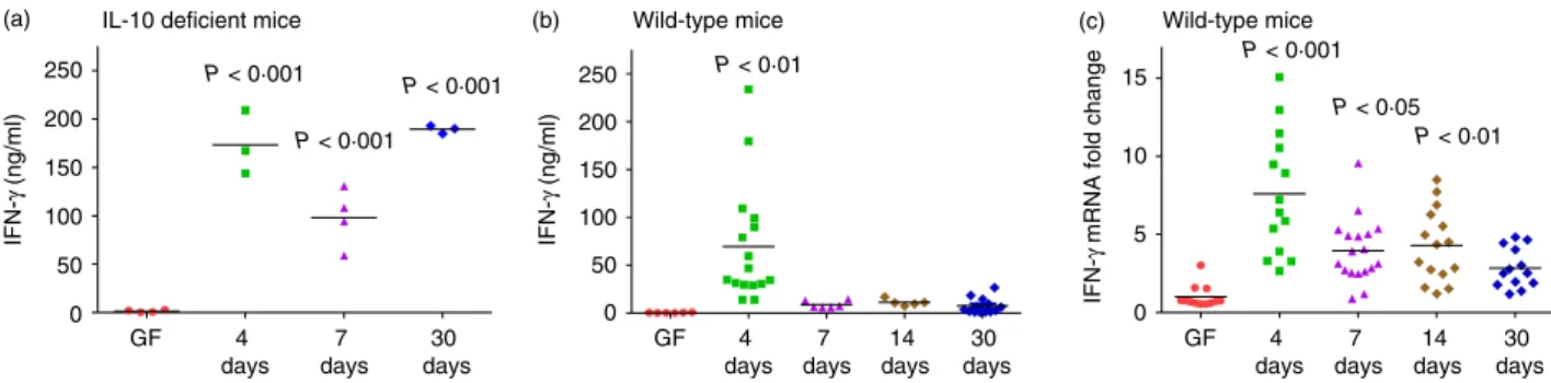
Kinetics of IFN-cproduction inE. coliNC101
monoassociated mice
To evaluate intestinal bacteria-induced immune responses in the intestinal tract upon bacterial colonization, we used 129SvEv WT and IL-10-deficient mice that were born in GF conditions and monoassociated with a non-patho-genic murine strain of Escherichia coli, designated E. coli NC101.28 Escherichia coli NC101 shares phylogenetic and genomic characteristics with several human and canine strains of adherent/invasive E. coli.29 However, our previ-ous results show that this organism is not pathogenic because inflammation does not develop in WT mice.28
in the supernatants of stimulated MLN cells and IL-12p40 production in colon explant cultures. Interleukin-17 secre-tion by MLN cells from IL-10-deficient mice followed the same kinetics afterE. colimonoassociation as IFN-c; how-ever, MLN cells from WT mice did not produce detectable IL-17 (data not shown). Likewise, IL-12p40 in colonic cul-ture supernatants from IL-10-deficient mice increased with time after monoassociation but was undetectable in the supernatants of colon tissue from WT mice (data not shown).
We next investigated IFN-c mRNA expression in the transverse colon of GF and E. coli monoassociated WT mice. The IFN-cmRNA expressed in the transverse colon showed the same pattern as IFN-cproduction in the super-natants of bacterial lysate-stimulated WT MLN cells, with significantly higher IFN-c mRNA levels 4 days after monoassociation withE. colicompared with GF and lower amounts at later time-points (Fig. 1c). Together, these data show that IFN-cis produced in normal, WT mice at early time-points in response to resident intestinal bacteria.
Kinetics of IL-10, IL-1band IL-6 production inE. coli
NC101 monoassociated mice
Interleukin-10-mediated inhibition of IFN-c production has been established in numerous model systems.12,31,32 Therefore, we explored IL-10 production in the same kinetic study. We measured IL-10 secretion in supernatants ofE. colilysate-stimulated unseparated MLN cells and IL-10 mRNA expression in transverse colon from the same monoassociated WT mice. We found that IL-10 secretion in the supernatants was higher at day 4 after colonization compared with GF levels and decreased thereafter (Fig. 2a).
Interleukin-10 mRNA expression in transverse colon from WT mice showed the same patterns with higher levels of IL-10 mRNA at day 4 after monoassociation compared with GF mice, progressively decreasing at later time-points (Fig. 2b). Together, these results show that IFN-cand IL-10 are produced rapidly and concomitantly in normal hosts upon bacterial monoassociation.
We also quantified IL-1b and IL-6 mRNA expression in the transverse colon from monoassociated WT mice (Fig. 3). The IL-1b mRNA expression was approximately fourfold higher 4 days after colonization compared with GF, then decreased at later time-points (Fig. 3a). Inter-leukin-6 mRNA expression (Fig. 3b) was similar to IL-1b, with fivefold higher levels at day 4 compared with GF fol-lowed by a subsequent decline. These data show that like IFN-c and IL-10, both IL-1b and IL-6 are activated rapidly after monoassociation of normal mice with a sin-gle non-pathogenic bacterial species, then are subse-quently down-regulated.
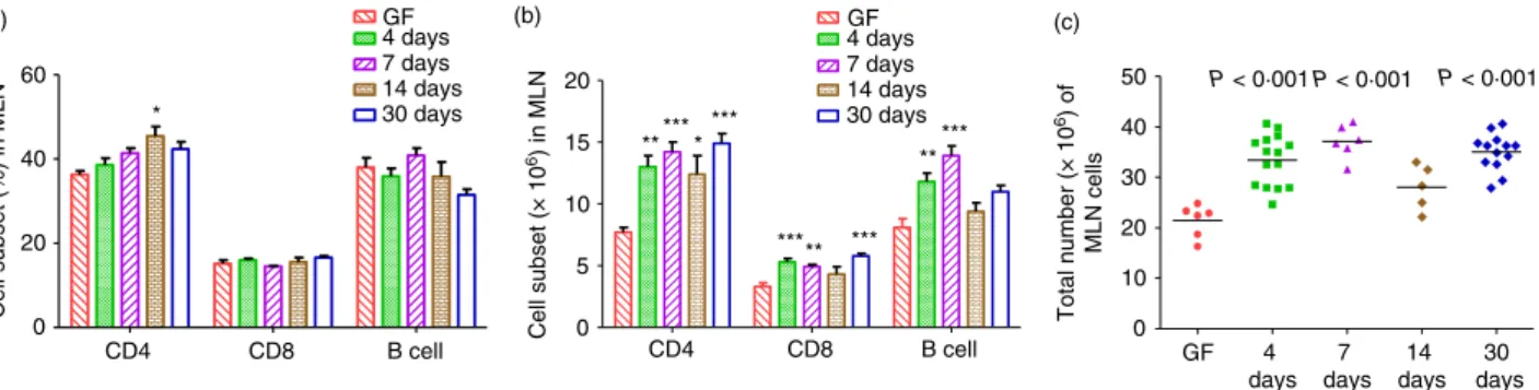
Cell number and phenotype
While the total number of cells obtained from MLN of E. coli monoassociated mice was significantly higher than the number harvested from MLN of GF mice (Fig. 4c), the proportions of CD4-positive cells, CD8-positive cells and B cells in MLN of GF andE. colimonoassociated mice were essentially the same (Fig. 4a), with the exception of a slightly higher percentage of CD4-positive cells at 14 days after colonization with E. coli. Therefore, the increased amounts of IFN-c(Fig. 1b) and of IL-10 (Fig. 2a) secreted by E. coli lysate-stimulated MLN cells obtained at day 4 afterE. colimonoassociation is not the result of changes in
GF GF 4
days 7 days
14 days
30 days 4
days 7 days
30 days
GF 4 days
7 days
14 days
30 days 0
50 100 150 200 250
P < 0·001
P < 0·001 P < 0·001 IL-10 deficient mice
(a)
IFN-γ
(ng/ml)
0 50 100 150 200 250
IFN-γ
(ng/ml)
P < 0·01 Wild-type mice (b)
0 5 10 15
IFN-γ
mRNA fold change
P < 0·05
P < 0·001
P < 0·01 Wild-type mice
(c)
Figure 1. Kinetics of interferon-c(IFN-c) production in 129SvEv wild-type (WT) and interleukin-10 (IL-10) -deficient mice monoassociated with
cell subset proportions among the fixed number of MLN cells (49 105 per well) from GF andE. coli monoassoci-ated mice that were stimulmonoassoci-atedin vitro.
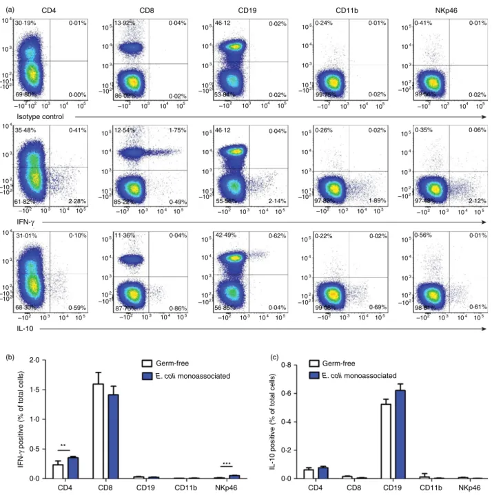
Cell types that produce IFN-cand IL-10
Next we designed experiments to determine which types of cells in MLN produce IFN-c and IL-10 after normal mice are monoassociated with intestinal bacteria. We har-vested MLN cells from WT mice monoassociated with E. coli NC101 for 4 days and re-stimulated them with E. coli lysate overnight, followed by a 5-hr stimulation
with ionomycin plus PMA. For comparison, we also eval-uated MLN cells from GF WT mice stimulated according to the same activation protocol. Representative intracellu-lar cytokine analysis using MLN cells from E. coli monoassociated mice (Fig. 5a) shows that a small propor-tion of CD4+cells and a higher proportion of CD8+cells produce IFN-c. Cells that express the NK cell and innate lymphoid cell marker NKp4633,34also produce IFN-c. IL-10 production can be readily detected in cells that express CD19; a smaller proportion CD4+ cells produce IL-10. Interestingly, the potent activation signals provided by in vitro stimulation with E. coli lysate followed by
0·0 0·5 1·0 1·5 2·0
IL-10 (ng/ml)
P < 0·01 (a)
0 2 4 6 8
IL-10 mRNA fold change
P < 0·01
P < 0·001 (b)
GF 4 days
7 days
14 days
30 days
GF 4 days
7 days
14 days
30 days
Figure 2. Kinetics of interleukin-10 (IL-10) production in 129SvEv wild-type (WT) mice monoassociated withEscherichia coliNC101. (a) IL-10
production by unseparated mesenteric lymph node (MLN) cells from germ-free (GF) orE. coliNC101 monoassociated 129SvEv WT mice. Day after monoassociation is indicated on thex-axis. MLN cells were stimulatedin vitrowith 10lg/mlE. coliNC 101 lysate. Supernatants were col-lected 72 hr later and IL-10 was quantified by ELISA. (b) IL-10 mRNA expression in transverse colon of GF orE. coliNC101 monoassociated 129SvEv WT mice determined by quantitative PCR. Copy number is normalized to GAPDH and expressed as fold change relative to GF. Each dot represents an individual mouse evaluated in experiments described for Figure 1. Data were analysed using one-way analysis of variance with Tukey’s multiple-comparison post-test.P-values indicate statistically significant differences between IL-10 produced byE. coliNC 101 lysate-sti-mulated MLN cells in (a) or IL-10 mRNA expressed in transverse colon in (b) from GF mice compared with mice at 4, 7, 14 or 30 days after E. coliNC101 monoassociation. In addition, statistically significant differences in (a), 4-day versus 7-day not significant, 4-day versus 14-day not significant, 4-day versus 30-dayP<0001; in (b) 4-day versus 7-day not significant, 4-day versus 14-dayP<001, 4-day versus 30-dayP<005.
0 2 4 6 8
IL-1
β
mRNA fold change
P < 0·001
P < 0·01
P < 0·001 (a)
0 5 10 15
IL-6 mRNA fold change
P < 0·001
P < 0·001 P < 0·05 (b)
GF 4 days
7 days
14 days
30 days
GF 4 days
7 days
14 days
30 days
Figure 3. Kinetics of interleukin-1b (IL-1b) and IL-6 expression in transverse colon of 129SvEv wild-type (WT) mice monoassociated with
ionomycin plus PMA are sufficient for detection of cyto-kine-producing cells in MLN of GF mice (Fig. 5b,c). Although the amounts of IFN-c in supernatants of MLN cells from GF mice stimulated withE. colilysate for 72 hr ranged from undetectable to ~06 ng/ml (Fig. 1b), the intracellular IFN-c results show that MLN cells from GF mice have the capacity to produce this cytokine. How-ever, MLN from GF mice contain fewer IFN-c producing CD4-positive and NKp46-positive cells compared with MLN fromE. colimonoassociated mice (Fig. 5b).
We have also determined cytokine-producing cells as a proportion of each MLN cell subset. The results in Table 1 show that although only 044% of MLN cells from mice monoassociated with E. coli express NKp46, an average of 12% of these cells produce IFN-c. A similar proportion of CD8-positive cells produce this cytokine, whereas IL-10 is produced by much lower proportions of CD4-positive (025%) or CD19-positive (14%) cells.
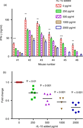
Recombinant IL-10 treatmentin vitro
We then evaluated the effects of exogenous IL-10 on IFN-c production in vitroin our model system. We har-vested MLN cells from WT mice monoassociated with E. coli NC101 for 4 days and stimulated the cells with E. coli NC101 lysate in the presence of 0, 250, 500, 1000 or 2000 pg/ml recombinant IL-10. As shown in Fig. 6, addition of recombinant IL-10 caused a dose-dependent decrease in IFN-c production by MLN cells from each mouse evaluated.
Interleukin-10 receptor blockadein vivo
To determine the role of endogenous IL-10 in the immune response that develops in vivo, we investigated whether blockade of IL-10 signalling will regulate IFN-c
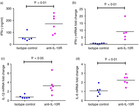
secretion. Either anti-IL-10R-specific antibody or isotype control antibody was given 1 day before, 2 days after and 5 days after colonization of GF mice with E. coli NC101 and the mice were evaluated 7 days after monoassocia-tion. Interferon-c measured in supernatants of E. coli NC101 lysate-activated MLN cells from anti-IL-10R-trea-ted mice was significantly higher than in supernatants of MLN cells from mice given isotype control antibody (Fig. 7a). The IFN-c mRNA expression in the transverse colon from anti-IL-10R antibody-treated mice was also significantly higher compared with isotype-control-treated mice (Fig. 7b). In addition, we detected higher expression of both IL-1band IL-6 mRNA in intestinal tissue of anti-IL10R-treated mice compared with isotype-control-treated mice (Fig. 7c,d). These data show that blockade of IL-10 signalling in vivo by anti-IL-10 receptor antibody increased IFN-c production and also mRNA expression of several cytokines, and indicate that endogenous IL-10, produced at early time-points after exposure to commen-sal bacteria, plays a role in suppressing these immune responses.
Discussion
The intestine of normal adult hosts contains a fully com-petent immune system and the lumen is occupied by enormous numbers of microorganisms that produce numerous components with the potential to activate innate and acquired immune responses. However, the normal intestinal tract is in a state of “physiologic inflam-mation” devoid of overt aggressive responses to luminal microbes and their products. Here we report the results of our studies that were designed to elucidate the immune response in the intestinal tract after initial expo-sure of normal lamina propria host cells and tissues to resident microorganisms. We demonstrate that a single
Cell subset (%) in MLN
CD4 CD8 B cell 0
20 40 60
GF 4 days 7 days (a)
14 days 30 days
GF 4 days 7 days 14 days 30 days *
Cell subset (× 10
6) in MLN
CD4 CD8 B cell 0
5 10 15 20 (b)
** *** ***
*
*** ** ***
** ***
0 10 20 30 40 50
Total number (× 10
6) of
MLN cells
P < 0·001
P < 0·001 (c)
P < 0·001
GF 4 days
7 days
14 days
30 days
Figure 4. Total cell number and cell subsets in mesenteric lymph nodes (MLN) of 129SvEv wild-type (WT) mice monoassociated with
strain of non-pathogenic E. coli rapidly induces a tran-sient innate and adaptive effector immune response in previously GF mice, and that IL-10 produced by B cells and CD4+ cells is a key mediator of the subsequent sup-pression of this response.
We initially embarked on the studies described in this report because we observed an early and then rapidly down-regulated immune response in the WT mice that served as controls for our experiments investigating intestinal inflammation in IL-10-deficient mice colonized
IFN-γ
positive (% of total cells)
IL-10 positive (% of total cells)
2·0 CD4 Isotype control IFN-γ IL-10 6 4 p K N b 1 1 D C 9 1 D C 8 D C 104 105 104 103 103
30·19% 0·01% 13·92%
86·02% 0·02%
0·04% 46·12 0·24%
99·75% 0·41% 99·56% 0·02% 0·02% 0·01% 0·02% 0·01% 53·84% 0·00% 0·35% 97·48% 0·56% 98·81% 0·61% 0·01% 2·12% 0·06% 1·89% 97·83% 2·14% 0·62% 55·56% 85·22% 2·28% 61·82% 69·80% 102
102 103 104 105 –101
–102
–102
–10102 1
105
104
103
–10102 0
105
104
103
–10102 1
105
104
103
–10102 2 104 103 102 –101 –102 104 103 102 –101 –102 0·8 0·6 0·4 0·2 0·0 Germ-free
E. coli monoassociated
Germ-free
E. coli monoassociated
** *** 1·5 1·0 0·5 0·0
CD4 CD8 CD19 CD11b NKp46 CD4 CD8 CD19 CD11b NKp46
105
104
103
–10102 1 105 104 103 102 –102 105 104 103
–10102 1
105
104
103
–10102 1
105
104
103
–10102 2
105
104
103
–10102 2
105
104
103
–10102 2 105 104 103 –102 102
103 104 105
–102 –102 103 104 105 –102 103 104 105 –102 103 104 105
103 104 105
–102
103 104 105
–102 –102 103 104 105 –102 103 104 105
103 104 105
–102
103 104 105
–102 –102 103 104 105 –102 103 104 105 –102 103 104 105 –102 103 104 105
0·22% 42·49% 56·85% 87·75% 11·36% 31·01% 68·30% 0·04% 0·10% 0·49% 0·04%
0·59% 0·86% 99·08% 0·69%
0·02% (a)
(b) (c)
0·02%
35·48% 0·41% 12·54% 1·75% 46·12 0·04% 0·26% 0·02%
Figure 5. Cell types that produce interferon-c(IFN-c) and interleukin-10 (IL-10). Unseparated mesenteric lymph node (MLN) cells from 129SvEv
with normal resident bacteria. Subsequently, in a separate study, we demonstrated a similar phenomenon at the ear-liest time-point evaluated (1 week) after adoptive transfer of IL-10-deficient CD4+T cells into Rag2 / immunode-ficient SPF recipients.13 The Rag2 / recipients were crossed to normal mice or to IL-10-deficient mice provid-ing either Rag2 / 9IL-10+/+ or Rag2 / 9IL-10 / recipients. These mice were born and raised under SPF conditions and therefore harbour a full complement of resident microorganisms, but they lack lymphoid cells before adoptive transfer and so provide an environment for homeostatic proliferation of donor cells. Relatively high and also very similar amounts of IFN-c were pro-duced by MLN cells obtained from both recipient types 1 week after T-cell transfer. By 2 weeks, the MLN cells obtained from IL-10-replete but not IL-10-deficient recip-ients no longer produced IFN-c. These results indicate that the transferred T cells that undergo homeostatic pro-liferation in SPF Rag2 / recipients are rapidly activated to produce IFN-cin vivo by components of the intestinal microbiota, and this early IFN-c response is inhibited by recipient-derived IL-10 produced by non-lymphoid cells.
In the experiments described in the current report, all of the components of the immune system are simultane-ously exposed to luminal bacteria, whereas in the adop-tive transfer model we were able to observe the adapadop-tive immune response of the transferred CD4+ T cells upon their activation in vivo by resident microbiota. The con-clusion is similar in both model systems, namely that
resident bacteria rapidly activate lymphoid cells (primarily CD8-positive cells but also cells that express CD4 or NKp46) to produce IFN-cand that IL-10 plays a key role in subsequent suppression of this response. In the adop-tive transfer model, we concluded that non-lymphoid cells of the Rag2 / recipients produce immunoregulatory IL-10 because the transferred CD4+T cells were obtained from IL-10-deficient donors.13 In the model using E. coli monoassociated WT mice described in the current report, we identified MLN CD4+ cells and B cells that produce IL-10 afterin vitrore-stimulation with E. colilysates. It is likely that other cell types, including myeloid cells and possibly also NK cells and innate lymphoid cells are cap-able of producing 10; however, we did not detect IL-10 production by CD11b-positive MLN cells or NKp46-positive MLN cells fromE. colimonoassociated mice.
Numerous studies have identified a critical role for IL-10 in maintaining intestinal homeostasis.14,35 Both
IL-10-deficient mice18 and IL-10R-deficient mice19 sponta-neously develop colitis that can be ameliorated by admin-istration of exogenous IL-10 immunoglobulin fusion protein that mimics IL-10 activity.36 Our focus on IL-10 in the present study stems from previous investigations using SPF and gnotobiotic IL-10-deficient mice as rodent models of chronic intestinal inflammation.13,23,28 There-fore, in the current experiments, we either provided exogenous recombinant IL-10in vitroor injected anti-IL-10R-specific antibodyin vivo to directly evaluate the role of IL-10 during the initial period after monoassociation
Table 1. Proportion of interferon-c- or IL-10-positive cells in mesenteric lymph node cell subsets
IFN-cpositive cells
Germ-free E. colimonoassociated for 4 days
Percent of subset in MLN1
Percent IFN-c-positive of subset2
Percent of subset in MLN
Percent IFN-c-positive of subset
CD4 32816 074006 31519 113006**
CD8 14807 1068083 12307 1146055
NKp46 039001 355083 044001 1242123***
IL-10 positive cells
Germ-free E. colimonoassociated for 4 days
Percent of subset in MLN
Percent IL-10 positive of subset3
Percent of subset in MLN
Percent IL-10 positive of subset
CD4 32816 017004 31519 025004
CD19 37520 140005 43517 143007
1Values represent meanSEM of cells expressing the indicated cell surface marker evaluated after overnight stimulation withE. coliNC101
lysate followed by 5 hr incubation with ionomycin plus PMA, with Golgi stop during the last 4 hr. MLN fromE. coliNC101 monoassociated micen=4; MLN from GF micen=6.
2Values represent meanSEM of the proportion of IFN-cpositive cells determined after gating on each subset. 3Values represent meanSEM of the proportion of IL-10 positive cells determined after gating on each subset.
of GF mice withE. coliNC101. The results of these stud-ies indicate that IL-10 signalling indeed inhibits IFN-c production and are therefore consistent with the conclu-sion that IL-10 plays a key role in maintaining a quies-cent immune response in the intestinal tract. This conclusion is supported by the observations of Berg
et al.37that systemic administration of recombinant IL-10 in vivocan attenuate the onset of colitis in IL-10-deficient mice and prevent progression of inflammation, but not reverse established colitis.
Our results confirm and extend the comprehensive analyses published by El Aidyet al.26,27 showing immune activation at early time-points after colonizing GF mice with complex faecal microbiota obtained from con-ventionally housed normal mice. Both our study and El Aidy’s results show that (i) the mucosal immune system recognizes and reacts to the components of bacteria that normally reside in the intestinal tract, and (ii) that GF mice have a fully functional immune system, with the capacity to respond rapidly, demonstrated by activation of both innate and adaptive immune responses following bacterial colonization. The maturation from a naive to an activated state proceeds very rapidly upon microbial exposure of GF mice, even when the mice are monoasso-ciated with a single species of non-pathogenic intestinal bacteria, namely E. coliNC101. Of note, E. coli NC101 is an example of an adherent/invasive E. coli strain29 that has the capacity to induce bacterial antigen-specific chronic T-cell-mediated colitis in IL-10-deficient mice.28 As we show in the current report, the same organism can also activate regulatory responses, including production of IL-10 in IL-10 replete WT mice.
Our study expands on that of El Aidy et al. in several key areas. We investigated the kinetics of immune sys-tem activation of normal GF mice elicited by exposure to a single well-studied strain of non-pathogenic E. coli rather than to the multitude of normal resident microbes. In addition, our in vitro and in vivo results identify the key role of IL-10 in suppressing the initial activation of the immune system of normal germ-free mice after bacterial colonization, and we identified B cells and CD4+cells in MLN as the sources of regulatory IL-10. It is also important to note that in our study, GF inbred 129S6/SvEv mice are the hosts. El Aidyet al.used GF C57BL/6 mice. There are many genetic and func-tional differences between these two strains. For example, SPF IL-10-deficient mice backcrossed to C57BL/6 mice are relatively resistant to the development of intestinal inflammation compared with SPF IL-10-deficient mice on the 129S6/SvEv background.37 As another example of the difference between these two inbred strains, the ileum of SPF and GF C57BL/6 mice contains a higher proportion of lysozyme-containing Paneth cells and higher levels of mRNA expression of several isoforms of
a-defensin compared with the same tissue obtained from SPF and GF 129SvEv mice.38 Therefore, demonstrating consistent results using a different inbred mouse strain strengthens the overall conclusion of our study and that of El Aidy et al. that non-pathogenic bacteria first acti-vate and then induce immune regulation in the intestinal tract of normal hosts.
Mouse number
IFN-γ
(ng/ml)
#1 #2 #3 #4 #5 #6
0 50 100
0 pg/ml
250 pg/ml
500 pg/ml
1000 pg/ml
2000 pg/ml rIL-10 added
*** ***
** **
**
** (a)
rIL-10 added pg/ml
Fold change
0 250 500 1000 2000 0·0
0·5
1·0 P < 0·01
P < 0·001
P < 0·001
P < 0·001 (b)
Figure 6. Interferon-c (IFN-c) production by mesenteric lymph
Taken together, our results demonstrate that the gut-associated immune system of normal GF mice recognizes and rapidly responds to a non-pathogenic strain of E. coli, with an initial response that includes production of inflammatory cytokines. This response rapidly pro-gresses to a regulated homeostatic immune profile with low IFN-c, IL-1b and IL-6 levels in the presence of sus-tained bacterial activation of immunoregulatory IL-10. Hence intestinal IL-10 signalling is a key factor in sup-pressing the host’s mucosal effector immune response to resident bacterial components.
Acknowledgements
The authors thank Maureen Bower of the Gnotobiotic Core of the CGIBD and National Gnotobiotic Rodent Resource Center, University of North Carolina at Chapel Hill and Maria Stone, Gnotobiotic Core of the CGIBD at North Carolina State University for GF andE. coliNC101 monoassociated mice. We also thank Anna Yedinak for expert technical assistance and Sarah Schuett, Manager of the Flow Cytometry and Cell Sorting Core, College of Veterinary Medicine, North Carolina State University for flow cytometry assistance.
This work was supported by the National Institutes of Health grants P01 DK094779 and P40 OD010995 (to RBS), by the Center for Gastrointestinal Biology and Dis-ease grant P30 DK034987, the Crohn’s and Colitis Foun-dation of America, and by the China Scholarship Council 2011685032 (to CW).
Author contributions
CW, SLT and RBS conceived and designed the experi-ments; CW and SLT performed the experiments, analysed the data, and wrote the manuscript; RBS provided intel-lectual input, financial support and edited the manu-script; KH provided financial support and reviewed the manuscript.
Disclosures
The authors declare no financial conflicts of interest.
References
1 Kaiko GE, Stappenbeck TS. Host–microbe interactions shaping the gastrointestinal environment.Trends Immunol2014;35:538–48.
IFN-γ
(ng/ml)
Isotype control anti-IL-10R 0
100 200 300
P < 0·01 (a)
IFN-γ
mRNA fold change
Isotype control anti-IL-10R 0
5 10 15 20 25
P < 0·01 (b)
IL-1
β
mRNA fold change
Isotype control anti-IL-10R 0
2 4 6 8
P < 0·05 (c)
IL-6 mRNA fold change
Isotype control anti-IL-10R 0
1 2 3 4
P < 0·01 (d)
Figure 7. Interferon-c(IFN-c) production in 129SvEv wild-type (WT) mice treatedin vivowith interleukin-10 (IL-10) receptor specific
2 Lozupone CA, Stombaugh JI, Gordon JI, Jansson JK, Knight R. Diversity, stability and resilience of the human gut microbiota.Nature2012;489:220–30.
3 Ley RE, Peterson DA, Gordon JI. Ecological and evolutionary forces shaping microbial diversity in the human intestine.Cell2006;124:837–48.
4 Eckburg PB, Bik EM, Bernstein CN, Purdom E, Dethlefsen L, Sargent Met al.Diversity of the human intestinal microbial flora.Science2005;308:1635–8.
5 Savage DC. Microbial ecology of the gastrointestinal tract.Annu Rev Microbiol1977; 31:107–33.
6 Sartor RB. Mechanisms of disease: pathogenesis of Crohn’s disease and ulcerative coli-tis.Nat Clin Pract Gastroenterol Hepatol2006;3:390–407.
7 Rivollier A, He J, Kole A, Valatas V, Kelsall BL. Inflammation switches the differentia-tion program of Ly6Chi monocytes from antiinflammatory macrophages to inflamma-tory dendritic cells in the colon.J Exp Med2012;209:139–55.
8 Saraiva M, O’Garra A. The regulation of IL-10 production by immune cells.Nat Rev Immunol2010;10:170–81.
9 Jarry A, Bossard C, Bou-Hanna C, Masson D, Espaze E, Denis MGet al.Mucosal IL-10 and TGF-bplay crucial roles in preventing LPS-driven, IFN-c-mediated epithelial dam-age in human colon explants.J Clin Invest2008;118:1132–42.
10 Jankovic D, Kullberg MC, Feng CG, Goldszmid RS, Collazo CM, Wilson Met al. Conventional T-bet+Foxp3 Th1 cells are the major source of host-protective regulatory
IL-10 during intracellular protozoan infection.J Exp Med2007;204:273–83. 11 Chirdo FG, Millington OR, Beacock-Sharp H, Mowat AM. Immunomodulatory
den-dritic cells in intestinal lamina propria.Eur J Immunol2005;35:1831–40.
12 Fiorentino DF, Bond MW, Mosmann TR. Two types of mouse T helper cell. IV. Th2 clones secrete a factor that inhibits cytokine production by Th1 clones.J Exp Med1989; 170:2081–95.
13 Liu B, Tonkonogy SL, Sartor RB. Antigen-presenting cell production of IL-10 inhibits T-helper 1 and 17 cell responses and suppresses colitis in mice.Gastroenterology2011; 141:653–62.
14 Shouval DS, Ouahed J, Biswas A, Goettel JA, Horwitz BH, Klein Cet al.Interleukin 10 receptor signaling: master regulator of intestinal mucosal homeostasis in mice and humans.Adv Immunol2014;122:177–210.
15 Moore KW, de Waal Malefyt R, Coffman RL, O’Garra A. Interleukin-10 and the inter-leukin-10 receptor.Annu Rev Immunol2001;19:683–765.
16 O’Garra A, Vieira P. Regulatory T cells and mechanisms of immune system control. Nat Med2004;10:801–5.
17 Roncarolo MG, Gregori S, Battaglia M, Bacchetta R, Fleischhauer K, Levings MK. Inter-leukin-10-secreting type 1 regulatory T cells in rodents and humans.Immunol Rev 2006;212:28–50.
18 Kuhn R, Lohler J, Rennick D, Rajewsky K, Muller W. Interleukin-10-deficient mice develop chronic enterocolitis.Cell1993;75:263–74.
19 Spencer SD, Di Marco F, Hooley J, Pitts-Meek S, Bauer M, Ryan AMet al.The orphan receptor CRF2-4 is an essential subunit of the interleukin 10 receptor.J Exp Med1998; 187:571–8.
20 Asseman C, Mauze S, Leach MW, Coffman RL, Powrie F. An essential role for inter-leukin 10 in the function of regulatory T cells that inhibit intestinal inflammation.J Exp Med1999;190:995–1004.
21 Mishima Y, Liu B, Hansen JJ, Sartor RB. Resident bacteria-stimulated interleukin-10-secreting B cells ameliorate T-cell-mediated colitis by inducing T-regulatory-1 cells that require interleukin-27 signaling.Cell Mol Gastroenterol Hepatol2015;1:295–310.
22 Mizoguchi A, Mizoguchi E, Takedatsu H, Blumberg RS, Bhan AK. Chronic intestinal inflammatory condition generates IL-10-producing regulatory B cell subset character-ized by CD1d upregulation.Immunity2002;16:219–30.
23 Sellon RK, Tonkonogy S, Schultz M, Dieleman LA, Grenther W, Balish Eet al.Resident enteric bacteria are necessary for development of spontaneous colitis and immune system activation in interleukin-10-deficient mice.Infect Immun1998;66:5224–31. 24 Mittal SK, Roche PA. Suppression of antigen presentation by IL-10.Curr Opin
Immu-nol2015;34:22–7.
25 Sabat R, Grutz G, Warszawska K, Kirsch S, Witte E, Wolk Ket al.Biology of inter-leukin-10.Cytokine Growth Factor Rev2010;21:331–44.
26 El Aidy S, van Baarlen P, Derrien M, Lindenbergh-Kortleve DJ, Hooiveld G, Levenez F et al.Temporal and spatial interplay of microbiota and intestinal mucosa drive estab-lishment of immune homeostasis in conventionalized mice.Mucosal Immunol2012; 5:567–79.
27 El Aidy S, Derrien M, Aardema R, Hooiveld G, Richards SE, Dane Aet al.Transient inflammatory-like state and microbial dysbiosis are pivotal in establishment of mucosal homeostasis during colonisation of germ-free mice.Benef Microbes2014;5:67–77. 28 Kim SC, Tonkonogy SL, Albright CA, Tsang J, Balish EJ, Braun Jet al.Variable
pheno-types of enterocolitis in interleukin 10-deficient mice monoassociated with two different commensal bacteria.Gastroenterology2005;128:891–906.
29 Dogan B, Suzuki H, Herlekar D, Sartor RB, Campbell BJ, Roberts CLet al. Inflamma-tion-associated adherent-invasiveEscherichia coli are enriched in pathways for use of propanediol and iron and M-cell translocation. Inflamm Bowel Dis 2014; 20: 1919–32.
30 Patwa LG, Fan TJ, Tchaptchet S, Liu Y, Lussier YA, Sartor RBet al.Chronic intestinal inflammation induces stress-response genes in commensalEscherichia coli. Gastroen-terology2011;141:1842–51.
31 Wolfe DN, Karanikas AT, Hester SE, Kennett MJ, Harvill ET. IL-10 induction by Bordetella parapertussislimits a protective IFN-cresponse.J Immunol2010;184:1392–
400.
32 D’Andrea A, Aste-Amezaga M, Valiante NM, Ma X, Kubin M, Trinchieri G. Interleukin 10 (IL-10) inhibits human lymphocyte interferonc-production by suppressing natural killer cell stimulatory factor/IL-12 synthesis in accessory cells. J Exp Med 1993; 178:1041–8.
33 Spits H, Artis D, Colonna M, Diefenbach A, Di Santo JP, Eberl Get al.Innate lym-phoid cells–a proposal for uniform nomenclature.Nat Rev Immunol2013;13:145–9. 34 Narni-Mancinelli E, Chaix J, Fenis A, Kerdiles YM, Yessaad N, Reynders Aet al.Fate
mapping analysis of lymphoid cells expressing the NKp46 cell surface receptor.Proc Natl Acad Sci USA2011;108:18324–9.
35 Kole A, Maloy KJ. Control of intestinal inflammation by interleukin-10.Curr Top Microbiol Immunol2014;380:19–38.
36 Nguyen DD, Wurbel MA, Goettel JA, Eston MA, Ahmed OS, Marin Ret al.Wiskott–
Aldrich syndrome protein deficiency in innate immune cells leads to mucosal immune dysregulation and colitis in mice.Gastroenterology2012;143:719–29.
37 Berg DJ, Davidson N, Kuhn R, Muller W, Menon S, Holland Get al.Enterocolitis and colon cancer in interleukin-10-deficient mice are associated with aberrant cytokine pro-duction and CD4+TH1-like responses.J Clin Invest1996;98:1010–20.