Brief Definitive Report
Borrelia burgdo~eri
O s p A Is an A r t h r o p o d - s p e c i f i c
T r a n s m i s s i o n - b l o c k i n g L y m e D i s e a s e V a c c i n e
By Aravinda M. de Silva,* Sam R.Telford III,~ Laura Rosa Brunetfl
Stephen W. Barthold,* and Erol Fikrig*
From the *Section of Rheumatology, Department of Internal Medicine, and r Section of Comparative Medicine, Yale University School of Medicine, New Haven, Connecticut 06520-8031; and ~Department of Tropical Public Health, Harvard University School of Public Health, Boston, Massachusetts 02115
Summary
Borrelia burgdo~feri, the spirochetal agent o f L y m e disease, is transmitted by Ixodes ticks. A vac- cine based on B. burgdo~feri outer surface p r o t e i n (Osp) A protects mice from spirochete infec- tion. H e r e w e r e p o r t on the expression o f O s p A on spirochetes inside engorging ticks and re- late O s p A expression to antispirochetal i m m u n i t y . Spirochetes in the gut o f unfed nymphal ticks were stained by an O s p A antibody, whereas in feeding ticks, the majority o f spirochetes in the gut and salivary glands did not stain w i t h the antibody. Thus, O s p A was not expressed on most spirochetes during transmission from the vector to the vertebrate host. T o examine the mechanism o f p r o t e c t i o n afforded b y O s p A antibody, mice w e r e passively i m m u n i z e d w i t h O s p A a n t i b o d y at different times relative to tick attachment. W h e n O s p A antibody was a d m i n - istered to mice before o r at the time o f tick attachment, spirochetal d e v e l o p m e n t events in the vector, such as g r o w t h and salivary gland invasion, were b l o c k e d and the mice were p r o t e c t e d from B. burgdotferi infection. W h e n O s p A antibody was administered to mice 48 h after tick at- tachment, spirochetes persisted in the n y m p h s and the mice were n o t p r o t e c t e d despite the presence o f circulating antibodies in the host as well as in the tick b l o o d meal. Thus, O s p A i m - m u n i t y appears to be effective only during a n a r r o w w i n d o w time at the b e g i n n i n g o f the b l o o d meal w h e n antibodies b i n d to OspA-expressing spirochetes in the tick gut and block transmission from the v e c t o r to the host.
T
he spirochetal agent o f L y m e disease, Borrelia burgdooCeri is transmitted b y Ixodes ticks. In unfed n y m p h a l ticks, spirochetes are f o u n d in the gut (l). D u r i n g tick feeding, the bacteria disseminate to the salivary glands and infect the vertebrate host (2-4). A vaccine based o n B. burgdo~ri outer surface p r o t e i n (Osp) A w h i c h protects mice from in- fection is currently being tested in h u m a n trials (5-7). H e r e w e examine the expression o f O s p A on spirochetes w i t h i n engorging n y m p h s and relate O s p A expression to p r o t e c - tion afforded by O s p A antibody.Materials and M e t h o d s
Mice. Pathogen-free C 3 H / H e N (C3H) mice were purchased from the National Institutes of Health (Bethesda, MD).
Maintenance and Infection of Ticks. Ixodes dammini (also known as L scapularis) were derived from a colony in its second genera- tion from field-collected adults, and were free of inherited infec- tion. Larvae were allowed to engorge on CD-1 mice that had been infected 2 wk previously by the bites of three to five nymphal I. dammini containing the N40 strain of B. burgdotferi. Engorged larvae were collected, held in mesh-covered plaster of
paris containing vials, and were allowed to molt at 95% relative humidity at 21~ The infected nymphs were held under the same conditions before their use in experiments, usually within 2 mo of molting.
Antibody Staining of OspA-expressing Spirochetes in Nymphal Ticks. Guts and salivary glands were dissected out of nymphs and prepared for antibody staining as previously described (2). mAb CIII.78, which binds to a COOH-terminal protective epitope on OspA, was used to assess OspA expression while a rab- bit polyclonal antibody raised against the JD-1 strain of B. burgdor- fen" was used for visualizing spirochetes. The tissues were imaged on a confocal scanning laser microscope (MtLC 600; Bio tkad Laboratories, Microscience Division, Cambridge, MA) equipped with an argon/krypton laser (2). When examining tick organs for spirochetes, the whole organ, including the entire thickness, was evaluated for the presence of spirochetes.
Estimation of Number of Spirochetes Expressing OspA inside Ticks. All internal organs were dissected out of infected nymphs that had either not fed or fed for 70 h on C 3 H / H e N mice. Or- gans from 10 nymphs were pooled in PBS and the tissues were ground using a 1-ml ounce homogenizer (Wheaton Instruments, Millville, NJ) to disrupt the tick cells and tissues while causing minimal damage to the much smaller spirochetes. From each ho- mogenate, a total of four individual 10-1xl drops were placed on
silylated glass slides (PGC Scientific, Gaithersburg, MD) and al- lowed to air dry. Each slide was dipped in acetone for 5 min be- fore staining with an anti-B, burgdo{feri FITC-conjugated rabbit polyclonal antibody or polyclonal rabbit sera against OspA or OspB. Using a conventional fluorescence microscope, the num- ber of spirochetes stained in 10 microscope fields (40• objective) was counted in each 10q*l drop. Four such drops were counted for each antisera and the mean number of spirochetes stained in a 10->1 drop was calculated for each antibody. The antiserum against whole spirochetes was used to estimate the total number of spirochetes in 10 I*1, while the OspA and OspB antisera were used to assess the percentage of the total expressing these Osps.
Evaluation of Mice for B. Burgdotferi Infection and Disease. 2 wk after the detachment of ticks, the mice were killed and selected tissues (blood, spleen, urinary bladder, ear punch, and skin near tick attachment site) were aseptically collected and cultured in Barbour-Stoenner-Kelley II media and examined for spirochetes. For experiments involving the assessment of disease, knees, tibio- tarsi, and hearts were formalin fixed, paraffin embedded, sec- tioned, and examined for inflammation. The sections were exam- ined blindly and an animal was considered to have arthritis or carditis when at least one joint or the heart showed evidence of inflammation.
R e s u l t s a n d D i s c u s s i o n
A l t h o u g h O s p A is an abundant protein on B. burgdofferi g r o w n in culture, vertebrate hosts infected by tick bite rarely seroconvert to O s p A (8). F u r t h e r m o r e , once spiro- chetes have adapted to the host, they are n o t vulnerable to B. burgdo~ri or O s p A antisera (9, 10). O s p A may therefore be differentially expressed during the life cycle o f B. burg~ dotferi in the tick and vertebrate host. Accordingly, w e used confocal fluorescence m i c r o s c o p y to examine O s p A ex- pression on B. burgdo~feri w i t h i n unfed and feeding ticks.
I. dammini were infected w i t h a clonal p o p u l a t i o n o f B. burgdo~ri strain N40. Guts and salivary glands were dis- sected from unfed nymphs and from ticks r e m o v e d from mice after 60 h o f feeding. Spirochetes were restricted to the gut o f unfed nymphs, and were labeled with an O s p A m A b and B. burgdo~eri antiserum, indicating that O s p A was expressed before tick attachment (Fig. 1, A and/3). D u r i n g tick feeding, the nymphs had m a n y spirochetes in c o n c e n - trated arrays in the gut, and smaller numbers in the salivary glands, w h i c h stained w i t h B. burgdo~feri antiserum (Fig. 1, C and E ) but did not react w i t h the O s p A m A b (Fig. 1, D and F ) . A few O s p A - l a b e l e d spirochetes were observed in the guts o f engorged nymphs (data not shown). Thus, O s p A was readily expressed b y B. burgdotferi before tick feeding, but the protein was not present in the majority o f spirochetes during engorgement.
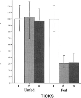
A n estimation o f the p r o p o r t i o n o f B. burgdo~feri express- ing O s p A in engorging n y m p h s was made by directly counting spirochetes that b o u n d different antisera. Equal aliquots o f tick h o m o g e n a t e s were spotted onto glass slides and stained with B. burgdo~eri, OspA, or OspB antisera. T h e B. burgdo{feri antiserum reflected the total n u m b e r o f spirochetes, whereas the O s p A or OspB antisera assessed the percentage o f spirochetes expressing these Osps (Fig. 2). In unfed nymphs, all spirochetes were stained w i t h the
O s p A o r OspB antisera (Fig. 2). In contrast, only 30 and 31% o f B. burgdo~eri from engorging ticks b o u n d the O s p A or OspB antisera respectively (Fig. 2). T h e lack o f b o t h O s p A and OspB staining on the majority o f spirochetes during tick feeding suggests that expression o f these p r o - teins may be regulated at the level o f transcription because the genes e n c o d i n g these two antigens are in the same o p - eron (11).
T h e absence o f O s p A on the surface o f spirochetes d u r - ing transmission p r o m p t e d us to investigate the mechanism b y w h i c h the O s p A vaccine p r o t e c t e d mice. O s p A antise- r u m was injected into C 3 H / H e N mice 24 h before placing four to seven infected n y m p h s o n each mouse. 60 h after attachment, the partially engorged ticks w e r e r e m o v e d from the mice and examined for B. burgdo~ri. 2 w k later, the mice w e r e evaluated for infection. N y m p h s that fed on mice treated w i t h the control (glutathione transferase [GT ]) antiserum were heavily infected and spirochetes were found in the salivary glands (Table 1). In contrast, only two o f seven n y m p h s that had fed on mice injected with O s p A antiserum contained a few B.
burgdo~feri
in the guts. N o n e o f these ticks had spirochetes in the salivary glands (Table 1). M o r e o v e r , mice treated w i t h O s p A antiserum were p r o t e c t e d from B. burgdo~ri infection (Table 1). Therefore, in the presence o f O s p A antiserum, spirochetes in the tick gut were destroyed, and replication and salivary gland inva- sion, critical parts o f the transmission process, were blocked. O s p A antiserum primarily p r o t e c t e d mice b y b l o c k i n g transmission within the vector.Table 1. Effect of OspA Antibody on Transmission of B. Burgdofferi by Nymphal Ticks
Ticks* Infected tick organs*
(antisera used for Infection
murine immunization) Midguts Salivary glands ofmice~
Unfed nymphs 3/4 0/4 NA
Fed nymphs (GT) 6/6 6/6 6/6
Fed nymphs (GT-OspA) 2/711 0/7 0/6
* B. burgdotferi N40--infected L dammini nymphs were divided into three groups. The first group was set aside as an unfed control group. The second group was placed on six C3H/HeN mice (four to seven nymphs per mouse) which, 24 h before the placement of ticks, had re- ceived a control rabbit antiserum raised against .GT (Fed nymphs, GT). The third group of nymphs were placed on C3H/HeN mice (four to seven nymphs per mouse), which had received serum from a rabbit im- munized with a OspA-GT fusion protein (Fed nymphs, GT-OspA). Each mouse received 200 p,1 (100 Ixl i.p. and 100 txl s.c.) of the anti- serum.
r Ticks were removed by forceps 60 h after placement, and midguts and salivary glands were dissected out and examined for spirochetes by staining with a FITC-conjugated rabbit polyclonal antibody raised against B. burgdorferi and performing confocal microscopy.
-~ The mice were killed 2 wk after tick removal and checked for infec- tion by attempting to culture spirochetes from blood, spleen, urinary bladder, and ear skin near the tick attachment site.
II Very few spirochetes were observed in these two infected midguts. NA, not applicable.
Figure 1. Confocal fluorescence images of B. burgdooCeri in tick organs stained for OspA expression. (A and B) The same field of a gut from an unfed in- fected nymph. (C and D) The same field of a gut from an infected nymph that had fed for 60 h. (E and F) are the same field of a salivary acinus from an infected nymph that had fed for 60 h. A, C, and E display the FITC signal from a rabbit serum raised against whole spirochetes, while B, D, and F display the Texas red signal from an mAb that binds to OspA. OspA antibody staining was readily detected on spirochetes before tick feeding. During engorge- ment, the majority failed to stain with the OspA antibody. Bar, 12.5 p~m.
B e c a u s e O s p A is differentially expressed d u r i n g tick f e e d i n g (Figs. 1 and 2), w e assessed B. burgdooCeri transmis- sion w h e n O s p A a n t i s e r u m was a d m i n i s t e r e d at various times d u r i n g tick e n g o r g e m e n t . I n f e c t e d n y m p h s w e r e p l a c e d o n m i c e and at various times relative to a t t a c h m e n t
( - - 2 4 , 0, 24, 48, and 72 h) O s p A a n t i s e r u m was passively a d m i n i s t e r e d to t h e animals. T h e n y m p h s fed to r e p l e t i o n , d e t a c h e d f r o m t h e m i c e , and 10 d later, t h e y w e r e e x a m - i n e d b y i m m u n o f l u o r e s c e n c e m i c r o s c o p y for spirochetes.
Figure 2. OspA and OspB are both not expressed on spirochetes within feeding nymphs. To estimate the number of spirochetes that have OspA and B on the surface before and during tick feeding, the organs were dissected out of infected nymphs that had not fed (10 nymphs) or that had fed for 70 h (10 nymphs). Homogenates were prepared from the dissected organs and stained with a polyclonal antibody raised against whole spirochetes (1), or polyclonal sera raised against OspA (2) or OspB (3) to estimate the percentage of spirochetes expressing OspA and OspB.
from mice given O s p A antiserum 24 h before or during tick placement (Table 2). W h e n O s p A antiserum was ad- ministered to mice at the 24-, 48- and 72-h time points, spirochetes remained in all nymphs, indicating that the dis- appearance o f O s p A begins b e t w e e n 0 and 24 h after at- tachment. Infected n y m p h s at the 2 4 - h time p o i n t had l o w - g r a d e infections (Table 2), suggesting that these ticks had a mixture o f B. burgdooCeri, including organisms w i t h and w i t h o u t OspA. Selective destruction o f the spirochetes
expressing O s p A m a y therefore have led to a r e d u c t i o n in the overall n u m b e r o f B. burgdo~feri. A t later time points (48 and 72 h), as m o r e spirochetes failed to express OspA, the n y m p h s remained heavily infected (Table 2).
B. burgdo~feri were not cultured from mice that w e r e treated w i t h O s p A antiserum at - 2 4 and 0 h relative to tick attachment (Table 2). M i c e w e r e p r o t e c t e d even w h e n antiserum was administered 24 h after attachment, despite a l o w level o f spirochete infection w i t h i n the n y m p h s (Table 2). T h e absence o f host infection at the 2 4 - h time p o i n t may be caused, in part, b y the large decrease in the p o p u l a - tion o f spirochetes in these n y m p h s (Table 2). In contrast to the 2 4 - h time point, w h e n O s p A antiserum was adminis- tered 48 and 72 h after tick attachment, all mice b e c a m e in- fected and d e v e l o p e d disease (Table 2), suggesting that the spirochetes entering mice did not express O s p A and were unaffected b y circulating O s p A antibody.
After n y m p h a l tick attachment, spirochete transmission requires ,'~ h, and during this time period, B. burgdogCeri multiply and cross the gut epithelial barrier into the h e m o - lymph, disseminate to the salivary glands, and infect the host via tick saliva (2, 3, 12). O u r results d o c u m e n t that during tick engorgement, OspA, w h i c h is abundantly ex- pressed on spirochetes in unfed ticks, is no longer expressed on the majority o f B. burgdooCeri in the vector. T h e loss o f O s p A probably begins ,'~ h after tick attachment, since spirochetes resistant to O s p A antiserum were first detected at this time. T h e absence o f O s p A appears to continue even after the spirochetes enter the m a m m a l i a n host because mice were infected w h e n O s p A antiserum was adminis- tered 48 and 72 h after tick attachment (Table 2). A recent report demonstrated that O s p C is absent from the spiro- chetes in unfed ticks, and is present in feeding ticks and in- fected mice (13). D u r i n g tick engorgement, w e observed that spirochetes in the gut and salivary glands that do not express O s p A react w i t h O s p C antiserum (data not shown).
T a b l e 2. Effect of OspA Antibody Administered to Mice at Different Times after Tick Attachment
Antibody*
(time administered relative to tick placement)
Infected nymphs* (severity of infectionll
Infection of mice~
Culture Disease
Anti-GT serum ( - 2 4 h) A n t i - G T - O s p A serum ( - 2 4 h) A n t i - G T - O s p A serum (0 h) A n t i - G T - O s p A serum (24 h) A n t i - G T - O s p A serum (48 h) A n t i - G T - O s p A serum (72 h)
5/7 (3, heavy; 2, moderate 0/6
1/7 (1, low) 6/6 (6, low)
9/9 (7, heavy; 2, moderate 7/7 (2, heavy; 2, moderate; 3, low)
4/4 3/3
0/4 0/4
0/4 0/4
0/4 0/4
4/4 4/4
4/4 4/4
*Four mice were used in each group and four B. burgdotferi N40-infected I. dammini nymphs were placed on each mouse. The groups of mice were infused with 200 txl (100 txl i.p. and 100 Ixl s.c.) of serum from a rabbit immunized with a rt OspA-GT fusion protein (GT-OspA) at the indicated times before or after tick attachment. A control rabbit serum raised against GT was used at the - 2 4 h time point.
* 10 d after detachment, ticks were examined by indirect immunofluorescence microscopy for spirochetes.
The mice were killed 2 wk after tick detachment for culture (blood, spleen, urinary bladder, ear, and tick attachment site) and histopathology (eval- utation of the joints and hearts for arthritis and/or carditis).
II To distinguish nymphs with low, moderate and heavy infections the number of spirochetes in 10 (100X objective) microscope fields were counted. Nymphs with <20 B. burgdo~eri were characterized as having low, 21-50 as moderate, and 51-200 as heavy infection, respectively.
Thus, as spirochetes m o v e from the vector, m a n y appear to be " c h a n g i n g their coat" by, at least in part, differential ex- pression o f O s p A and O s p C . D u r i n g engorgement, t e m - perature shifts and b i o c h e m i c a l changes in the tick m a y di- rectly or indirectly influence Osp expression (13, 14).
T h e loss o f O s p A expression b y spirochetes during trans- mission has revealed the m e c h a n i s m by w h i c h the O s p A vaccine protects vertebrate hosts: antibody binds to the OspA-expressing B. burgdo~feri in the tick gut and prevents their replication and subsequent dissemination to the sali- vary glands and, ultimately, to the vertebrate host. This is
the first example o f a vaccine u n d e r g o i n g h u m a n clinical trims that protects b y blocking transmission from the v e c - tor. A p p r o x i m a t e l y 24 h after tick attachment, some o f the spirochetes in the v e c t o r b e c o m e resistant to vaccine- i n d u c e d O s p A i m m u n i t y , probably because they no longer express O s p A on their surface. By 48 h after tick e n g o r g e - ment, B. burgdooCeri that had entered mice w e r e fully resis- tant to O s p A antiserum. Thus, the O s p A vaccine is likely to be effective only during a n a r r o w w i n d o w o f 24 h after tick attachment, w h e n O s p A antibody comes into contact w i t h OspA-expressing spirochetes in the tick gut.
We thank D. Beck and R. Bangham for technical assistance.
This work was supported by grants from the National Institutes of Health (AI-49387 to E. Fikrig, AI- 30548 to S. W. Barthold, and AI 19693 to S. R. Telford), NIH training grant A R 07107 (A. de Silva), Ar- thritis Foundation (E. Fikrig), Pew Scholars Program in the Biomedical Sciences (E. Fikrig), and Smith- Kline Beecham Pharmaceuticals (to S. R. Telford and L. R. Brunet).
Address correspondence to Erol Fikrig, Yale University School of Medicine, Section of Rheumatology, P.O. Box 208031, 333 Cedar Street, New Haven, CT 06520-8031.
Received for publication 9 August 1995.
R e f e r e n c e s
1. Burgdoffer, W., J.F. Anderson, L. Gem, R.S. Lane, J. pies- man, and A. Spielman. 1991. Relationship ofBorrelia burgdor- feri to its arthropod vectors. Scand. J. Infect. Dis. Suppl. 77:35-
40.
2. de Silva, A.M., and E. Fikrig. 1995. The growth and migra- tion of Borrelia burgdotferi in Ixodes ticks during blood feeding. Am.J. Trop. Med. Hyg. 2909-2913.
3. Ribeiro, J.M., T.N. Mather, J. Piesman, and A. Spielman. 1987. Dissemination and salivary delivery of Lyme disease spirochetes in vector ticks (Acari: Ixodidae). J. Med. Entomot. 24:201-205.
4. Zung, J.L., S. Lewengrug, M.A. Rudzibnska, A. Spielman, S.R. Telford, and J. piesman. 1989. Fine structural evidence for the penetration of the Lyme disease spirochete Borrelia
burgdo~feri through the gut and salivary tissues of Ixodes dam- mini. Can.]. Zool. 67:1737-1748.
5. Barbour, A.G., S.L. Tessier, and W.J. Todd. 1983. Lyme dis- ease spirochetes and Ixodid tick spirochetes share a common surface antigenic determinant defined by a monoclonal anti- body. Infect. Immun. 41:795-804.
6. Fikrig, E., S.W. Barthold, F.S. Kantor, and R.A. Flavell. 1990. Protection of mice against the Lyme disease agent by immunizing with recombinant OspA. Science (Wash. DC). 250:553-556.
7. Simon, M.M., U.E. Schaible, M.D. Kramer, C. Eckerskom, C. Museteanu, H.K. Muller-Hermelink, and R. Wallich.
1991. Recombinant outer surface protein A from Borrelia burgdopCeri induces antibodies protective against spirochetal in- fection in mice.J. Infect. Dis. 164:123-132.
8. Aguero-Rosenfeld, M.E., J. Nowakowski, D.F. McKenna, C.A. Carbonaro, and G.P. Wormser. 1993. Serodiagnosis in early Lyme disease. J. Clin. Microbiol. 31:3090-3095.
9. Barthold, S.W., E. Fikrig, L.K. Bockenstedt, and D.H. Pers- ins. 1995. Circumvention of outer surface protein A immu- nity by host-adapted Borrelia burgdo~eri. Infect. Immun. 63: 2255-2261.
10. Shih, C., A. Spielman, and S.R. Telford. 1995. Short report: mode of action of protective immunity to Lyme disease spi- rochetes. Am.J. Trop. Med. Hyg. 52:72-74.
11. Margolis, N., and P.A. Rosa. 1993. Regulation of expression of major outer surface proteins in Borrelia burgdooCeri. Infect. Immun. 61:2207-2210.
12. Piesman, J. 1993. Dynamics of Borrelia burgdooCeri transmission by nymphal Ixodes dammini ticks. J. Infect. Dis. 167:1082- 1085.
13. Schwan, T.G., J. Piesman, W.T. Golde, M.C. Dolan, and P.A. Rosa. 1995. Induction of an outer surface protein on Borrelia burgdotferi during tick feeding. Pro& Natl. Acad. Sci. USA. 53:397-404.