Incidental Findings Found In Typical Screening Populations Undergoing Colorectal Cancer Screening with Computed Tomography Colonography: A Systematic Review
By
Andrew M. Moon
A Master’s Paper submitted to the faculty of the University of North Carolina at Chapel Hill
in partial fulfillment of the requirements for the degree of Master of Public Health in
the Public Health Leadership Program
Chapel Hill 2012
Advisor Signature
Advisor Printed Name Date
Second Reader Signature
- 1 -
Abstract:
Background: CT colonography (CTC) is a noninvasive technology used to screen for colorectal cancer. Unlike other screening modalities, CTC provides a view of the abdomen and pelvis allowing radiologists to detect lesions in extracolonic organs. There is much debate on the
balance of potential benefits versus potential harms of discovering, working up and treating these extracolonic findings. This debate might be especially relevant for asymptomatic populations receiving screening with CTC.
Purpose: This systematic review aims to determine the frequency and clinical implications of finding incidental, extracolonic lesions during CT colonography (CT) in asymptomatic, screening populations. In addition, this review reports the frequency and clinical outcomes of clinically important lesions. Lastly, this review summarizes the various methods studies used to define the clinical significance of incidental findings.
Data Sources: I carried out a systematic search of MEDLINE, Embase, the Cochrane Clinical Trials databases and published reviews up to March 2012.
Study Selection: Two investigators independently reviewed 282 abstracts and 53 full text articles using a set of predefined inclusion and exclusion criteria. Both reviewers carried out independent critical appraisals of each study using criteria developed by the United States Preventive Services Task Force.
Data Extraction: One reviewer extracted information on study samples, designs, populations, interventions and outcomes from six studies. A second reviewer verified this information for accuracy.
Data Synthesis: The frequency of extracolonic findings (ECFs) ranged from 27.2% to 68.9% (mean 49.3%). Included studies used similar classification systems of clinical importance, which were primarily based on the likelihood of clinical workup. Studies reported that 5.6% of the reported ECFs were of high clinical importance and 15.5% of lesions were either moderate- or high-importance. A minority of these findings represented lesions that could have benefitted from early diagnosis and intervention. Studies reported that 0.09% to 1.2% of subjects were diagnosed with AAAs and 0.23% to 0.88% were diagnosed with extracolonic cancers. Studies used widely varying lengths and methods of following ECFs, making it difficult to estimate the true clinical implications of incidental findings. However, the range of moderate/high to high-importance findings (5.6% to 15.5%) provides a good estimate of the number of subjects requiring some clinical workup.
- 2 -
the included literature does not address how ECFs are handled in non-academic settings. This systematic review also had several weaknesses. The decision to limit the review to screening populations might reduce the strength of my findings. I attempted to compensate by including populations at high risk of CRC and studies conducted outside the US, but this might have reduced the generalizability of my findings. Furthermore, I were unable to adjust for different follow-up time periods, making it difficult to compare the clinical outcomes of ECFs among included studies. Lastly, I attempted to develop an outcomes table for ECFs from screening CTC, but were unable to do so because of the imprecision of results, variable periods of follow-up and gaps in reported outcomes.
Conclusions: Based on these results, a large proportion of individuals receiving CT colonography for colorectal cancer screening will have an extracolonic lesion discovered. Roughly one-fifth to one-third of these findings will receive some clinical workup and the majority of these will ultimately be diagnosed as benign. Since a small percentage of potentially important findings will result in clinical benefit, it is possible that the classification systems are overly sensitive. In addition, the reporting of all extracolonic findings might result in
- 3 -
Introduction
Statement of Purpose:
CT colonography (CTC) is a noninvasive technology used to screen for colorectal
cancer.1 Unlike alternative screening modalities such as colonoscopy, CTC provides a view of the extracolonic abdomen and pelvis, which allows radiologists to detect lesions in several
abdominal organs including the kidneys, liver and pancreas. For some, such incidental findings
might be viewed as serendipitous discoveries that allow earlier intervention and improved
outcomes. Alternatively, detecting such lesions could lead to harms such as unnecessary
diagnostic workups, interventions and patient anxiety. In addition, the radiologic surveillance or
intervention for extracolonic findings could carry significant financial costs that might affect the
cost-effectiveness of CTC as a screening tool.2
In this systematic review, I examined the frequency and clinical implications of
incidental lesions detected during screening CT colonography (CTC). Characteristics of study
populations and CTC techniques are reported to indicate which factors might influence the
frequency of incidental findings. In addition to reporting the overall frequency of these findings,
I provide estimates of the reported clinical importance of these findings. Many studies on
incidental findings have categorized lesions as high, moderate and low clinical importance and
the Working Group on Virtual Colonoscopy have proposed a similar classification system.3 These categories are designed to inform clinical care with higher importance findings requiring
surveillance or immediate intervention. In this review, I systematically assessed the methods
study authors used to classify findings into categories of clinical importance. The methods used
for categorizing incidental findings provides important context for assessing the reported
- 4 -
systems to determine how they might affect the frequency and workup of extracolonic findings
and whether they are likely to reduce variability in clinical practice.
This systematic review aims to inform radiologists and primary care physicians of the
likelihood of finding incidental lesions during screening CTC, the clinical outcomes of detecting
these lesions and the usefulness of classifying these findings by clinical importance. The last
systematic review on the frequency and implications of incidental lesions was published in 2005
and included 3 studies focusing on screening populations, although some of these studies
included a large percentage of symptomatic patients.4 This previous review was thus unable to make a conclusion on the frequency and clinical implications of extracolonic findings in pure
screening populations. My review, on the other hand, focuses specifically on asymptomatic
populations that more closely resemble a general screening population for CTC. I thought it was
particularly useful to focus on an asymptomatic population since it is possible that patients’
symptoms could be attributable to incidental lesions. In addition, there are many more ethical
issues raised when diagnosing asymptomatic, healthy patients with findings detected
incidentally. My review should also provide a unique view on classification systems for judging
the clinical importance of extracolonic findings. Only one review, published in the Journal of
Law, Medicine, and Ethics,5 has systematically assessed how study authors judge clinical
significance of incidental findings. Therefore, this systematic review will provide an updated
view of the frequency and clinical outcomes of incidental findings from CTC in a screening
population and report how study authors classify the clinical importance of extracolonic lesions.
I believe that such a review is important as lower radiation doses are used for CTC,6 as recommendation statements on interpreting and managing extracolonic findings are
- 5 -
Problems with Current Screening Technologies for Colorectal Cancer:
Colorectal cancer (CRC) is the third most common cancer and the second leading cause
of cancer deaths in the United States.9 CRC has a well-characterized preclinical period during
which most tumors develop from precursor lesions. In addition, early detection and treatment of
CRC reduces its mortality. All of these characteristics make it an appropriate candidate for
screening.
Screening for CRC has been a significant factor in its declining incidence and mortality
in recent years.10,11 Consequently, the United States Preventive Services Task Force, the
American College of Radiology and the US Multi-Society Task Force on Colorectal Cancer
(USMSTF) all recommend colorectal cancer screening for individuals starting at age 50 or
earlier for those with certain inherited syndromes or inflammatory bowel disease.12,13 Despite its
demonstrated benefit and supporting recommendations, CRC screening is being underutilized by
huge numbers of American adults aged 50 and older.14 There are many reasons for this,
including the drawbacks associated with each individual screening technique.
Colonoscopy is an increasingly preferred screening technology,14 potentially because of negative coverage decision by the Centers for Medicare and Medicaid Services15 and similar
decisions by many other private insurers to cover colonoscopy.16 Colonoscopy also has high public perceptions of its accuracy17 and the demonstrated benefits of high sensitivity, high
specificity and ability for immediate polyp removal.13 A long-term follow-up study of the National Polyp Study cohort reported a 53% reduction in mortality from colonoscopy and
polypectomy.18 However, colonoscopy has several drawbacks that might reduce compliance
- 6 -
during the procedure and the risk of serious complications such as bowel perforation and serious
bleeding.19 In a recent trial,20 patients who were recommended colonoscopy for CRC screening
had a significantly lower rate of adherence compared to those offered FOBT or given a choice
between FOBT and colonoscopy. In addition, colonoscopy might not be available for some due
to its expense and the limited number of trained endoscopists.21
Sigmoidoscopy might limit patient discomfort and the need for sedation, but it shares many
of colonoscopy’s drawbacks. In addition, some have raised concerns that sigmoidoscopy misses
important lesions in the proximal colon.22,23 One way to compensate for this lost sensitivity is pairing sigmoidoscopy with fecal occult blood tests (FOBT). Sigmoidoscopy every five years
combined with yearly FOBT screening has been recommended as an alternative to
colonoscopy.12 But FOBT has its own shortcomings, including its high risk of false positives, resulting in unnecessary additional endoscopies.24
Double contrast enhanced barium enema (DCBE) is a CRC screening technique with good
safety profile and a moderate cost25 but concerns about DCBE’s poor sensitivity to detect polyps
have contributed to its decreased use in recent years.26
Techniques of Screening CTC:
After first being described in 1994,27 CT colonography or virtual colonoscopy has emerged as a new, non-invasive technique for CRC screening.28 CTC involves a helical,
thin-section CT of a distended and cleansed colon, providing data that can be reconstructed into two-
and three-dimensional images.29 Bowel cleansing for CTC consists of patients maintaining a clear liquid diet for 24 hours and cathartic cleansing with laxatives, similar to conventional
- 7 -
and/or iodine contrast.29 For CTC it is necessary to distend the bowel by blowing room air or carbon dioxide through a catheter into the rectum.30 Bowel distension helps avoid missing
lesions hidden by undistended or collapsed segments of bowel. After preparation, CT scanning is
performed with the patient in supine and prone positions to help differentiate between polyps and
stool.
There are several CT characteristics that must be considered by radiologists carrying out
CTC, including slice thickness, radiation dose and the use of IV contrast. Recommendations on
slice thickness state that it should not exceed 3 mm when using multi-detector computed
tomography (MDCT) on screening populations.31 There are several ways to change the radiation
dose in CTC including increasing pitch and slice collimation, decreasing the voltage (kVp) or
current (mAs) or employing automatic exposure control in smaller patients.32 The appropriate radiation dose has been a moving target, as researchers attempt to reduce the potential for
iatrogenic injury. Investigators have taken advantage of the high contrast between the colonic
mucosa and air in order to reduce radiation doses.6,33 A recent study by Macari et al. reported
excellent detection of polyps larger than 10 mm with significantly lower radiation doses.34 Ultra-low dose protocols, which set the current to the Ultra-lowest setting possible (10 effective mAs or 40
electric mAs) and cut the dose used by Macari et al. by 80%, have also proven effective for
detecting polyps.35 While these studies show that ultra-low doses of radiation can be used to detect polyps, such doses might not be sufficient for detecting extracolonic lesions. Using low
tube currents decreases the number of photons that reach detectors and thus increases image
noise, which is less of an issue for detecting polyps due to the high contrast between the
intracolonic air and the colonic mucosa. In addition, low radiation doses might provide adequate
- 8 -
might improve the diagnostic quality of CTC. But due to its extra cost, requirement for IV access
and the risk of anaphylactic allergic reactions, IV contrast is not currently recommended for
screening CTC.31
Acceptance of CTC in the United States:
CTC for colorectal cancer screening had delayed early acceptance, likely resulting from
conflicting reports on its sensitivity for detecting polyps.8 Some concerns were addressed by the
National CTC Trial,36 a trial with 2,531 individuals in 15 centers, reported 90% sensitivity and 86% specificity for detecting large adenomas. Nevertheless, this study involved only specially
trained radiologists and did not report detection of lesions < 5 mm. In addition, reported
sensitivities and specificities of CTC for screening have been more heterogeneous and less
encouraging in low-risk populations.29
These weaknesses, in addition to concerns of the implications of extracolonic findings,
contributed to the USPSTF’s negative recommendation for CTC as a cancer screening technique
in 2008.19 The same conclusion was reached by the American College of Physicians in their updated recommendations in 2012.37 In contrast, in 2008 CTC was endorsed as an appropriate CRC screening technique by the American Cancer Society, American College of Radiology
(ACR) and the U.S. Multisociety Task Force on Colorectal Cancer.38
There are indications that CTC has not won over primary care physicians, who play a
large role in recommending screening tests for colorectal cancer. Only a minority (23%) of
surveyed primary care physicians in the U.S. felt that CT colonography was very effective at
reducing colorectal cancer mortality. Less than 5 percent of these surveyed physicians said they
- 9 -
would be dramatically affected by a positive recommendation for CTC by the USPSTF or a
change in Medicare reimbursement for CTC.
Indications and Advantages of Screening CTC:
There are several reported indications for screening CTC. For instance, CTC is a useful
option after a failed optical colonoscopy.40,41 Failed colonoscopy occurs in roughly 5% of patients as a result of patient discomfort, colon tortuosity, adhesions from previous surgeries or
hernias.28 CTC can also be used to evaluate the colon proximal to an obstructing colon cancer, although bowel preparation can be challenging if obstruction is near-complete.42,43 CTC might
also be used for patients with contraindications to endoscopy. Common contraindications for
colonoscopy include advanced patient age, severe comorbidities, predisposition to severe
bleeding or prior adverse reaction to sedation.28 Due to its need for colonic distension, CTC also
has several contraindications including acute abdominal pain, recent abdominal surgery,
entrapment of colonic loops from abdominal wall hernia or acute inflammatory conditions (acute
diverticulitis, active Crohn’s disease or ulcerative colitis and toxic megacolon).44-46 CTC can also be used for patients who refuse other CRC screening options. Lastly, it’s possible that CTC
could be considered a primary option for colorectal cancer screening in the near future.47
There are several advantages of CTC that might make it more acceptable to some patients
and providers. First, CTC shows sensitivity and specificity for adenomas >10 mm comparable to
colonoscopy, which is currently the gold standard.19 This might make CTC more acceptable to those who worry about the variable sensitivity and specificity of FOBT and sigmoidoscopy.
There is the promise of advances in technology that could lead to improved computer-aided
- 10 -
CTC might also be more acceptable to patients concerned about the discomfort of
colonoscopy, although there are some inconsistencies in studies looking at this issue. Some
studies have shown that patients receiving CTC report less pain than during colonoscopy,26,50,51 although other studies report just the opposite.52-54 These differences are likely explained by
whether investigators used spasmodic bowel agents or sedation.55 If fecal and fluid tagging procedures improve, the pendulum of patient acceptance might swing in favor of CTC,
especially since bowel preparation is viewed as one of the most onerous features of CTC.56
Another advantage to CTC is the decreased risk of serious complications such as bowel
perforation, serious bleeding and adverse effects of sedation.28 Since sedation is not required for
CTC, patients are not required to secure a ride from the procedure and, compared to
colonoscopy, can return to work sooner.
Concerns with CTC:
In addition to the need for full bowel preparation and the need for colonic insufflation,
there are several concerns with using CT colonography for CRC screening. First, it is very
difficult, if not impossible, to detect flat or small (< 5 mm) colonic lesions with CTC.57 CTC also involves radiation exposure and the associated risks of iatrogenic malignancy,58 although these
risks will probably be reduced with low-dose protocols for screening CTC.29 Furthermore, past cost-effectiveness analyses of CTC report that it is the most expensive modality for detecting an
adenoma.59
Incidental findings are one feature of CTC that has been described as both an advantage60 and a flaw61 of this technology. Early detection of lesions such as abdominal aortic aneurysms or
- 11 -
detection of some AAAs or cancers might lead to unnecessary patient anxiety from being labeled
with a serious condition, overdiagnosis and overtreatment. It is difficult to weigh these potential
benefits and harms of detecting such life-threatening lesions. CTC might also detect benign
lesions that could be misidentified as being clinically important. It is possible that a patient could
be subjected to significant inconvenience, radiation exposure and possibly surgery for what turns
out to be a benign finding. In the face of these uncertainties, it is difficult for radiologists and
primary care physicians to know how to properly address incidental findings detected during
screening CTC.
Important Factors in the Frequency of Incidental Findings:
There are several factors that might influence the frequency of extracolonic findings
including CT technique, patient features and radiologists’ detection thresholds. These factors are
important when comparing the relative frequencies of incidental findings reported in the
literature. In addition, these factors might affect the generalizability of findings for different
screening populations in the United States.
Radiation dose is one aspect of the CT technique that might influence the ability to detect
incidental lesions. In light of reductions in radiation for CTC, the frequency of extracolonic
findings might decrease with emphasis on reduction of radiation doses for screening CTC.
Low-radiation protocols might also limit the specificity of initial diagnoses of incidental lesions,
potentially making it difficult to determine the clinical significance of an extracolonic finding.
Another important CT factor is the use of IV contrast. Since IV contrast is also not recommended
for screening CTC, its ability to correctly diagnosis incidental findings may be reduced. One
- 12 -
reported that lesions were found in 71% of those who received IV contrast compared to 29% of
those who underwent an unenhanced scan.62 Lastly, CT slice thickness might influence the
detection of extracolonic findings, with thinner slices leading to increased detection of lesions.
There are several patient factors that might affect the detection of incidental findings. It is
important to take patient age into consideration, especially since CRC screening is generally
recommended for patients age 50 or older. In addition, the presence of symptoms might
influence the detection of incidental findings or the likelihood of these lesions being reported.
Therefore, reported frequencies from cohorts of symptomatic patients might be less applicable to
screening populations. Lastly, the a-priori risk of colorectal cancer could affect the frequency of
extracolonic findings. The accuracy of various screening techniques for detecting polyps might
also vary for low- and high-risk patients. Lastly, the ACR White Paper on CTC states that
screening CTC is contraindicated for certain high-risk patients (e.g. hereditary polyposis or
nonpolyposis cancer syndromes).32
The radiologist’s level of training might impact the likelihood of following up on an
incidental finding. In a retrospective analysis of radiologist reports in the U.S., the odds of
recommending additional imaging decreased by 15% with each decade of radiologist
experience.63 In addition, experience in community settings have shown that CTC experience
does not substitute for proper training.64 In light of these findings, frequencies of extracolonic findings from studies performed at academic medical centers might be less applicable to
community hospitals or outpatient endoscopy suites. There might also be a temporal trend
towards increased detection of incidental findings. After adjusting for potential confounders,
radiologists in 2008 were 2.16 times as likely to recommend additional imaging than radiologists
- 13 -
detection of incidental findings. Supporting this, large, prospective studies have reported large
interobserver variability among radiologists interpreting CTC.65-67
Pressures to Address Incidental Findings:
The American College of Radiology’s Incidental Findings Committee wrote that many
physicians are unwilling to accept diagnostic uncertainty in the face of incomplete data, a lack of
clear diagnostic and treatment algorithms, fear of litigation and a desire to adhere to the “better
safe than sorry” philosophy.7
To help address these concerns, the ACR developed a set of rules
with the aim of “optimizing” utilization of imaging when addressing incidental lesions.7 They
have released specific diagnostic and treatment guidelines for incidental findings in the kidneys,
liver, adrenal glands and pancreas. The recommendations include separate considerations for
low-dose, unenhanced CT examinations, like CTC. While the recommendations attempt to
provide straightforward guidance to radiologists in order to reduce unnecessary further workup,
there are several weaknesses with their coverage of low-dose unenhanced CT. First, there were
many gaps in the evidence, making it difficult for them to develop truly evidence-based
recommendations. For instance, they found no studies addressing the management of lesions
found during unenhanced CT including lesions in the kidneys, liver, adrenal glands and pancreas.
Furthermore, the White Paper did not address how to handle lesions of the lungs, stomach, small
bowel, ovaries, gallbladder, retroperitoneum, uterus, prostate, urinary bladder and bone. In
addition, the ACR recommendations concede that formulaic recommendations are not always
appropriate since patient factors, such as age or comorbidities, might change a physician’s
approach to workup and treatment. This is especially true for screening CTC, since screening
- 14 -
require a more conservative surveillance and treatment strategies.39 In light of these concerns, the recommendations provided by the ACR might be less helpful for guiding clinicians who discover
incidental findings during CRC screening with CT colonography.
The Working Group on Virtual Colonoscopy has published a classification system of
incidental findings (Table 1) as part of the CT Colonography Reporting and Data System
(C-RADS).3 This effort was designed to provide a standardized method for characterizing the clinical importance of incidental lesions on CTC in order to minimize excessive costs and
unnecessary patient anxiety. Incidental lesions are classified as E0-E4, in a way similar to the
BI-RADS classification system for screening mammography.68 As indicated in Table 1, E2 findings
should not receive workup, by definition. E3 findings are incompletely characterized lesions with
work-up that is subject to local physician practice and patient preference. E4 findings are
potentially important findings that should be communicated to the referring physician (e.g.
primary care provider or gastroenterologist). These are likely to require further workup or
immediate treatment.
While the C-RADS system provides a helpful framework, it is unclear how often these
definitions are used in clinical practice or research protocols. Since this standardized system has
only been introduced fairly recently, it is likely that many of the studies reporting incidental
findings from CTC have used different definitions for the clinical importance of findings.
Table 1. C-RADS Classification of Extracolonic Findings on CTC (Adapted from Zalis et al.3)
Description Examples
E0
Limited Exam
Compromised by artifact; evaluation of extracolonic soft tissues is severely limited
- 15 - E1 Normal Exam or Anatomic Variant
No extracolonic abnormalities visible. Retroaortic left renal vein
E2 Clinically Unimportant Finding No workup indicated
a. Liver, Kidney: simple cysts
b. Gallbladder: cholelithiasis without cholecystitis c. Vertebra: hemangioma
d. Hiatal hernia
E3 Likely Unimportant Finding, Incompletely Characterized
a. Kidney: minimally complex or homogenously hyperattenuating cyst
b. Small lung nodule
E4 Potentially Important Finding
a. Kidney: solid renal mass b. Ovarian mass
c. Lymphadenopathy
d. Vasculature: aortic aneurysm
e. Lung: non-uniformly calcified parenchymal nodule ≥ 1 cm
It is unclear how these classification systems were developed and how they might impact
clinical practice. For instance, non-uniformly calcified, large (≥ 1 cm) lung nodules are
considered potentially important, but it is unclear how to classify small, calcified nodules or
large, uniformly calcified nodules. In addition, it is unclear which classifications require workup
or intervention. Two of the most serious clinical findings, extracolonic malignancies and
abdominal aortic aneurysms (AAAs), have significant clinical heterogeneity that might require a
more nuanced workup and treatment. Lastly, the reliability or validity of this classification
system has not been independently assessed. As a result, this classification system has not been
endorsed or widely applied in practice. A survey of 1600 radiologists asking about the approach
to incidental findings at chest CT demonstrated a wide variability in practices and a substantial
deviation from recommended medical practice.69 It is therefore possible that even the best
classification systems for addressing incidental findings has a limited effectiveness on clinical
practice. Furthermore, there are indications that non-radiologist physicians have a poor grasp of
recommendations for CTC.70 This is important since primary care physicians, not radiologists,
- 16 - Summary:
To address the low uptake of colorectal cancer screening, CT colonography might be
used increasingly to screen average risk adults. In fact, President Obama underwent a CTC for
colorectal cancer screening during his most recent presidential physical.71 Third-party payers
might soon reimburse colorectal cancer screening of average-risk adults with CTC, leading to its
widespread adoption. Such a development would increase the number of incidental, extracolonic
lesions detected during screening CTC. Currently, radiologists and primary care physicians face
conflicting information regarding the proper approach to extracolonic findings. In the face of this
uncertainty, this systematic review reports the frequency of extracolonic findings in CTC used
for screening for colorectal cancer, including how many of these findings were considered to be
clinically important. This review also reports how study authors classified the clinical
importance of lesions and the potential effects these determinations have on the workup and
treatment of incidental findings. It would be informative for both radiologists and primary care
providers to know the extent of testing required to achieve diagnostic certainty. Standardized
classification systems and algorithms will not necessarily improve care, even if they standardize
care, since they have not been validated. In addition, large inter-rater variability might limit the
usefulness of classification systems and reduce the generalizability of reported frequencies of
clinically important incidental findings.
Key Questions:
This review aims to address the following questions:
(1) What is the overall frequency of incidental lesions detected during screening CT
- 17 -
(2) What is the frequency and clinical outcomes of incidental lesions deemed to be of
high, moderate and low clinical importance?
(3) How do the authors of studies determine how to classify the clinical importance of
incidental lesions?
Methods
In addition to reporting the frequency and clinical implications of extracolonic findings
on CTC, I aimed to determine whether each study’s distribution of high, moderate and low
importance findings and the rationale provided for these classifications. In addition, I explored
the potential benefits and harms of detecting extracolonic findings. While it is impossible to
determine definitively whether detecting incidental findings will result in a net benefit,72 in this review I have attempted to use information on the natural history of individual lesions to develop
probabilistic estimates of potential harms and benefits to patients. A search of systematic reviews
was conducted and revealed that no recent reviews have focused specifically on the frequency
and clinical implications of incidental findings specifically for screening CT colonography.
Eligibility criteria:
I limited the review to randomized controlled trials, cohort studies and high-quality case
series. I reviewed systematic reviews and meta-analyses for references and commented on any
their unique conclusions, but their findings were not included in my results to avoid duplicate
counting of studies. Studies were required to identify the incidence of extracolonic lesions
- 18 -
I based my inclusion/exclusion criteria for study populations to find patients that would
best represent a screening population. As such, I excluded studies with 15% or more
symptomatic patients. I also limited my review to studies with at least 75% patients between the
age of 50 and 74 years old. When not enough information was provided to determine whether the
study met these age criteria, I estimated the percentage of subjects aged 50 to 74 years old
assuming uniform distribution. This age range was chosen to match the USPSTF
recommendations for colorectal cancer screening.73 I also chose to include only studies looking
at CT colonography without IV contrast, in accordance with guidelines for screening CTC.32 I considered applying a cut-off value for CTC radiation doses, but opted against doing so since
radiation doses for CTC continue to change as technologies improve. I excluded studies
including patients with a personal history of colorectal cancer. However, I included studies with
subjects at higher risk for colorectal cancer, including those with a personal history of polyps,
polyposis syndrome or family history of cancer. The studies that included high-risk subjects were
considered separately from studies with low-risk screening populations. I considering limiting
this review to studies looking at U.S. practices, given the heterogeneity of patient populations
and the variability in radiologist experience and training.74 However, the implications of incidental findings affect CTC programs beyond the U.S. In addition, many of these sources of
variability, including radiologist training, experience and type of institution, are present within
the United States. I therefore included studies from all settings and countries. To account for the
potential variability introduced by including non-U.S. studies, I collected information on the
setting of the study intervention and training of the radiologists. I did not consider any
comparators for CT colonography since extracolonic findings are not detected during any other
- 19 -
incidental findings and only reported frequency of extracolonic lesions from initial CTC.
Furthermore, I only considered RCTs and cohort studies for inclusion in the final review. The
full PICOTTS inclusion and exclusion criteria are listed below in Table 2.
I excluded studies with inadequate reporting of methods, population, interventions or
outcomes. The full reporting criteria used to judge studies are included in Appendix B.
Two reviewers independently reviewed the titles and abstracts of studies identified during
the searches and included studies based on the criteria listed above. When the reviewers
disagreed or the abstract did not contain enough information to apply the criteria, the full article
- 20 -
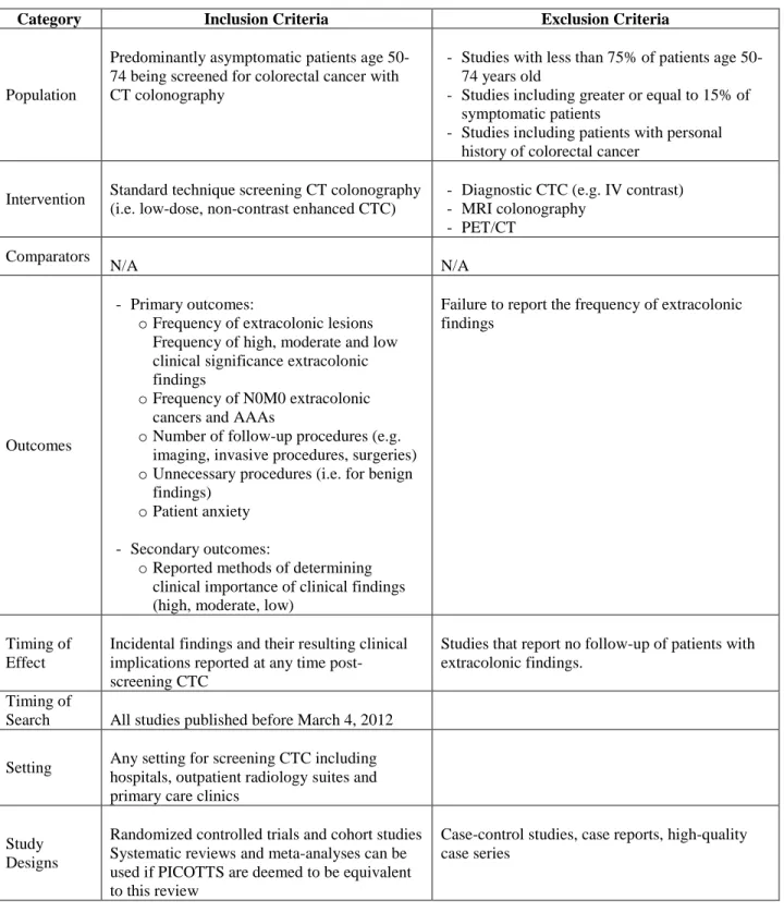
Table 2. PICOTTS framework for review of incidental findings in CTC
Category Inclusion Criteria Exclusion Criteria
Population
Predominantly asymptomatic patients age 50-74 being screened for colorectal cancer with CT colonography
- Studies with less than 75% of patients age 50-74 years old
- Studies including greater or equal to 15% of symptomatic patients
- Studies including patients with personal history of colorectal cancer
Intervention Standard technique screening CT colonography (i.e. low-dose, non-contrast enhanced CTC)
- Diagnostic CTC (e.g. IV contrast) - MRI colonography
- PET/CT
Comparators
N/A N/A
Outcomes
- Primary outcomes:
o Frequency of extracolonic lesions Frequency of high, moderate and low clinical significance extracolonic findings
o Frequency of N0M0 extracolonic cancers and AAAs
o Number of follow-up procedures (e.g. imaging, invasive procedures, surgeries) o Unnecessary procedures (i.e. for benign
findings) o Patient anxiety
- Secondary outcomes:
o Reported methods of determining clinical importance of clinical findings (high, moderate, low)
Failure to report the frequency of extracolonic findings
Timing of Effect
Incidental findings and their resulting clinical implications reported at any time post-screening CTC
Studies that report no follow-up of patients with extracolonic findings.
Timing of
Search All studies published before March 4, 2012
Setting Any setting for screening CTC including hospitals, outpatient radiology suites and primary care clinics
Study Designs
- Randomized controlled trials and cohort studies - Systematic reviews and meta-analyses can be
used if PICOTTS are deemed to be equivalent to this review
- 21 - Search strategy:
To identify original research on this topic, I conducted a systematic search of MEDLINE,
Embase and the Cochrane Clinical Trials databases. I searched PubMed on March 4, 2012 using
the following search: “(colonography[tw] OR virtual colonoscopy[tw] OR colography[tw] OR
CT colonoscopy[tw] OR virtual endoscopy[tw]) AND (extracolonic[tw] OR incidental*[tw] OR
incidentaloma*[tw] OR serendipitous[tw]).” The search was adapted for Embase, which was
accessed via Elsevier. A research librarian was consulted for the development of the search
terms, which can be found below in Appendix A. I also performed manual searches of systematic
reviews, included studies and background articles to find additional studies missed by my search
strategy.
I placed no date or language limits on the search to avoid missing studies that had not yet
been indexed. I performed an updated search, using the same search parameters, three months
following the initial search to identify any studies published since the initial search. I imported
citations into an Endnote (Thompson Reuters, New York, NY) electronic database.
Data extraction:
One reviewer extracted data on study samples, designs, populations, interventions and
outcomes using a standardized spreadsheet. These data were verified by a second reviewer and
discrepancies were resolved by consensus.
I extracted information on study methodologies such as the method of sampling (e.g.
whether subjects were selectively studied or were consecutive cases), whether data collection
was prospective or retrospective and the funding sources or potential conflicts of interest. I
- 22 -
extracted the study’s methods for determining classifications of clinical importance. I recorded
the number of observers (radiologists), the training level of radiologists, whether the study was
set in a community or academic setting and where the study was based. Lastly, I recorded the
clinical specialty of study authors and any potential conflicts of interest.
I also collected information on study populations including age, ethnicity, presence of
symptoms, risk of colorectal cancer. I obtained information on interventions including CT factors
such as slice thickness, radiation dose, use of supine and/or prone exam and whether IV contrast
was used. I extracted information on outcomes including the frequency of extracolonic findings
from selected studies and the number of lesions with high, moderate and low clinical importance.
Since these definitions varied among authors, I collected information on findings that might be
considered life-extending: the number of “early” (N0M0) cancers and AAAs.4 For AAAs, I used the definition from the USPSTF, which defines AAAs as expansions of the aorta below the renal
arteries to a diameter greater or equal to 3 cm.75 This is likely a conservative definition for potential clinical importance since the only two conditions with evidence supporting survival
benefit from screening with abdominal imaging are AAA76 and renal cell carcinoma.77 In addition, many of these diagnoses will represent aneurysms and cancers that would have never
have otherwise become clinically apparent (i.e. are “overdiagnosed”). Lastly, I gathered
information on the number of recommendations for additional imaging (RAIs), surgeries,
biochemical investigations, medical treatments and clinical appointments.
Assessing the Internal Validity of Studies:
I assessed the quality, or internal validity, of studies by applying the rules used by the
- 23 -
bias of studies as good, fair or poor quality. These ratings were based on my assessment of the
recruitment of patients (i.e. consecutive sampling), maintenance of groups (i.e. attrition,
cross-overs, adherence, contamination), the measurement of outcomes (i.e. equal, valid and reliable),
consideration of all important outcomes, description of study populations, description of
intervention, consideration of potential confounders and sample size. In addition, I developed
quality criteria specific to my topic. The full quality criteria are included in Appendix C. Poor
quality studies were defined as those with a fatal methodological flaw, such as more than 40%
patient dropout. Fair quality studies had a few methodological flaws but no fatal flaws. Good
quality studies had one to no methodological flaws. I hypothesized that the methodological
quality of studies contributed to the likely heterogeneity of reported outcomes. I explored this
hypothesis after grading each study’s internal validity. I also assessed the power of studies as
part of my quality assessment. I excluded studies with inadequate power (n < 50), since these
results would be more susceptible to random variation.
Two reviewers independently made judgments on internal validity and analysis/power of
studies. Disagreements were settled through consultation with a third investigator.
Assessing the Generalizability of Studies:
To assess the external validity of studies, I used the guidance provided in the USPSTF
procedure manual with some minor adjustments.78 I used information gathered for each study’s
populations, settings and interventions to assess how closely each resembled an asymptomatic
population receiving colorectal cancer screening with CTC. I also used the GRADE Working
Group’s definition of directness to help guide this assessment of generalizability.79
I judged each
- 24 -
symptoms and risk of colorectal cancer. I also considered each study’s setting, training and
number of radiologists and CT colonography technique. Two reviewers independently graded the
external validity of studies as good, fair or poor (full descriptions in Appendix D).
Disagreements were settled through consultation with a third investigator.
Data Synthesis and Analysis:
For outcomes data, I collected the reported frequencies of extracolonic findings from
CTC reported in each study, including the classification of findings into high, moderate and low
clinical importance. I reported the number of recommendations for additional imaging, surgeries,
medical treatments, confirmed diagnoses and unknown diagnoses for findings. Lastly, I
attempted to estimate the potential harms of the workup, treatment and surgeries.
I also analyzed patient and CT factors to judge whether there were any clear variables
that might affect the frequency of extracolonic findings or the likelihood of recommending
additional imaging or treatment. When reported, the prevalence of extracolonic findings in those
determined to have colorectal cancer was considered separately, since these patients require an
abdominal CT scan for staging purposes.
Given the subjectivity of interpreting CT colonography scans for extracolonic findings, I
decided that the heterogeneity of findings would preclude a quantitative data synthesis. The
heterogeneity of included studies was not formally assessed but I did collect information on the
various methodologies and their risks of bias to provide some indication on the potential sources
- 25 -
I did not carry out a formal assessment of potential publication bias. I did, however,
assess published studies for selective reporting of results by assessing the outcomes reported in
the methods section compared to the results section.
Strength of Evidence:
To assess the strength of evidence, I applied the approach used by the USPSTF.78 When applicable, I assessed each of the six critical appraisal questions used by the USPSTF (see
Appendix E). Since this review considered no comparators, I considered well-done, large,
prospective cohort studies to be of near-equal value to randomized controlled trials. By
evaluating the internal and external validity grades of individual studies, I assessed the aggregate
internal and external validity of the body of evidence for each key question. I judged the
consistency of evidence by looking at the variability in outcomes between studies and whether
there were clear differences in study methodologies or populations that accounted for the
differences. Lastly, I assessed directness by asking how well study populations, interventions and
outcome measures fit my key questions. In other words, I evaluated how generalizable each
study was to a typical screening population receiving CT colonography for colorectal cancer
screening. Based on these elements, I graded the quality of evidence as high, moderate, low or
very low. The full description of these ratings can be found in Appendix F.
Results
Searches, performed between March 4, 2012 and March 8, 2012, identified 282 titles and
abstracts, including 44 past reviews, commentaries or letters. An additional 15 studies were
- 26 -
After both reviewers performed a title and abstract review, 53 titles remained for full text review.
During the full text review, 43 studies were excluded for invalid publication type/study design (n
= 11), wrong intervention (n = 5), invalid study population (n = 14), incorrect outcomes (n = 6),
poor quality (n = 4) and inadequate reporting (n = 3). After an extensive review of reporting and
quality criteria, an additional 3 studies were excluded for inadequate follow-up of patients
following CTC. Disagreements between reviewers on inclusion/exclusion of 6 studies were
settled by consensus. Two studies82,83 performed at the University of Wisconsin had overlapping
dates of enrollment. Study authors concluded that these studies had overlapping datasets, so I
decided to exclude the study published in 201083 since the reporting of outcomes was more
complete for my population of interest in the 2008 study.82 After removing this duplicate study, 6 cohort studies (2 prospective and 4 retrospective) met my final inclusion criteria and were
included in the systematic review. An updated search performed on May 28, 2012 identified 4
new studies, which were all excluded for incorrect PICOTTS. A flow diagram of my search
- 28 - Included Studies:
The six studies included a total of 6,316 subjects with a mean age of 59.9 years old
(range 26-90). All of these six studies were cohort studies (4 retrospective, 2 prospective), five
were conducted in the United States and one was based in Australia. The studies included
subjects with varying levels of risk for colorectal cancer. Table 3 contains summaries of these
studies’ characteristics.
Internal Validity
I assessed the quality of included studies using the USPSTF criteria for internal validity
including measurement bias, confounding bias and selection bias.
Included studies had varying potentials for measurement bias resulting from the CTC
technique and method of reading scans. Equality of measurement was judged based on the use of
a single CTC technique and standardized system of clinical importance. Half of studies included
CTC scans performed on different scanners with varying radiation dose and slice thickness,
increasing the risk of measurement bias. In addition, only half of studies used a standardized
system for judging the clinical importance of ECFs.
I judged the validity of measurements based on the use of a classification system based
on the system of judging clinical importance, the exclusion of previously-diagnosed ECFs and
the method of following ECFs. Almost no studies used valid measurement criteria for reading
and interpreting CTC scans. Only one study60 assessed each lesion’s likelihood of benefiting from diagnosis or treatment. Furthermore, several studies failed to exclude previously diagnosed
ECFs, which are likely to be treated differently than newly diagnosed findings. Furthermore,
- 29 -
Most studies relied on their institution’s electronic medical record without accounting for
investigations, treatments or procedures delivered at other institutions.
The reliability of measurements was also highly variable. Most studies employed
adequately trained radiologists. However, only three studies60,84,85 used duplicate reading of CTC
scans. Only one of these studies86 specified that the two radiologists performed their
interpretations of scans independently. Moreover, only two studies33,85 explicitly stated that radiologists were blinded to past radiological scans and patient history.
Potential confounding bias was noted in two studies82,84 which reported higher average age, more comorbidities and a higher likelihood of intracolonic findings among those with ECFs.
These same studies noted that older, sicker populations were less likely to receive clinical
workup for their ECFs. While the risk of confounding bias is likely to be small, there was not
enough information provided to determine its magnitude in most studies. Three out of the six
studies33,85,86 failed to report any information on potential confounders and the remaining studies reported only a few relevant variables. Patient enrollment was unlikely to contribute bias, as
most studies consecutively enrolled individuals referred to their institutions for screening CTC.
No studies provided information on the subjects who were lost to follow-up (i.e. did not receive
all or part of their clinical workup at the same institution), making it difficult to assess the
potential for selection bias introduced by differential loss to follow-up. However, all studies were
able to follow at least 70% of their population.
The full quality assessment and final grades of internal validity can be found in Appendix
G below.
- 30 -
Almost all studies included a group of consecutive patients who took part in their
institution’s CTC screening program. The only study that did not enroll consecutive patients84
invited a randomly-selected group of patients from the community to participate. One study60 required that patients be referred from a gastroenterology clinic in the local area and, similarly,
another recruited patients referred to their institution for CRC screening, guaiac-positive stool or
incomplete colonoscopy.
Settings and Description of Radiologists
These studies were predominantly conducted in the United States, with the exception of
one study conducted in Australia.84 All studies were conducted at academic medical centers, including one60 conducted at a military medical center.
The degree of CTC-specific training varied greatly between studies. Two studies60,86
enrolled participating radiologists in a training program in which they read 50 pathology-proven
CTC cases, in accordance with the American College of Radiology’s recommendations for CTC
training.87 Other studies82,84 reported that participating radiologists had reached this
recommended threshold through their clinical experience. The remaining two studies33,85 stated that their radiologists were experienced and board-certified. However, these studies did not
report the radiologists’ CTC-specific training.
Half of the six included studies60,84,85 had multiple radiologists review the CTCs and this
review was conducted independently in only one of these three studies.85 This review was performed retrospectively on the initial reads of CTC scans. Those subjects with “clinically
important” (C-RADS E3 or E4) extracolonic findings discovered retrospectively were reassessed
- 31 -
radiologists were blinded to patient’s previous imaging and medical history. In addition, none of
the included studies reported whether CTC readers were blinded to the purpose of their project.
In fact, several studies82,84,86 employed study authors to read and interpret CTCs for the study.
CTC Parameters
All studies used multidetector CT scanners (4-, 8-, 16-, 64- and/or 124-detector rows) and
some used single-slice detector CT scanners as well.33,85 Each study scanned patients in both the
supine and prone positions and none used IV contrast material. Studies showed wide variations
in slice thickness, reconstruction interval and pitch. Radiation levels also varied, with currents
ranging from 40-100 mA. Each study used a CT scanner with a peak voltage of 120 kV. Only
one study84 calculated the total radiation dose, reporting that each scan resulted in less than 5 mSv (total effective body dose)2.
Three of the studies60,82,84 used a standardized CTC technique for all subjects. Two of the studies without a standardized technique85,86 were retrospective and had a range of CTC
parameters corresponding to the ones used by different radiologists in their institution. In the
remaining study,33 subjects underwent multi- or single-detector scans. The currents of the multidetector scans were adjusted to match the image noise of the single-slice technology.
Definitions of Clinical Importance of ECFs
Most of the included studies60,85,86 applied the C-RADS classifications published by the Working Group on Virtual Colonoscopy.3 These were applied retrospectively in these studies, with the exception of one60 which began applying it prospectively while their study was ongoing.
- 32 -
additional workup85 and the other labeled lesions that were particularly high-risk.60 Two of the studies that did not use C-RADS were conducted before the Working Group on Virtual
Colonoscopy published this classification system. These studies33,84 classified extracolonic lesions based on their need for further workup, similar to the C-RADS system. In the final
study,82 radiologists prospectively labeled findings as moderate or greater importance or minimal or no potential importance depending on the need for further workup or clinical importance of
the findings themselves, if diagnostic. The full descriptions of clinical importance classification
systems used are found in Table 4 below.
Despite the homogeneity in classification systems, similar clinical findings were often
classified differently in different studies. For instance, lung nodules were described as both
moderate- and high-importance. Most studies did not further characterize lung nodules by size or
appearance (i.e. calcification or speculation). In addition, one study60 characterized cystic masses
of the ovary as high importance but classified complex ovarian cysts as moderate importance.
Similarly, cystic pancreatic lesions were classified as high-importance33 while pancreatic cysts
were labeled as moderate importance60. Osteoblastic and osteolytic bone lesions were classified as both moderate and high clinical importance. Conflicting classification was also applied to
mesenteric lymph nodes and splenic artery aneurysms. As was the case with pulmonary nodules,
lesions of the kidneys, liver, adrenal glands and ovaries were inadequately characterized by study
authors. Lastly, studies rarely provided an explicit definition for AAAs. One study84 reported at
least 1 AAA with a diameter < 3 cm, others85 only reported aneurysms ≥ 3 cm and another study60 separately reported “high-risk” AAAs as those with a diameter ≥ 5 cm.
- 33 -
The studies had widely varying study lengths ranging from a mean follow-up time of 1 to
2 years. Only one study84 set a defined follow-up duration (2 years) from the time of CTC. Other
studies based their follow-up duration and clinical endpoints. For instance, one study86 searched subjects’ medical records solely to confirm completion of the imaging studies recommended
during the initial CTC read. Another study33 focused only on post-CTC imaging studies and did not report on subsequent clinical appointments, biochemical investigations, medical treatments,
surgeries or other invasive procedures. On the other hand, four studies33,60,82,84 reported on the
surgeries performed subsequent to CTC. Some of the retrospective studies tried to collect
information on all medical workup performed subsequent to the subject’s CTC, but their
methods for collecting this information were not always clear. Most studies failed to explicitly
report a duration or specific endpoint that would end their follow-up of ECFs.
Some studies reported the follow-up investigations received by all study subjects. Others
limited the scope to subjects with high clinical importance findings (i.e. C-RADS E3 and E4)
lesions since, by definition, lesions below this threshold were not supposed to receive workup.
One study84 determined that subjects follow up with a radiologist, general practitioner or appropriate specialist depending on the clinical importance of their finding (e.g. specialist for
high clinical importance lesions) and the recommended workup (e.g. radiologist for imaging). In
another study,85 information on ECFs was passed on to subjects’ primary care physicians at the time of the initial CTC and all subsequent medical workup initiated by the PCP was collected
from subjects’ medical records.
The included studies used several different sources of information to determine what
follow-up investigations occurred. For instance, most33,60,82,85,86 employed a review of subjects’
- 34 -
studies60 reported the percentage of subjects whose subsequent workup would be captured by this system. For the others, it was unclear how often subjects received follow-up that would not
be captured by their electronic medical record. Conversely, one study84 had all subjects receive their follow-up exams at a single institution thus ensuring that they could account for all
subsequent imaging, procedures and treatments.
Frequency of Incidental Findings
The method of tallying extracolonic findings varied among studies. For instance, some
studies included previously detected extracolonic findings while others excluded them or
reported them separately. One study84 included only extracolonic findings that had changed significantly since the last time they were imaged. In addition, other studies only reported
extracolonic findings they deemed to be of high or moderate clinical significance. The full
description of methods of tallying ECFs and number of ECFs detected is included in Table 5
below.
Four of the six studies33,60,84,86 reported the frequency of subjects with extracolonic findings. Among these studies, the frequency of subjects with at least one ECF during their
initial CTC ranged from 27.2% to 68.9% (mean 49.3%). Two of these studies33,84 reported the
total number of ECFs, ranging from 1.2 to 1.8 ECFs per subject with an extracolonic lesion
detected. Three of the other studies60,83,86 did not report the overall frequency of ECFs at all. The
other85 only reported the number of ECFs for the entire population, which included symptomatic and high-risk subjects. This study reported that among all subjects, 272 of 376 (72.5%) had at
least one extracolonic finding. Among these 272 subjects, investigators found 520 incidental
- 35 -
Two studies reported the number of extracolonic findings found that were missed on the
initial CTC. One study85 reviewed all initial CTC scans and reports, stating that 144 E2, 29 E3
and 4 E4 lesions were missed by the initial reader. Based on these numbers, they calculated an
8.8% miss rate for E3 and E4 findings. Another study33 reported the ECFs not reported on the
initial CTC that were discovered during the subsequent radiological workup. They noted 44
lesions (23 of medium- or high-importance) that were not reported on the initial CTC report.
Their retrospective read of the initial scan was able to see 25% of these findings on the initial
CTC. The remaining 75% could only be seen on scans taken as part of the radiological workup.
Frequency of Clinically Important Lesions
The method of determining clinical significance varied somewhat among studies. Study
authors sometimes based clinical importance on the lesion’s characteristics and some based it
retrospectively on the final diagnosis. For studies using the C-RADSs classification system
(Table 1), I considered E4 findings to be high significance and E3 findings to be moderate
significance. By definition, E2 lesions do not require clinical workup3 and therefore were not included in either of these groups. The methods of reporting ECFs and reported frequencies can
be found in Table 5 below.
In the four studies reporting high-importance lesions separately,33,60,84,85 their frequency ranged from 1.5% to 10.4% (mean 5.6%). In one of these studies (frequency of high-importance
findings = 7.4%),84 there was no separate category for moderate importance extracolonic findings. Other than this study, all others reported the combined frequency of moderate- and
high-importance ECFs. The frequency of moderate- and high-importance findings ranged from
- 36 -
To address the heterogeneity between classification systems, I also looked at the number
of abdominal aortic aneurysms, diagnosed early (N0M0) cancers, total cancers and lesions
suspicious for cancer. In the three studies33,60,84 that reported the number of abdominal aortic aneurysms diagnosed during the initial CTC the frequency of AAAs ranged from 0.088% to
1.2%. The study84 reporting a frequency of 1.2% (5 of 432 subjects) stated that they found 6 AAAs with diameters of 2.8 to 4.5 cm. Since I defined AAAs as ≥ 3 cm and this study did not
report the individual diameters of each aneurysm, only 5 of these AAAs were counted although
there might have been fewer that were ≥ 3 cm. In the study including symptomatic patients, there
was no separate reporting of outcomes for their average-risk population. However, they reported
a newly diagnosed AAA in 1 subject (0.19%). The remaining two studies either stated total
number of aneurysms in the aortoiliac system82 or did not report individual outcomes.86 Four studies33,60,82,84 reported the number of newly discovered cancers, which were
diagnosed in 0.23% to 0.88% of subjects (mean 0.45%). The frequency of N0M0 cancers was
discussed in two studies,60,84 which reported early stage cancers among 0.13% and 0.23% of
subjects. The frequency of lesions suspicious for malignancy had wide variation and was often
unclear due to poor characterization of lesions. The complete list of diagnosed AAAs,
malignancies and suspicious lesions are included in Table 7 below.
Chin et al.84 reported that 5 subjects (1.2%) had AAAs, 1 subject (0.23%) had a newly discovered cancer and no additional subjects had lesions suspicious for malignancy. This newly
discovered cancer was a noninvasive renal cell carcinoma. Veerappan et al.60 reported AAAs in 2 subjects (0.088%) and early-stage cancers in 3 subjects (0.13%), including one stage 1a lung
adenocarcinoma and two stage 1 renal cell carcinomas. In addition, this study reported subjects
- 37 -
carcinoma, totaling 6 subjects (0.26%) receiving cancer diagnoses. Gluecker et al.33 reported 4 subjects (0.59%) with AAAs and 6 subjects (0.88%) with cancer diagnoses, including 1
squamous cell carcinoma of the lung, 1 renal adenocarcinoma, 1 renal oncocytoma and 3 ovarian
serous cystadenomas. In addition, they reported 77 lesions suspicious for malignancy,
representing 8.9% of all ECFs. This study did not report the number of N0M0 cancers.
Clinical Implications of Incidental Findings
The duration, methodology and reporting of follow-up varied greatly among studies. The
number of subjects requiring some workup ranged from 2.0% to 8.7% (mean 5.4%). After
excluding studies that did not report surgeries or invasive procedures,85,86 the mean increases to 7.4%. One study33 did not state how many subjects required workup but did report the number of individual tests and procedures required.
Most studies reported primarily the imaging workup required. CTs and ultrasound scans
encompassed the bulk of recommended or performed imaging tests, making up 52.8% and 35.0%
of all imaging tests, respectively. The complete list of imaging studies can be found in Table 8
below. In addition to the imaging reported in Table 8, Veerappan et al.60 noted that subjects received 6 bone scans, 3 upper endoscopies and 1 bronchoscopy. There were also some scans
reported in Pickhardt et al.82 that are not listed in Table 8, including 1 skeletal scintigraphy scan, 2 renal scintigraphy scans and 3 small-bowel capsule endoscopies.
The four studies that listed surgeries reported that, of the 370 subjects requiring workup,
48 surgical procedures were performed. The majority of reported surgeries were for AAA repair
or treatment of suspected malignancy. The full summary of these findings can be found in Table
- 38 -
There were also several procedures for lesions that were ultimately found to be benign.
One study84 specifically reported this number, stating that 75% (24 of 32) of findings receiving
workup were ultimately diagnosed as benign. Another study82 reported that while adnexal lesions accounted for 45% (10 out of 22) of all surgeries, all lesions proved to be benign and all 10 liver
lesions receiving contrast-enhanced CT were eventually diagnosed as benign. The same study
stated that pulmonary nodules discovered on CTC led to chest CT in 6 subjects, CT-guided lung
biopsy in 4 subjects and thorascopic resection in 2 subjects. One of these pulmonary nodules was
found to be malignant. In a separate study,60 none of the 8 patients receiving surgery for a pelvic mass ultimately had a malignancy and one of the 2 AAA repairs were on aneurysms they
considered to be “low risk.” However, the majority of studies did not explicitly report the
number of potentially unnecessary procedures performed for benign findings.
There were many other clinical outcomes that were omitted from the majority of these
studies. For instance, only two studies33,84 reported the number of medical treatments required. One of these studies84 reported that no subjects required medical treatment. The other study33
reported that one subject required chemotherapy for thyroid cancer metastases to the lungs and
another received antihelmintic treatment for ileal ascaris.
An especially glaring omission was discussion of harms from this additional workup,
especially considering the description of its potential benefits. For instance, only one study82 discussed the complications resulting directly from workup, stating that there were no reported
complications of surgeries or invasive procedures. In addition, no studies calculated subjects’
exposure to ionizing radiation during the subsequent radiological workup or discussed the
potential harms of surgeries for benign findings. These surgeries carry the possibility of
- 39 -
recovery. Lastly, the studies failed to collect data on the potential psychological harms from
extracolonic findings, such as the anxiety of a potentially serious diagnosis.
Funding Sources and Potential Conflicts of Interest
None of the six studies reported their funding sources and only one included study86 reported conflicts of interest. This particular study reported that authors had no potential conflicts
of interest. However, the study that was excluded for duplicate data83 reported relevant conflicts
of interest for some of the authors from the study I included.82 These authors were consultants for companies that develop computer software for CTC and the cofounders for a company that
provides educational materials and trainings on CT colonography.
Generalizability of Studies:
I assessed the external validity of each study using criteria developed from the USPSTF78 and the GRADE working group.79 I judged the generalizability of study populations, settings and
interventions to a typical screening population.
I defined a typical population as one that was primary asymptomatic, at average-risk of
colorectal cancer and within the recommended screening ages of 50-74. Three studies60,82,84 were
rated as having good population external validity. The one study with a fair rating86 primarily enrolled screening patients but also included those with an incomplete colonoscopy. I gave the
remaining two studies33,85 poor generalizability ratings due to their inclusion of symptomatic and high-risk patients without any separate reporting of outcomes.
I defined a typical setting as some mixture of academic and non-academic institutions.