Investigation of Mutagenicity due to Compovnds other than MX in Chlorinated Hiimic and Drinking Water.
by
Ravinder Singh
(Under the direction of Dr. L.M. Ball).
ABSTRACT
3-Chloro-4-(dichloromethyl)-5-hydroxy-2(5H)-furanone, i.e MX, is recognized as a principal mutagen contributing to
(on an average) 30% of the mutagenicity of chlorinated
drinking water in the Ames assay. Compounds with structural similarities to MX were suggested as probable mutagens which could account for the remaining 7 0% of the mutagenicity of
chlorinated water. The theoretical basis for the formation
and activity of these MX analogues was discussed. Three such candidate mutagens, red-MX, ox-MX and ox-EMX were synthesized, and their mutagenic activities determined (in the Ames assay, strain TA 100) to be 0.13 net revs./ng; 0.36 net revs./ng and 0.03 net revs./ng respectively. It would appear that the aldehyde group is a critical structural feature that governs the mutagenicity of MX. The
concentration of MX in samples of chlorinated fulvic acids,
humic and drinking water was determined. MX accounted for 36%, 32% and 17% of the overall mutagenicity of these
samples, respectively. SIM mode GC/MS analyses on
derivatized and underivatized extracts of the chlorinated
waters revealed that red-MX and ox-MX were present in
ii
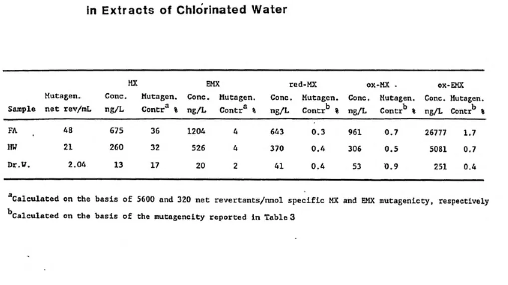
times higher. Due to their weak mutagenicities however, all
three compounds were found to account for (individually)
less than 1% of the overall activity in each sample.Mutagenicity-directed HPLC fractionation was performed on a chlorinated solution of fulvic acids, selected because it was highly mutagenic but contained a low amount of MX
(72% non-MX mutagenicity), After the second level of
separation, all the activity had concentrated into a single
sub-fraction (31 000 net revs./liter) which was free of MX.
The responsible mutagens seem to be less polar compounds in
Ill
ACKNOWLE DGEMENTS
The successful completion of this work was possible because of the support and input from many individuals - professors,
colleagues and friends.
I would like to thank my research adviser. Dr. Louise M. Ball for her time, energy and guidance. Her input had a tremendous positive influence on this work, as well as the path of my graduate study. Her insight into all aspects of
this subject is greatly appreciated.
I also appreciate the counsel of my advisory committee
members. Dr. M. Judith Charles, Dr. Russell F. Christman and
Dr. Alvis G. Turner.
This work benefitted very much from the expertise of Dr. Leif Kronberg of The Abo Akademi, Turku, Finland. It was both a pleasure and a privilege to work with Dr. Kronberg
during his time here as a visiting scholar.
My parents, Pergas Singh and Baljit Kaur, and the rest of my family have in many ways enabled me to come this far in my
education and in my life.
Finally, I am grateful for the encouragement and the help from my colleagues Heidi Bojes, Dave Cozzie, Tom Holloway, Peter Kelsey, Lisa Stocking, and all my friends, especially Nathan and Vivian Cobb. For seeing me through this work, right through to its completion, I owe a lot to Christine
iv
CONTENTS
I. INTRODUCTION ...1
II. BACKGROUND...4
2.1. Chlorination By-products in Drinking Water 2.1 (a) . Occurrence in Drinking Water...4
2.1 (b) . Formation During Chlorination...7
2.1 (c) . Stability of MX in Water...9
2.2. Structure-Activity Relationships...11
2.3. Genotoxic Activity 2.3 (a). Genotoxicity of Drinking Water Concentrates...13
2 . 3 (b) . Genotoxicity of MX...16
2.4. Analytical Techniques 2.4 (a). Concentration and Extraction of Organics...18
2.4 (b) . GC/MS Analyses and Quantitation...20
2.4 (c). HPLC Fractionation-Bioassay (coupled) Procedure...23
2.4 (d). Mutagenicity Analysis: The Ames assay...24
III. APPROACH...28
3.1. Formation of MX and the MX Analogues during Chlorination...29
3.2. Mutagenicity-Directed Sub-fractionation...32
IV. EXPERIMENTAL..."...33
4.1. Materials...33
4.2. Chlorination and Extraction of Samples 4.2 (a). Humic Water Sample...34
4.2 (b) . Fulvic Acids Sample...35
4.2 (c) . Drinking Water Sample...36
4.3. High Performance Liquid Chromatographic Separation...36
4.4 (b) . red-MX...39
4.4 (c) . OX-EMX...41
4.5. Derivatization and Gas Chromatography/Mass Spectrometry Analyses...44
4.6. Mutagenicity (Ames) Assay...49
V. RESULTS...51
5.1. Mutagenicity of the MX Analogues...51
5.2. Quantitative Determination of the MX Analogues in the Mutagenic Extracts...56
5.2 (a). Concentration and Activity in the Chlorinated Humic Water...57
5.2 (b). Concentration and Activity in the Chlorinated Fulvic Acids...61
5.2 (c). Concentration and Activity in the Chlorinated Drinking Water...61
5.3. HPLC Fractionation of Chlorinated Fulvic Acid. 5.3 (a). Chlorination...62
5.3 (b). Separation and Activity of Fractions.... 66
5.3 (c) . Additive Effect of the Mutagenic. Activity...75
VI. DISCUSSION, CONCLUSIONS AND RECOMMENDATIONS 6.1. Discussion...79
6.2. Conclusions...87
6.3. Recommendations for Future Work...90
vi
LIST OF FIGURES.
FIG. 1: Formation of MX Analogues in Water...10
FIG. 2: Stability of MX in Water...31
FIG. 3: Mass Spectrum of ox-MX anhydride...40
FIG. 4: Mass Spectrum of red-MX...42
FIG. 5: Mass Spectrum of ox-EMX...43
FIG. 6: Mutagenic Response of ox-MX in TA 100 ...52
FIG. 7: Mutagenic Response of red-MX in TA 100 ...53
FIG. 8: Mutagenic Response of ox-EMX in TA 100 ...54
FIG. 9: Mass Spectrum of Methylated ox-MX...58
FIG. 10: Mass Spectrum of Methylated ox-EMX...59
FIG. 11: Activities of Mutagens in Chapel Hill Drinking Water...63
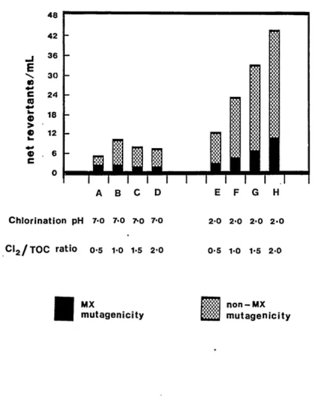
FIG. 12: MX and non-MX Mutagenicity in Solutions of Chlorinated Fulvic Acids...65
FIG. 13: Effect of pH on the Extractability of Mutagenic Activity from Solutions of . Chlorinated Fulvic Acids...67
FIG. 14: Scheme for the Separation and Purification of Sample H...70
FIG. 15: HPLC Profile of Sample H Separation...71
FIG. 16: MX and non-MX Mutagenicity in Sample H Sub-fractions...72
FIG. 17: HPLC Profile of Fraction HI Separation...73
Vll
LIST OF TABLES.
TABLE 1: Derivatization Agents and GC Conditions
for SIM Mode GC/MS Analyses...46
TABLE 2: Ion Peaks Used and Relative Ratios Found
for the SIM Mode GC/MS Analyses of
MX and the MX Analogues in Chlorinated
Water...47 TABLE 3: Mutagenic Potencies of MX and the
MX Analogues...55
TABLE 4: Concentrations and Activities of MX
and MX Analogues in Extracts of
Chlorinated Water...60
TABLE 5: Mutagenic Activity and MX, E-MX Concentrations in Aqueous Solutions
of Chlorinated Fulvic Acids...64
TABLE 6: Total Activity and MX, E-MX Concentrations in Sample H Extracted at Various pH Values..68 TABLE 7: Total Activity and MX Concentration in
HPLC Sub-fractions of Sample H...74 TABLE 8: Additive Effect of the Mutagenic Activity
^-^-*.V--^^-WPE;-'
I: INTRODUCTION
Drinking water contains a complex mixture of organic chemicals, many of which are known to be potentially harmful to human health. Some of these compounds are introduced
into source waters by industrial, agricultural and municipal effluents. Others however, are formed de novo by the very disinfection process meant to make our drinking water
microbiologically safe. In fact, emphasis on the problem of organic micropollutants in drinking water has shifted
markedly to the issue of these chlorination by-products. In the last 15 years or so, great strides have been made in detecting these by-product compounds in drinking water (Rook,1974; Bellar, 1974; Symons et. al.,1975), recognizing their genotoxic and carcinogenic potential
(Simmon and Tardiff, 1976; Nat. Cancer Inst., 1976;
Loper,1978; Nestman et. al., 1979; Lang et. al., 1980; Cheh et. al., 1980; Kool et. al., 1985; Wilcox and Williamson, 1986) and understanding the mechanism of their formation
(Cheh et. al., 1980; Christman et. al., 1983; Coleman et. al., 1984; Holmbom et. al., 1984; Kronberg et. al., 1985b; Meier et. al., 1986; Maruoka, 1986). From the regulatory
and examine the principal disinfection by-products in order
to promulgate standards under Phase IV of the Revised
National Primary Drinking Water Regulations (Cotruvo and
Vogt, 1985). Thus, much of the recent research has
concentrated on identifying and structurally characterizing
the mutagens present in drinking water, particularly the
non-volatile organohalides.
Although most of the mutagenic activity of drinking
water can be attributed to organics in the non-volatile
fraction, only a single major mutagen, 3-chloro-4-'
(dichloromethyl)-5-hydroxy-2(5H)-furanone, i.e MX, has been
identified so far (Kronberg et. al., 1987; Meier et. al.,
1987b). On an average, this compound can account for about
30% of the total mutagenicity of the organic fraction. In
order to get a more comprehensive understanding of the
health hazards posed by the presence of mutagens in drinking water, chemical identities of compounds responsible for the
residual two-thirds of the mutagenicity must be deteirmined.
This would allow syntheses of the pure compounds to be used
in long-term animal toxicity and carcinogenicity studies as
is being done with MX.
The objective of this research was to elucidate the
structures and activities of compounds which could possibly be responsible for the yet uncharacterized mutagenicity
(non-MX mutagenicity) of chlorinated drinking water. This objective was approached through two specific tasks:
i.e the MX analogues: Synthesize compounds with
(critical) structural similarities to MX, determine their mutagenic potencies and
subsequently search for these compounds in
chlorinated fulvic acids, humic water and drinking
water.
(II) Mutagenicity directed sub-fractionation:
Chromatographic separation (with respect to mutagenicity) of a highly mutagenic
extract where the amount of MX has been
II: BACKGROUND.
2.1 : Chlorination By-Products in Drinking Water.
More than a decade of research has gone into
understanding the formation and occurrence of these
genotoxic compounds in drinking water. Some of the
important aspects are discussed below.
2.1 (a) : Occurrence in Drinking Waters.
The initial concern about the presence of chlorination
by-products was sparked by the discovery of trihalomethanes
(THMs) and other volatile organohalides in drinking water in
The Netherlands (Rook, 1974) and in the United States
(Bellar et. al., 1974). The first of such chemicals
identified were primarily the bromo- and chloro- substituted
methanes. The widespread occurrence of these compounds in
U.S. drinking waters was confirmed by the National Organics Reconnaissance Survey (Symons et. al., 1975). Since then, numerous other by-product compounds have been identified and detected in water. It is now known that a majority of these compounds are genotoxic/carcinogenic in nature, and that they are formed in the chlorination process during drinking
Although volatile organic compounds were the center of
attention initially, more recent research has shown that the non-volatile compounds may be of greater concern. About 90%
of the dissolved organic content in drinking water is
comprised of relatively non-volatile (high molecular weight,
non-purgeable) organic compounds (NAS, 1977). These
compounds are extractable with organic solvents or can be
adsorbed onto XAD resin material. Since chlorine binds
mainly to organics in the non-volatile fraction, most of the
mutagenic activity can be attributed to non-volatile
organohalides. Thus, organic extracts or XAD concentrates
of chlorinated drinking water are most often mutagenic.
Within this concentrate itself, one can distinguish between
neutral, relatively non-polar compounds (which account for
only 10% of the total mutagenicity), and polar, acidic
compounds (accounting for the rest of the mutagenicity).
Almost all the compounds identified in water are
present at concentrations of less than 1 ug/liter. These
levels are far below those which would be required to
account for the total observed mutagenic activity, based on
known individual mutagenic potencies. Fielding and Horth (1986) demonstrated that 14 such compounds together could account for less than 10% of the mutagenicity of the extract itself (a mixture of 14 compounds, at 10 times the
concentration typical in water could only equal the
Against this background, it is easy to understand the importance of chlorinated hydroxyfuranones.
3-chroro-4-(dichloromethyl)-5-hydroxy-2(5H)-furanone i.e MX was the first single mutagen identified in drinking water which could account for a relatively large portion of the total
(extracted) mutagenicity (Kronberg et. al., 1987; Meier et. al., 1987b). Although present at very low concentrations, MX contributes to about 5-60% of the total mutagenicity of drinking water. This is because MX is among the most potent known mutagens ever tested in the Ames TA 100 strain. The estimated activity of MX has ranged from 5600 to 13000 net revertants/nmole in TA 100 (Padmapriya et. al., 1985; Meier
et. al., 1987a, 1987b; Kronberg et. al., 1987). in
comparison, the most mutagenic among the THMs, i.e chlorodibromomethane, induces only about 0.004 net
revertants/nmole in TA 100 (Simmon et. al., 1977). The concentration of MX in drinking waters varies considerably. Water samples from 2 3 cities in Finland had MX levels
relative to MX, and accounts for only a few percent of the total mutagenicity (Kronberg et. al., 1987).
2.1 (b) : Formation During Chlorination.
Various investigators have shown that it is the interaction of aquatic organic matter with chlorine that leads to the formation of a mixture of halogenated and
non-halogenated by-products. A variety of non-volatile
aliphatic halogenated products can result from the exposure of aquatic humic and fulvic acid fractions to chlorine
(Christman et. al., 1983). Dominant among them are the C,
halogenated acids (principally di- and trichloroacetic acids), shown also to be present in chlorinated municipal drinking water (Uden and Miller, 1983). The mechanisms of by-product formation have been studied by employing isolated humic and fulvic acids (the fulvic acid fraction is a major part of aquatic humic substances) as well as some other model compounds resembling the complex molecules of natural humic matter. The general finding has been that the
specific by-products formed will depend on the molecular structure of the substrate involved, its concentration, the carbon-to-chlorine ratio and the pH of chlorination, among
other factors (NRC, 1987). Nevertheless , two categories of
byproducts can be recognized : volatile hydrophobic and non¬
volatile hydrophilic compounds. The mutagenicity associated
with chlorinated humic/fulvic acid solutions is however
8
Working with extractions at different pH values and with solvents of different polarity, Kronberg et. al. (1985b) demonstrated that the principal mutagens, in a chlorinated humic acid solution, were relatively polar compounds. In support of this are other investigations showing that a
majority of the mutagenicity of chlorinated humic and fulvic material resides in the strong acid fraction (Meir et. al.,
1986) and that almost 65-75% of the recovered activity was due to polar mutagenic products (Maruoka, 1986). Among the commonly occurring mutagens, as resulting from humic and fulvic acid chlorination are chlorinated acetones, ^'
chlorinated propenals such as 1,3-dichloroacetone and 2-chloropropenal, dichloroacetonitrile, dichloro- and
trichloroacetic acids, choral, and chloropicrin. Most of these significant by-products have been summarized in a review by Christman et. al. (1984). This review also discusses the involvement of the 1,3-dihydroxybenzene or
resorcinol structures (found within the humic
macrostructure) in the formation of the chlorination by¬ products. Another group of by-products of particular
interest currently are the chlorinated hydroxyfuranones,
which include MX and E-MX (Holmbom et. al., 1984).
Since organic nitrogenous compounds can account for a
considerable part of the total organic carbon (TOC) in natural waters, it is also possible for mutagens to be
possess mutagenic activity, and some of the compounds
identified in such extracts are haloacetonitriles (Trehy and
Bieber, 1981), trichloroaldehyde (Trehy et. al., 1986) and
MX and E-MX (Horth et. al., 1987). Other substrates
implicated in the production of mutagenic chlorination by¬
products are proteins, peptides and phenolic compounds.
2.1 (c) : Stability of MX in Water
The stability of MX, and other possible acidi.c
mutagens, in water is discussed at length by Kronberg and
Christman (1988), Kronberg (1987), and Holmbom et. al. (1984). The pKa value of MX in aqueous solutions has been determined to be approximately 5.25, assuming that the hydration/dehydration reaction between the closed and
protonated open form is much faster compared to the proton
transfer. Thus, at pH values common in drinking water (pH
7.0-8.5), MX exists in its open form.
MX is stable in water at low pH (pH 2.0), but as the pH increases to about 7 or 8, it can partly convert to its
geometric isomer E-MX, and partly undergo hydrolytic
degradation reactions. The second reaction dominates at pH
8-9. At pH 8, the half-life of MX at 23 degrees Celsius is
about six days. Under neutral or alkaline conditions, E-MX
does not isomerize to MX. However, if the pH is lowered toabout 2.0, E-MX is quantitatively converted to MX (see Fig.
1, modified from Kronberg and Christman, 1988) . These
10
MX acyclic tautomer
CI2CH CI
\ /
HO-'^O'^'^O
pKa=5-25
CLCH CI
\ /
/ \
CHO COO"
pH<4 pH 5-7// pH»8
CI2CH COO"
/ \ CHO CI
+ degradation
products
E-MX
11
chlorination are alkali-labile and (b) a majority "of the MX
formed will persist throughout most of the water
distribution system. Theoretically, it may be possible for
E-MX to isomerize to MX in-vivo as the drinking water passes through highly acidic conditions in the gastrointestinal tract. However, the rate of isomerization is too slow to cause any substantial increase in the mutagenicity of the
consumed water (due to the more potent MX).
2.2 : Structure-Activity Relationships.
The high mutagenic potency of MX has prompted attempts
to study and define the structural features (particularly in
chlorofuranones) that are critical in rendering MX and
related compounds mutagenic.
Both the type of substituent groups as well as their position(s) seem to be important. Compounds which differ
from MX only in a substitution on the 5-position of the furanone ring (i.e 4-position of the acyclic tautomer) display mutagenic potencies almost equal to that of MX. Streicher (1987) explained the potencies of three such
compounds by suggesting their susceptibility to hydrolyse to
MX. However, a change in the 4-position substituent of the
MX furanone ring can dramatically influence the potency of
the compound. Replacing the dichloromethyl group of MX with
a chlorine, a methyl group and a chloromethyl group reduced
12
1987). It has been postulated that the dichloromethyl group
of MX is in fact, the site of nucleophilic attack," and does
not merely serve as an electronegative activator.
Another decisive factor governing the activity of MX is
the cls_arrangement of the CHCl^ and CI substituents on the
carbon-carbon double bond, which is present in both the furanone ring as well as the acyclic forms of MX (Ishiguro et. al., 1988). This fact is most evident in the
mutagenicity displayed by E-MX, which has the CI substituent trans to the dichloromethyl group. The activity of E-MX is, at the most, about 10% of that of MX (Kronberg et. al.,
1988). Further, it is important for the dichloromethyl group to be alpha to the aldehyde group in the rin'g-opened form of MX. In this position, it is conceived to enhance the electrophilicity (and hence the mutagenicity) of the enone system through its electron withdrawing effect
(Ishiguro et. al., 1988). Finally, there is the obvious contribution of the three chlorine substituents, as the furanone structure devoid of any chlorine is essentially
non-mutagenic.
2.3 : Genotoxic Activity.
The current concern about chlorination by-products
stems from the recognition that these compounds could pose a significant health hazard to humans. Numerous studies have compared the mutagenic and carcinogenic properties of
ͣ
'SSSW^SI^WW^
13
common finding is that the chlorination process itself is
responsible for the introduction of mutagens which are generally not present prior to this process (Cheh et. al., 1980; Loper et. al., 1985; Nestman et. al.; 1979). The gravity of this problem came to light when a study by the National Cancer Institute implicated chloroform (one of the by-products formed) as being capable of inducing
hepatocellular carcinoma in mice and renal tumors in rats. The most extensively used tool for obtaining toxicity information on drinking water samples has been the Ames
Salmonella assay. Almost all the strains recommended by Dr. Ames and co-workers (Ames, McCann and Yamasaki, 1975; Maron
and Ames, 1983) for routine screening (TA 1535, TA 1538, TA 98, TA 97, TA 100, TA 102) have been used. TA 98 and TA 100 seem to be the most sensitive in their ability to detect the presence and potency of mutagenic compounds in drinking
water (Meier, 1988) and the highest response is almost
always in the absence of exogenous metabolizing enzymes, S9. The mutagens would thus appear to be direct acting
frame-shift or base-pair substitution types.
2.3 (a) : Genotoxicity of Drinking Water/Concentrates. Organic extracts and XAD resin concentrates of
chlorinated fulvic acid solutions, humic water and drinking water have been shown to produce a dose-related increase in the reversion, well above the spontaneous rate, in the
14
modified fluctuation assay. Another genotoxic endpoint
shown by drinking water concentrates in a microbial assay is the induction of SOS response in E. coli (Bourbigot et. al.,
1986).
Drinking water concentrates have also been tested in
some of the commonly used eukaryotic assay systems,
considered by some to be more relevant to human health effects than microbial assays. Lang et. al. (1980) showed
that drinking water concentrates were able to transform
BALB/C3T3 cells inzyitro. When transplanted to athymic mice, these transformed cells could produce tumors in-vivo
(Kurzepa et. al., 1984). This ability to transform a mouse fibroblast cell line (BALB/3T3) in a dose related manner was also shown by Loper (1978) using a reverse-osmosis
concentrate which was earlier shown to be mutagenic in the Ames assay strains TA 98 and TA 100. Robinson et. al.
(1981) working with a reverse-osmosis concentrate of
drinking water from five cities, were able to demonstrate that the chemicals present were primarily initiators in the
mouse skin assay rather than promoters or complete
carcinogens. However, the duration of the experiment was probably not long enough to detect carcinogenicity, even at doses up to 30 mg of organic material per mouse.
For purposes of comparison, other investigators have
15
surface source was positive in the Ames assay and was able
to induce SCE and chromosomal aberrations in CHO cells.
Wilcox and Williamson (1986) tested the same XAD-2
concentrates of drinking water using vitro as well as
in-vivo assays. While the concentrates were clastogenic in CHO
cells and cultured human lymphocytes, they were not able to induce chromosomal aberrations in mouse bone-marrow cells following in-vivo-oral administration. The authors
suggested that perhaps the clastogens were inactivated
before they could reach the target cells in the bone-marrow
This explanation is supported by In-vitro evidence as well.
Meier and Bull (1985) reported the ability of humic acids, chlorinated at pH conditions relevant to drinking
water chlorination, to induce SCE in a mammalian cell line.
However, these same samples could not increase the percent
of micronucleated cells in the mouse micronucleus assay.
They were negative in the mouse sperm morphology assay as
well. A long term carcinogenicity study to address the
effects of chronic exposure to chlorinated drinking water
was done in The Netherlands. After 24 months of receiving
organic concentrates (XAD-4/8) of drinking water (mutagenic
in the Ames Assay) up to 68 times the estimated human
exposure, the test rats displayed no increases in the
incidence of tumors (Kool et. al., 1985)
16
It is well established that among the identified
chlorination by-products in water, MX is the single most
potent mutagen, contributing significantly to the total
mutagenicity of the extract. Thus, recent work on the
mutagenicity and carcinogenicity of drinking water has
focused on such properties of the pure compound (MX).
Meier, Blazak and Knohl (1987) investigated the mutagenic
and clastogenic properties of MX, and confirmed that its
genotoxicity in-vitro is not restricted to bacterial cells
alone. MX was found to induce a dose-related increase in
the reversion rates of Ames Salmonella strains TA 1535, TA
1538, TA 92, TA 97, TA 98, TA 100, TA 102, all in the absence of S9. This would indicate that MX is a
direct-acting mutagen capable of effecting both base-pair
substitutions as well as frame-shift mutations. MX was also
able to induce chromosomal aberrations (chromatid deletions
and chromatid exchanges) in mammalian (CHO) cells. This
work also described the acute toxicity of MX, the acute oral
LD50 (to Swiss-Webster mice) being reported as 128
rog/kg/day.
Although MX can be considered a clastogen of
comparatively high activity, it failed to induce micronuclei
in mouse bone-marrow in-vivo even at the highest
administered dose (70% of the LD^q, sacrificed after 72
hours). Thus, the authors concluded that "...MX fits into a
mutagenic in~vitro but are inactive for rodent bone-marrow
in-vivo."
The genotoxic potential of MX was further confirmed by
Brunborg et. al. (1988). They indicated that MX could
induce SCE in cultured mammalian cells (V79) at very low
levels (2-5 uM). Higher concentrations of MX blocked normal
mitosis and therefore, gene mutations could not be
registered. MX was also found to induce DNA damage in rat
testicular cells.
Finally, the fate of MX in~vivo has been recently
reported by Kopfler et. al. (1988). They concluded that MX
was not active in the mouse micronucleus assay (in which the
target cells are developing erythrocytes in the bone-marrow)
probably because it is inactivated before reaching the
target cells. MX is a very reactive electrophile and can be
inactivated by reactions with nucleophiles likediethyldithiocarbamate and gluthathione. As for its
absorption/distribution, most of the 8 mg gavage dose was
excreted or expired. After sacrifice at 48 hours, the
carcass and internal organs of the rats contained only about
8% of the dose (mainly in the kidney and liver). The
gastrointestinal tract had nuclear anomalies following oral
administration.
Based on a literature review, it would appear that
although chlorinated drinking water contains compounds
having mutagenic and clastogenic potential, their in-vivo
18
2.4 : Analytical Techniques.
2.4 (a) : Concentration and Extraction of Organics. Organic compounds in drinking water are present at very low concentrations (1 ug/liter or less). Loper (1981)
pointed out that at such typical concentrations, the
mutagens are not likely to be detected by the Ames assay.
Even the use of the modified microscale fluctuation assay
which directly incorporates unconcentrated water samples , has proven unsuccessful in detecting mutagens (Foster et. al., 1983; Monarco et. al., 1985). Thus, to make compounds
amenable to analytical and mutagenicity testing, they have
to be isolated and concentrated from the water samples. Two
very important features of the concentration method were
outlined by Kronberg (1987); (a) the method should give high
recoveries of a wide range of organics since the attempt is
to "capture" compounds with unknown structures and
properties, and (b) the organic solvent to be used' in the
concentration procedure should be compatible with both the
intended bioassay and the analytical method (or be easily
exchangeable to another solvent since the chemical as well
as the mutagenicity analyses are to be done on the sameconcentrate).
Some of the methods which have been in common use are
freeze-drying, reverse-osmosis, liquid-liquid extraction and
binding to different solid-phase adsorbents. Concentration
ͣ
^S?^^SSf75g^?^':f'?3^^.r ͣ
19
recovered. Wilcox et. al. (1986) noted that differences in
the types and levels of genotoxic activity are probably a
reflection of this fact rather than due to real differencesin the composition of the water samples. The most widely
used concentration method seems to be adsorption of the
organics onto macroreticular XAD resins (Kronberg, 1987;
Meier, 1988). These non-ionic resins give efficient
recoveries for both non-polar compounds (XAD-2 and" XAD-4)
and also compounds of intermediate polarity (7 and
XAD-8). The adsorbed material is then eluted with an organic
solvent which subsequently can be evaporated to obtain the
extract. It appears that an eqi-weight mixture of XAD-4 and
XAD-8 resins is the most effective in terms of recovering
mutagenicity. Liquid-liquid extraction with diethyl ether
is also considered as effective as using XAD. The pH of the
water sample however seems to be critical in governing this
efficiency (Kronberg, 1987).
Sample acidification prior to concentration (by XAD or
liquid-liquid extraction) results in a higher recovery of
organics and a higher mutagenic activity as well. This
preferential isolation of mutagenicity (from chlorinated
water) at acidic pH as compared to neutral pH suggests that
acidic organic compounds are responsible for the activity.
The ionization of acidic compounds is suppressed at low pH
conditions, making their adsorption to XAD resins stronger.
The adsorption of neutral compounds however occurs
'isss^^^.-t; t. ^^sr-'^r^i^ r
20
polarity of the solvent being used is also important in addition to pH. While acidic mutagens are recovered by
polar solvents like diethyl ether, the neutral compounds are
extractable with non-polar solvents like hexane.
These adsorption and extractability studies have
indicated the existence of two distinct classes of mutagens in chlorinated drinking and humic waters, one with acidic and the other with neutral properties (Wigilius et. al.,
1985; Maruoka, 1986; Kronberg, 1987; Ringhand et. al., 1987). A comparison of neutral and acidic concentrates
indicates that the acidic fraction is about 7-10 times more
mutagenic (Kronberg et. al., 1985; Monarca et. al., 1985;
Meier et. al., 1986 Ringhand et. al., 1987). The
identification of MX, an acidic chlorinated furanone, as a major contributor to the mutagenicity of drinking water has
further confirmed this view.
2.4 (b) : GC/MS Analyses and Quantitation.
Non-volatile compounds are responsible for most of the
mutagenic activity found in chlorinated water extracts. The
earlier emphasis on the trihalomethanes (THMs) was' partlydue to the fact that these compounds were easily quantified
by GC procedures. However, by comparing the total amount of
organic halogen (TOX) produced in drinking water treatment
to the levels of THMs formed, Oliver (1978) and Glaze et.al.(1980) found the amounts of TOX to be larger than the THMs.
21
non-volatile by-product compounds as well. Kopfler et. al.
(1985) were able to show that most of the mutagenicity is
not associated with compounds which can be identified by
GC/MS analyses (at least not in their underivatized state).
They were able to trap the gas stream exiting the GC system
and show that it contained less than 10% of the mutagenicity
in the original extract (of a chlorinated humic acidsolution). Lyophilization of this same material however
recovered about 90% of the mutagenic activity. This inability to recover most of the mutagenic activity
following direct GC injection has also been reported by
other investigators (Coleman et. al., 1984; Meier et. al.,
1985; Kringstad et. al., 1983). Hence, in order to identify
the mutagens by GC/MS analyses, properties such as their
non-volatility, heat lability, polarity and acidic nature
must be circumvented. One principal method used to overcome
this problem has been solvent extraction followed by derivatization and then GC/MS (Norwood et. al., 1983).
Suitable derivatives can be prepared which will provide
enhanced volatility and thus convert the mutagens into gas
chromatographable compounds. Some of the derivatization
choices are acetylation of the hydroxyl groups and
methylation of the free acids. A large number of non¬
volatile aliphatic halogenated by-products have beenidentified by such extraction-methylation-GC/MS analysis
procedures. Christman and co-workers (Christman et. al.,
22
identified more than 100 different by-products (from the chlorination of isolated aquatic humic and fulvic acids) by GC/MS methods. The dominant compounds were chloroform, bromodichloromethane, chloral (trichloroethanal),
dichloroacetic acid (DCA), trichloroacetic acid (TCA), dichlorosuccinic acid and dichloromalonic acid. A further
example is the finding that the identification of MX in the
mutagenic fractions required derivatization by methylation
and analysis as the methyl ether (Holmbom et. al., 1984). The use of GC/MS analyses to detect and quantify MX and E-MX in water has been well described in Kronberg's work
(Kronberg et. al., 1987; Kronberg, 1987). Identification of
MX and E-MX was carried out by SIM (selected ion m'onitoring)
mode GC/MS analyses. The mass spectrometer was operated alternatively in the electron impact (EI) positive or
negative ion chemical ionization modes. Presence of these compounds was confirmed by positive matching of the
retention times of the ions and the methylated standard MX
or E-MX (in the reconstructed ion chromatograms) as well as
positive matching of the relative peak area ratios. Quantitation of MX and E-MX was done by reference to an
internal standard, mucobromic acid (MBA), which was added to
the extracts or fractions prior to methylation. It should
be recognized here that even after derivatization of
compounds to increase volatility, over 80% of the organic
23
2.4 (c) : HPLC Separation-Bioassay (coupled) Procedure In the preceding discussion, it was pointed out that
drinking water contains a large portion of organic chemicals
which are not amenable to GC-MS directly due to their insufficient volatily and thermal instability (Fawell and Fielding, 1985). High performance liquid chromatography
(HPLC) is a separation technique which by operating at ambient temperatures with liquid mobile phases, overcomes
this limitation. Since water contains a very large amount
of organic compounds, it becomes necessary to separate the
mutagenic compounds from the non-mutagenic compounds. HPLC
separation of extracts of water provides fractions of
interest (fractionated in semi-preparative or analytical
columns) which can then be examined directly, or with derivatization by MS. HPLC has been widely used to
fractionate drinking water organics (Crathorne et. al.,
1979, 1984) and to obtain fractions of chlorinated drinking
water for mutagenicity testing (Horth et. al., 1985).
An effective way of eliminating those compounds which
are not mutagenic from a complex mixture is to fractionate
the material with respect to mutagenicity using HPLC. This
bioassay- directed separation can pinpoint a fraction
containing only a few relatively potent compounds which,
while being present at low concentrations, may still be the
main contributors to the overall mutagenicity of the
generally starts with sorption/desorption on XAD resins,
liquid-liquid extraction followed by repeated TLC and/or
HPLC separations into smaller active subtractions. The
structural determination of the mutagen(s) is then attempted
with spectrometric studies of the mutagenic subtraction.
Some of the major mutagens identified using this approach
have been 3-(2-chloroethoxy)-1,2-dichloropropene in a
chloroform extract of drinking water (Tabor and Loper,
1980), MX in kraft chlorination liquors (Holmbom et. al.,
1984), and MX and E-MX in chlorinated humic water (Kronberg
et. al., 1987). Thus, the HPLC technique provides a way to
deal with the non-volatile compounds. Fielding and Horth
(1986) pointed out that the mutagenic fractions obtained by
their HPLC separation work did not contain the volatilemutagens (ex. chloral, chlorodibromomethane and chloroform)
which had been identified earlier in extracts of the same
drinking water by GC/MS techniques.
2.4 (d) ; Mutagenicity Analysis: The Ames Assay.
The presence of genotoxic materials in drinking water
may cause potentially adverse human health effects. Bull
et. al. (1982) suggested that appropriate bioassays be used
to screen raw and finished drinking water for genotoxic
activity. This, used in addition to chemical analysis for
known individual mutagens and carcinogens, allows for the
complex mixture of chemicals (in water) to be characterized
25
Information on the genotoxicity and mutagenicity of
drinking water samples has been obtained, to date, primarily
by the use of the Ames Salmonella/microsome mutagenicityassay (Kool et. al.,1983; Meier et. al., 1985; Meier, 1988).
However, evidence from mammalian and eukaryotic assaysindicates that the genotoxic activity is not restricted to
just bacterial mutagenicity alone (Meier, 1988). The Ames
assay has been used to demonstrate the mutagenicity produced
by chlorination of drinking water, humic substances (Kronberg et. al., 1985) and pulping process effluents (Holmbom et. al., 1984). The common choice of the Ames
assay in preliminary screening of unknowns is a reflection
of its utility in being able to detect a range of chemicals
quickly and economically. It may also be possible to use
the exogenous xenobiotic metabolizing enzymes system (S9),
to determine the effect of mammalian metabolic
activation/inactivation on the mutagen. Initial validation
of the Ames assay showed that it was considerably sensitive
and specific (estimated at 85-90% and 74-87% respectively)
in detecting carcinogens as being mutagens (Mccann and Ames,
1976; Sugimura et. al., 1976). Recent evidence however
indicates a substantially lower correlation. Tennant et.
al. (1987) evaluated four in-vitro short-term genotoxic
assays (including the Ames assay) for their ability to
predict carcinogenicity of a number of chemicals in rodents.
The Ames assay alone had a positive predictive value of 62%,
26
improved upon by a battery of the tests together. However, it must be noted that certain known human carcinogens, as
well as epigenetic carcinogens are not detected by the Ames
assay.
The Ames assay utilizes Salmonella typhimurium strains which carry a specific mutation in certain genes that code
for enzymes required in histidine production (histidine operon). Thus, the cells are unable to grow in the absence
of histidine in the culture media. The assay essentially
measures back mutation, wherein restoration of the normal
gene function occurs. Such cells, regaining
histidine-independence, are able to grow and form visible colonies on
histidine-free media. Exposure of these constructed
Salmonella strains to mutagenic agents induces an increase
in the frequency of back-mutation, distinctly above the spontaneous rate. In addition to the histidine mutation,
the standard tester strains also contain other mutations
that greatly enhance their ability to detect mutagens (Maron
and Ames, 1983).
There are a number of different standard tester strains
of Salmonella typhimurium which respond to different types
of changes in their genetic material. The strain TA 98
detects agents causing frame-shift mutations while TA 100
and TA 102, base-pair substitutions. TA 100 detects
mutagens which effect base-pair substitution at the G-C
pairs while TA 102 detects those affecting the A-T pairs.
27
base-pair (Levin et. al., 1982). In addition, TA 102 can be used to identify mutagens which operate through oxidative damage. It is now recommended that TA 102, in addition to TA 98 and TA 100, be used for all routine screening. This is especially important in evaluating mutagenic by-products from the use of disinfectants other than chlorine (Meier,
28
III: APPROACH.
Non-MX mutagenicity is a term used here to describe the
mutagenic activity of chlorinated fulvic acids, humic water
or drinking water that is not accounted for by the
identified mutagens. The most potent mutagen among the
known compounds, MX, accounts for (on an average) about
one-third of the total mutagenicity present in an organic
extract of chlorinated water. The remaining identified compounds are relatively weak mutagens, and even
collectively, account for barely a few percent of the
mutagenic activity. Thus, the remaining two-thirds of the activity can be considered "non-MX mutagenicity",
contributed by compounds yet to be identified.
Some of the responsible compounds may be similar to MX, particularly in having the structural configurations
critical for high activity. It is conceivable that such MX
analogues exist in water with MX. On the other hand, the
unknown mutagens while not structurally related, may just
share some of the physico-chemical properties of MX. Hence,
it may be possible to separate and identify them using a
scheme similar to that which was successful in identifying
29
3.1: Formation of MX and MX Analogues during
Chlorination.
The activity of MX, like many other potent mutagens, is dictated by certain critical structural arrangements.
Previous research has shown that the two most important features which bestow upon MX its extreme mutagenicity are the dichloromethyl group at the 3-position, which has to be alpha to the aldehyde group in the open form of MX and the
CIS arrangement of the CHCI2 and Cl groups around the
carbon-carbon double bond (see section 2.2). Considering its potency, it is conceivable that the unknown mutagens may be structurally similar to MX. The only compounds possibly
fulfilling the structural requirements mentioned above are the MX analogues in which the aldehyde has been reduced to an alcohol group (red-MX), or oxidized to a carboxyl group
(ox-MX).
The mechanisms of mutagen formation known so far indicate that MX is an intermediate of certain oxidation reactions in water. It is also known that MX results from the oxidation of humic macromolecular carbon, presumably through intermediate steps and compounds. Therefore, it is conceivable that red-MX could be present as an oxidized form of the humic macromolecule but as a reduced precursor of MX.
30
(oxidized MX). In water, this red-ox relationship could be
described as
Macromolecule --^ red-MX --^ MX __^ ox-MX Chlorine, being a good oxidant, would thus force the reaction to the right, forming red-MX, MX, and ox-MX among perhaps other intermediates. The actual humic and chlorine concentrations would affect the equilibrium conditions and at equilibrium, one or more of these compounds may be
present in very small quantities. Another aspect of this
equilibrium to consider is that when present in water, MX is
always accompanied by its geometric isomer, E-MX. It is likely that the reduced and oxidized forms of E-MX (red-EMX and ox-EMX respectively) are also formed as intermediates. Further, since MX and E-MX do isomerize in aqueous
solutions, red-MX and ox-MX can also be expected to transform into their respective E-isomers. The entire relationship, taking both red-ox and isomerization into
consideration, can be written as
Macromolecule —^ red-MX —ͨ MX —ͨ ox-MX
i
red-EMX —ͨ E-MX --ͨ ox-EMX
Thus, the structures which may be present in water are
illustrated in Fig. 2. It must be borne in mind however,
FIG.2 Formation of MX Analogues in Water.
red-MX
HCCL CI
\ /
f
HCCI, CI
\_/ / \
HOHoC COOH
MX
isom
oxid
HCCI, CI
\ ' /
HO/^0^%
'll pH = 5-25 ox-MX
HCCI2 ^Cl
oxidHCCI2 CI
/ \
CHO COOH
^ / \
HOOC COOH
isom pH 5-7 isom
w
HCCI 2 COOH \ /
/ \ HOH2C CI
red-EMX
oxid
HCCI 2 COOH \ /
/ \
CHO CI
E-MX
oxid
HCCI 2 ^COOH
/ \
HOOC CI
32
and therefore, definitive reaction schemes cannot be
written. Furthermore, we are very limited in our knowledge
of the structure and influence of the precursor humic
macromolecules, and their role in the formation of- such by¬
product compounds.
3.2: Mutagenicity Directed Sub-fractionation.
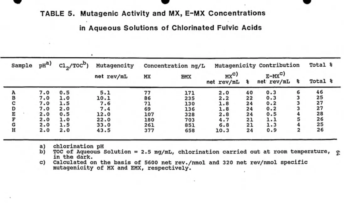
Depending on the chlorination conditions and the TOC content, it is possible to obtain a chlorinated fulvic acid sample which is high in mutagenicity but (simultaneously) low in MX. A fulvic acid solution, with a high TOC content, will, upon chlorination, yield a comparatively large number of organo-halide compounds. If the concentration of MX is small in comparison to a relatively high overall
mutagenicity, this would indicate that the extract, contains many compounds (mutagens) of interest. The coupled
HPLC-Bioassay procedure can thus be used to obtain active subtractions which are relatively free of MX as well as other non-mutagenic organic material. When sufficiently
"pure", these active sub-fractions can be subjected to GC/MS
33
IV: EXPERIMENTAL
4.1 : Materials.
For the Ames assay, Salmonella typhimurium strains TA 98, TA 100 and TA 102 were obtained from Dr. Bruce- Ames, University of California at Berkeley. Sodium azide and 2-nitrofluorene were purchased from Aldrich (Milwaukee, WI), 2-anthramine and 1,8 dihydroxyanthraquinone (Danthron) from Sigma Chemical Co. (St. Louis, MO) and Daunomycin from Fluka Chemical Corp. (Ronkokoma, NY). Oxoid Nutrient Broth #2 was obtained from Oxoid, Ltd. (Basingstoke, Hants, England) and Bacto agar from Difco Laboratories (Detroit, MI). Molecular biology grade DMSO was purchased from Fisher Biotech.
Chemicals for the VBME solution were all obtained from
Fisher Scientific (Fairlawn, NJ). The NADP and Glucose-6-Phosphate were ordered from Boehringer Mannheim (West
Germany). The 39 used was from the livers of
Aroclor-1254-treated male Sprague-Dawley rats and was purchased from
Moltox (College Park, MD).
HPLC grade acetonitrile and ethyl acetate were obtained
from Fisher Scientific (Fairlawn, NJ). Ethyl ether was from
Burdick & Jackson (Muskegon, MI). The sodium hypochlorite
34
Scientific (Pittsburgh, PA). The XAD resins were Xmberlite
(Rohm & Haas, Philadelphia).
The MX analogues were synthesized by Dr. Leif Kronberg using MX which in turn had been synthesized by the method of
Padmapriya, et.al. (1985).
4.2 : Chlorination and Extraction of Seunples.
All the chlorination was done with sodium hypochlorite. The concentration of the sodium hypochlorite solution was determined by the DPD Ferrous Titrimetric Method (Std. Methods, 408D) as was the residual chlorine in the
chlorinated samples. The TOC content of the samples was analysed by the persulfate oxidation method using 01 Corp.
TOC700 TOC analyzer. The CI2/TOC ratios were on a weight to
weight basis.
4.2 (a) : Humic Water Sample.
The humic water sample was natural freshwater with a high content of humic substances (TOC =20 mg/L) collected
from a marshy lake (Lake Savojaeri) in southwest Finland. The lake did not receive any municipal or industrial water
effluents. The water was chlorinated at a 1:1 CI2/TOC ratio
(on a weight to weight basis) while being buffered at
pH 7.0 ±0.2 (with potassium phosphate buffer). The reaction
was allowed to go on for 60 hours at ambient temperature in
35
pH 2.0 by adding 4N HCl. The mutagens were concentrated by passing the acidified humic water through a column of XAD-4 and XAD-8 resins (1:1 volume mixture) at a flow rate of one bedvolume/min (20 ml/min). Residual water remaining in the column was removed with a gentle stream of nitrogen. The absorbed organics were subsequently eluted in the reverse direction with 3 bedvolumes of ethyl acetate. The extract was then concentrated further by evaporation and its final volume adjusted such that 1 ml of ethyl acetate equalled
1 liter of original humic water. This extract of
chlorinated humic water is referred to, in parts of this
report, as HW.
4.2 (b) : Fulvic Acids Seunple.
The fulvic acids were extracted from a highly colored natural lake water (Lake Drummond, VA) using the isolation
method of Thurman and Malcolm (1981).
They were then dissolved in distilled water to give a
working sample with a TOC of 2.5 g/liter. A portion of this
sample was subdivided into 8 aliquots , 4 of which were
adjusted to pH 7.0 using potassium phosphate buffer, while
the other 4 were adjusted to pH 2.0 with 4N HCl. At eachpH, the 4 aliquots were chlorinated (with sodium
hypochlorite solution) at CI2/TOC ratios (wt./wt.) of 0.5,
1.0, 1.5, and 2.0 respectively. These were designated
samples A, B, C, D for pH 7.0 and samples E, F, G, H for pH
35
pH 2.0 by adding 4N HCl. The mutagens were concentrated by
passing the acidified humic water through a column of XAD-4
and XAD-8 resins (1:1 volume mixture) at a flow rate of one
bedvolume/min (20 ml/min). Residual water remaining in the
column was removed with a gentle stream of nitrogen. The
absorbed organics were subsequently eluted in the reverse
direction with 3 bedvolumes of ethyl acetate. The extract
was then concentrated further by evaporation and its final
volume adjusted such that 1 ml of ethyl acetate equalled
1 liter of original humic water. This extract of
chlorinated humic water is referred to, in parts of this
report, as HW.
4.2 (b) : Fulvic Acids Sample.
The fulvic acids were extracted from a highly colored
natural lake water (Lake Drummond, VA) using the isolation
method of Thurman and Malcolm (1981).
They were then dissolved in distilled water to give a
working sample with a TOC of 2.5 g/liter. A portion of this
sample was subdivided into 8 aliquots , 4 of which were
adjusted to pH 7.0 using potassium phosphate buffer, while
the other 4 were adjusted to pH 2.0 with 4N HCl. At each
pH, the 4 aliquots were chlorinated (with sodium
hypochlorite solution) at CI2/TOC ratios (wt./wt.) of 0.5,
1.0, 1.5, and 2.0 respectively. These were designated
samples A, B, C, D for pH 7.0 and samples E, F, G, H for pH
allowed to proceed at ambient temperatures in the dark for
60 hours. All the samples were then acidified to pH 2.0
with 4N HCl and extracted 3 times with diethyl ether. The
extracts were combined, evaporated to dryness, and
redissolved in ethyl acetate. The volume was adjusted such
that 1 ml of ethyl acetate = 1 liter of water with a TOC =
20 ing/liter. The fulvic acid sample, chlorinated at pH 2.0
and at a CI2/TOC ratio of 2, is referred to as FA or as
Sample H.
4.2 (c) : Drinking Water Sample.
The drinking water samples were collected directly from
the distribution system (i.e., tap water) in Chapel Hill,
North Carolina. The water treatment plant (OWASA) uses
surface water with a TOC of approximately 5 mg/liter and
chlorinates at approximately 5 mg CI2 per liter of raw
water. The water samples were stored for 24 hours in a
decanter glass in order to evaporate the residual chlorine.
Subsequently, the pH of the water samples was adjusted to
pH 2.0 with 4N HCl. The extracts were obtained by the
procedure described for the humic water sample. This
extract of drinking water is referred to hereafter as DrW.
4.3 : High Performance Liquid chromatographic
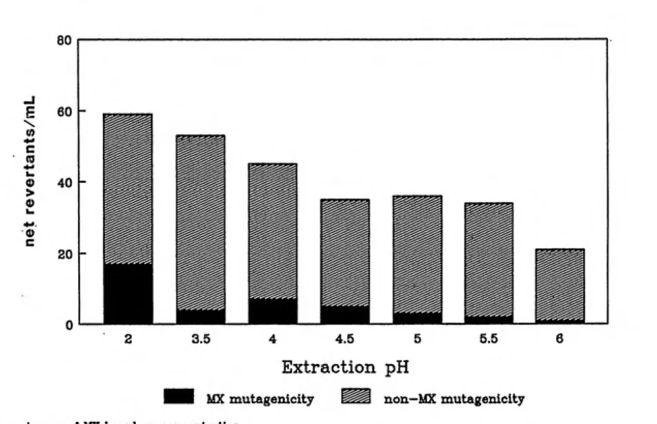
Separation.
First, aliquots of the aqueous sample H were adjusted
37
were extracted with diethyl ether. The mutagenic activity of each extract was determined by the Ames assay, while the MX concentration was determined by GC/MS analysis.
The FA or sample H extract was fractionated using Reverse-Phase HPLC. Separation was achieved on a Varian LC-5000 (Palo Alto, CA) equipped with a six port Rheodyne
injector. The first fractionation was done on a Cg
analytical column (Phase Sep. Spherisorb 5Cg, 4.5 x 250 mm)
fitted with a 4.5 x 50 mm pellicular ODS precolumn. The eluent system for this separation is described as Program I.
The second fractionation was achieved on a C^s analytical
column (Alltech, Econosphere 5C-j^g, 4.5 x 250 mm) also fitted
with a 4.5 X 50 mm pellicular ODS precolumn. The successful eluent system is described as Program II. HPLC effluent was monitored at 230 nm wavelength using a Perkin-Elmer LC-85B
Spectrophotometric Detector with an LC-85 autocontrol
(Norwalk, CT), and recorded on a Perkin-Elmer 561 recorder. A suitable portion of the extract (equivalent to 1000-1500 mL of the original solution) was evaporated and
redissolved in a 1:1 mixture of acetonitrile (ACN) and the 0.1 M potassium phosphate buffer. Then, about 33 0 ul was injected via a 500 jjlI loop. Chromatographic fractions were collected manually based on UV absorbance peaks obtained during a prior trial separation. Three portions of the
extract (33 0 ul each) were injected and separated
38
run were then combined. Each fraction was subsequently acidified to pH 2.0 with 4N HCl and repeatedly extracted using diethyl ether. The ether extracts were evaporated to dryness, the residue redissolved in ethyl acetate and
refrigerated until further use.
Program I : Separation of Sample H.
Cg analytical column; Flow: 1 ml/min
Isocratic elution:- 20% Acetonitrile (ACN) and 80% potassium phosphate buffer
(0.1 M, pH 6.0)
Program II : Separation of H3.
Cng analytical column; Flow: 1 ml/min
Stepwise gradient elution with Acetoni¬
trile (ACN) and 0.1 M potassium phosphate
buffer (pH 6.0)
0-11 min: 100% buffer
11-21 min: 90% buffer 10% ACN 22-32 min: 80% buffer 20% ACN 33-43 min: 70% buffer 30% ACN 44-54 min: 60% buffer 40% ACN
54-64 min: 60% buffer up to 100% buffer.
4.4 : Synthesis.
The MX analogues were synthesized, characterized (section 4.4), and quantified in the chlorinated samples (section 4.5) by Dr. Leif Kronberg, Abo Akademi, Turku, Finland; visiting scholar (1988-1989), Department of
Environmental Sciences and Engineering, University of North
Carolina at Chapel Hill.
4.4 (a) : ox-MX.
(Z)-2-Chloro-3-(dichloromethyl)-butenedioic acid
(ox-MX) was obtained by the oxidation of MX. 48 mg
39
at 70°C for 24 hours. After cooling in ice and diluting it
with 20 ml of ice-cold water, the reaction mixture was extracted 3 times with diethyl ether. The ether extracts were combined, washed with 0.01 M HCl and subsequently
evaporated to dryness. Recrystallization from dichloromethane yielded the pure compound (9.1 mg,
yield = 21%). The electron-impact mass spectrum of the compound is presented in Figure 3. This mass spectrum represents the anhydride of ox-MX since ox-MX loses a molecule of water upon heating in the MS inlet probe (or GC-in^ector). The H NMR spectrum of the compound (obtained at 400 MHz, XL-400, Varian Associates, Palo Alto, CA) showed the resonance signal of the proton in the dichloromethyl
group to appear at 6 6.2.
4.4 (b) : red-MX.
3-Chloro-4-(dichloromethyl)-2(5H)-furanone (red-MX) was obtained by the reduction of MX. 20 mg (93 jumol) of MX was treated with aluminum isopropoxide (225 /imol) in isopropanol
(the Meerwein-Ponndorf reduction) for 2 hours at 70°C. The
reaction was stopped by adding ice and 4N HCl. The
acidified mixture was then heated to 50°C for a few minutes
and then cooled again. Subsequently, it was extracted 3 times with diethyl ether. The extracts were combined,
washed with 0.01 N HCl, and the ether evaporated to obtain
FIG.3 Mass Spectrum of ox-MX anhydride
100
lU u 2 <
Q 50
ͣ
z.
ͣ
D a <
>
LU
72
ͣ
ii ,11 hlllUi
87
iiii,.>>|jil liU
107
HCCIz CI
142
L-^____r^m___. I |llli|illl|lii. 1. 11
179
170
li |l'H|i H'Ml
186
214(M*)
o
41
the pure compound was 17.3%. The electron-impact mass
spectrum of red-MX is shown in Figure 4. The
ͣ
'"H NMR
resonance signal of the dichloromethyl group was observed at
S 6.74 (IH) and of the protons in the lactone ring at 8 5.16
(2H).
4.4 (c) : ox-EMX.
(E)-2-Chloro-3-(dichloromethyl)-butenedioic acid
(ox-EMX) was obtained from the oxidation of E-MX. 10 mg
(46.3 /imol) of E-MX was treated with 5 mg (56 /Ltmol) ofNaC102 in water with resorcinol as chlorine scavenger. The
reaction was allowed to proceed at pH 3.5 for 2.5 hours.
Subsequently, the pH of the mixture was raised to 4.5 and
the first diethyl-ether extraction was carried out. This
ether extract was discarded, the pH of the solution was
lowered to pH 2.0 and the diethyl ether extraction was
repeated. This second extract was washed with 0.01 N HCl
and, upon evaporation of the ether, the crude compound was
recrystallized from dichloromethane. Finally, the crystals
were washed with CCl^. The yield of ox-EMX was 20%. The
electron-impact mass spectrum of ox-EMX is given in
Figure 5. The ^H NMR resonance signal of the dichloromethyl
group was observed at S 5.6.
The attempts to synthesize
(E)-2-Chloro-3-(dichloromethyl)-4-hydroxy-butenoic acid (red-EMX) were
FIG.5 Mass Spectrum of ox-EMX UJ u < Q z CQ < > g Ui a 100 35. 9fl. 85. ee. 75 J ». ES. ee. ss. 50 45 J 46. 35. 38. 25. i 28. 15. | 18 5. 107 73 lUjl
Hccr2 COOH
HOOC XCI
jiiiiy
142
uiMii Ail.
170
i l...,I.I.M..I ill
I m
JlUiLuJl...J214
44
isopropoxide in isopropanol and with NaBH^ in a mixture of
isopropanol and water did not yield red-EMX as indicated by GC-MS analyses. The second reaction nevertheless produced a compound which could be an isomer of red-MX. It had an
identical mass spectrum plus a chlorine ion cluster at
mass/charge ratios of 121 and 12 3.
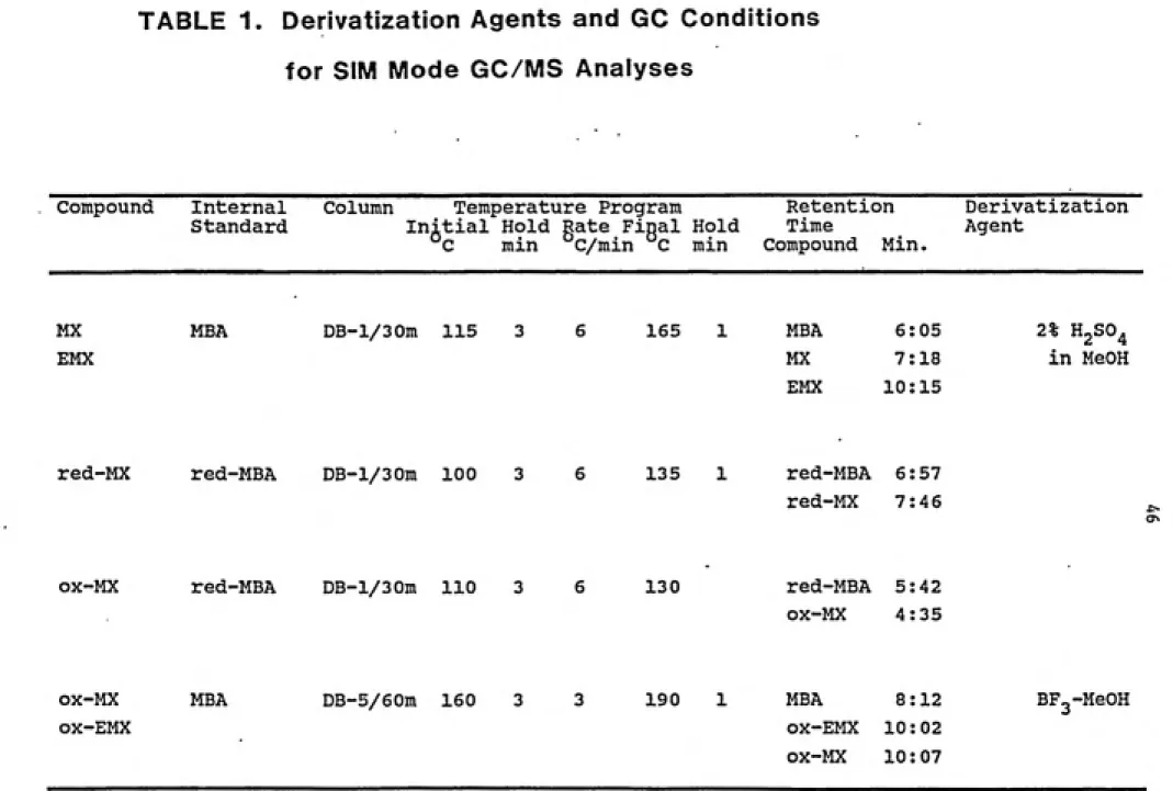
4.5 : Derivatization and Gas-Chromatography/Mass
Spectrometry Analyses.
To prepare the chlorinated humic water, fulvic acid, and drinking water samples for GC/MS analyses, their
extracts were evaporated to dryness and the residues were
methylated using 250 fiL of the respective methylation agent (see Table 1). In order to detect and quantify MX and E-MX by SIM mode GC/MS analysis, the extracts (ether or XAD
extracts) were methylated using 2% (volume/volume)" H2SO4 in
methanol. The reaction was allowed to proceed for 1 hour at
70°C. The reaction mixture was neutralized by the addition
of 2% aqueous NaHC03 and subsequently extracted twice using
n-hexane (2 x 250 /xL) . For the determination of the
synthesized ox-MX and ox-EMX in the samples, the extracts
were methylated with BF3 (12% by weight) in methanol. This
reaction was allowed to go on for 12 hours at 70°C. The
reaction mixture was similarly neutralized and extracted. The hexane extracts, combined for each sample, were concentrated under a stream of nitrogen before being
45
compounds was carried out by reference to an internal standard, mucobromic acid (MBA), added to the extracts in
known amounts.
The analysis of red-MX was carried out in unmethylated extracts using 2, 3-dibromo-2(5H)-furanone (red-MBA) as an
internal standard. Attempts were also made to analyze ox-MX
in unmethylated extracts, again, using red-MBA as the
internal standard.
The GC/MS analyses of these extracts were performed on a Hewlett-Packard 5890 capillary gas chromatograph
interfaced with a VG 70 - 250 SEQ mass spectrometer. The conditions for the analyses are given in Table 1. For qualitative and quantitative determination, the MS was
operated in the selected ion monitoring (SIM) mode, and the
ion peaks monitored are listed in Table 2. The SIM data was
recorded and computed using the standard SIM routine of the
VG 11-250J data system. Response factors for the ions were
calculated versus the respective internal standard, (Table 2)
and the identification of the compounds in the extracts was
based on positive matching of retention times and of
relative ion peak area ratios.
For the synthesized compounds (reaction mixtures), the
GC analysis was done on a Carlo-Erba HRGC 5160 capillary gas
chromatograph equipped with a DB-1 fused silica capillary
column (30 m long). Separation of ox-MX and ox-EMX was
attempted on DB-l/30m, DB-17/30m, DB-1701/15m, SP-2340/30m,
TABLE 1. Derivatization Agents and GC Conditions
for SIM Mode GC/MS Analyses
Compound Internal
Standard
Column Temperature Program Retention Derivatization Initial Hold Rate Final Hold Time Agent
C min C/min C min Compound Min.
MX
EMX
MBA DB-l/30m 115 3 165 1
red-MX red-MBA DB-l/30m 100 3 6 135 1
MBA 6:05 2% H2SO4
MX 7:18 in MeOH
EMX 10:15
red-MBA 6:57
red-MX 7:46
OX-MX red-MBA DB-l/30m 110 3 130 red-MBA 5:42
ox-MX 4:35
OX-MX ox-EMX
MBA DB-5/60m 160 3 3 190 1 MBA 8:12 ox-EMX 10:02
ox-MX 10:07
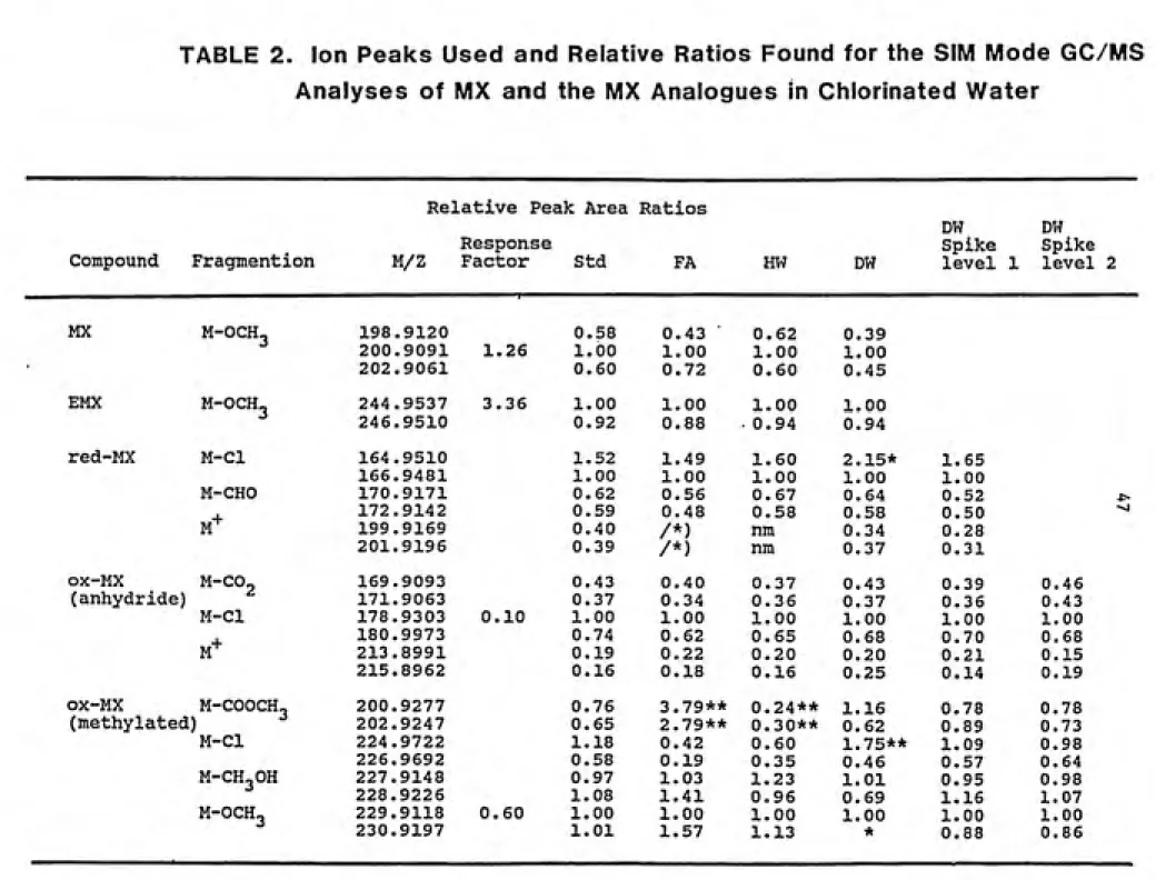
TABLE 2. Ion Peaks Used and Relative Ratios Found for the SIM Mode GC/MS Analyses of MX and the MX Analogues in Chlorinated Water
Relative Peak Area Ratios
DW DW
Response Spike Spike
Compound Fragmention M/Z Factor Std FA HW DW level 1 level 2
MX M-OCH3 198.9120 0.58 0.43 ͣ 0.62 0.39
200.9091 1.26 1.00 1.00 1.00 1.00
202.9061 0.60 0.72 0.60 0.45
EMX
M-OCH^ 244.9537 3.36 1.00 1.00 1.00 1.00
246.9510 0.92 0.88 0.94 0.94
red-MX M-Cl 164.9510 1.52 1.49 1.60 2.15* 1.65
166.9481 1.00 1.00 1.00 1.00 1.00
M-CHO 170.9171 0.62 0.56 0.67 0.64 0.52 *>
M-*-172.9142 0.59 0.48 0.58 0.58 0.50 ^
199.9169 0.40 /*) nm 0.34 0.28
201.9196 0.39 /*) nm 0.37 0.31
ox-MX
M-CO2 169.9093 0.43 0.40 0.37 0.43 0.39 0.46
(anhydride) 171.9063 0.37 0.34 0.36 0.37 0.36 0.43
M-Cl 178.9303 0.10 1.00 1.00 1.00 1.00 1.00 1.00
M+
180.9973 0.74 0.62 0.65 0.68 0.70 0.68
213.8991 0.19 0.22 0.20 0.20 0.21 0.15
215.8962 0.16 0.18 0.16 0.25 0.14 0.19
ox-MX M-COOCH^
) ^
200.9277 0.76 3.79** 0.24** 1.16 0.78 0.78 (methylated 202.9247 0.65 2.79** 0.30** 0.62 0.89 0.73
M-Cl 224,9722 1.18 0.42 0.60 1.75** 1.09 0.98
226.9692 0.58 0.19 0.35 0.46 0.57 0.64
M-CH3OH 227.9148 0.97 1.03 1.23 1.01 0.95 0.98
228.9226 1.08 1.41 0.96 0.69 1.16 1.07
M-OCH3 229.9118 0.60 1.00 1.00 1.00 1.00 1.00 1.00
"^
Table 2 (Continued)
Relative Peak Area Ratios
Compound Fragmention
Response
M/Z Factor Std FA HW DW
DW DW
Spike Spike
Level 1 Level 2
ox-EMX M-COOH3 200.9277 0.96 1.03 1.11 1.10
202.9247 0.50 1.00 1.00 1.00 1.00
M-OCH3 224.9722226.9692 0.870.39 0.700.43 0.710.48 0.790.40
MBA M-OCH3 240.8325
red-MBA M-Br 160.9239
) Interference
nm= not measured
ͣ
kit
) Interference from ox-EMX
*** _i _i
) Response factor= (Acomp x Ccomp ) x (Cstd. x Astd. ) where
C=concentration and A=ion peak area ratio
49
4.6 : Mutagenicity (Ames) Assay.
The bacterial mutagenicity of red-MX, ox-MX, and ox-EMX was tested in the constructed Salmonella typhimurium strains TA 100, TA 98, and TA 102 according to the standard plate
incorporation procedure of Maron and Ames (1983). The genotypes of these strains are as follows:
TA 100 - his G46, rfa, uvrB, pKM 101
TA 98 - his D3052, rfa, uvrB, pKM 101 TA 102 - his'G428, rfa, pKM 101, pAQl
The strains were kept in storage at -70°C, and from these
master-plates were prepared and kept at 4°C. The presence
of genetic markers as well as spontaneous reversion rates and positive control responses were verified for each
master-plate before it was used to grow overnight-cultures of the strain. The positive control and spontaneous
responses were also tested along with every experiment. Experiments giving values outside the historically
acceptable range were rejected. The positive control chemicals and the amount added per plate were as follows:
TA 100 (-S9) TA 100 (+S9)
TA 98 (-S9)
TA 98 (+S9) TA 102 (-S9)
TA 102 (+S9)
1.5 /xg Sodium Azide
0.5 /ig 2-anthramine 3.0 Mg 2-nitrofluorene 0.5 /ig 2-anthramine
6.0 jug Daunomycin
3 0 Mg 1/8 dihydroxyanthraquinone
(Danthron)
The effect of exogenous xenobiotic metabolizing enzymes
on the mutagenicity of the MX analogues was tested using
Aroclor 1254 - induced rat liver homogenate fraction, S9